Patients › Hand
Infecciones de la mano
Paronychia, deep-space and flexor sheath infections — recognition, antibiotics, drainage.
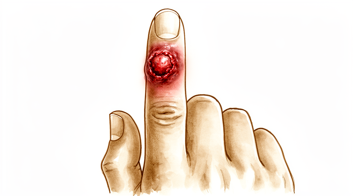
Qué está sintiendo
Una infección en la mano suele doler en un solo punto. La piel sobre esa zona puede estar roja, caliente e hinchada. El dolor suele intensificarse al mover el dedo o al agarrar algo. Descansar la mano alivia un poco el malestar, pero el dolor no desaparece por sí solo.
Las tareas cotidianas se vuelven difíciles: abrochar botones, girar una llave, sostener una taza de té o escribir en el teclado pueden resultar dolorosos. Si la infección afecta a la vaina tendinosa de un dedo (el túnel por donde se desliza el tendón), todo el dedo puede hincharse y quedar ligeramente doblado. Enderezarlo duele, y el dolor persiste incluso cuando otra persona intenta enderezarlo con suavidad. Si la articulación está infectada, esta se hincha, es sensible al tacto y está caliente; moverla en cualquier dirección resulta muy doloroso.
Algunas infecciones se extienden por la piel en forma de rayas rojas y sensibles; además, los ganglios de la axila pueden inflamarse y doler. La hinchazón puede hacer que el dorso de la mano parezca abultado, y a veces es difícil determinar dónde se ha acumulado el líquido. Por la noche, el malestar aumenta, pues la hinchazón se intensifica cuando la mano permanece inmóvil.
La mayoría de las infecciones aparecen en cuestión de horas o días, no semanas. Si el enrojecimiento, la hinchazón y el dolor empeoran en lugar de mejorar, eso indica infección y no una simple distensión. Algunas afecciones presentan síntomas muy similares: la gota, una picadura de insecto, una reacción a una astilla o un brote de artritis; por eso, al principio no siempre es fácil identificar la causa.
Informe a su cirujano si padece diabetes, enfermedad renal o alguna condición o medicamento que debilite su sistema inmunitario. Estos factores influyen en el comportamiento de la infección y en el tratamiento necesario. Lo mismo aplica a cualquier infección que no haya mejorado tras tomar antibióticos o tras limpiar la herida, ya que algunas infecciones de crecimiento lento requieren pruebas especiales para ser identificadas.
Con un tratamiento oportuno, la mayoría de las infecciones de tejidos blandos de la mano se resuelven por completo. El riesgo de demorar el tratamiento es que pueda aparecer rigidez, flexión permanente del dedo o, en casos graves, incluso la pérdida del dedo.
¿Qué está ocurriendo realmente?
La mano es un espacio muy concurrido. Veintisiete huesos se encuentran muy juntos bajo una piel que es delgada en el dorso de la mano y está firmemente adherida a la palma. Los tendones, esas “cuerdas” que mueven los dedos, se deslizan a través de túneles estrechos. Uno de esos túneles, la vaina flexora, rodea al tendón que permite el movimiento de flexión del dedo; su interior está revestido por una capa lisa que nutre y lubrica el tendón. Cuando las bacterias penetran mediante un corte, una astilla o una herida punzante, ese espacio cerrado no les permite propagarse más que a lo largo del túnel.
La hinchazón es el verdadero problema. En un túnel así no hay espacio para que se produzca hinchazón, por lo que la presión aumenta. Si esa presión sigue subiendo, puede interrumpir el flujo sanguíneo hacia el propio tendón. Entonces el tendón se cicatriza adhiriéndose a la vaina que lo rodea, lo que deja el dedo rígido incluso después de que la infección haya desaparecido. Por eso una infección de la vaina tendinosa requiere tratamiento inmediato, sin esperar unos días.
Otros lugares también presentan sus propios riesgos. La almohadilla de grasa en la punta del dedo está dividida en pequeñas cavidades selladas por fibras resistentes; por eso una infección allí (llamada “felon”) genera presión rápidamente y puede extenderse al hueso o a la articulación cercana. La piel junto a la uña también puede albergar una infección lenta y persistente (paroniquia crónica), a menudo provocada por hongos; la humedad se filtra bajo la cutícula y mantiene la zona inflamada. En algunas personas, los túneles tendinosos del pulgar y del dedo meñique están conectados, de modo que la infección puede propagarse de un lado de la palma al otro.
La mayoría de las infecciones de la mano son causadas por bacterias que normalmente viven en la piel y penetran a través de alguna lesión. Otras provienen de mordeduras, ya sean humanas o de animales. La diabetes, los corticoides o un sistema inmunitario debilitado facilitan que la infección se instale y alcance estructuras más profundas como tendones, huesos o articulaciones. Por eso su cirujano le preguntará sobre su historial médico y por qué el tratamiento temprano es crucial: con atención oportuna, la mayoría de las infecciones de tejidos blandos se resuelven por completo; en cambio, la demora conlleva riesgo de rigidez permanente.
¿Qué podemos hacer al respecto?
El Dr. Kieran Hirpara, cirujano de extremidades superiores en el Mater Private Hospital Rockhampton, trata las infecciones de la mano actuando con rapidez, ya que la demora es lo que provoca daños duraderos. Por lo general, los pacientes son derivados a nuestra clínica por su médico de cabecera; si un fisioterapeuta le ha sugerido que nos consulte, igualmente necesitará una derivación de su médico de cabecera para poder acceder al reembolso de Medicare. En esa consulta tomamos la historia clínica, examinamos su mano y solicitamos los exámenes necesarios para determinar qué tipo de microorganismo está causando la infección y hasta qué profundidad ha llegado.
En muchas infecciones en fase inicial, el primer paso es no quirúrgico. Si una infección de la vaina tendinosa se detecta dentro de las 48 horas posteriores al inicio de los síntomas, los antibióticos y un entablillado pueden impedir su propagación. Luego se realiza un seguimiento estrecho; si la situación no mejora rápidamente, procedemos a la cirugía en lugar de esperar. Tras cualquier tratamiento, mantener la mano en una posición protectora mediante entablillado y mover los dedos tempranamente ayuda a prevenir la rigidez. La rehabilitación es fundamental: una mano infectada que no se mueve se vuelve rígida, por lo que la terapia forma parte del plan de tratamiento, no es un complemento opcional.
Los antibióticos constituyen la otra vertiente del tratamiento médico. La primera prescripción se elige para cubrir a las bacterias más probables de causar su infección; una vez que los resultados de laboratorio indican exactamente cuál es el patógeno, cambiamos al antibiótico adecuado. Algunas infecciones requieren antibióticos por vía intravenosa en lugar de comprimidos; en casos graves, el tratamiento puede durar entre 4 y 6 semanas. Si su sistema inmunológico está debilitado por alguna enfermedad o por medicamentos como los esteroides, tratamos con mayor rapidez e intensidad, pues estas infecciones pueden agravarse con mayor facilidad. Las mordeduras, ya sean humanas o de animales, también suelen requerir antibióticos preventivos.
La cirugía se considera cuando hay pus que drenar o cuando la infección ha alcanzado tendones, huesos o articulaciones. Un absceso o acumulación de líquido bajo la piel debe liberarse para aliviar la presión; con frecuencia, la herida se rellena inicialmente para favorecer el drenaje. Una infección en la punta del dedo (llamada “felon”) o una infección junto a la uña (paroniquia) se drena mediante una pequeña incisión, a veces extirpando parte de la uña. La infección de la vaina tendinosa requiere lavar dicha vaina, mientras que la infección ósea (osteomielitis) implica eliminar el tejido necrótico y administrar antibióticos durante un período prolongado. Una excepción importante: si una ampolla en el dedo resulta ser una infección viral (panadizo herpético), no se recurre a la cirugía, pues abrirla podría empeorar el cuadro.
Qué esperar
La mayoría de las infecciones de la mano detectadas a tiempo se resuelven por completo con tratamiento. Con una atención oportuna, la mayor parte de las infecciones de los tejidos blandos desaparecen totalmente y la mano vuelve a la normalidad. Cuanto antes reciba tratamiento, mayores serán sus posibilidades de evitar rigidez o daños duraderos.
El tiempo de recuperación depende de la profundidad a la que haya llegado la infección. Una infección de la piel o de la uña puede mejorar en cuestión de días tras el drenaje o el uso de antibióticos. En cambio, una infección de la vaina tendinosa, una infección ósea o una infección articular requieren más tiempo; algunas necesitan semanas de tratamiento antibiótico. Incluso después de que la infección haya desaparecido, el dedo puede permanecer rígido durante un tiempo. Mover los dedos tempranamente, una vez que su cirujano lo autorice, ayuda a evitar que esa rigidez se vuelva permanente.
Si el tratamiento se retrasa, el pronóstico es menos favorable. Una infección de la vaina tendinosa que persiste durante varios días puede provocar cicatrización del tendón dentro de su canal, dejando el dedo rígido incluso tras la curación de la infección. Las infecciones graves pueden causar pérdida permanente de la función; en algunos casos, incluso la pérdida del dedo, a pesar de un tratamiento exhaustivo y oportuno. Por eso estas infecciones se consideran casos de urgencia y no se dejan en observación por unos días.
Algunas infecciones se comportan de manera distinta. En personas con diabetes o con el sistema inmunitario debilitado, las infecciones pueden propagarse más rápidamente y afectar estructuras más profundas; por ello requieren un tratamiento más agresivo y, a veces, más de una intervención quirúrgica. Las infecciones de crecimiento lento, como las causadas por micobacterias (un grupo de microorganismos relacionados con el que produce la tuberculosis), necesitan tratamientos antibióticos prolongados combinados con cirugía para erradicarlas; su recuperación puede tardar meses en lugar de días.
Por lo general, se le pedirá que regrese después del tratamiento para comprobar que la infección está desapareciendo y que su mano se mueve correctamente. Asista a esas citas aunque se sienta mejor, ya que algunos problemas solo se manifiestan cuando la hinchazón disminuye. Si el dolor, el enrojecimiento o la hinchazón reaparecen tras haber parecido remitir, comuníquese con su cirujano en lugar de esperar a ver qué sucede.
¿Cuándo consultar a un especialista?
Las infecciones de la mano requieren atención inmediata. El daño causado por la demora se produce rápidamente; por eso, no espere a ver si la situación mejora por sí sola.
Acuda a urgencias si observa rayas rojas que se extienden desde la mano hacia el brazo, si todo el dedo está hinchado, doblado y demasiado doloroso para enderezarlo, o si la piel se está oscureciendo, formando ampollas o desgarrándose. Lo mismo aplica si presenta fiebre y malestar general junto con los síntomas en la mano, o si la mano sufrió una lesión y ahora está muy hinchada y tensa. Estos signos pueden indicar una infección profunda o en expansión que requiere evaluación y tratamiento el mismo día.
Solicite una valoración urgente, preferiblemente el mismo día, si el dolor es intenso y empeora, si una articulación del dedo está caliente, hinchada y demasiado dolorosa para moverse, o si se observa acumulación de pus bajo la piel o junto a una uña. Una infección que no mejora tras uno o dos días de tratamiento antibiótico también requiere revisión; no basta con esperar a que termine la receta.
Consulte a su médico de cabecera sin demora ante cualquier infección en la mano, incluso si parece leve. Esto es especialmente importante si padece diabetes, enfermedad renal o alguna afección o toma medicamentos que debiliten su sistema inmunitario, pues en esos casos las infecciones pueden propagarse más rápido y afectar zonas más profundas. Informe a su médico sobre cualquier mordedura reciente de animal o persona, herida punzante, o antecedentes laborales o de viaje que pudieran sugerir la presencia de un patógeno poco común.
Si una infección se repite con frecuencia o nunca se resuelve por completo a pesar del tratamiento, solicite una evaluación por parte de un especialista, ya que algunas infecciones de crecimiento lento requieren pruebas específicas para su identificación.
En mayor profundidad
Advanced reading: the deeper science (optional)
Esta sección profundiza más de lo necesario para que usted tome sus propias decisiones de tratamiento. Las infecciones de la mano merecen una lectura adicional, ya que los antibióticos que generalmente se administran en primer lugar son, en este contexto específico, los incorrectos; además, ciertas infecciones de la mano constituyen una emergencia quirúrgica que con frecuencia se confunde con un caso leve.
El microorganismo más común resiste a los antibióticos más habituales
Un estudio longitudinal de diez años realizado con 815 infecciones de mano en entornos urbanos reveló que, aunque la incidencia anual de MRSA ha disminuido en general, este sigue siendo el patógeno más frecuente. Por otro lado, la resistencia a la clindamicina y la levofloxacina ha aumentado de manera constante. La conclusión de los autores es clara: el tratamiento antibiótico empírico para infecciones de mano debe evitar la penicilina, los betalactámicos, la clindamicina y la levofloxacina [1].
Esta lista de antibióticos a evitar es más extensa de lo que la mayoría espera; además, incluye muchos de los fármacos que habitualmente se recetan para infecciones cutáneas en otras partes del cuerpo. En la práctica, esto significa que el hecho de que una infección de mano no mejore con el antibiótico de primera línea es algo común y predecible, no una excepción; por ello, es recomendable consultar a un especialista en lugar de persistir con el mismo tratamiento.
Los patrones de resistencia varían según la zona, por lo que este criterio debe entenderse como un principio general y no como una prescripción rígida. No obstante, explica por qué, con frecuencia, las infecciones de mano requieren un tratamiento más intensivo de lo que su gravedad aparente sugiere.
Infección de la vaina flexora: detalles que determinan el desenlace
La tenosinovitis flexora piogénica, es decir, la infección que se produce dentro del conducto cerrado por el que discurre el tendón flexor, es la que se comporta de manera distinta respecto a otras infecciones de la mano. Dicha vaina constituye un espacio confinado con escasa irrigación sanguínea; por ello, el pus bajo presión que se acumula en su interior puede destruir la superficie de deslizamiento del tendón en cuestión de días.
En un total de 763 pacientes, dos factores contribuyeron a mejorar el rango de movimiento: el uso de antibióticos como parte del tratamiento y la irrigación mediante catéter en lugar del lavado abierto. Las evidencias respaldan tanto el tratamiento precoz como el uso sistémico de antibióticos [2].
Cabe señalar que el desenlace evaluado es el rango de movimiento, no la erradicación total de la infección. Es posible curar la infección sin que el dedo recupere su movilidad plena, y ese es el verdadero costo de cualquier retraso en el tratamiento. Los cuatro signos clásicos —dedo hinchado en forma de “salchicha”, ligeramente flexionado; sensibilidad a lo largo de toda la vaina tendinosa; y dolor intenso al intentar estirarlo pasivamente— merecen ser reconocidos, pues su presencia justifica una evaluación inmediata en lugar de limitarse a administrar medicamentos por vía oral.
El caso que constituye una verdadera emergencia
La fascitis necrotizante de origen mano es poco frecuente y extremadamente destructiva. La revisión sistemática de 161 casos concluye que el diagnóstico precoz reviste una importancia capital, y que la intervención quirúrgica temprana y decidida debe realizarse con un umbral bajo, especialmente cuando existen factores de riesgo [3].
El término “umbral bajo” se emplea intencionadamente. En este diagnóstico, esperar a obtener certeza puede resultar en la pérdida de extremidades. Los signos que la diferencian de la celulitis común son: dolor desproporcionado respecto a la apariencia de la lesión, progresión rápida en cuestión de horas y no días, así como síntomas sistémicos como fiebre, confusión y sensación de malestar general, todo ello acompañado de una zona de enrojecimiento que puede parecer de tamaño moderado.
¿Por qué una herida pequeña puede ser engañosa?
Existen dos mecanismos que provocan infecciones mucho más profundas de lo que la herida visible indica. Una mordedura en la articulación de un dedo, producida al golpear una boca con el puño, permite que microorganismos bucales penetren a través de la piel, el tendón extensor y la cápsula articular en un solo movimiento; posteriormente, la piel se cierra sobre dicha contaminación. Asimismo, una punción en la palma de la mano puede introducir gérmenes en la vaina del flexor mediante un punto de entrada que se cierra en el transcurso de un día.
En ambos casos, la herida visible no constituye un indicador fiable de la profundidad del problema. El patrón del dolor —en particular el dolor al mover el dedo, en lugar del dolor en la propia herida— resulta más revelador que el aspecto de la piel.
Referencias
[1] Kistler JM, Thoder JJ, Ilyas AM. Incidencia de SARM y tendencias en el uso de antibióticos en infecciones de la mano en entornos urbanos: un estudio longitudinal de 10 años. Hand (N Y). 2018;14(4):449-54. https://doi.org/10.1177/1558944717750921
[2] Giladi AM, Malay S, Chung KC. Revisión sistemática sobre el tratamiento de la tenosinovitis flexora piogénica aguda. J Hand Surg Eur Vol. 2015;40(7):720-8. https://doi.org/10.1177/1753193415570248
[3] Christopoulos G, Khoury A, Johnson M, Sergentanis TN. Fascitis necrotizante de origen mano: revisión sistemática y metaanálisis. Hand (N Y). 2022;19(4):568-74. https://doi.org/10.1177/15589447221141486
Evidence & references
This is the clinical evidence summary written for health professionals. It is technical, and it lists the research this page was built from. You do not need to read it to understand your treatment or to make a decision about it.
Overview
- Treatment of acute hand infections requires a combination of surgical intervention (incision and drainage) and appropriate antibiotic therapy tailored to the organism and infection severity [1].
- Fungal infections of the hand are most commonly cutaneous infections involving the skin and nails [2].
- Cutaneous fungal infections of the hand can be treated with topical or local therapy [2].
- Mycobacterium tuberculosis must be maintained in the differential diagnosis for subacute presentations of flexor tenosynovitis or carpal tunnel syndrome of the hand [3].
- Mycobacterium tuberculosis must be considered in the differential diagnosis of subacute flexor tenosynovitis or carpal tunnel syndrome even in nonendemic areas and in the absence of past tuberculosis infection or exposure [3].
- Stiffness and contracture are the most common sequela of pyogenic flexor tenosynovitis [4].
- Stiffness and contracture resulting from pyogenic flexor tenosynovitis benefit from early motion and hand therapy [4].
- Tuberculous tenosynovitis can be overlooked as a cause of chronic tenosynovitis [5].
- Tuberculous tenosynovitis is particularly easily overlooked in immunocompetent young people lacking risk factors [5].
- The majority of patients with fungal tenosynovitis of the hand and upper extremity were successfully treated with surgical debridement and antimicrobial therapy [6].
- Fungal tenosynovitis of the hand and upper extremity has a recurrence rate of 30% [6].
- A recurrence rate of 30% in fungal tenosynovitis highlights the need for close post-treatment follow-up [6].
- Management of fungal osteomyelitis and fungal septic arthritis is challenging, especially in immunocompromised patients [7].
- Outcomes for fungal osteomyelitis and fungal septic arthritis have historically been poor due to a lack of evidence-based treatment guidelines [7].
- Invasive infections may require a combination of serial surgical debridement and a multidrug antibiotic regimen [8].
Anatomy & Pathophysiology
General Principles & Diagnosis
- Chronic hand infections can be caused by viruses, bacteria, mycobacteria, fungi, Prototheca, protozoa, parasites, and insects [14].
- Hand infections may be superficial (skin or nails), subcutaneous, or deep (nerves, tendons/tenosynovium, joints/synovium, bone, and rarely muscles) [14].
- Chronic lesions of the hand have a nonspecific presentation, and early biopsy and cultures facilitate diagnosis [14].
- Infection must be included in the differential diagnosis for any chronic lesion of the hand, particularly in immunocompromised patients [14].
- An infection that does not respond to antibiotics, incision, drainage, or debridement is suspect for a chronic or atypical etiology [14].
- The adage "culture a tumor and biopsy an infection" is recommended when an unusual lesion is encountered to avoid diagnostic delay [14].
- Nontuberculous mycobacterial (NTM) infections of the hand are now more common than Mycobacterium tuberculosis infections [14].
- Tenosynovial infections are far more common than joint and bone infections in mycobacterial hand disease [14].
- Hansen disease is the most common chronic infection affecting the hand in developing countries and infects peripheral nerves [14].
- High suspicion for Hansen disease is warranted when peripheral neuropathy of the ulnar nerve, with or without nerve enlargement, is seen in an immigrant [14].
- Consultation with an infectious disease specialist, microbiology personnel, and a pathologist improves diagnostic accuracy for chronic hand infections where organisms are scarce or slow-growing [14].
Bacterial Pathophysiology
- Paronychia contains polymicrobial aerobic and anaerobic flora, with mixed aerobic/anaerobic flora isolated in half of the patients [9].
- Human bite injuries to the hand involve 42 different bacterial species identified in normal human mouth flora [48].
- The mechanism for introducing anaerobic bacteria into the joint during a clenched-fist injury involves the lacerated tendon gliding proximally when the finger is extended, creating a closed, anaerobic environment [48].
- Actinomycosis is caused by endogenous human flora, most commonly Actinomyces israelii, which is a normal inhabitant of the oral cavity [32].
- Actinomycosis spreads contiguously in a slow but progressive manner, ignoring tissue planes [32].
- The identification of "yellow sulfur granules" from a draining sinus or pus is diagnostic for actinomycosis [32].
- Actinomycosis cultures are positive in only 25% of cases, often because specimens were not sent in anaerobic media [32].
- Mycetoma presents with a clinical triad of tumification, draining sinuses, and granules in the discharging pus [33].
- Mycetoma grains are 0.2 to 5.0 mm in diameter and may be black, white, yellow, pink, or red depending on the causal organism [33].
- Mycetoma infection begins in the skin and subcutaneous tissues and tends to follow fascial planes as it spreads proximally and mediolaterally [33].
- With increasing depth, mycetoma progressively infects and destroys all connective tissues and eventually bone [33].
Fungal Pathophysiology
- Cutaneous fungal infections are caused by fungi that infect and metabolize keratin, preventing invasion beneath the skin [29].
- Candida albicans and dermatophytes (Trichophyton, Microsporum, and Epidermophyton) cause the majority of chronic cutaneous and nail infections [29].
- Chronic cutaneous C. albicans infection occurs in the moist palms and webs of a "clenched fist" in patients with spasticity [29].
- C. albicans involvement in chronic paronychia is characterized by mycelium in the outer epidermis with no involvement of the dermis [10].
- Fungal tenosynovitis has a recurrence rate of 30% despite treatment with surgical debridement and antimicrobial therapy [6].
- Management of fungal osteomyelitis and fungal septic arthritis is challenging, especially in immunocompromised patients, with historically poor outcomes due to a lack of evidence-based treatment guidelines [7].
Mycobacterial Pathophysiology
- Mycobacterium tuberculosis must be maintained in the differential diagnosis for subacute flexor tenosynovitis or carpal tunnel syndrome, even in nonendemic areas and without past exposure [3].
- Tuberculous tenosynovitis can be overlooked as a cause of chronic tenosynovitis, particularly in immunocompetent young people lacking risk factors [5].
- Extrapulmonary TB accounts for almost 20% of all TB cases in the general population and 72% of all cases in U.S. AIDS patients [30].
- Skeletal TB represents 1% of all TB and 10% of all extrapulmonary TB in the United States [30].
- Of skeletal TB cases, 10% occur in the upper extremity, with 4% specifically in the hand and fingers and an additional 3% in the hand accompanied by multifocal sites elsewhere [30].
- Tuberculous osteomyelitis of the hand typically occurs in the phalanges (tuberculous dactylitis) and metacarpals [30].
Anatomical Structures & Compartments
- The hand consists of 19 bones, 17 articulations, and 19 muscles situated entirely within the hand, plus approximately the same number of tendons activated by forearm muscles [11].
- The skeleton of the hand and wrist consists of 27 bones, of which 19 are long bones [19].
- The thumb ray is the shortest, composed of a metacarpal and two phalanges, and is more mobile and proximal than the other rays [19].
- The trapezium is angled out in front of the carpal plane, allowing the first metacarpal to make an angle of about 45 degrees with the second metacarpal in the sagittal plane [19].
- The transverse axis of the palm is oblique, forming an acute angle of approximately 75 degrees with the longitudinal axis [19].
- The extrinsic extensors run through six different fibroosseous retinacular compartments at the wrist level [18].
- The first extensor compartment contains the abductor pollicis longus and the extensor pollicis brevis [18].
- The second extensor compartment contains the extensor carpi radialis longus and the extensor carpi radialis brevis [18].
- The third extensor compartment contains the extensor pollicis longus, which turns abruptly radialward about Lister tubercle [18].
- The fourth extensor compartment contains the extensor indicis proprius lying deep to the four tendons of the extensor digitorum communis [18].
- The fifth extensor compartment contains the extensor digiti quinti [18].
- The sixth extensor compartment contains the extensor carpi ulnaris [18].
- The sagittal bands stabilize the digital extensor tendons over the midline of the metacarpophalangeal joint and transmit proximal extrinsic extensor tension to the proximal phalanx [18].
- Rupture or attenuation of the sagittal band fibers allows the extrinsic extensor tendon to sublux to the ulnar side of the metacarpal head, causing ulnar deviation of the finger [18].
- The flexor digitorum profundus inserts on the proximal volar aspect of the distal phalanx and flexes the distal, proximal interphalangeal, and metacarpophalangeal joints [18].
- The flexor digitorum superficialis inserts via radial and ulnar slips into the proximal metaphysis of the middle phalanx and primarily flexes the proximal interphalangeal joint [18].
- The tenosynovial sheath of the flexor pollicis longus is continuous with the radial bursa, and the sheath to the little finger is continuous with the ulnar digital bursa [28].
- In some patients, the radial and ulnar bursae communicate, allowing a horseshoe abscess to spread between the thumb and little finger if infection occurs in either flexor tendon sheath [28].
- The fibroosseous tunnel of the digital flexor sheath consists of annular pulleys for mechanical stability and cruciate pulleys for flexibility [28].
- The A2 and A4 annular pulleys are the most essential in maintaining the mechanical advantage of the flexor tendons [28].
- The tenosynovium lining the fibroosseous tunnel supplies nutrition and lubrication to the poorly vascularized flexor tendons [28].
- Within the flexor tendon sheath, tendon vascularity is supplied via the vincula system, specifically the vinculum longus and brevis [28].
- There are seven interosseous muscles in the hand: four dorsal and three volar [24].
- The dorsal interossei are abductors, while the volar interossei are adductors [24].
- The middle finger has two dorsal interossei (abductors) and no volar interossei (adductors) because the central axis of the hand lies within it [24].
- The deep head of each dorsal interosseous muscle forms a lateral band that flexes and weakly abducts the proximal phalanx while extending the middle and distal phalanges [24].
- The volar interossei form the ulnar lateral band of the index finger and the radial lateral band of the ring and little fingers [24].
- The abductor digiti quinti and flexor digiti quinti brevis are structurally and functionally similar to the superficial and deep heads of the dorsal interossei, respectively [24].
- The opponens digiti quinti arises from the pisohamate ligament and the hook of the hamate and inserts onto the ulnar side of the diaphysis of the fifth metacarpal [24].
- The flexor retinaculum maintains and restrains the tendons of the extrinsic flexors within the carpal canal, keeping them close to the axis of flexion–extension of the wrist [26].
- The metacarpal arch is adaptable due to the mobility of the peripheral metacarpals, which can deepen the palmar concavity as they approach each other [26].
- The index metacarpal is the most firmly fixed, while the fifth metacarpal is semi-independent with a range of flexion–extension of approximately 20 degrees [26].
- The metacarpophalangeal joints are the keystones of the longitudinal arches and are stabilized by collateral ligaments and the thick volar articular capsule (volar plate) [26].
- The volar plates are interconnected by the transverse interglenoid ligament, which ties together the anterior glenoid ligaments of the metacarpophalangeal articulations [26].
- The palmar skin is subdivided into two zones by the oppositional crease of the thumb: a radial mobile portion and an ulnar/distal fixed portion [23].
- The central triangular part of the palm has fixed, poorly vascularized skin covering almost directly the superficial palmar aponeurosis [23].
- Incisions made along the sides of the diamond-shaped cutaneous contact zones in flexed digits present a minimal chance of retraction [23].
- The dorsal slope of the web spaces has supple, non-adherent skin, while the palmar surface is flat and densely adherent to the commissural skeleton [23].
- The "princeps pollicis" artery is the terminal branch of the radial artery that crosses the first intermetacarpal space and runs along the ulnar side of the first metacarpal [27].
- Only 15% of anatomical dissections of the thumb palmar arteries fall into the classical "typical" category [27].
- In the second segment of the thumb, the ulnar collateral artery is often easier to dissect and larger than the radial collateral artery [27].
- The dorsal arteries of the thumb originate from palmar arteries (princeps, commissural, or anastomoses) at the level of the first metacarpal and head distally along the sides of the distal phalanges [27].
Classification
Flexor Tenosynovitis
- Kanavel identified four cardinal signs of suppurative flexor tenosynovitis: tenderness over the involved sheath, rigid positioning of the finger in flexion, pain on attempts to hyperextend the fingers, and swelling of the involved part [16].
- Of the four cardinal signs of suppurative flexor tenosynovitis, tenderness over the flexor sheath is considered the most significant [16].
- The Michon classification categorizes flexor tenosynovitis into three intraoperative stages based on characteristic findings [16].
- Michon stage I flexor tenosynovitis is characterized by increased fluid in the sheath, primarily serous exudate [16].
- Michon stage II flexor tenosynovitis is characterized by cloudy or purulent fluid and granulomatous synovium [16].
- Michon stage III flexor tenosynovitis is characterized by septic necrosis of the tendon, pulleys, or tendon sheath [16].
- The Michon classification has not been validated [16].
- Direct inoculation is the most common cause of infectious flexor tenosynovitis [16].
- Adjacent spread from a local infection or hematogenous spread are possible causes of infectious flexor tenosynovitis [16].
- While the flexor sheath is usually involved in infectious tenosynovitis, the radial and ulnar bursae may also be involved [16].
Subcutaneous Fungal Infections
- There are three major subcutaneous fungal infections of the hand: chronic paronychia, sporotrichosis, and phaeomycotic cysts [44].
- Chronic paronychia is usually initially misdiagnosed as a bacterial infection [44].
- A phaeomycotic cyst is a deep dermal or subcutaneous infection resulting from the traumatic implantation of a dematiaceous (pigmented) fungus such as Exophiala or Phialophora [44].
Mycetoma
- Mycetoma of the hand and upper extremity has been graded into five stages [47].
- Mycetoma stage I is the nodular stage, characterized by a small, firm, painless subcutaneous nodule or nodules under the skin [47].
- The duration of mycetoma stage I is 2 to 3 months [47].
- Mycetoma stage II is the sinusoidal stage, where nodules become abscesses and drain granules through sinuses to the surface of the skin or to the surface of an ulcerated nodule [47].
- The duration of mycetoma stage II is 4 to 12 months [47].
- Mycetoma stage III is the skeletal stage, where the infection progresses to osteomyelitis [47].
- Mycetoma stage IV is the skeletal stage, where limb deformity occurs over the course of a year or more [47].
- Mycetoma stage V is the metastatic stage, where lesions of the hand may metastasize to the axilla and the chest wall [47].
- Radiographic signs of mycetoma include thinning of the metacarpal, bone erosions and cavities, sclerosis, and periosteal reaction [47].
- A network of connected sinuses is present in the soft tissues from the bone to the skin in mycetoma [47].
- Constitutional symptoms are absent in mycetoma unless there is superimposed bacterial infection [47].
- Pain is not a predominant symptom in mycetoma [47].
- In 80% of cases, patients postpone seeking medical care for mycetoma until the second or third stage is reached due to lack of pain [47].
Clinical Presentation
General Principles and Diagnostic Approach
- Chronic lesions of the hand, both superficial and deep, have a nonspecific presentation [14].
- Early biopsy and cultures facilitate diagnosis of chronic hand infections [14].
- Infection must be included in the differential diagnosis for any chronic lesion of the hand [14].
- In an immunocompromised patient, infection must always be included in the differential diagnosis [14].
- An infection that does not respond to antibiotics, incision, drainage, or debridement is suspect [14].
- "Culture a tumor and biopsy an infection" is a useful adage when an unusual lesion is encountered [14].
- With a careful history and physical examination, the location of the infection, the extent of spread, and the presence of swelling, lymphangitis, lymphadenitis, and joint involvement can be determined [38].
- Fluctuance can be difficult to identify in the hand [38].
- Radiographs are helpful in revealing bone injury [38].
- Radionuclide scanning may show bone infection [38].
- MRI and ultrasound may localize an abscess [38].
- The specificity of all inflammation markers (WBC, C-reactive protein, ESR) was inadequate for diagnosis of finger infections [38].
- If any fluid or tissue is obtained, it is sent to the laboratory for Gram stain, crystals, culture, and antibiotic sensitivity determinations [38].
- Specific requests are usually made of the laboratory to culture for aerobic and anaerobic bacteria, mycobacteria, and fungi [38].
Specific Infection Types
- Tuberculous tenosynovitis can easily be overlooked as a cause of chronic tenosynovitis, particularly in immunocompetent young people lacking risk factors [5].
- M tuberculosis should be maintained in the differential diagnosis of a subacute presentation of flexor tenosynovitis or carpal tunnel syndrome of the hand, even in nonendemic areas and in the absence of past tuberculosis infection or exposure [3].
- In chronic paronychia lesions studied, C. albicans involvement was present with mycelium in the outer epidermis but no involvement of the dermis [10].
- Finger joint infections usually result from the spread of infection in adjacent structures, direct penetration of the joint, and less commonly, hematogenous spread [34].
- The involved joints in finger joint infections are usually swollen, tender, and warm, and the finger is usually held in slight flexion [34].
- Careful inspection and palpation may reveal a fluctuant joint effusion in finger joint infections [34].
- Active and passive motions are usually quite painful in finger joint infections [34].
- Fluid obtained from a septic joint is usually turbid, opaque, or grossly purulent [34].
- The joint fluid WBC is usually greater than 50,000/mm³ in septic joints [34].
- Lowering the cell count threshold to 17,500 increases the sensitivity of the diagnosis of septic arthritis to 83% [34].
- The polymorphonuclear count is usually greater than 75% in septic joint fluid [34].
- The synovial fluid glucose is 40 mg or less in septic joints [34].
- Blue nails and clubbing are both a sign of HIV infection [21].
- Red fingers syndrome has been reported in patients with HIV and hepatitis C infection [21].
- Fingers of patients with AIDS may be red with painless erythema and periungual telangiectasia [21].
- Nails in patients with AIDS may be blue with painless clubbing [21].
- Diagnosis of AIDS should be considered when there is an unusual hand infection [21].
- Diagnosis of AIDS should be taken into consideration for any patient who needs a repeat drainage or debridement procedure [21].
Differential Diagnosis Considerations
- Conditions that can be confused with hand infections include gout, acute calcium deposition, pseudogout, pyogenic granuloma, insect bites, pyoderma gangrenosum, foreign bodies, factitious lesions, herpetic gangrenes, metastatic lesions, silicone synovitis, granuloma annulare, rheumatoid arthritis, nonspecific tenosynovitis, reactions to intravenous medications (e.g., chemotherapeutic agents), and Sweet syndrome [38].
Investigations
- MRI, CT, and 3-phase bone scans may add to the radiographic diagnosis made on plain radiographs for hand osteomyelitis [42].
- MRI, CT, and 3-phase bone scans have poor sensitivity and specificity for the diagnosis of hand osteomyelitis [42].
- Deep tissue cultures are the most accurate method of diagnosis for hand osteomyelitis [42].
- The presence of an indolent course in hand osteomyelitis suggests a fungal or mycobacterial cause [42].
- Incisional biopsy is indicated for patients with lytic destructive lesions of the bones of the hand [46].
- Biopsy must be strongly considered before the initiation of treatment for presumed deep infection because misdiagnosis is the most common error in diagnosis [46].
- MRI is particularly useful in delineating the extent of bone and soft tissue involvement in Ewing’s sarcoma that may not be apparent on plain radiographs [46].
Treatment
General Principles
- Early suspicion, biopsy, and diagnosis of a chronic infection is the mainstay of all ensuing treatment principles [14].
- Pharmacologic treatment of a chronic hand infection requires close monitoring for serious side effects and drug resistance [14].
- Consultation with an infectious disease specialist, microbiology personnel, and a pathologist improves the accuracy of a diagnosis when organisms are scarce, slow to grow, and require special media and temperature to grow [14].
Fingertip Infections
- Early-stage paronychia can be managed with warm soaks and antistaphylococcal antibiotics [35].
- When paronychia has progressed to abscess formation, drainage, with or without removal of the involved portion of the nail plate from the fold, is required [35].
- Following drainage of paronychia, cultures should be taken whenever possible, and antibiotics as well as warm soapy soaks should be recommended [35].
- Routine oral antibiotics usually are not effective for chronic paronychia [35].
- Nail plate removal and eponychial marsupialization is the recommended treatment for chronic paronychia [35].
- The “Swiss roll” technique has also been described to treat chronic paronychia [35].
- Surgical drainage is the mainstay of treatment for felon [35].
- Drainage of a felon should be accomplished without violating the flexor sheath or DIP joint [35].
- A midaxial incision along the non–pressure-bearing side of the digit or a longitudinal incision over the volar pulp skin is preferred for felon drainage [35].
- The wound from a felon drainage is left open, and warm soapy soaks are initiated to allow drainage [35].
- Surgical drainage or débridement of herpetic whitlow lesions is contraindicated [35].
- When administered early, oral acyclovir may lessen symptom severity in herpetic whitlow [35].
- In children with herpetic whitlow, a 10-day course of a penicillinase-resistant oral antibiotic is required if cultures from blistering dactylitis reveal growth [35].
Cellulitis and Abscesses
- Empiric coverage for CA-MRSA should be provided if local prevalence rates exceed 10% to 15% [15].
- Ultrasonography has a 78.4% positive predictive value of identifying an abscess and a negative predictive value of 90% to rule out an abscess [15].
- Early antibiotic administration has not been shown to greatly reduce bacterial culture growth from hand abscess so long as decompression is performed within 24 hours [15].
- After surgical decompression, débridement, and irrigation of the abscess, packing is often used, at least initially, to allow for continued drainage [15].
- No difference has been shown between different soaks and daily dressing changes in clearing the infection postoperatively [15].
- Infections in immunosuppressed patients are more likely to involve deeper structures such as joints, bone, tendons, and muscle [15].
- Immunosuppressed patients with hand infections should be treated rapidly and aggressively as their potential for increased morbidity is high [15].
Flexor Tenosynovitis
- When early tenosynovitis is suspected, immediate treatment with antibiotics and splinting may abort the spread of infection if the patient’s symptoms have been present for less than 48 hours [16].
- If nonsurgical treatment is selected for early tenosynovitis, patients should be followed closely with a low threshold for hospital admission [16].
- Good results have been reported in patients with pyogenic flexor tenosynovitis treated with surgical drainage, followed by outpatient management with intravenous antibiotics, wound care, and rehabilitation [16].
- If gross pus is obtained from the aspiration of the digital flexor sheath, surgical drainage usually is indicated [16].
- Vancomycin is effective for infections caused by gram-positive bacteria [16].
- Ciprofloxacin is most effective for gram-negative organisms, including Pseudomonas [16].
- The Michon classification recommends minimally invasive drainage and catheter irrigation for stage I or II flexor tenosynovitis [16].
- The Michon classification recommends extensile open debridement and possible amputation for stage III flexor tenosynovitis [16].
- The use of a continuous postoperative irrigation catheter has not been shown to improve outcomes, but rather increases postoperative pain and adds difficulty to postoperative care [16].
- Stiffness and contracture are the most common sequela of pyogenic flexor tenosynovitis and benefit from early motion and hand therapy [4].
- Invasive infections may require a combination of a serial surgical debridement and a multidrug antibiotic regimen [8].
Fungal Infections
- Fungal infections of the hand are most commonly cutaneous infections involving the skin and nails and can be treated with topical or local therapy [2].
- Although the majority of patients with fungal tenosynovitis were successfully treated with surgical debridement and antimicrobial therapy, a recurrence rate of 30% highlights the need for close post-treatment follow-up [6].
- Fluconazole and AmB are the drugs of choice for use in patients with deep candidiasis [20].
- For periprosthetic fungal infection, radical synovectomy, tenosynovectomy, and removal of the implant are recommended in addition to chemotherapy combination of amphotericin B and 5-fluorouracil [20].
Mycobacterial Infections
- The case highlights the importance of maintaining M tuberculosis in the differential diagnosis of a subacute presentation of flexor tenosynovitis or carpal tunnel syndrome of the hand, even in nonendemic areas and in the absence of past tuberculosis infection or exposure [3].
Complications
Flexor Tenosynovitis
- Persistent tenosynovial infection can cause pressures within the flexor sheath to exceed 30 mm Hg, rendering tendons ischemic [16].
- Delay in treatment of flexor tenosynovitis may lead to damage to the flexor tendon, resulting in adhesion, loss of excursion, finger stiffness, and impaired function [16].
- The prognosis for function is poor if a flexor tenosynovitis infection produces pus that must be drained [16].
- If an open technique is used for flexor tenosynovitis drainage, healing and rehabilitation are prolonged and full motion may not be regained [16].
- Patients presenting with ischemic changes in flexor tenosynovitis had amputation rates of 59% [16].
- Patients presenting with subcutaneous purulence in flexor tenosynovitis had amputation rates of 8% [16].
- The use of a continuous postoperative irrigation catheter for flexor tenosynovitis has not been shown to improve outcomes, but rather increases postoperative pain and adds difficulty to postoperative care [16].
Fungal Infections
- Fungal infections following organ transplantation can have an associated mortality as high as 76% [40].
- Histoplasmosis necrotizing myofasciitis in severe burns of the upper extremity can result in fatality [40].
- Cutaneous histoplasmosis in a renal transplant patient with hand and forearm necrotizing myofasciitis required below-elbow amputation [40].
- Recurrent histoplasmosis infection after multiple antifungal medications and debridements may require above-elbow amputation to save the patient's life [40].
General Infection Complications
- Hand involvement is an independent risk factor for hospital admission for patients presenting to the emergency department with cellulitis [15].
- Patients with immunosuppression are at a higher risk for atypical infections [15].
- Patients with immunosuppression have a high potential for increased morbidity from hand infections [15].
- M tuberculosis must be maintained in the differential diagnosis of subacute flexor tenosynovitis or carpal tunnel syndrome, even in nonendemic areas and in the absence of past tuberculosis infection or exposure [3].
Recovery
- Stiffness and contracture following pyogenic flexor tenosynovitis benefit from early motion and hand therapy [4].
- A recurrence rate of 30% was observed in patients treated for fungal tenosynovitis of the hand and upper extremity [6].
- The 30% recurrence rate in fungal tenosynovitis highlights the need for close post-treatment follow-up [6].
- After surgical decompression, débridement, and irrigation of a hand abscess, packing is often used initially to allow for continued drainage [17].
- No difference has been shown between different soaks and daily dressing changes in clearing the infection postoperatively after hand abscess drainage [17].
- In a matched cohort study of upper extremity infections, the most frequent immunosuppressive medication was glucocorticoids [17].
- Infections in immunosuppressed patients were more likely to involve deeper structures such as joints, bone, tendons, and muscle [17].
- In a review of 74 HIV-seropositive patients treated for upper extremity infections, 26 infections (29%) required more than one operation [21].
- In a review of 74 HIV-seropositive patients treated for upper extremity infections, 11 infections (12%) resulted in amputation [21].
- Among 14 AIDS patients with hand infections, almost one-third needed multiple debridements and resulted in amputation of a finger or hand [21].
Key Evidence
- [L5] Treatment requires a combination of surgical intervention (incision and drainage) and appropriate antibiotic therapy tailored to the organism and infection severity. [1] (10.1016/j.jhsa.2014.03.031)
- [L5] Fungal infections of the hand are most commonly cutaneous infections involving the skin and nails and can be treated with topical or local therapy. [2] (10.1016/j.hcl.2020.03.009)
- [L5] The case highlights the importance of maintaining M tuberculosis in the differential diagnosis of a subacute presentation of flexor tenosynovitis or carpal tunnel syndrome of the hand, even in nonendemic areas and in the absence of past tuberculosis infection or exposure. [3] (10.5435/jaaosglobal-d-17-00083)
- [L5] Stiffness and contracture are the most common sequela and benefit from early motion and hand therapy. [4] (10.2106/jbjs.rvw.26.00015)
- [L5] Tuberculous tenosynovitis can easily be overlooked as a cause of chronic tenosynovitis, particularly in immunocompetent young people lacking risk factors. [5] (10.1007/s00402-012-1527-2)
- [L4] Although the majority of patients were successfully treated with surgical debridement and antimicrobial therapy, a recurrence rate of 30% highlights the need for close post-treatment follow-up. [6] (10.1016/j.jhsa.2016.11.014)
- [L5] Management of fungal osteomyelitis and fungal septic arthritis is challenging, especially in immunocompromised patients, and historically outcomes have been poor due to a lack of evidence-based treatment guidelines. [7] (10.5435/jaaos-22-06-390)
- [L5] Invasive infections may require a combination of a serial surgical debridement and a multidrug antibiotic regimen. [8] (10.5435/jaaosglobal-d-19-00024)
- [L4] Paronychia contains polymicrobial aerobic and anaerobic flora, with mixed aerobic/anaerobic flora isolated in half of the patients. [9] (10.1016/0266-7681(93)90063-l)
- [L4] Each lesion studied had C. albicans involvement with mycelium in the outer epidermis but no involvement of the dermis. [10] (10.1001/archderm.1962.01590090066015)
References
[1] Acute Hand Infections. The Journal of Hand Surgery. 2014. DOI: 10.1016/j.jhsa.2014.03.031
[2] Fungal Infections of the Hand. Hand Clinics. 2020. DOI: 10.1016/j.hcl.2020.03.009
[3] Flexor Tenosynovitis of the Hand Caused by Mycobacterium tuberculosis. JAAOS: Global Research and Reviews. 2018. DOI: 10.5435/jaaosglobal-d-17-00083
[4] Management of Pyogenic Flexor Tenosynovitis. JBJS Reviews. 2026. DOI: 10.2106/jbjs.rvw.26.00015
[5] Tuberculous extensor tenosynovitis of the hand. Archives of Orthopaedic and Trauma Surgery. 2012. DOI: 10.1007/s00402-012-1527-2
[6] A Rare Diagnosis: Recognizing and Managing Fungal Tenosynovitis of the Hand and Upper Extremity. The Journal of Hand Surgery. 2017. DOI: 10.1016/j.jhsa.2016.11.014
[7] Fungal Osteomyelitis and Septic Arthritis. Journal of the American Academy of Orthopaedic Surgeons. 2014. DOI: 10.5435/jaaos-22-06-390
[8] Finger Flexor Tenosynovitis From Stonefish Envenomation Injury. JAAOS: Global Research and Reviews. 2019. DOI: 10.5435/jaaosglobal-d-19-00024
[9] Paronychia: a Mixed Infection. Journal of Hand Surgery. 1993. DOI: 10.1016/0266-7681(93)90063-l
[10] Chronic Paronychia. Archives of Dermatology. 1962. DOI: 10.1001/archderm.1962.01590090066015
[11] Exam Of The Hand Wrist 2Ed. INTRODUCTION.
[14] Green S Operative Hand Surgery. GENERAL PRINCIPLES.
[15] Orthopaedic Knowledge Update 13 Ebook Without Multimedia. Bone and Soft-Tissue Infections of the Hand and Wrist > Soft-Tissue Infections > Hand Cellulitis and Abscesses.
[16] Campbell S Operative Orthopaedics 4 Volume Set. TUMORS AND TUMOROUS CONDITIONS OF THE HAND > TENOSYNOVITIS.
[17] Exam Of The Hand Wrist 2Ed. REFERENCES.
[18] A Lange Medical Book Current Diagnosis Treatment In Orthopedics Fifth Edition. 9Hand Surgery > Image DISORDERS OF THE MUSCULATURE OF THE HAND.
[19] Exam Of The Hand Wrist 2Ed. 1.1 SKELETON OF THE HAND > The osseous skeleton.
[20] Green S Operative Hand Surgery. Candidiasis.
[21] Green S Operative Hand Surgery. Clinical Manifestations of AIDS Infection in the Hands.
[23] Exam Of The Hand Wrist 2Ed. Functional cutaneous units.
[24] Green S Operative Hand Surgery. Interosseous and Hypothenar Muscles.
[26] Exam Of The Hand Wrist 2Ed. The arches of the hand > The metacarpal arch.
[27] Exam Of The Hand Wrist 2Ed. Techniques of investigation of the arterial supply by J P Melki > Vascularization of the thumb > Palmar aspect.
[28] A Lange Medical Book Current Diagnosis Treatment In Orthopedics Fifth Edition. 9Hand Surgery > FLEXOR TENDON INJURY.
[29] Green S Operative Hand Surgery. Cutaneous Fungal Infections > Etiology and Epidemiology.
[30] Green S Operative Hand Surgery. Mycobacterial Osteomyelitis.
[32] Green S Operative Hand Surgery. CHRONIC BACTERIAL INFECTIONS > Actinomycosis.
[33] Green S Operative Hand Surgery. Clinical Findings.
[34] Campbell S Operative Orthopaedics 4 Volume Set. TUMORS AND TUMOROUS CONDITIONS OF THE HAND > SEPTIC ARTHRITIS.
[35] Aaos Comprehensive Orthopaedic Review 3. Infections of the Hand* > I Fingertip Infections.
[38] Campbell S Operative Orthopaedics 4 Volume Set. TUMORS AND TUMOROUS CONDITIONS OF THE HAND > GENERAL APPROACH TO HAND INFECTIONS.
[40] Green S Operative Hand Surgery. Histoplasmosis.
[42] Orthopaedic Knowledge Update 13 Ebook Without Multimedia. Bone and Soft-Tissue Infections of the Hand and Wrist > Bone Infections.
[44] Green S Operative Hand Surgery. Subcutaneous Fungal Infections.
[46] Green S Operative Hand Surgery. BOX 59.1 Ganglions of the Hand and Wrist > Ewing's Sarcoma.
[47] Green S Operative Hand Surgery. Staging.
[48] Campbell S Operative Orthopaedics 4 Volume Set. TUMORS AND TUMOROUS CONDITIONS OF THE HAND > HUMAN BITE INJURIES.