Patients › Shoulder
Fractura del húmero proximal: osteosíntesis abierta (fijación con placa y clavo)
¿Por qué se ha recomendado esta operación?
El Dr. Kieran Hirpara, cirujano de extremidades superiores en el Mater Private Hospital Rockhampton, adapta el tratamiento a su lesión específica. Por lo general, los pacientes son derivados a nuestra clínica por su médico de cabecera; si un fisioterapeuta le ha sugerido que nos consulte, igualmente necesitará una derivación de su médico de cabecera para poder acceder al reembolso de Medicare. En su primera visita, tomamos su historia clínica, examinamos su hombro y solicitamos estudios de imagen si es necesario para determinar cuál es el problema.
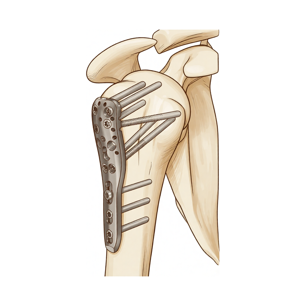
Esta operación se denomina reducción abierta y fijación interna. Consiste en volver a colocar en su posición el hueso fracturado en la parte superior del brazo y mantenerlo así, normalmente mediante una placa metálica y tornillos o, en ocasiones, con una varilla dentro del hueso. Normalmente la recomendamos cuando la fractura está muy desplazada o cuando el hueso se ha roto en varios fragmentos, ya que esas fracturas rara vez permanecen en su sitio por sí solas. Muchas fracturas de hombro sanan sin cirugía, por lo que siempre evaluamos ambas opciones junto con usted. Para algunas personas, especialmente aquellas que gozan de buena salud, la cirugía ofrece mayores posibilidades de mantener el hombro móvil y funcional. El objetivo es sencillo: un hombro estable, menos doloroso y capaz de cumplir con sus necesidades.
Antes de la operación
Una vez planificada la cirugía, organizaremos las pruebas de imagen necesarias para delinear la fractura. Por lo general, esto implica radiografías tomadas desde varios ángulos; en algunos casos, también se realiza una tomografía computarizada que genera una imagen detallada del hueso. Si es preciso examinar con mayor precisión los tejidos blandos alrededor de la articulación, como los tendones, podríamos recurrir a una resonancia magnética o a una ecografía. En la mayoría de los casos no se requieren otras pruebas. No obstante, si padece otras enfermedades, podría ser necesario realizar análisis de sangre o una consulta con el anestesista, el médico encargado de su cuidado durante la intervención. En los días previos a la cirugía, le indicaremos qué medicamentos debe suspender y cuáles debe seguir tomando. Deberá abstenerse de ingerir alimentos y líquidos durante siete horas antes de la operación. Pedimos que sea un periodo de siete horas en lugar de uno más breve para poder adelantar su intervención si el programa quirúrgico lo permite. Coordine con alguien que lo lleve a casa después de la cirugía, ya que no podrá conducir por sí mismo. No olvide llevar una lista de los medicamentos que toma actualmente, y vístase con ropa holgada y cómoda que sea fácil de quitarse.
El día de la intervención
Llegará a la unidad de admisiones quirúrgicas del hospital, donde se le registrará y preparará para la cirugía. Allí conocerá al anestesista. Esta operación se realiza bajo anestesia general combinada con un bloqueo nervioso regional. El anestesista se reunirá con usted antes de la intervención y le explicará ambos procedimientos.
A continuación, será llevado al quirófano, donde se realiza la operación. Después, despertará en la sala de recuperación, donde las enfermeras le vigilarán mientras la anestesia va desapareciendo. Una vez que su estado sea estable, será trasladado a la planta de hospitalización.
Descripción del procedimiento quirúrgico
La operación se denomina reducción abierta y fijación interna. “Abierta” significa que el cirujano realiza una incisión en la zona a operar para acceder al hueso fracturado. “Reducción” consiste en volver a colocar los fragmentos rotos en su posición normal. “Fijación interna” implica mantenerlos en esa posición mediante elementos metálicos.
Lo más habitual es que el cirujano utilice una placa fijada al hueso mediante tornillos; en ocasiones se emplea en su lugar una varilla insertada en el canal medular del hueso del brazo. Ambos métodos mantienen la fractura estable mientras cicatriza. La elección depende de la forma de la fractura y de la calidad ósea. En ocasiones se emplea una placa más larga si la fractura se extiende a lo largo del hueso. Si el hueso es delgado, el cirujano podría añadir soportes adicionales para reforzar la reparación.
Una vez alineados y fijados los fragmentos óseos, se cierran los bordes de la incisión con puntos de sutura. A continuación se coloca un apósito sobre la zona; este debe permanecer en su lugar durante unos 10 días.
Después de la operación
La mayoría de los pacientes permanecen una o dos noches en el hospital tras esta operación. Despertará en la sala de recuperación y luego será trasladado a la planta de hospitalización. Las enfermeras lo revisarán con regularidad y le administrarán medicamentos para mantenerlo cómodo. Su brazo descansará en un cabestrillo sencillo, el cual se retira para realizar ejercicios y para lavarse. Una enfermera le enseñará cómo moverse de forma segura antes de que regrese a casa. Alguien debe acompañarlo durante las primeras 24 horas después de llegar a casa. Dejamos el vendaje puesto durante unos 10 días; por favor, no lo retire antes de ese plazo a menos que se lo indiquemos. Lo cambiamos o lo retiramos cuando venga a la consulta.
Recuperación
Es normal experimentar algo de dolor e hinchazón durante los primeros días y semanas. Esto forma parte del proceso de curación y suele disminuir a medida que el hueso se une. El descanso, las compresas frías y los analgésicos recetados le ayudarán a aliviar la molestia. Comience a mover la mano, la muñeca y el codo desde el principio, tal como le enseñará su fisioterapeuta.
Para mayor comodidad, su brazo se mantiene en un cabestrillo sencillo. Este se retira durante los ejercicios y para lavarse. Su fisioterapeuta le guiará en movimientos suaves al inicio, para luego ir aumentando la intensidad a medida que la fractura cicatriza. En casa, podrá caminar, preparar comidas sencillas y realizar tareas ligeras con la otra mano. Evite levantar objetos pesados con el brazo afectado y no cargue peso sobre él hasta que se le indique lo contrario.
Al principio, dormir puede resultar incómodo. Muchas personas encuentran más cómodo descansar apoyadas en una silla o con almohadas adicionales.
Los hitos de recuperación se definen por acontecimientos, no por fechas concretas. Una vez que la hinchazón disminuya, los movimientos suelen resultar más fáciles. Cuando el cirujano considere que el hueso ha sanado adecuadamente, se retirará el cabestrillo definitivamente. Podrá volver a conducir una vez que el cirujano lo autorice, generalmente en la revisión a las seis semanas; consulte nuestra guía sobre conducción tras una cirugía de extremidad superior. A medida que recupere la fuerza, podrá estirar el brazo por encima de la cabeza y realizar más actividades con él.
La recuperación varía según cada persona. Su cronograma puede ser distinto, y su cirujano y fisioterapeuta le guiarán durante todo el proceso.
Qué puede salir mal
La mayoría de los pacientes evolucionan bien, pero en ocasiones pueden surgir problemas. Su cirujano y el equipo lo vigilarán de cerca para detectar cualquier incidencia a tiempo.
En algunos casos, el hueso no se une como debería. Es posible que note dolor persistente en el lugar de la fractura, o la sensación de que el brazo no gana fuerza con el tiempo. Informe a su médico en la próxima revisión si el dolor no mejora.
La lesión a veces afecta el riego sanguíneo de la articulación del hombro. Si esto ocurre, meses después la articulación puede volverse dolorosa y rígida, y el rango de movimiento podría no mejorar como se esperaba. Mencione este hecho en su revisión para que podamos solicitar estudios de imagen.
El material metálico que fija el hueso puede desplazarse o aflojarse. Es posible percibir un nuevo chasquido, una sensación de roce o dolor que reaparece tras un período de mejoría. Algunas personas sienten la presencia de un tornillo o placa cerca de la piel. Si algo de esto sucede, comuníquese con la clínica; a veces es necesaria una pequeña intervención para retirar o ajustar el material metálico.
Los tendones alrededor del hombro pueden irritarse. Es posible notar dolor al levantar el brazo o dificultad para moverlo en ciertos ángulos. Hágalo saber en su revisión.
El hombro puede volverse rígido y tenso; movimientos sencillos, como llevar la mano detrás de la espalda, pueden resultar difíciles. Los ejercicios tempranos ayudan a prevenir esto; por ello, continúe con la fisioterapia e informe a su fisioterapeuta si observa que el movimiento no mejora.
También pueden aparecer problemas en la herida quirúrgica. Esté atento a enrojecimiento que se extienda desde la zona de la incisión, secreción de líquido o dolor profundo y palpitante que no ceda con analgésicos comunes. Estos signos requieren llamar a la clínica de inmediato. Asimismo, una pequeña acumulación de sangre bajo la herida puede provocar hinchazón; avísenos si esto ocurre.
Durante la cirugía, los nervios cercanos al brazo pueden sufrir contusión. Es posible notar entumecimiento, hormigueo o debilidad en la muñeca o la mano que antes no existían. Comuníquelo sin demora.
Tras la cirugía de hombro, pueden formarse coágulos sanguíneos. Si aparece hinchazón repentina y sensibilidad en la pantorrilla, o dificultad para respirar, debe acudir al servicio de urgencias.
En la tabla de complicaciones de esta página se detallan las tasas habituales; si desea conocer los datos específicos, puede consultarla.
¿Cuándo deben llamarnos?
Llámenos de inmediato si tiene fiebre, o si la piel alrededor de la herida se vuelve más roja, hinchada o comienza a exudar líquido. Acuda a urgencias si experimenta dolor intenso y repentino, hinchazón y sensibilidad en la pantorrilla, o dificultad para respirar; estos pueden ser signos de un coágulo sanguíneo. Llámenos si su mano o brazo se entumecen, se sienten fríos o no puede moverlos. Cualquier nuevo entumecimiento u hormigueo en la muñeca o la mano también requiere una llamada urgente. Si no está seguro, llame a la clínica y le orientaremos.
¿Dónde leer más sobre esta afección?
Esta página trata sobre la operación en sí. La afección que se trata con ella, incluyendo lo que demuestran las evidencias sobre cuándo la cirugía es útil y cuándo no, se explica con mayor detalle en la página de Fractura del húmero proximal.
Evidence & references
This is the clinical evidence summary written for health professionals. It is technical, and it lists the research this page was built from. You do not need to read it to understand your treatment or to make a decision about it.
Overview
- Treatment for proximal humerus fractures remains controversial [4].
- Nonsurgical management of proximal humerus fractures demonstrates successful outcomes and union rates greater than 90% [4].
- Both intramedullary nails and locking plates can effectively restore shoulder function in the treatment of displaced proximal humeral fractures [1].
- The superiority of intramedullary nails over locking plates for restoring shoulder function in displaced proximal humeral fractures is unclear [1].
- Modern proximal humeral nail designs and techniques have demonstrated promising outcomes [2].
- Modern proximal humeral nail designs and techniques can provide stable fixation [2].
- Intramedullary nails are superior to locking plates in reducing total complication rates for proximal humerus fractures [3].
- Intramedullary nails are superior to locking plates in reducing intraoperative blood loss for proximal humerus fractures [3].
- Intramedullary nails are superior to locking plates in reducing operative time for proximal humerus fractures [3].
- Intramedullary nails are superior to locking plates in reducing postoperative fracture healing time for proximal humerus fractures [3].
- Intramedullary nails are superior to locking plates in reducing the postoperative humeral head necrosis rate for proximal humerus fractures [3].
- No superior treatment was suggested between locking plates and intramedullary nails for displaced proximal humeral fractures [5].
- Intramedullary nailing and plating demonstrate equivalent clinical outcomes for the surgical management of displaced proximal humerus fractures in adults [6].
- Patients undergoing ORIF for proximal humerus fracture dislocations have reasonable functional outcomes [7].
- Patients undergoing ORIF for proximal humerus fracture dislocations have relatively high avascular necrosis rates [7].
- Patients undergoing ORIF for proximal humerus fracture dislocations have relatively high reoperation rates [7].
- Intramedullary fixation represents an alternative treatment option for proximal humeral fractures [8].
- Intramedullary fixation for proximal humeral fractures has specific fixation and biologic advantages [8].
- Intramedullary fixation for proximal humeral fractures has reported outcomes comparable with other techniques [8].
- Fixation of proximal humerus fractures with proximal humerus locking plates is associated with a high rate of complications [10].
- Fixation of proximal humerus fractures with proximal humerus locking plates is associated with a high rate of reoperation [10].
- Limited evidence suggests that locking plate and intramedullary nail are both valuable options for the treatment of proximal humeral fractures [11].
- No single fixation method is a panacea for proximal humeral fractures [17].
- The choice of implant and method for proximal humeral fractures should be selected according to individual patient and fracture pattern characteristics based on clearly defined indications and contraindications [17].
- Augmentation of plate fixation for proximal humeral fractures seems to be a reliable and safe procedure [21].
- Augmentation of plate fixation for proximal humeral fractures mechanically increases construct stability [21].
- Augmentation of plate fixation for proximal humeral fractures reduces complication rates [21].
- Augmentation of plate fixation for proximal humeral fractures improves patient outcomes [21].
Anatomy & Pathophysiology
Bony Anatomy
- The proximal humeral anatomy comprises four main parts: the humeral head, greater tuberosity (GT), lesser tuberosity (LT), and humeral shaft [35].
- The articular head is spherical and has a diameter of 37 to 57 mm [35].
- The most superior portion of the articular surface of the humeral head averages 8 mm above the GT [35].
- Humeral version averages 29.8 degrees, with a range of 10 to 55 degrees [35].
- The humeral head is inclined approximately 130 degrees with respect to the humeral shaft [35].
- The bicipital groove lies between the GT and LT and serves as a pathway for the long head of the biceps [35].
- The distal aspect of the bicipital groove is internally rotated with respect to the proximal portion [35].
- The anatomic neck of the proximal humerus is located at the junction of the articular surface and the tuberosities [35].
- The surgical neck represents an indistinct region (metadiaphyseal junction) below the tuberosities but above the humeral shaft [35].
- The GT is located in a posterior-superior location with respect to the humeral shaft and serves as the attachment site for the supraspinatus, infraspinatus, and teres minor tendons [35].
- The LT is located on the anterior aspect of the proximal humerus and serves as the attachment site for the subscapularis tendon [35].
- The glenoid is a convex structure of shallow depth shaped like an inverted pear [35].
- The acromion, coracoacromial ligament, and coracoid process form the coracoacromial arch, a rigid bony-ligamentous structure that imparts stability to the shoulder girdle [35].
- The humeral head averages 19° of retroversion and 41° of inclination (neck-shaft angle) [38].
- The neck-shaft angle measures an average of 135 degrees, and the humeral head is retroverted an average of 30 degrees [36].
- The proximal humerus has three centers of ossification: the humeral head (4 to 6 months), the greater tuberosity (1 to 3 years), and the lesser tuberosity (3 to 5 years) [38].
- The ossification centers of the proximal humerus fuse to the shaft at age 17 to 20 years [38].
- The glenoid cavity is a shallow socket, approximately one third the size of the humeral head [36].
Vascular Supply
- The proximal humerus receives its blood supply from the anterior and posterior humeral circumflex branches from the third division of the axillary artery [35].
- The posterior humeral circumflex artery travels with the axillary nerve, enters the quadrilateral space posteriorly, and anastomoses with a branch of the anterior circumflex to supply the posterior cuff [35].
- The anterior humeral circumflex artery (AHCA) arises from the axillary artery at the inferior border of the subscapularis [35].
- The AHCA provides vascular inflow to the humeral head by way of its terminal anterolateral branch known as the artery of Laing (also known as the arcuate artery) [35].
- The ascending branch of the AHCA courses parallel to the lateral aspect of the long head biceps tendon and enters the humeral head at the interface of the bicipital groove and GT [35].
- Injury to the arcuate artery may result in osteonecrosis of the humeral head [35].
- Additional extraosseous collateral branches can permit humeral head perfusion despite complete ligation of the arcuate artery [35].
- The major blood supply to the humeral head is through the ascending branch of the anterior humeral circumflex artery, which penetrates the head at the bicipital groove and becomes the arcuate artery [36].
- The anterolateral ascending branch of the anterior humeral circumflex artery provides the primary blood supply to the humeral head [38].
- The terminal intraosseous portion of the anterior humeral circumflex artery enters at the proximal aspect of the intertubercular groove as the arcuate artery [38].
Pathophysiology and Displacement
- Following a fracture of the proximal humerus, displacement of each "part" occurs in a predictable manner based on the deforming forces created by the tendinous insertions of the pectoralis major, subscapularis, supraspinatus, and infraspinatus [35].
- The subscapularis inserts on the lesser tuberosity and causes medial displacement [35].
- The supraspinatus and infraspinatus insert on the greater tuberosity and cause superior and posterior displacement [35].
- The pectoralis major inserts on the humeral shaft and displaces it medially [35].
- A fracture involving the anatomic neck is prognostically worse than fractures involving other regions of the proximal humerus with respect to the potential disruption of the vascular supply to the humeral head and the subsequent development of avascular necrosis [35].
- Fractures of the anatomic neck have a poor prognosis because of complete disruption of the blood supply to the head [36].
- Surgical neck fractures are common, and with these, the blood supply to the head is preserved [36].
- PHFs alter complex interactions of joint surface anatomy, joint volume, atmospheric pressure, and joint fluid cohesion and adhesion, resulting in pain, decreased ROM and stiffness, and disability [35].
- Displaced PHFs can impede normal movement of the rotator cuff, subacromial bursa, and subdeltoid bursa passing underneath the coracoacromial arch, causing impingement and disruption of normal glenohumeral motion [35].
- In PHFs (displaced and nondisplaced fractures), the subdeltoid and subacromial bursae can become thickened and fibrotic, forming adhesions that can limit normal glenohumeral motion [35].
- Early ROM exercises after a fracture have been hypothesized to decrease the formation of such adhesions [35].
- The rotator cuff muscles serve as depressors of the humeral head to allow the deltoid to efficiently abduct the humerus [36].
- The infraspinatus and teres minor are external rotators, while the subscapularis is an internal rotator of the humerus [36].
- The deltoid and pectoralis major muscles, along with the rotator cuff, cause predictable displacement of fractures around the proximal humerus [36].
Classification
- Observer agreement for classifying proximal humeral fractures according to the AO-classification is low, with reported mean kappa values for interobserver agreement varying between 0.26 and 0.53 [51].
- Mean kappa values for interobserver agreement decreased from 0.53 for AO Types to 0.2 for AO Groups, suggesting decreased agreement with an increasing number of classification units [51].
- No study has assessed observer agreement on AO-subgroups [51].
- In a systematic review of locking plate fixation, the classification procedure was reported in only five out of twelve studies [51].
- In three studies within a systematic review of locking plate fixation, the classification was performed by one surgeon, and in two studies, it was performed by two or three surgeons [51].
- Classification type and group seem to be of minor importance for clinical outcome in most studies [51].
- Outcome after locking plate osteosynthesis in AO/OTA Type C fractures was comparable with outcome reported in displaced 4-part fractures [51].
- According to the ICD-10 classification system, fractures of the humeral head were the most common fracture type for proximal humerus fractures [54].
- Intramedullary nail fixation was utilized maximally (~ 20%) in fractures of the surgical neck (S42.22) [54].
- Intramedullary nail fixation was least likely used in humeral head fractures (S42.21) [54].
- Reverse shoulder arthroplasty (RSA) showed its highest utilization rate in humeral head fractures and fractures of the anatomical neck (S42.23) [54].
- Fractures of the greater tuberosity (S42.24) were mainly managed by screw fixation (40.4%) [54].
Clinical Presentation
- Adult proximal humeral fractures occur at an estimated annual rate of 6 per 10,000 persons in the United States [13].
- Proximal humeral fractures vary in location and complexity, potentially involving any combination of the surgical and anatomic necks of the humerus, as well as the greater and lesser tuberosities [13].
- The choice of treatment for proximal humeral fractures depends on the fracture type and severity, surgeon expertise, patient age, and patient health status [30].
- Nonsurgical management for proximal humerus fractures demonstrates successful outcomes and union rates greater than 90% [4].
- Most proximal humeral fractures in elderly patients can be treated nonoperatively with good functional outcomes [9].
- Over the past decade, most older adults who sustain proximal humerus fractures continue to receive nonoperative treatment [19].
- Multiple studies comparing nonoperative and operative treatment for displaced proximal humeral fractures in the geriatric population have demonstrated minimal differences in functional outcomes [13].
- Factors such as surgeon experience as well as the quality and maintenance of the reduction may influence operative outcomes for displaced proximal humeral fractures in the geriatric population [13].
- In the treatment of 2 and 3-part fractures involving the surgical neck, intramedullary nailing has demonstrated functional outcomes that are comparable with those of open reduction and internal fixation (ORIF) [13].
- Several authors have demonstrated the negative effect of osteopenia on outcomes after ORIF of proximal humeral fractures [13].
- Optimal management of osteoporotic proximal humeral fractures has evolved to include the use of locking plates and augmentation with intramedullary fibular grafts, calcium phosphate or sulfate cement, and iliac crest bone graft [66].
- Patients undergoing ORIF for proximal humerus fracture dislocations have reasonable functional outcomes but relatively high avascular necrosis and reoperation rates [7].
- Fixation of proximal humerus fractures with proximal humerus locking plates is associated with a high rate of complications and reoperation [10].
- Plate fixation was associated with a higher risk of avascular necrosis development than conservative treatment in patients with proximal humeral fractures [16].
- Considerable variability exists in the use of outcome measures across the proximal humerus fracture literature, making treatment comparison challenging [32].
Investigations
Imaging Protocols and Modalities
- At least two X-ray views should be obtained for shoulder imaging: an anteroposterior view in the plane of the glenoid and an axillary projection with the arm in abduction [43].
- The anteroposterior view in the plane of the scapula shows the superoinferior position of the humeral head relative to the glenoid, presence of osteophytes, joint space narrowing, degree of medial displacement, bone quality, loose bodies, and humeral head collapse or deformity [23].
- The axillary view taken with the arm in the functional position of elevation in the plane of the scapula is referred to as the "truth view" because it demonstrates glenohumeral relationships in the functional position of elevation [23].
- The standardized axillary "truth view" enables the measurement of posterior subluxation or "functional decentering" that is not evident in images taken with the arm at the side [23].
- Computed tomography (CT) is helpful for planning fracture surgery and shoulder joint replacement [43].
- Magnetic resonance imaging (MRI) is useful to identify osteonecrosis of the humeral head, bone tumours, labral tears, and rotator cuff tears [43].
- Ultrasound is a simple and accurate test for identifying rotator cuff tears and calcific tendinitis [43].
- The purpose of imaging the shoulder is to help establish the diagnosis, determine the severity of the pathoanatomy, assist in surgical planning, and enable the surgeon to illustrate the condition of the shoulder to the patient [23].
- Standardized plain films are almost always sufficient to garner the information needed for shoulder evaluation [23].
- Three-dimensional reconstructions can reveal fine details of shoulder anatomy, but this additional information rarely changes the planning or conduct of arthroplasty [23].
- Surgeons need to develop a judicious approach to imaging that yields necessary information while avoiding the tendency to "over-image" [45].
Preoperative Assessment and Fracture Characterization
- All available open reduction and internal fixation (ORIF) techniques require careful analysis of fracture type, fragment displacement, and bone quality, making preoperative CT extremely valuable for three- and four-part proximal humeral fractures [74].
- Imaging-based assessment of fracture stability does not reliably predict outcomes in patients with two-part proximal humeral fractures and may lead to unnecessary surgeries [73].
Outcome Measures
Treatment
Non-Operative Management
- Non-operative treatment is advocated for the majority of non-displaced and minimally displaced isolated tuberosity fractures with generally good outcomes [64].
- Most pediatric patients with proximal humerus fractures have favorable results, and complications are infrequent [14].
Operative Management: General Principles
- Treatment of displaced proximal humerus fractures must be individualized based on patient and fracture characteristics [25].
- No single fixation method is a panacea for proximal humeral fractures; choice of implant and method should be selected according to individual patient and fracture pattern characteristics based on clearly defined indications and contraindications [17].
- Surgical management of proximal humeral fractures in younger patients is challenging due to high expectations and the lack of a single device providing reproducible results [33].
- Technical strategies to maximize the success of surgical treatment for proximal humerus fractures emphasize innovations in technique and implant design to mitigate high complication rates [62].
Operative Management: Intramedullary Nailing
- Modern proximal humeral nail designs and techniques have demonstrated promising outcomes and can provide stable fixation [2].
- Intramedullary fixation represents an alternative treatment option for proximal humeral fractures with specific fixation and biologic advantages, including reported outcomes comparable with other techniques [8].
- Regaining full range of shoulder and elbow movements in combination with absence of complains regarding the shoulder or elbow joints has been striking, indicating that the unreamed technique and avoidance of locking screws could be important factors towards optimum outcomes [12].
- Retrograde elastic stable intramedullary nailing (ESIN) has become the method of choice for surgical treatment of proximal humerus fractures in children and adolescents based on many studies comparing this technique to direct percutaneous pinning [71].
Operative Management: Locking Plate Fixation
- Augmentation of plate fixation for proximal humeral fractures seems to be a reliable and safe procedure that mechanically increases construct stability and reduces complication rates while improving patient outcomes [21].
- Fixation of proximal humeral fractures in elderly patients using locked plates with or without cement augmentation has no significant difference in revision rate, but the implant failure and total complication rates may be lesser on using the cement-augmented locked plate for fixation than on using a locked plate alone [31].
- Augmentative procedures, including cortical strut augmentation, are being investigated to address the issue of osteopenia in proximal humeral fracture treatment; their role in the treatment of these fractures is unclear at this time [13].
Comparative Effectiveness: Nails vs. Plates
- The available evidence suggests that both intramedullary nails and locking plates can effectively restore shoulder function in the treatment of displaced proximal humeral fractures, with unclear superiority of either method [1].
- The intramedullary nail is superior to locking plate in reducing the total complication, intraoperative blood loss, operative time, postoperative fracture healing time and postoperative humeral head necrosis rate of PHF [3].
Operative Management: Fracture Dislocations and Arthroplasty
- In the geriatric population, reverse total shoulder arthroplasty has demonstrated improved functional outcomes, with a decreased rate of reoperation, compared with hemiarthroplasty [13].
- Tuberosity repair has been shown to improve functional outcomes and range of motion after both reverse total shoulder arthroplasty and hemiarthroplasty and should be performed at the time of arthroplasty [13].
- Comparative studies support the use of reverse shoulder arthroplasty in elderly patients with complex proximal humerus fractures because the functional outcomes and relief of pain are reliably improved [72].
Anesthesia
- Regional anaesthesia is a good option for postoperative analgesia in patients undergoing surgical repair of a proximal humerus fracture and is associated with fewer adverse events, a shorter recovery time, and a better functional outcome than those achieved by general anaesthesia alone [67].
Complications
General Complication Rates and Outcomes
- In a systematic review of late screw-related complications in locking plating, 33% of reported cases had at least one complication, with 11% of all complications being screw-related [20].
- Patients undergoing ORIF for proximal humerus fracture dislocations have relatively high avascular necrosis and reoperation rates [7].
- Open fractures and 4-part proximal humerus fractures had the highest complication rates following intramedullary nailing [79].
- A meta-analysis of randomized controlled trials did not support the treatment of open reduction and internal fixation to improve the functional outcome when compared with nonoperative treatment for treating elderly patients with displaced 3-part or 4-part proximal humeral fractures [75].
- In most studies of proximal humeral fractures, only 1 or 2 patients experiencing an alternative outcome or lost to follow-up would change the conclusions for the dichotomous outcome studied [29].
Avascular Necrosis
- The intramedullary nail is superior to locking plate in reducing the postoperative humeral head necrosis rate of proximal humerus fractures [3].
Screw and Implant-Related Complications
- Most late screw-related complications in locking plating were secondary screw perforations and screw cut-outs, being predominantly linked to poor bone quality [20].
- Screw loosening and retraction were found less frequently as a result of locking mechanism failure in locking plating of proximal humerus fractures [20].
- Screw perforation was the most frequent screw-related complication in locking plating, mostly reported in female patients older than 50 years, following four-part or AO/OTA type C fractures, and detected four weeks postoperatively [20].
- Locked humeral stems provide reliable diaphyseal fixation with a low incidence of screw-related complications in reverse total shoulder arthroplasty for complex proximal humerus fractures [78].
Augmentation and Allograft Outcomes
- Augmentation of plate fixation for proximal humeral fractures mechanically increases construct stability and reduces complication rates while improving patient outcomes [21].
- Patients with proximal humerus fractures treated with a locking compression plate augmented with a fibular allograft have decreased odds of a major complication when compared with patients treated with a locking compression plate alone [81].
Venous Thromboembolism
- Venous thromboembolism was the most frequently reported complication after shoulder arthroplasty when compared to ORIF, with reverse shoulder arthroplasty having the highest venous thromboembolism rate [70].
Salvage and Revision Surgery
- The failed fixation group performed significantly better than the failed hemiarthroplasty group in postoperative constant and shoulder abduction after salvage reverse shoulder arthroplasty [27].
- Revision surgery for failed arthroplasty of proximal humerus fracture is complex with a high likelihood of inferior outcomes compared with primary arthroplasty [77].
Non-Operative Management
- Complications following non-surgical management of proximal humeral fractures are described using heterogeneous terminology and definitions, calling for standardized definitions to improve evidence synthesis [34].
Recovery
- A systematic review of rehabilitation protocols in proximal humerus fracture management included 3507 patients and 3519 proximal humerus fractures [24].
- In the systematic review of rehabilitation protocols, 65.9% of the patients were female [24].
- The weighted mean age of patients in the rehabilitation protocol systematic review was 63.5 years [24].
- The follow-up duration in the rehabilitation protocol systematic review was 22.4 months [24].
- Of the 45 treatment cohorts included in the rehabilitation protocol systematic review, 33 were treated with ORIF with plate fixation and 5 were treated with ORIF with intramedullary nail [24].
- Of the included proximal humerus fractures in the rehabilitation protocol systematic review, 2220 were treated with ORIF with plating and 208 were treated with a nail [24].
- Ten studies included in the rehabilitation protocol systematic review included fracture dislocations in their cohorts [24].
- The levels of evidence in the rehabilitation protocol systematic review were Level I (15%), Level II (8%), Level III (25%), and Level IV (53%) [24].
- Patients 65 years of age with 3- or 4-part proximal humerus fractures achieve the most benefit in terms of range of motion, postoperative functional outcomes, tuberosity union, and overall complication rate when undergoing reverse total shoulder arthroplasty with a noncemented stem and early postoperative range of motion compared to reverse total shoulder arthroplasty with a cemented stem and delayed rehabilitation [69].
Key Evidence
- [L5] The available evidence suggests that both intramedullary nails and locking plates can effectively restore shoulder function in the treatment of displaced proximal humeral fractures, with unclear superiority of either method. [1] (10.1016/j.xrrt.2024.01.001)
- [L5] Modern proximal humeral nail designs and techniques have demonstrated promising outcomes and can provide stable fixation. [2] (10.1016/j.jse.2015.11.016)
- [L1] The intramedullary nail is superior to locking plate in reducing the total complication, intraoperative blood loss, operative time, postoperative fracture healing time and postoperative humeral head necrosis rate of PHF. [3] (10.1186/s13018-019-1345-0)
- [L5] Treatment for proximal humerus fractures remains controversial, with nonsurgical management demonstrating successful outcomes and union rates greater than 90%. [4] (10.5435/jaaos-d-24-01073)
- [L1] No superior treatment was suggested between locking plates and intramedullary nails for displaced proximal humeral fractures. [5] (10.1007/s00264-017-3683-z)
- [L1] Intramedullary nailing and plating demonstrate equivalent clinical outcomes for the surgical management of displaced proximal humerus fractures in adults. [6] (10.1016/j.jse.2026.02.016)
- [L4] Patients undergoing ORIF for proximal humerus fracture dislocations have reasonable functional outcomes but relatively high avascular necrosis and reoperation rates. [7] (10.1016/j.jse.2022.04.018)
- [L4] Intramedullary fixation represents an alternative treatment option for proximal humeral fractures with specific fixation and biologic advantages, including reported outcomes comparable with other techniques. [8] (10.5435/jaaos-d-18-00360)
- [L5] Most proximal humeral fractures in elderly patients can be treated nonoperatively with good functional outcomes. [9] (10.2106/jbjs.l.01293)
- [L4] Fixation of proximal humerus fractures with proximal humerus locking plates is associated with a high rate of complications and reoperation. [10] (10.1016/j.injury.2010.11.058)
- [L1] Limited evidence suggests that locking plate and intramedullary nail are both valuable options for the treatment of proximal humeral fractures. [11] (10.1186/s13018-015-0242-4)
- [L4] Regaining full range of shoulder and elbow movements in combination with absence of complains regarding the shoulder or elbow joints has been striking, indicating that the unreamed technique and avoidance of locking screws could be important factors towards optimum outcomes. [12] (10.1016/s0020-1383(13)70037-8)
- [L5] [13] (10.2106/jbjs.20.00665)
- [L5] Most pediatric patients with proximal humerus fractures have favorable results, and complications are infrequent. [14] (10.5435/jaaos-d-14-00033)
- [L1] Plate fixation was associated with a higher risk of AVN development than conservative treatment in patients with proximal humeral fractures. [16] (10.1186/1749-799x-9-31)
- [L4] No single fixation method is a panacea for proximal humeral fractures; choice of implant and method should be selected according to individual patient and fracture pattern characteristics based on clearly defined indications and contraindications. [17] (10.1016/j.injury.2010.10.016)
- [L4] Over the past decade, most older adults who sustain proximal humerus fractures continue to receive nonoperative treatment. [19] (10.1016/j.jseint.2021.08.006)
- [L2] [20] (10.1016/j.injury.2019.11.002)
- [L1] Augmentation of plate fixation for proximal humeral fractures seems to be a reliable and safe procedure that mechanically increases construct stability and reduces complication rates while improving patient outcomes. [21] (10.1007/s00402-019-03162-2)
- [L4] [24] (10.1177/17585732231182374)
- [L5] Treatment of displaced proximal humerus fractures must be individualized based on patient and fracture characteristics, with a general evolution toward humeral head preservation using options ranging from nonsurgical immobilization to various surgical techniques including locking plate fixation and hemiarthroplasty. [25] (10.5435/00124635-200701000-00003)
- [L2] The failed fixation group performed significantly better than the failed HA group in postoperative constant and shoulder abduction. [27] (10.1177/17585732221099200)
- [L2] In most studies of proximal humeral fractures, only 1 or 2 patients experiencing an alternative outcome or lost to follow-up would change the conclusions for the dichotomous outcome studied. [29] (10.1016/j.jse.2022.01.141)
- [L4] The choice of treatment for proximal humeral fractures depends on the fracture type and severity, surgeon expertise, patient age, and patient health status. [30] (10.5435/jaaos-d-15-00240)
- [L1] Fixation of proximal humeral fractures in elderly patients using locked plates with or without cement augmentation has no significant difference in revision rate, but the implant failure and total complication rates may be lesser on using the cement-augmented locked plate for fixation than on using a locked plate alone. [31] (10.1186/s12891-024-07502-1)
- [L4] Considerable variability exists in the use of outcome measures across the proximal humerus fracture literature, making treatment comparison challenging. [32] (10.1016/j.jse.2020.04.006)
- [L5] The paper concludes that surgical management of proximal humeral fractures in younger patients is challenging due to high expectations and the lack of a single device providing reproducible results. [33] (10.1016/j.jse.2010.12.006)
- [L1] This systematic review highlights significant heterogeneity in the terminology and definitions used to describe complications following non-surgical management of proximal humeral fractures, calling for standardized definitions to improve evidence synthesis. [34] (10.1186/s12891-019-2459-6)
- [L2] [51] (10.1016/j.injury.2011.08.025)
- [L4] [54] (10.1007/s00402-019-03252-1)
- [L5] This review highlights various technical strategies to maximize the success of surgical treatment for proximal humerus fractures, emphasizing innovations in technique and implant design to mitigate high complication rates. [62] (10.5435/jaaos-d-22-01211)
- [L4] Non-operative treatment is advocated for the majority of non-displaced and minimally displaced fractures with generally good outcomes, while displaced fractures may require arthroscopically assisted fixation or open/percutaneous reduction and internal fixation depending on fracture type and patient factors. [64] (10.1016/j.injury.2007.09.022)
- [L4] Optimal management of osteoporotic proximal humeral fractures has evolved to include the use of locking plates and augmentation with intramedullary fibular grafts, calcium phosphate or sulfate cement, and iliac crest bone graft. [66] (10.1016/j.jse.2012.04.003)
- [L1] This systematic review suggests that RA is a good option for postoperative analgesia in patients undergoing surgical repair of a proximal humerus fracture and is associated with fewer adverse events, a shorter recovery time, and a better functional outcome than those achieved by general anaesthesia alone. [67] (10.1007/s00402-019-03253-0)
- [L1] Patients 65 years of age with 3- or 4-part proximal humerus fractures achieve the most benefit in terms of ROM, postoperative functional outcomes, tuberosity union, and overall complication rate when undergoing rTSA with a noncemented stem and early postoperative ROM compared to rTSA with cemented stem and delayed rehabilitation. [69] (10.1016/j.jse.2024.03.040)
- [L4] Among the various procedures, VTE was the most frequently reported after SA when compared to ORIF, with RSA having the highest VTE rate. [70] (10.1016/j.xrrt.2023.06.003)
- [L5] [71] (10.1016/j.otsr.2013.06.010)
- [L4] Comparative studies support the use of reverse shoulder arthroplasty in elderly patients with complex proximal humerus fractures because the functional outcomes and relief of pain are reliably improved. [72] (10.5435/jaaos-d-13-00190)
- [L5] Imaging-based assessment of fracture stability does not reliably predict outcomes in patients with two-part proximal humeral fractures and may lead to unnecessary surgeries. [73] (10.1530/eor-2026-0043)
- [L4] All available ORIF techniques require careful analysis of fracture type, fragment displacement, and bone quality, making preoperative CT extremely valuable. [74] (10.1016/j.otsr.2012.12.006)
- [L1] The meta-analysis did not support the treatment of open reduction and internal fixation to improve the functional outcome when compared with nonoperative treatment for treating elderly patients with displaced 3-part or 4-part proximal humeral fractures. [75] (10.1371/journal.pone.0075464)
- [L5] Revision surgery for failed arthroplasty of proximal humerus fracture is complex with a high likelihood of inferior outcomes compared with primary arthroplasty. [77] (10.5435/jaaos-d-17-00051)
- [L4] Locked humeral stems provide reliable diaphyseal fixation with a low incidence of screw-related complications in reverse total shoulder arthroplasty for complex proximal humerus fractures. [78] (10.1016/j.xrrt.2025.100625)
- [L4] Open fractures and 4-part proximal humerus fractures had the highest complication rates. [79] (10.1016/j.jse.2024.07.049)
- [L1] The pooled WMD and prediction interval suggest that 95% of patients with proximal humerus fractures treated with an LCP augmented with a fibular allograft will have improved radiographic outcomes, improved ASES clinical outcome scores, and decreased odds of a major complication when compared with patients treated with an LCP alone. [81] (10.1016/j.jse.2021.11.004)
References
[1] Intramedullary versus locking plate fixation for proximal humerus fractures: indications and technical considerations. JSES Reviews, Reports, and Techniques. 2024. DOI: 10.1016/j.xrrt.2024.01.001
[2] Intramedullary nailing of the proximal humerus: evolution, technique, and results. Journal of Shoulder and Elbow Surgery. 2016. DOI: 10.1016/j.jse.2015.11.016
[3] Effect of intramedullary nail and locking plate in the treatment of proximal humerus fracture: an update systematic review and meta-analysis. Journal of Orthopaedic Surgery and Research. 2019. DOI: 10.1186/s13018-019-1345-0
[4] Contemporary Management of Proximal Humeral Fractures. Journal of the American Academy of Orthopaedic Surgeons. 2025. DOI: 10.5435/jaaos-d-24-01073
[5] Locking plates versus intramedullary nails in the management of displaced proximal humeral fractures: a systematic review and meta-analysis. International Orthopaedics. 2017. DOI: 10.1007/s00264-017-3683-z
[6] Effectiveness of intramedullary nailing vs. locked plating (open reduction and internal fixation) in adult displaced proximal humerus fractures: a systematic review and meta-analysis. Journal of Shoulder and Elbow Surgery. 2026. DOI: 10.1016/j.jse.2026.02.016
[7] Fracture dislocations of the proximal humerus treated with open reduction and internal fixation: a systematic review. Journal of Shoulder and Elbow Surgery. 2022. DOI: 10.1016/j.jse.2022.04.018
[8] Intramedullary Fixation for Proximal Humeral Fractures. Journal of the American Academy of Orthopaedic Surgeons. 2020. DOI: 10.5435/jaaos-d-18-00360
[9] Proximal Humeral Fracture Treatment in Adults. Journal of Bone and Joint Surgery. 2014. DOI: 10.2106/jbjs.l.01293
[10] A systematic review of locking plate fixation of proximal humerus fractures. Injury. 2011. DOI: 10.1016/j.injury.2010.11.058
[11] Meta-analysis of locking plate versus intramedullary nail for treatment of proximal humeral fractures. Journal of Orthopaedic Surgery and Research. 2015. DOI: 10.1186/s13018-015-0242-4
[12] T1.11 The management of 100 humeral shaft fractures with “Garnavos” humeral nail. Injury. 2013. DOI: 10.1016/s0020-1383(13)70037-8
[13] Current Controversies in the Treatment of Geriatric Proximal Humeral Fractures. Journal of Bone and Joint Surgery. 2021. DOI: 10.2106/jbjs.20.00665
[14] Evaluation and Management of Pediatric Proximal Humerus Fractures. Journal of the American Academy of Orthopaedic Surgeons. 2015. DOI: 10.5435/jaaos-d-14-00033
[16] Avascular necrosis in proximal humeral fractures in patients treated with operative fixation: a meta-analysis. Journal of Orthopaedic Surgery and Research. 2014. DOI: 10.1186/1749-799x-9-31
[17] New trends in fixation of proximal humeral fractures: A review. Injury. 2011. DOI: 10.1016/j.injury.2010.10.016
[19] Trending a decade of proximal humerus fracture management in older adults. JSES International. 2022. DOI: 10.1016/j.jseint.2021.08.006
[20] Late screw-related complications in locking plating of proximal humerus fractures: A systematic review. Injury. 2019. DOI: 10.1016/j.injury.2019.11.002
[21] Augmentation of plate osteosynthesis for proximal humeral fractures: a systematic review of current biomechanical and clinical studies. Archives of Orthopaedic and Trauma Surgery. 2019. DOI: 10.1007/s00402-019-03162-2
[23] Rockwood And Matsen S The Shoulder. Arthroscopic Management of Prearthritic and Arthritic Conditions of the Shoulder and the Postarthroplasty Shoulder > Radiographic Evaluation.
[24] Rehabilitation protocols in proximal humerus fracture management: A systematic review. Shoulder & Elbow. 2023. DOI: 10.1177/17585732231182374
[25] Innovations in the Management of Displaced Proximal Humerus Fractures. Journal of the American Academy of Orthopaedic Surgeons. 2007. DOI: 10.5435/00124635-200701000-00003
[27] Outcomes after salvage reverse shoulder arthroplasty for failed primary fixation or hemiarthroplasty for proximal humerus fractures: A systematic review. Shoulder & Elbow. 2022. DOI: 10.1177/17585732221099200
[29] Fragility of randomized controlled trials on treatment of proximal humeral fracture. Journal of Shoulder and Elbow Surgery. 2022. DOI: 10.1016/j.jse.2022.01.141
[30] Management of Acute Proximal Humeral Fractures. Journal of the American Academy of Orthopaedic Surgeons. 2017. DOI: 10.5435/jaaos-d-15-00240
[31] Cement-augmented locked plate fixation proximal humerus fractures in elderly patient: a systematic review and meta-analysis. BMC Musculoskeletal Disorders. 2024. DOI: 10.1186/s12891-024-07502-1
[32] Outcome measures reported for the management of proximal humeral fractures: a systematic review. Journal of Shoulder and Elbow Surgery. 2020. DOI: 10.1016/j.jse.2020.04.006
[33] Proximal humeral fractures in younger patients: fixation techniques and arthroplasty. Journal of Shoulder and Elbow Surgery. 2011. DOI: 10.1016/j.jse.2010.12.006
[34] Complications after non-surgical management of proximal humeral fractures: a systematic review of terms and definitions. BMC Musculoskeletal Disorders. 2019. DOI: 10.1186/s12891-019-2459-6
[35] Rockwood And Matsen S The Shoulder. Shoulder and Elbow Specialty Clinic Workers’ Survey > ANATOMY.
[36] A Lange Medical Book Current Diagnosis Treatment In Orthopedics Fifth Edition. 2Musculoskeletal Trauma Surgery > SHOULDER AND ARM INJURIES.
[38] Aaos Comprehensive Orthopaedic Review 3. Anatomy of the Shoulder, Arm, and Elbow > I. Shoulder.
[43] Apley And Solomon S Concise System Of Orthopaedics And Trauma. INVESTIGATION.
[45] Rockwood And Matsen S The Shoulder. Developmental Anatomy of the Shoulder and Anatomy of the Glenohumeral Joint > SENIOR EDITOR COMMENTARY.
[51] Benefits and harms of locking plate osteosynthesis in intraarticular (OTA Type C) fractures of the proximal humerus: A systematic review. Injury. 2012. DOI: 10.1016/j.injury.2011.08.025
[54] Trends in surgical management of proximal humeral fractures in adults: a nationwide study of records in Germany from 2007 to 2016. Archives of Orthopaedic and Trauma Surgery. 2019. DOI: 10.1007/s00402-019-03252-1
[62] Technical Tips for Reduction and Stable Fixation of Proximal Humerus Fractures. Journal of the American Academy of Orthopaedic Surgeons. 2023. DOI: 10.5435/jaaos-d-22-01211
[64] Isolated tuberosity fractures of the proximal humerus: Current concepts. Injury. 2008. DOI: 10.1016/j.injury.2007.09.022
[66] Evaluation of the osteoporotic proximal humeral fracture and strategies for structural augmentation during surgical treatment. Journal of Shoulder and Elbow Surgery. 2012. DOI: 10.1016/j.jse.2012.04.003
[67] Regional anaesthesia for surgical repair of proximal humerus fractures: a systematic review and critical appraisal. Archives of Orthopaedic and Trauma Surgery. 2019. DOI: 10.1007/s00402-019-03253-0
[69] Optimal combination of arthroplasty type, fixation method, and postoperative rehabilitation protocol for complex proximal humerus fractures in the elderly: a network meta-analysis. Journal of Shoulder and Elbow Surgery. 2024. DOI: 10.1016/j.jse.2024.03.040
[70] Venous thromboembolism following surgical management of proximal humerus fractures: a systematic review. JSES Reviews, Reports, and Techniques. 2023. DOI: 10.1016/j.xrrt.2023.06.003
[71] Proximal humerus fractures in children and adolescents. Orthopaedics & Traumatology: Surgery & Research. 2014. DOI: 10.1016/j.otsr.2013.06.010
[72] Reverse Shoulder Arthroplasty for the Management of Proximal Humerus Fractures. Journal of the American Academy of Orthopaedic Surgeons. 2015. DOI: 10.5435/jaaos-d-13-00190
[73] Imaging-based assessment of fracture stability does not reliably predict outcomes in patients with two-part proximal humeral fractures and may lead to unnecessary surgeries. EFORT Open Reviews. 2026. DOI: 10.1530/eor-2026-0043
[74] Surgical treatment of three and four-part proximal humeral fractures. Orthopaedics & Traumatology: Surgery & Research. 2013. DOI: 10.1016/j.otsr.2012.12.006
[75] Internal Fixation Versus Nonoperative Treatment for Displaced 3-Part or 4-Part Proximal Humeral Fractures in Elderly Patients: A Meta-Analysis of Randomized Controlled Trials. PLoS ONE. 2013. DOI: 10.1371/journal.pone.0075464
[77] Management of the Failed Arthroplasty for Proximal Humerus Fracture. Journal of the American Academy of Orthopaedic Surgeons. 2019. DOI: 10.5435/jaaos-d-17-00051
[78] Use of locking humeral stems in reverse total shoulder arthroplasty for complex proximal humerus fractures: a scoping review. JSES Reviews, Reports, and Techniques. 2026. DOI: 10.1016/j.xrrt.2025.100625
[79] Complications following intramedullary nailing of proximal humerus and humeral shaft fractures: a systematic review. Journal of Shoulder and Elbow Surgery. 2025. DOI: 10.1016/j.jse.2024.07.049
[81] Improved outcomes for proximal humerus fracture open reduction internal fixation augmented with a fibular allograft in elderly patients: a systematic review and meta-analysis. Journal of Shoulder and Elbow Surgery. 2022. DOI: 10.1016/j.jse.2021.11.004