Patients › Shoulder
Artritis del hombro
Shoulder arthritis causes pain, stiffness, and reduced range of motion — diagnosis and treatment options explored.
Qué está sintiendo
La artritis del hombro es una artritis por desgaste en la principal articulación de esfera y cavidad del hombro. El dolor suele ir aumentando a lo largo de años, en lugar de aparecer de repente. Se localiza en lo profundo del propio hombro y suele acompañarse de una rigidez que poco a poco va reduciendo el rango de movimiento de la articulación.
Algunos movimientos y posturas lo empeoran. Levantar el brazo por encima de la cabeza, separarlo hacia un lado o empujar con el hombro pueden agravarlo. Después de la actividad, el dolor sordo suele instalarse. La noche es un momento frecuente de empeoramiento, y muchas personas notan que el hombro las despierta o les dificulta encontrar una postura cómoda para dormir. Al despertar, la articulación puede sentirse rígida y tardar en soltarse.
Las tareas cotidianas que requieren alcanzar algo se vuelven las más difíciles. Lavarse el cabello, apagar una luz que está detrás de usted o subir y rodear con el brazo para vestirse pueden convertirse en tareas realmente pesadas. A menudo aparece debilidad junto con la rigidez, de modo que levantar y cargar objetos se siente menos seguro que antes.
La rigidez de la artritis es diferente de la del hombro congelado, que se desarrolla en semanas o meses, no en años. Otras afecciones también pueden imitar a la artritis, como la inflamación de la artritis reumatoide, que a menudo provoca cansancio, dolor en otras articulaciones y pérdida de peso, además del dolor y la hinchazón del hombro. Su cirujano distingue estas afecciones mediante su historia clínica y la exploración, a veces con estudios de imagen o análisis de sangre.
Algunos signos requieren atención más rápida. Si el hombro se vuelve caliente, rojo, hinchado y muy doloroso, sobre todo si tiene fiebre, debe acudir al servicio de urgencias ese mismo día, y no necesita una derivación de su médico de cabecera. Si los síntomas del hombro no mejoran, empeoran a lo largo de semanas, le despiertan por la noche o le impiden usar el brazo o trabajar, consulte a su médico de cabecera o solicite una valoración por un especialista. Si no logra contactar con la clínica fuera del horario de atención o un fin de semana, acuda al servicio de urgencias más cercano.
¿Qué está ocurriendo realmente?
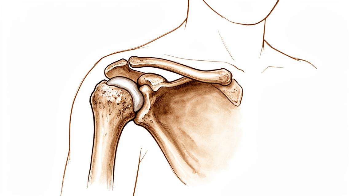
El hombro es una articulación de esfera y cavidad. La esfera es la parte superior redondeada del hueso del brazo, y la cavidad es un plato poco profundo en la escápula. La cavidad es pequeña por naturaleza: tiene alrededor de un tercio del tamaño de la esfera que sostiene. Un anillo de cartílago blando alrededor de su borde, llamado labrum, aumenta la profundidad de la cavidad para que la esfera se mantenga en su sitio. Ambas superficies están recubiertas por una capa de cartílago lisa y resbaladiza que permite que los dos huesos se deslicen uno sobre otro casi sin fricción.
En la artritis, ese cartílago liso se desgasta. Imagínelo como el amortiguador entre los dos huesos, que se va adelgazando hasta que los huesos empiezan a rozar entre sí. El cuerpo responde formando hueso adicional en los bordes de la articulación, y estos espolones óseos aumentan la rigidez. El revestimiento de la articulación también puede inflamarse e hincharse. El roce de hueso contra hueso, los espolones y la inflamación, en conjunto, explican el dolor sordo y profundo, la reducción del rango de movimiento y el dolor nocturno descritos más arriba.
El desgaste no siempre es uniforme. En muchos hombros con artritis, la esfera se desplaza hacia la parte posterior de la cavidad, de modo que el borde posterior de la cavidad se desgasta más rápido que el resto. Los tejidos de la parte delantera de la articulación también se tensan con el tiempo, lo que gira la esfera aún más hacia atrás y carga todavía más la zona desgastada. Los tendones que rodean la articulación, conocidos como manguito rotador, suelen seguir intactos en este tipo de artritis, aunque pueden verse afectados en otras formas.
La artritis tiene varios puntos de partida. La mayoría de los casos no tienen una única causa clara, aunque la tendencia familiar puede influir. Otros aparecen tras una fractura antigua, una cirugía previa en la articulación, luxaciones repetidas o enfermedades inflamatorias como la artritis reumatoide, en la que el propio sistema de defensa del cuerpo ataca el revestimiento de la articulación y destruye el cartílago. Sea cual sea el punto de partida, el resultado final en la articulación es muy similar, y es lo que provoca sus síntomas.
Qué podemos hacer al respecto
El Dr. Kieran Hirpara, cirujano de extremidad superior en el Mater Private Hospital Rockhampton, comienza con las opciones menos invasivas que se adapten a su condición. Por lo general, los pacientes son derivados a nuestra clínica por su médico de cabecera; si un fisioterapeuta le ha sugerido que nos consulte, igualmente necesitará una derivación de su médico de cabecera para poder acceder al reembolso de Medicare. En su primera visita, tomamos su historia clínica, examinamos su hombro y solicitamos estudios de imagen cuando son necesarios. Las radiografías casi siempre son suficientes para planificar el tratamiento. Una tomografía computarizada (TC) o una resonancia magnética aportan más detalle en algunos casos, por ejemplo para revisar el manguito rotador o medir el desgaste óseo antes de la cirugía.
En la artritis leve a moderada, normalmente probamos primero el tratamiento no quirúrgico. Modificar la forma en que utiliza el hombro ayuda, así que analizamos qué movimientos y tareas cargan la articulación y buscamos maneras de evitarlos. La fisioterapia tiene como objetivo mantener la articulación en movimiento y fortalecer la musculatura que la rodea. Los medicamentos antiinflamatorios pueden aliviar el dolor sordo, y una inyección de cortisona en la articulación puede calmar la inflamación durante un periodo de semanas a meses. Damos a cualquiera de estas opciones una prueba adecuada antes de hablar de cirugía, porque la cirugía funciona mejor cuando se ha hecho antes un intento real de tratamiento conservador.
La cirugía se plantea cuando el tratamiento no quirúrgico no ha aportado suficiente mejoría y el dolor o la rigidez siguen limitando lo que usted puede hacer. La operación principal es el reemplazo de hombro, en el que las superficies desgastadas de la esfera y la cavidad se sustituyen por piezas artificiales. El tipo que comentemos depende de su edad, del estado de su manguito rotador y del hueso de la cavidad, y tomaremos esa decisión junto con usted como una decisión compartida.
Qué esperar
La artritis no retrocede. El cartílago desgastado no vuelve a crecer, de modo que el desgaste de fondo permanece. Lo que sí cambia es cuánto le molesta el hombro en el día a día, y eso puede inclinarse a su favor.
Sin tratamiento, el patrón suele ser un deterioro lento a lo largo de los años, más que un empeoramiento repentino. El dolor sordo y la rigidez tienden a aumentar de forma gradual, y las cosas que notó al principio, como alcanzar objetos por encima de la cabeza o dormir sobre ese lado, suelen volverse más difíciles en lugar de más fáciles. Algunas personas se mantienen estables durante largos periodos, pero la tendencia general de un hombro con artritis sin tratar es empeorar.
Con un buen manejo, la mayoría de las personas evolucionan mejor que eso. Mantener la articulación en movimiento y fortalecer la musculatura que la rodea, con la guía de su fisioterapeuta, puede calmar los síntomas y permitirle seguir haciendo más cosas durante más tiempo. Muchas personas manejan su artritis durante años sin llegar a necesitar cirugía.
Si la cirugía llega a ser la respuesta adecuada, el reemplazo de hombro tiene una larga trayectoria para este mismo problema. Es el tratamiento con los resultados más predecibles para aliviar el dolor y mejorar la función en la artritis avanzada del hombro. Las personas suelen notar que el hombro funciona mejor y que su salud en general mejora en un plazo relativamente corto después de la operación. La fuerza también mejora durante los dos primeros años, aunque el tendón de la parte delantera del hombro no siempre recupera por completo su fuerza normal.
Ningún tratamiento es una garantía. La mayoría de las personas obtienen un alivio real, pero algunas se quedan con rigidez o debilidad, y una minoría necesita otra operación más adelante. Los implantes son piezas artificiales y pueden aflojarse o desgastarse con el tiempo, algo que pesa más cuanto más joven y activo sea usted. Su cirujano le explicará cómo es un resultado realista en el caso concreto de su hombro.
Algo que conviene saber: si su artritis todavía es leve en la radiografía, la cirugía suele aportar menos mejoría que en la artritis grave. Eso no es un motivo para evitar la cirugía, pero sí para tener claro qué espera conseguir con ella.
Si los síntomas del hombro no mejoran, empeoran a lo largo de semanas, le despiertan por la noche o le impiden usar el brazo o trabajar, consulte a su médico de cabecera o solicite una valoración por un especialista.
¿Cuándo consultar a un especialista?
La mayoría de los hombros con artritis pueden esperar a una cita programada, pero algunos signos no. Si el hombro se vuelve caliente, rojo, hinchado y muy doloroso, sobre todo si tiene fiebre, acuda a un servicio de urgencias ese mismo día. Una infección en la articulación es urgente, y no necesita una derivación de su médico de cabecera. Lo mismo se aplica si no logra contactar con la clínica fuera del horario de atención o un fin de semana: acuda al servicio de urgencias más cercano.
Acuda a su médico de cabecera si el hombro no mejora, empeora a lo largo de semanas, le despierta por la noche o le impide trabajar o usar el brazo. Solicite una valoración por un especialista si el dolor y la rigidez han ido aumentando durante años y ahora le limitan para lavarse el cabello, vestirse o llevar la mano hacia atrás.
Algunos patrones merecen una evaluación más temprana. Un desgaste rápido y grave puede afectar a mujeres mayores con un dolor de hombro que fue apareciendo de forma silenciosa. Una erosión grave en alguien que ha tenido gota indica gota en el propio hombro. Y si el hombro está caliente y doloroso con fiebre, es el signo de atención el mismo día descrito más arriba.
Su cirujano también querrá saber si tiene dolor de cuello, o entumecimiento y hormigueo que bajan por debajo del codo, porque pueden tener su origen en el cuello y no en el hombro.
En profundidad
Advanced reading: the deeper science (optional)
Esta sección va más allá de lo necesario para que usted tome sus propias decisiones de tratamiento. La artritis del hombro merece una lectura adicional, ya que la decisión principal —qué tipo de prótesis utilizar— se ha convertido recientemente en una verdadera elección entre opciones, sin una respuesta clara; además, dicha elección varía según la edad del paciente.
Reemplazo anatómico versus inverso, en pacientes mayores de 70 años
El reemplazo anatómico reproduce la disposición anatómica normal y depende de que el manguito rotador funcione correctamente. En cambio, el reemplazo inverso invierte la posición de la esfera y la cavidad, permitiendo que el deltoides realice el levantamiento; no requiere, por tanto, un manguito rotador funcional. Históricamente, el reemplazo inverso se reservaba para pacientes sin manguito rotador; hoy en día, su uso es mucho más extendido.
La comparación realizada en 1,716 pacientes mayores de 70 años con un manguito rotador íntegro arrojó resultados claros. El reemplazo anatómico proporcionó una movilidad rotacional superior, superando la diferencia mínima clínicamente relevante, lo cual podría facilitar actividades cotidianas como vestirse o asearse; en tanto, el reemplazo inverso ofreció una mayor supervivencia del implante y tasas de revisión más bajas [1].
Se trata de dos beneficios distintos: el reemplazo anatómico otorga una movilidad rotacional perceptible a diario, mientras que el inverso ofrece mayor durabilidad del implante. Ninguno de los dos resulta claramente superior, motivo por el cual hoy en día esta elección se debate en lugar de ser automática.
El implante inverso presenta más complicaciones tempranas en este contexto
La ventaja en cuanto a durabilidad conlleva un costo inmediato cuando se utiliza el implante inverso para tratar la artritis común.
En un total de 8,846 pacientes, se observó una tasa más alta de complicaciones tempranas con el implante inverso en comparación con el implante anatómico en casos de artritis glenohumeral primaria; esto incluye inestabilidad, fractura escapular, infección y complicaciones de cualquier causa. No obstante, no hubo diferencia en la tasa de revisiones durante ese período de seguimiento [2].
En conjunto con los datos sobre supervivencia mencionados anteriormente, se observa que el implante inverso implica un mayor riesgo de problemas tempranos a cambio de una menor probabilidad de complicaciones tardías. Qué aspecto resulta más relevante depende en gran medida de cuántos años se requiere que el implante funcione correctamente.
Reemplazo tanto de la cavidad glenoidea como de la cabeza humeral
La hemiartroplastia reemplaza únicamente la cabeza humeral, dejando intacta la cavidad glenoidea desgastada. Se trata de una intervención menos compleja que evita el uso del componente glenoideo, que es la parte más propensa a aflojarse con el paso del tiempo.
Sin embargo, la evidencia científica no respalda este procedimiento. En un estudio realizado con 1,317 pacientes que padecían artrosis glenohumeral primaria y tenían el manguito rotador intacto, la artroplastia total de hombro resultó superior a la hemiartroplastia en cuanto a resultados clínicos, riesgo de revisión quirúrgica y complicaciones postoperatorias [3].
La razón es que la cavidad glenoidea desgastada sigue generando dolor al entrar en contacto con la nueva cabeza humeral. Al prescindir del componente glenoideo, se evita un modo de fallo a largo plazo, aunque se acepta la posibilidad de complicaciones a corto plazo.
En pacientes más jóvenes, el cálculo cambia por completo
En personas menores de 60 años, cada opción terapéutica resulta insatisfactoria por distintas razones; esto se refleja claramente en la literatura médica, sin que dicha literatura ofrezca soluciones definitivas. Es muy probable que cualquier prótesis implantada requiera una revisión durante la vida del paciente, y la cirugía de revisión de hombro es considerablemente más compleja que la intervención inicial. Las opciones que preservan la articulación —como el desbridamiento artroscópico, la liberación capsular o la eliminación de osteofitos— no detienen el avance de la artritis, pero pueden retrasar la necesidad de una prótesis; para un paciente joven, este objetivo en sí mismo resulta coherente.
Un hallazgo relevante para quienes se han sometido a cirugía de estabilización
La artritis posterior a la estabilización del hombro es frecuente en las imágenes diagnósticas y, en la mayoría de los casos, no genera síntomas. Tras la reparación artroscópica de Bankart, la prevalencia fue del 60 % para cualquier tipo de cambio artrítico y del 28 % para cambios moderados a graves; sin embargo, generalmente es asintomática, sin que se haya encontrado ninguna correlación significativa con los factores de riesgo conocidos [4].
Si se ha sometido a una cirugía de estabilización y en un estudio posterior se detecta artritis, este hallazgo es común, normalmente no es la causa de los síntomas, y por sí solo no constituye motivo para tomar medidas.
Referencias
[1] Gupta MS, Krishan A, Rashid A, Lee MH. Artroplastia total inversa versus anatómica en pacientes mayores de 70 años con artrosis glenohumeral primaria: una revisión sistemática y metaanálisis. J Shoulder Elbow Surg. 2026;35(5):1370-86. https://doi.org/10.1016/j.jse.2025.10.015
[2] Givens JM, Malkani AL, Ong KL, Watson HN, Harreld KL. Tasas de complicaciones tras la artroplastia total inversa y anatómica en pacientes con artrosis glenohumeral primaria. J Shoulder Elbow Surg. 2024;33(2):273-80. https://doi.org/10.1016/j.jse.2023.06.017
[3] Singh Jagdev B, McGrath J, Cole A, Gomaa A, Chong HH, Singh HP. Artroplastia total versus hemiartroplastia en pacientes con artrosis glenohumeral primaria y manguito rotador intacto: metaanálisis mediante la herramienta revisada de evaluación del riesgo de sesgo de Cochrane. J Shoulder Elbow Surg. 2022;31(12):2657-70. https://doi.org/10.1016/j.jse.2022.07.012
[4] Yeo MH, Seah SJ, Ang G, Arce G, Lie D. Prevalencia y factores de riesgo para el desarrollo de artrosis glenohumeral tras la reparación artroscópica de Bankart: una revisión sistemática y metaanálisis. J Shoulder Elbow Surg. 2025;34(12):e1224-e1233. https://doi.org/10.1016/j.jse.2025.03.011
Evidence & references
This is the clinical evidence summary written for health professionals. It is technical, and it lists the research this page was built from. You do not need to read it to understand your treatment or to make a decision about it.
Overview
- Shoulder arthritis is common, and management strategies, especially in young patients, continue to evolve with significant improvements in implant design [2].
- Longevity remains a concern in more active patients with shoulder arthritis [2].
- The goal of the 2016 special issue on anatomic total shoulder arthroplasty was to provide an understanding of best practices and address current controversies in the field [4].
- In patients with primary glenohumeral degenerative joint disease, shoulder arthroplasty was effective within a relatively short time frame in improving patient assessment of both shoulder function and overall health status [5].
- Patients with glenohumeral osteoarthritis converted intraoperatively to reverse shoulder arthroplasty had outcomes comparable to those who underwent total shoulder arthroplasty [6].
- No treatment for the stiff shoulder has proved to be definitive [7].
- The literature supports many forms of treatment for the stiff shoulder, both operative and nonoperative [7].
- Treatment of advanced glenohumeral arthritis by total shoulder arthroplasty has historically provided the most predictable results for pain relief and improved function [8].
- Knowledge of the array of shoulder prostheses currently available and the indications for each, as well as the use of treatment algorithms, can lead to optimized patient outcomes [10].
- A clear standardised set of shoulder arthroplasty complication definitions is lacking [11].
- Reverse shoulder arthroplasty provides optimal outcomes with low complication rates across a short term of follow-up for glenohumeral osteoarthritis with an intact rotator cuff [13].
- Patients with mild radiographic signs of arthritis have about sevenfold higher odds of failing to achieve the minimum clinically important difference (MCID) after anatomic total shoulder replacement compared to patients with severe arthritis [15].
- The Western Ontario Osteoarthritis of the Shoulder Index (WOOS) is recommended for continued use in shoulder arthroplasty registries and observational studies [18].
- Total shoulder arthroplasty is a good procedure with a successful long-term record [19].
- Anatomic total shoulder arthroplasty (ATSA) is the benchmark for surgical treatment of glenohumeral arthritis with an intact cuff [20].
- Reverse total shoulder arthroplasty (RTSA) has gained popularity for rotator cuff arthropathy and other complex indications [20].
- Total shoulder arthroplasty did not provide a clinically important advantage over hemiarthroplasty in terms of patient-reported pain, function, nor adverse effects [39].
- The evidence regarding the comparison of total shoulder arthroplasty and hemiarthroplasty was of low quality [39].
- Under optimal circumstances, simultaneous shoulder arthroplasty is feasible [53].
- Reverse total shoulder arthroplasty (rTSA) provides comparable clinical outcomes across indications of irreparable rotator cuff tear, rotator cuff tear arthropathy, and primary osteoarthritis at 2 years [56].
- The broad applicability of rTSA is supported by comparable clinical outcomes across its various indications at 2 years [56].
- Registry data indicate a 2.5-year revision rate for resurfacing prostheses [62].
- The revision rate for resurfacing prostheses is more than three times the revision rate for stemmed hemiarthroplasty [62].
- Both augmented and standard anatomic total shoulder arthroplasty can provide satisfactory and sustained improvements in patient-reported outcomes in patients with acquired glenoid retroversion due to glenohumeral osteoarthritis [72].
- rTSA has seen a significant increase in utilization for glenohumeral osteoarthritis [168].
- Evidence suggests similar success in patient-reported outcomes and satisfaction for rTSA compared to anatomic total shoulder arthroplasty (aTSA) [168].
- aTSA may offer better range of motion compared to rTSA [168].
- rTSA demonstrates improved survivorship compared to aTSA [168].
- rTSA addresses common causes of failure such as rotator cuff failure and glenoid component loosening with improved survivorship compared to aTSA [168].
- Early results for meniscal allograft interposition arthroplasty for the arthritic shoulder appear promising [185].
- Meniscal allograft interposition arthroplasty does not preclude conversion to a total shoulder replacement or arthrodesis should this become necessary in the future [185].
- Acceptable results, similar to those of anatomic total shoulder arthroplasty, can be achieved with ream-and-run arthroplasty in challenging populations [190].
Anatomy & Pathophysiology
Bony Anatomy
- The glenoid cavity is a shallow socket approximately one-third the size of the humeral head [93].
- The glenoid is a convex structure of shallow depth shaped like an inverted pear [92].
- The subchondral bone of the glenoid is relatively flat, with articular concavity augmented by cartilage and a circumferential labrum [94].
- The glenoid averages 5° of retroversion in relation to the axis of the scapular body [94].
- The humeral head is spherical with a diameter of 37 to 57 mm [92].
- The most superior portion of the articular surface of the humeral head averages 8 mm above the greater tuberosity [92].
- Humeral version averages 29.8 degrees, with a range of 10 to 55 degrees [92].
- The humeral head is inclined approximately 130 degrees with respect to the humeral shaft [92].
- The average neck-shaft angle is 45 degrees (±5 degrees), with a range of 30 to 50 degrees [101].
- Arthritic shoulders have a flatter neck-shaft angle close to 50 degrees [101].
- The radius of curvature of the humeral head is approximately 25 mm and is slightly larger in men than in women [101].
- The glenoid articular surface radius of curvature is 2 to 3 mm larger than that of the humeral head [101].
- The superior margin of the humeral head articular surface is normally superior to the top of the greater tuberosity by 8 to 10 mm [101].
- The distance from the lateral base of the coracoid process to the lateral margin of the greater tuberosity is called the lateral humeral offset [101].
- A significant decrease in lateral humeral offset reduces the lever arms for the deltoid and supraspinatus muscles, weakening abduction [101].
- A significant increase in lateral humeral offset causes excessive tension on soft tissues, resulting in loss of motion and likely accelerated polyethylene wear [101].
- Humeral articular malposition of more than 4 mm leads to increased subacromial contact [101].
- An offset of 8 mm in any direction significantly decreases passive range of motion [101].
- Proximal humeral retroversion is highly variable, ranging from 0 to 55 degrees depending on the measurement method [101].
- The scapula is anteverted on the chest wall approximately 30 degrees relative to the body [103].
- The humeral head is retroverted 30 degrees relative to the transepicondylar axis of the humerus [103].
- Primary glenohumeral osteoarthritis is associated with distinct global scapular morphologic characteristics [9].
- Three reproducible scapular rotation types were identified that remained mostly constant postoperatively [80].
- There are no significant side-dependent differences in the osseous anatomy of the glenohumeral joint [189].
Soft Tissue Anatomy
- The glenoid labrum is composed of dense fibrocartilaginous tissue and increases the depth of the socket by 50% around the humeral head [102].
- The glenoid articular surface and the labrum combine to create a socket that is approximately 9 mm deep in the superoinferior direction and 5 mm deep in the anteroposterior direction [102].
- Adding the glenoid labrum increases the glenoid surface to 75% of the humeral head vertically and 57% horizontally [102].
- The shoulder capsule is large and has twice the surface area of the humeral head [105].
- The shoulder capsule typically accepts approximately 28 to 35 mL of fluid, with a greater amount in women than in men [105].
- In patients with adhesive capsulitis, the shoulder capsule accepts only 5 mL or less of fluid [105].
- The coracohumeral ligament originates from the base and lateral border of the coracoid process and inserts on the greater tuberosity [105].
- The coracohumeral ligament appears to have a static suspensory function for the humeral head in the glenoid cavity when the arm is in the dependent position [105].
- The transverse humeral ligament consists of transverse fibers of capsule extending between the greater and lesser tuberosities to contain the long head of the biceps tendon [105].
- The rotator cuff tendons blend into the capsule over varying lengths and average approximately 2.5 cm [105].
- The subscapular bursa lies between the subscapularis tendon and the neck of the scapula and communicates with the joint cavity between the superior and middle glenohumeral ligaments [95].
- The subscapular bursa often houses loose bodies in the shoulder and is a region where synovitis may be most intense [95].
- The rotator interval is defined medially by the base of the coracoid, superiorly by the supraspinatus tendon, and inferiorly by the subscapularis tendon [94].
- The rotator interval contains the coracohumeral ligament, the superior glenohumeral ligament, and the intra-articular portion of the long head of the biceps tendon [94].
- Laxity of the rotator interval results in inferior laxity (the sulcus sign), and contracture of the interval is seen with adhesive capsulitis [94].
- The superior glenohumeral ligament is a primary static restraint against anterior translation with the arm at the side [94].
- The middle glenohumeral ligament is a primary static restraint against anterior translation with the arm in external rotation and 45° of abduction [94].
- The anterior band of the inferior glenohumeral ligament is a primary static restraint against anterior-inferior dislocation in 90° of abduction and external rotation [94].
- The posterior band of the inferior glenohumeral ligament is a primary static restraint against posterior-inferior translation in internal rotation and adduction [94].
- The superior glenohumeral ligament is the primary restraint to inferior humeral subluxation in 0 degrees of abduction [102].
- The middle glenohumeral ligament limits external rotation when the arm is in the lower and middle ranges of abduction but has little effect at 90 degrees of abduction [102].
- The anteroinferior glenohumeral ligament complex is the main stabilizer to anterior and posterior stresses when the shoulder is abducted 45 degrees or more [102].
- The tendons of the infraspinatus and supraspinatus muscles join approximately 15 mm proximal to their insertion and cannot be readily separated by blunt dissection [102].
- The supraspinatus and subscapularis tendons join as a sheath that surrounds the biceps tendon at the entrance of the bicipital groove [102].
- The coracoacromial ligament contributes to anterosuperior stability in rotator cuff deficiency and should be preserved with irreparable cuff tears to prevent anterosuperior escape [103].
Vascular and Neural Anatomy
- The proximal humerus receives its blood supply from the anterior and posterior humeral circumflex branches from the third division of the axillary artery [92].
- The anterior humeral circumflex artery provides vascular inflow to the humeral head by way of its terminal anterolateral branch known as the artery of Laing or arcuate artery [92].
- The ascending branch of the anterior humeral circumflex artery courses parallel to the lateral aspect of the long head biceps tendon and enters the humeral head at the interface of the bicipital groove and greater tuberosity [92].
- Injury to the arcuate artery may result in osteonecrosis of the humeral head [92].
- Additional extraosseous collateral branches can permit humeral head perfusion despite complete ligation of the arcuate artery [92].
- 64% of the humeral head blood supply arises from the posterior humeral circumflex artery [98].
- The axillary nerve is a terminal branch coming off the posterior cord of the brachial plexus just proximal to the coracoid process [97].
- The axillary nerve passes beneath the conjoined tendon anterior to the subscapularis 3 to 5 mm medial to the musculotendinous junction [97].
- The axillary nerve splits into the anterior and posterior branches within the quadrangular space [97].
- The anterior and middle deltoid muscle receives sole innervation from the anterior branch of the axillary nerve [97].
- Posterior deltoid muscle innervation varies, with supply only from the anterior branch in 2.3% of cases, from the posterior branch in 8.5%, and from both branches in 89.1% [97].
- The posterior branch of the axillary nerve branches to supply the teres minor muscle and then terminates as the superior lateral brachial cutaneous nerve [97].
- The suprascapular artery runs superior to the superior transverse scapular ligament, while the suprascapular nerve runs deep to the ligament [94].
- Entrapment of the suprascapular nerve at the superior transverse scapular ligament causes denervation of both the supraspinatus and the infraspinatus [94].
- The spinoglenoid ligament overlies the suprascapular nerve at the spinoglenoid notch [94].
- Entrapment, traction, or compression of the suprascapular nerve at the spinoglenoid notch causes denervation of the infraspinatus [94].
Pathophysiology
- Glenohumeral arthritis is defined as a condition in which the normal articular surfaces of the humeral head and glenoid are compromised by degeneration, inflammation, or injury [134].
- The rotator cuff compresses the humeral articular surface into the glenoid concavity, a mechanism referred to as "concavity compression" [134].
- If the glenoid concavity is compromised, the humeral head is no longer stabilized [134].
- In degenerative joint disease, articular cartilage fails from heavy use, cumulative minor traumatic episodes, underlying structural defects, anomalies in carticular composition, or a combination of these factors [134].
- Capsulorrhaphy arthropathy is a complication of prior repair for glenohumeral instability involving overtightening of the anterior capsule causing posterior humeral subluxation and/or prominent hardware causing excoriation of the humeral articular cartilage [134].
- In rheumatoid and other types of inflammatory arthritis, cartilage is uniformly destroyed by an autoimmune reaction with characteristic periarticular osteopenia, marginal erosions, and minimal osteophyte formation [134].
- Chondrolysis is an iatrogenic condition in which glenohumeral articular cartilage is destroyed, most commonly due to the toxic effects of local anesthetics infused by a pain pump after arthroscopic surgery [134].
- In avascular necrosis, the bone supporting the humeral articular cartilage collapses, often because of corticosteroid use, alcoholism, or prior trauma [134].
- In posttraumatic arthritis, the anatomy of the glenohumeral joint is distorted due to a prior fracture with malunion or nonunion [134].
- Rotator cuff tear arthropathy is the combination of an irreparable rotator cuff defect and glenohumeral arthritis, which may lead to anterosuperior escape and pseudoparalysis [134].
- Neurotropic arthropathy arises in association with syringomyelia, diabetes, or other causes of joint denervation [134].
- The etiology of stiff shoulder development remains elusive, with current research focused on immunologic basis and the role of cell signaling and inflammatory mediators [112].
- Frozen shoulder is primarily a capsular pathology with signs of inflammation, fibroblast proliferation, and neovascularization [112].
- There is a clear association of diabetes mellitus and Dupuytren’s contracture with frozen shoulder [112].
- Current research is focused on defining the role of matrix metalloproteinases (MMPs), tissue inhibitors of metalloproteinases (TIMPs), other cytokines, and cytogenetics in frozen shoulder [112].
- Rheumatoid arthritis is a systemic autoimmune disorder that affects multiple joints [54].
- Erosive pannus formation within the joint and the release of inflammatory cytokines result in cartilage damage, bone resorption, and soft-tissue degradation in rheumatoid arthritis [54].
- Up to 75% of patients with rheumatoid arthritis eventually develop rotator cuff pathology [54].
- Between 25% and 30% of patients with rheumatoid arthritis have full-thickness rotator cuff defects at the time of surgery [54].
- Of patients with rheumatoid arthritis for more than 5 years, 91% develop shoulder symptoms [54].
- Classic radiographic findings for inflammatory arthritis of the shoulder include osteopenia, marginal erosions, and cyst formation [54].
- Advanced inflammatory arthritis is characterized by concentric joint space narrowing and medial glenoid wear [54].
- Large, irreparable rotator cuff tears in inflammatory arthritis may result in superior migration of the humeral head, "acetabularization" of the acromion, and rounding of the greater tuberosity [54].
- Neer identified three types of shoulder rheumatoid arthritis based on radiographic findings: dry, wet, and resorptive [54].
- The dry form of rheumatoid arthritis presents with joint space narrowing, subchondral cysts, erosions with marginal osteophytes [54].
- The wet form of rheumatoid arthritis presents with marginal erosions and a pointed contour of the proximal humerus [54].
- The resorptive form of rheumatoid arthritis presents with rapid bone and cartilage loss with centralization (medialization to the level of the coracoid process) of the glenohumeral joint [54].
- In the wet form of rheumatoid arthritis, exuberant granulations with marginal erosion cause the ends of the bone to become pointed [59].
- Severe destruction of the glenoid in wet rheumatoid arthritis occurs due to granulation erosion, disuse osteopenia, and pressure erosion from the pointed end of the humerus [59].
- Centralization in rheumatoid arthritis is associated with severe bone loss and loss of the contour of the shoulder, resembling a Burgundy wine bottle without shoulders [59].
- Primary osteoarthritis of the shoulder has an unknown cause, but a genetic predisposition may be present [119].
- Secondary causes of shoulder osteoarthritis can be posttraumatic, postsurgical, or a result of persistent or recurrent shoulder instability [119].
- Posterior glenoid wear and posterior humeral head subluxation occur in up to 45% of shoulders affected by primary osteoarthritis [119].
- The anterior soft tissues, including the anterior capsule and the subscapularis, become contracted in primary osteoarthritis, limiting external rotation [119].
- Joint space narrowing and periarticular osteophyte formation occur most commonly on the inferior aspects of the humeral head in primary osteoarthritis, referred to as a "goat’s beard" [119].
- Full-thickness rotator cuff tears are rarely (5% to 10%) associated with primary osteoarthritis [119].
- Walch et al identified three types of glenoid morphology associated with primary glenohumeral osteoarthritis based on erosion and bone loss [119].
- Type A glenoid morphology in primary osteoarthritis is concentric wear with no subluxation, occurring in 59% of cases [119].
- Type B glenoid morphology in primary osteoarthritis involves posterior humeral subluxation, occurring in 32% of cases [119].
- Type C glenoid morphology in primary osteoarthritis involves glenoid retroversion greater than 25° from dysplasia, occurring in 9% of cases [119].
- Type D glenoid morphology involves any level of glenoid anteversion or with humeral head subluxation of less than 40% (anterior subluxation) [119].
- Progressive contracture of the anterior capsule limits external rotation and leads to obligate posterior humeral translation, increased force on the posterior glenoid, and eventual posterior glenoid erosion [131].
- An excessively tight anterior capsular repair can force the head of the humerus posteriorly, accentuating posterior subluxation [131].
- The manifestation of early glenohumeral arthritis in the young adult is a devastating occurrence with a multifactorial nature that compounds treatment difficulty [24].
- Untreated subscapularis tears can lead to dynamic anterior instability and glenohumeral arthrosis [122].
- The subscapularis forms the anterior portion of the transverse plane "force couple" of the rotator cuff, balancing forces to maintain glenohumeral congruency [122].
- Shoulders with rotator cuff tears require considerable compensatory deltoid function to prevent abduction motion loss [160].
- The biomechanical shoulder model is consistent with clinical observations regarding glenoid inclination and acromion index effects on humeral head translation and cartilage strain [45].
- Critical shoulder angle should be considered as a "combined shoulder angle" with balanced contributions of glenoid inclination and acromial angle in shoulder
Classification
Glenoid Morphology and Wear Patterns
- Pathoanatomic metrics with identified threshold values can be used to discriminate glenoid types in shoulders with primary glenohumeral osteoarthritis [17].
- An anatomic pattern of glenoid bone loss exists for different classes of glenohumeral arthritis [25].
- Shoulders presenting with posterior subluxation (B types) remained posteriorly subluxed over a decade, while concentric arthritis developed an eccentric pattern 20% of the time [64].
- A small lateral extension and less posterior rotation of the acromion is associated with shoulder osteoarthritis and is present in almost all types and subtypes of glenoid morphology [128].
- Patients with primary osteoarthritis and glenoid type A show barely any differences in scapular morphology and scapulothoracic orientation compared to a healthy control group [86].
- The Walch Classification was used to assess OA grade at the time of conversion to shoulder arthroplasty, with 55.1% of patients exhibiting A1-type OA, 18.4% A2, 16.3% B1, and 10% B2 [37].
- Preoperative imaging in a cohort of shoulders with glenoid bone loss showed 4 type A2 glenoids and 2 type C glenoids according to the Walch classification [42].
- The mean preoperative glenoid retroversion was 18.5° (range, 9.5°-30°) in shoulders with severe glenoid bone loss [42].
- The humeral subluxation index showed a mean preoperative measurement of 53.0% and a postoperative measurement of 53.1% in shoulders with glenoid bone loss [42].
- A well-centered glenoid has a humeral subluxation index value of 45% to 55% [42].
- Measurement of humeral subluxation in the glenoid hull plane may be more accurate than in the scapular plane [30].
- Subluxation was greater in B2 glenoids compared to non-B2 glenoids using both the scapular method and the modified method [207].
- The mean subluxation percentage was 73.5% (SD 15.1%) using the scapular method and 59.7% (SD 11.2%) using the modified method [207].
- A 3D classification system using combined humeroscapular alignment and glenoid erosion can be applied to describe degenerative glenohumeral arthritis comprehensively [113].
- Current classifications exhibit poor reliability in categorizing glenoid defects post-reverse shoulder arthroplasty removal [121].
Humeral Head Morphology
- Osteoarthritic humeral head morphology varies significantly from normal, with larger spherical diameters [183].
- Osteoarthritic humeral head morphology does not vary as a function of the Walch classification between symmetric and asymmetric glenoids [183].
- The Samilson and Prieto classification system grades an inferior humeral head osteophyte by millimeters of extension, with <3 mm as grade I, 3 to 7 mm as grade II, and >7 mm as grade III [205].
- A modified Samilson and Prieto classification includes an osteophyte between 8 and 12 mm as grade III and >12 mm as grade IV [205].
- Mild dislocation arthropathy is indicated by an inferior humeral or glenoid exostosis measuring less than 3 mm in height [203].
- Moderate dislocation arthropathy is indicated by an inferior humeral or glenoid exostosis measuring between 3 and 7 mm in height, with slight glenohumeral-joint irregularity [203].
- Severe dislocation arthropathy is indicated by an inferior humeral or glenoid exostosis measuring more than 7 mm in height, with narrowing of the glenohumeral joint and sclerosis [203].
Rheumatoid Arthritis Classifications
- Rheumatoid arthritis involvement is classified clinically as low-grade, intermediate, or severe [59].
- The dry form of rheumatoid arthritis is characterized by sclerosis, subchondral cysts, loss of joint space, minimal margin erosion, and marginal osteophytes [59].
- The wet form of rheumatoid arthritis is characterized by exuberant granulations with marginal erosion, causing the ends of the bone to become pointed [59].
- The wet and resorptive form of rheumatoid arthritis is associated with severe bone loss and central migration of the humerus [59].
- "Centralization" in rheumatoid arthritis is characterized by severe loss of bone, loss of shoulder contour, and a flattened point resembling a Burgundy wine bottle [59].
- The "ascending" form of rheumatoid arthritis is characterized by upward migration of the humeral head preceding glenoid wear, with the head retaining sphericity [200].
- The "centred" form of rheumatoid arthritis is characterized by the absence of upward migration and uniform wear of the glenoid throughout its height [200].
- The "destructive" form of rheumatoid arthritis is characterized by destruction of the humeral head, loss of sphericity, and a "champagne cork" appearance at the anatomical neck [200].
- Radiographic assessment of glenoid wear in rheumatoid arthritis on a true AP view is staged as stage 1 (subchondral bone intact or minimally deformed), stage 2 (wear reaching the foot of the coracoid), and stage 3 (wear beyond the foot of the coracoid) [59].
Septic Arthritis Classifications
- Septic arthritis is divided into four arthroscopic stages: Stage I (opacity of fluid, redness, no radiological alterations), Stage II (severe inflammation, fibrinous deposition, pus, no radiological alterations), Stage III (thickening of synovial membrane, compartment formation, no radiological alterations), and Stage IV (aggressive pannus with cartilage infiltration, subchondral osteolysis, osseous erosions) [156].
- The Tan et al. classification system for septic joints is based on anatomic type, host class, and clinical setting [156].
- Anatomic type I in the Tan et al. classification is periarticular soft-tissue infection without pyarthrosis [156].
- Anatomic type II in the Tan et al. classification is isolated septic arthritis [156].
- Anatomic type III in the Tan et al. classification is septic arthritis with soft-tissue extension but no osteomyelitis [156].
- Anatomic type IV in the Tan et al. classification is septic arthritis with contiguous osteomyelitis [156].
- Host class A in the Tan et al. classification represents a patient with a normal immune system [156].
- Host class B in the Tan et al. classification represents a compromised system, either locally (B_L) or systemically (B_S) [156].
- Host class C in the Tan et al. classification is reserved for patients where risks of aggressive treatment outweigh the negative aspects of infection [156].
- Clinical setting 1 in the Tan et al. classification includes less than 5 days of symptoms and a nonvirulent organism [156].
- Clinical setting 2 in the Tan et al. classification includes symptoms for 5 days or more, or a virulent organism [156].
Frozen Shoulder Classifications
- Primary frozen shoulder is defined by total elevation restricted to 135° or less, restriction localized to the humero-scapular joint, and no findings explaining the decreased range of motion [196].
- Secondary frozen shoulder is defined by decreased range of motion following a traumatic lesion, such as soft tissue injury or fractures [196].
- Stage 1 of frozen shoulder according to Reeves is characterized by pain with a duration of 10 to 36 weeks [196].
Periprosthetic Fracture Classifications
- The Wright and Cofield classification divides periprosthetic humeral fractures into type A (propagate proximally from the distal stem), type B (centered over the distal stem), and type C (located distal to the tip of the stem) [181].
Rotator Cuff and Muscle Atrophy Classifications
- The Goutallier classification grades fatty infiltration of rotator cuff musculature from Grade 0 (normal muscle) to Grade 4 (more fat than muscle) [194].
- Fuchs simplified the Goutallier classification into three categories by combining grades 0 and 1 as normal and grades 3 and 4 as advanced degeneration [194].
- Zanetti’s “tangent sign” is a binary method where a positive sign indicates significant muscular atrophy if the supraspinatus muscle belly fails to intersect a line drawn from the superior border of the coracoid to the superior border of the scapular spine [194].
- The Thomazeau “occupation ratio” considers cross-sectional area of the muscle belly relative to fossa size, with 0.6–1.0 indicating normal or slight atrophy, 0.4–0.6 indicating moderate atrophy, and <0.4 indicating severe atrophy [194].
- The Patte classification for full-thickness rotator cuff tears categorizes tears by extent (Groups I–IV), topography in the sagittal plane (Segments 1–6), topography in the frontal plane (Stages 1–3), trophic quality, and state of the long head of biceps [194].
- The Ellman and Gartsman classification categorizes full-thickness rotator cuff tears as Crescent (A), reverse L (B), L-shaped (C), trapezoidal (D), or massive (E) [194].
Acromioclavicular Joint Injury Classifications
- Grade I acromioclavicular sprains involve a mild force with only a few fibres of the acromioclavicular ligament and capsule involved, with no laxity [195].
- Grade II acromioclavicular sprains involve rupture of the capsule and acromioclavicular ligament, frequently referred to as subluxation, with no rupture of coracoclavicular ligaments [195].
- Grade III acromioclavicular sprains involve rupture of both the acromioclavicular and coracoclavicular ligaments, frequently referred to as dislocation [195].
- Grade III acromioclavicular separations are described as 100% displacement of the clavicle on radiographs compared with the contralateral side [198].
- Grade V acromioclavicular separations are described as exaggerated superior dislocation between 100% and 300% as seen radiographically [198].
Clinical Presentation
General Presentation and Epidemiology
- Shoulder arthritis is common, and management strategies continue to evolve with significant improvements in implant design, though longevity remains a concern in more active patients [2].
- The incidence of primary glenohumeral osteoarthritis was 0.4% in patients with orthopaedic complications and 4.6% in patients who had shoulder diseases [16].
- Increased age is the main determinant of radiological changes in shoulder osteoarthritis, as well as pain [65].
- The manifestation of early glenohumeral arthritis in the young adult is a devastating occurrence, and the multifactorial nature of this disease process compounds the difficulty in overall treatment [24].
- Glenohumeral arthrosis after anterior repair affects younger patients than typical glenohumeral osteoarthritis [63].
- Chronic renal disease may lead to a general arthropathy which can be quite disabling in the shoulder joints [73].
- Septic arthritis of the shoulder joint is rare but increases in incidence with age, and delays in diagnosis of greater than a month are frequent because systemic illness is relatively uncommon [12].
- Rapidly destructive arthrosis of the shoulder joints should be considered in the differential diagnosis of elderly women with insidious shoulder pain [26].
- Physicians and orthopedic surgeons should consider gouty shoulder arthritis when severe erosion is present in patients with a history of gout [74].
History Taking
- The history and physical examination are paramount for the diagnosis of a stiff shoulder, and ancillary studies may also be helpful in certain circumstances [7].
- Treatment should begin with a dialogue between the surgeon and the patient regarding the diagnosis, the probable natural history of the condition if untreated, and the potential risks and benefits of different treatment options [49].
- Patients with glenohumeral arthritis often present with pain and stiffness that usually occurs over years, whereas frozen shoulder onset occurs over weeks to months [130].
- A single true anteroposterior radiograph of the shoulder can easily differentiate idiopathic frozen shoulder from glenohumeral arthritis [130].
- Most patients with rheumatoid arthritis report generalized fatigue, pain in other joints, intermittent fever, and weight loss [54].
- Pain, swelling, progressive loss of motion, and weakness commonly are seen in the affected shoulder in rheumatoid arthritis [54].
- The history should define the mechanism of injury, including the position of the arm, the amount of force applied, and the point of force application [129].
- Injury with the arm in extension, abduction, and external rotation favors anterior dislocation [129].
- Electoshock, seizures, or a fall on the flexed and adducted arm are commonly associated with posterior dislocation [129].
- If instability is recurrent, the history defines the initial injury, the position or action that results in instability, how long the shoulder stays out of joint, and what means have been necessary to reduce the shoulder [129].
- The history solicits evidence of neurologic or rotator cuff problems after previous episodes of shoulder instability [129].
- Previous treatment of recurrent instability, as well as the effectiveness of this treatment, should be documented in the history [129].
- A comprehensive history is the first and arguably the most important aspect of a complex decision-making process in evaluating a patient with a suspected rotator cuff tear [135].
- The patient’s physiologic and chronologic age should be considered throughout history and examination [135].
- Degenerative tearing typically occurs in older patients, while a greater injury is required to tear the cuff of persons at the younger end of the age distribution [135].
- Traumatic glenohumeral dislocations in persons older than 40 years have a strong association with rotator cuff tears [135].
- Activity level and expectations of treatment are an essential part of the overall decision-making process and discussion with the patient [135].
- In all patients, prior treatment including physical therapy, injections, and previous surgery should be noted in the history [135].
- Any neck pain, numbness and tingling in the arm, symptoms radiating below the elbow, or medial scapular pain may be a sign of cervical radiculopathy [135].
Physical Examination
- Common findings during early stages of rheumatoid arthritis include localized warmth and limited range of motion with pain [54].
- In a more chronic condition of rheumatoid arthritis, crepitus and weakness also may be encountered [54].
- Periscapular atrophy may be noted if an associated rotator cuff tear is present in rheumatoid arthritis [54].
- Sternoclavicular or acromioclavicular joint tenderness occurs in about one-third of patients with glenohumeral involvement in rheumatoid arthritis [54].
- An acutely dislocated shoulder is usually very painful, and muscles are in spasm in an attempt to stabilize the joint [129].
- The humeral head may be palpable anteriorly in an acutely dislocated shoulder [129].
- The posterior and lateral aspect of the shoulder shows a hollow beneath the acromion in an acutely dislocated shoulder [129].
- The arm is held in slight abduction in an acutely dislocated shoulder [129].
- Passive and active motions are limited by pain in an acutely dislocated shoulder [129].
- An essential part of the physical examination of an anteriorly dislocated shoulder is assessment of the neurovascular status of the upper extremity and charting of the findings before reduction [129].
- Recognition of a posterior dislocation may be impaired by the lack of a striking deformity of the shoulder and by the fact that the shoulder is held in the traditional sling position of adduction and internal rotation [129].
- Limited external rotation of the shoulder, often to <0 degrees, is a classic feature of posterior dislocation [129].
- Limited elevation of the arm, often to <90 degrees, is a classic feature of posterior dislocation [129].
- Posterior prominence and rounding of the shoulder in comparison to the normal side is a classic feature of posterior dislocation [129].
- Flattening of the anterior aspect of the shoulder is a classic feature of posterior dislocation [129].
- Prominence of the coracoid process on the dislocated side is a classic feature of posterior dislocation [129].
- Asymmetry of the shoulder contours can often best be visualized by viewing the shoulders from above while standing behind the patient [129].
- Motion is limited in posterior dislocation because the head of the humerus is fixed on the posterior glenoid rim by muscle forces, or the head might actually be impaled on the glenoid rim [129].
- Patients with old, unreduced posterior dislocations of the shoulder can have 30 to 40 degrees of glenohumeral abduction and some humeral rotation as a result of enlargement of the groove [129].
- With long-standing disuse of the muscles about the shoulder, atrophy will be present, which accentuates the flattening of the anterior portion of the shoulder, the prominence of the coracoid, and the fullness of the posterior portion of the shoulder [129].
- The hallmark of a frozen shoulder is the corresponding loss of both passive and active range of motion [141].
- A major reason why practitioners that are not experienced with shoulder pathology miss the diagnosis of frozen shoulder is that they do not test range of motion [141].
- The examiner often moves directly to more provocative maneuvers to assess the rotator cuff, which are often positive in frozen shoulder patients [141].
- A complete cervical examination should be performed, including neurologic testing of the extremities, when evaluating a frozen shoulder [141].
- The shoulder should be inspected for signs of trauma or previous surgery, which may provide clues for the diagnosis of acquired stiffness [141].
- Important landmarks, such as the acromioclavicular joint and bicipital groove, are palpated for tenderness [141].
- Range of motion, both active and passive, should be tested in all planes and recorded as objectively as possible [141].
- Range of motion measurements should be repeated in the contralateral shoulder [141].
- It is essential to differentiate glenohumeral motion from humeroscapular motion as many patients with glenohumeral stiffness can compensate with scapulothoracic motion [141].
- Glenohumeral motion is best examined by examining the patient in the supine position with the arm free so that the scapula is being compressed against the chest wall through gravity [141].
- Alternatively, the examiner can stabilize the scapula with one hand while passively moving the arm with the other to differentiate glenohumeral motion [141].
- Strength testing of the rotator cuff is performed using standard manual motor testing as well as special tests (belly press, lift-off, lag signs, hornblower’s sign) [141].
- Provocative tests for impingement, acromioclavicular joint pathology, labral tears, instability, and biceps pathology may also be indicated depending on the history and clinical suspicion [141].
- Patients with frozen shoulder have what is often described as achy discomfort at rest and severe pain with attempted movements, especially sudden movements [141].
- Difficulty with sleeping is an almost universal complaint in patients with frozen shoulder [141].
- During the frozen phase of frozen shoulder, the pain tends to abate, but motion becomes severely limited in all planes [141].
- Even simple tasks, such as turning off the light and washing hair, become chores during the frozen phase of frozen shoulder [141].
- Sleeping is usually problematic during the frozen phase of frozen shoulder [141].
- In the final stage of frozen shoulder, range of motion slowly returns and residual discomfort generally resolves [141].
- Return of flexibility in the final stage of frozen shoulder can take months to years [141].
- Motion restrictions often persist in the final stage of frozen shoulder, but are generally mild and do not cause significant impairment [141].
- Shoulder dysfunction can often be associated with subtle or overt neurologic deficits [154].
- Some degree of neurologic testing in the form of isolated muscle strength testing, sensory testing, and reflex testing is needed to fully evaluate most shoulders [154].
- Every assessment of shoulder muscle strength must be performed bilaterally to allow the comparison of the relative strength between the involved and uninvolved shoulders [154].
- A comprehensive evaluation of muscle strength can identify weakness resulting from pain-related muscle inhibition [154].
- A comprehensive evaluation of muscle strength can reveal weakness not anticipated based on patient history and functional assessments [154].
- A comprehensive evaluation of muscle strength can demonstrate better strength than expected for a patient’s level of functional impairment [154].
- The position of testing is selected to best isolate the function of each individual muscle group so that groups of muscles are not tested in conjunction with each other [154].
- Isometric muscle testing with the involved joint and muscle in a position of optimal mechanical advantage results in the most consistent, reproducible assessment of strength [154].
- Full symmetrical muscle strength is assessed with the involved muscle in its position of maximal shortening because this position accentuates subtle weakness within the muscle that might otherwise go undetected in stronger patients [154].
- Muscle strength testing of the deltoid should independently assess the anterior, middle, and posterior bundles of the deltoid [154].
- The anterior bundle of the deltoid is assessed by placing the shoulder in 90 degrees of forward flexion with the elbow extended and the arm in neutral rotation [154].
- The middle bundle of the deltoid is assessed by placing the shoulder in 90 degrees of abduction with the elbow extended and the palm of the hand facing up [154].
- The position of external rotation in abduction rotates the greater tuberosity out from under the acromion, decreasing the likelihood of supraspinatus impingement within the subacromial space [154].
- The posterior bundle of the deltoid is tested by placing the shoulder in extension with the elbow flexed to 90 degrees [154].
- Applying force at the flexed elbow eliminates confusion that might arise from pushing at the wrist with the elbow extension, which can lead to breaking of the elbow extension and a misperception that the isometric posterior deltoid is weak [154].
- The biceps is tested with the shoulder in neutral rotation, the elbow flexed fully, and the palm in full supination [154].
- The brachialis is tested with the forearm placed in full pronation to prevent co-contraction of the biceps, which would bring the forearm into supination [154].
- The triceps is tested with the arm in 90 degrees of forward elevation with the elbow fully extended and the hand in full supination [154].
- The patient resists elbow flexion while the examiner attempts to flex the elbow by stabilizing the arm with one hand on the biceps and applying force at the wrist with the other hand for triceps testing [154].
- Superior trapezius and levator scapulae are tested by having the patient perform a shoulder shrug while the examiner attempts to hold the shoulders in a depressed position [154].
- Middle trapezius and rhomboids are tested by having the patient pinch the shoulder blades together while the examiner places several fingers along the medial border of the scapula [154].
- Although absolute strength testing of the middle trapezius and rhomboids is not possible with this maneuver, the quality of the contraction can generally be assessed by direct palpation of the involved rhomboids and middle trapezius [154].
Diagnostic Assessment and Imaging
- There is no reliable laboratory test or imaging modality for primary idiopathic frozen shoulder, which is ultimately a clinical diagnosis [130].
- Usually the patient’s history and examination provide the only information required to diagnose frozen shoulder [130].
- The diagnosis of frozen shoulder should be considered in patients that present with some degree of pain associated with the corresponding loss of both passive and active range of motion [130].
- It is essential to consider the patient’s age, gender, and additional risk factors when considering the diagnosis of idiopathic frozen shoulder [130].
- The most relevant risk factors for idiopathic frozen shoulder are age between 40 and 60 years, female sex, and having diabetes [130].
- If a patient lacks the three risk factors of age between 40 and 60 years, female sex, and diabetes, then other diagnoses should strongly be considered [130].
- A complete blood cell count, erythrocyte sedimentation rate, C-reactive protein level, uric acid level, rheumatoid factor, serum complement, HLA-B27 screening, and antinuclear antibody titer are helpful in confirming the diagnosis of inflammatory arthritis [54].
- Arthrocentesis may be helpful in the setting of an acutely painful shoulder to rule out septic arthritis and crystalline arthropathies [54].
- Fluid tests for arthrocentesis include cell count, Gram stain, culture, and crystal analysis [54].
- Milwaukee shoulder aspirates contain blood-tinged fluid with debris, hydroxyapatite crystals, and inflammatory cells with a preponderance of monocytes [54].
- The diagnosis of Milwaukee shoulder is confirmed by positive staining of the crystals with alizarin red [54].
- Gout can be diagnosed by the characteristic negatively birefringent, needle-shaped deposition of sodium urate crystals [54].
- Pseudogout joint fluid is characterized by positively birefringent, rhomboid-shaped calcium pyrophosphate dihydrate crystals [54].
- Large, irreparable rotator cuff tears may result in superior migration of the humeral head, “acetabularization” of the acromion, and rounding of the greater tuberosity [54].
- CT should be performed when large bony defects or deformities are present in inflammatory arthritis [54].
- MRI is useful for evaluating the integrity of the rotator cuff tendons and muscle quality in inflammatory arthritis [54].
- A preoperative radiographic examination of the cervical spine is mandatory for patients with inflammatory arthritis to assess cervical spine stability before intubation [54].
- Dry rheumatoid arthritis is characterized by joint space narrowing, subchondral cysts, erosions with marginal osteophytes [54].
- Wet rheumatoid arthritis is characterized by marginal erosions (sometimes quite extreme) and a pointed contour of the proximal humerus [54].
- Resorptive rheumatoid arthritis is characterized by rapid bone and cartilage loss with centralization (medialization to the level of the coracoid process) of the glenohumeral joint [54].
- Plain radiographs at the advanced stage of septic arthritis can show changes from joint space narrowing and bone destruction, but these are insensitive and nonspecific in the early stage [52].
- Clinical signs such as redness, limited range of motion, serologic markers, aspirated joint fluid analysis, and culture can support a diagnosis of septic arthritis of the shoulder joint, but it cannot reflect the severity of the disease [52].
- F-18-FDG PET/CT effectively differentiates septic shoulder arthritis from varying stages of osteoarthritis [68].
- To reduce the severity of septic shoulder infection, timely diagnosis and treatment are essential [117].
- Preoperatively, the history and physical examination are most important for evaluating painful glenoid arthrosis after humeral head replacement [139].
- Current X-ray images, including an axillary
Investigations
Plain Radiography and Standardized Views
- The purpose of shoulder imaging is to establish diagnosis, determine pathoanatomic severity, assist in surgical planning, and illustrate the condition to the patient [46].
- Standardized plain films are almost always sufficient to garner the information needed for shoulder arthroplasty planning [46].
- CT scans may offer increased precision in measuring glenoid version, but this precision does not necessarily improve surgical quality or clinical outcome [46].
- The first key radiographic view is the anteroposterior (AP) view in the plane of the scapula, taken so the x-ray beam passes through the glenohumeral joint [46].
- The AP view in the scapular plane shows the superoinferior position of the humeral head relative to the glenoid, presence of osteophytes, joint space narrowing, medial displacement of the humerus, bone quality, loose bodies, and humeral head collapse or deformity [46].
- The second key radiographic view is the axillary view taken with the arm in the functional position of elevation in the plane of the scapula [46].
- The axillary view is oriented so that both the spinoglenoid notch and the scapular neck are visible [46].
- The axillary view demonstrates glenohumeral relationships in the functional position of elevation, referred to as the "truth view" [46].
- CT scans have the disadvantage of being taken with the arm in the adducted position, unlike the functional axillary view [46].
- Joint space narrowing is most evident on the axillary truth view because arthritis usually involves the central aspect of the humeral head [46].
- The standardized axillary view can show posterior subluxation or "functional decentering" that is not evident in images taken with the arm at the side [46].
- The degree of posterior subluxation can be measured by the position of the center of the humeral head in relation to the plane of the scapula [46].
- The degree of posterior subluxation can be measured by the position of the center of the humeral head in relation to the glenoid face [46].
- The degree of posterior subluxation can be measured by the point of contact of the humeral articular surface on the glenoid articular surface [46].
- The point of contact of the humeral articular surface on the glenoid reflects the degree of centering of the net humeral joint reaction force on the glenoid [46].
- Malcentering of the joint reaction force leads to posterior instability, posterior glenoid wear, and "rocking horse" loosening of prosthetic glenoid components [46].
- The standard shoulder series should include a true AP view in the scapular plane, an AP view, an axillary view, and a scapular Y view [114].
- The true AP view in the scapular plane visualizes the anterior greater tuberosity in profile with the x-ray beam perpendicular to the plane of the scapula [114].
- The AP view is taken with the arm in internal rotation to visualize the posterior aspect of the greater tuberosity and the lesser tuberosity in profile [114].
- The axillary view is necessary for evaluating glenohumeral joint instability and determining humeral head position in the glenoid fossa [114].
- The axillary view may detect occult, locked posterior shoulder dislocation in a patient who exhibits a lack of passive external rotation [114].
- The axillary view is helpful in evaluating glenoid morphology in glenohumeral osteoarthritis [114].
- The scapular Y view provides visualization of the coracoacromial arch and can reveal coracoacromial spurs associated with rotator cuff pathology [114].
- The scapular Y view is a reliable alternative for evaluating glenohumeral subluxation and dislocation [114].
- The acromiohumeral distance is normally 7 to 14 mm [114].
- The width of the glenohumeral joint space should be symmetric superiorly and inferiorly [114].
- The coracoclavicular distance is normally 1.1 to 1.3 cm [114].
- Neer classified acromial morphology as type I (flat), type II (curved), and type III (hooked) [114].
- Type III acromial morphology has been shown to have a correlation with the presence of rotator cuff disease, though no direct causal relationship has been demonstrated [114].
- The Neer trauma series consists of an AP view, a lateral view in the scapular plane, and a Velpeau modified axillary view [107].
- The lateral radiograph in the scapular plane is the tangential Y-view of the scapula [107].
- The combination of the three views in the Neer trauma series allows evaluation of the shoulder joint in three separate perpendicular planes [107].
- The axillary view is important for evaluating the glenoid articular surface and the relationship of the humeral head anteriorly and posteriorly [107].
- Radiographs at 6-month follow-up demonstrate a space between the humeral head and the glenoid in all cases of surgical treatment for glenohumeral arthritis in the young patient [21].
- A quantitative method using plain radiographs can determine the rate of medial migration of the humeral head after shoulder arthroplasty [224].
- Radiographic assessment of scapular notching is not reliable [220].
Computed Tomography (CT)
- CT imaging is frequently used to assess for bony lesions in recurrent instability cases or for preoperative templating for shoulder arthritis [111].
- CT with three-dimensional reconstructions is the advanced imaging study of choice for determining the extent of glenoid bone loss in the setting of shoulder instability [114].
- Three-dimensional CT reconstruction allows for reliable evaluation of the scapulohumeral relationship [219].
- 3D CT reconstruction reveals significant posterior translation of the humeral head in osteoarthritic shoulders compared to nonpathologic controls [219].
- This posterior translation supports the pathomechanism of glenoid component loosening [219].
- Patients with primary osteoarthritis and glenoid type A barely show differences in scapular morphology and scapulothoracic orientation compared to a healthy control group [86].
- Arthritic B2 glenoids are common, and their maximal erosion is usually posteroinferior [69].
- Three significantly differently oriented wear patterns (posterior-superior, posterior-central, and posterior-inferior) were distinguished in shoulders demonstrating posterior wear on axillary imaging [227].
- These data demonstrate an anatomic pattern of glenoid bone loss for different classes of glenohumeral arthritis [25].
- The critical shoulder angle is an effective radiographic parameter that is associated with rotator cuff tears and osteoarthritis [215].
- Surgeons may consider using reverse arthroplasty in cases of primary shoulder arthritis with a critical shoulder angle of 35 degrees or greater [87].
Magnetic Resonance Imaging (MRI)
- MRI is the modality of choice for evaluating the rotator cuff, biceps, and subacromial/subdeltoid bursa [111].
- T1-weighted MRI can reveal Hill-Sachs lesions and is often used with magnetic resonance arthrograms to provide a more detailed picture of the joint surfaces [111].
- T2-weighted MRI provides better visualization of full-thickness rotator cuff tears [111].
- MRI is useful for preoperative osseous imaging for total shoulder arthroplasty because it offers a more precise method of determining glenoid version compared with x-ray imaging [174].
- MRI effectively displays the pathologic changes that result from injuries and nontraumatic disorders that involve the acromioclavicular joint [197].
- The value of the routine use of MRI for the diagnosis and management of arthropathy of the acromioclavicular joint has been questioned [197].
- Degenerative findings of the acromioclavicular joint are just as likely to be present in asymptomatic persons [197].
- MRI is more sensitive for osteoarthritis of the acromioclavicular joint than conventional radiography [197].
- Reactive bone edema in the distal clavicle, medial acromion, or both is a more reliable predictor of acromioclavicular pathology than degenerative changes [197].
- Fluid in the acromioclavicular joint is a common MRI finding in patients with shoulder problems [197].
- Advancing age, acromioclavicular joint osteophytes, and glenohumeral joint fluid are notable associated findings that suggest a relationship between acromioclavicular joint fluid and osteoarthritis [197].
- Rapidly destructive arthrosis of the shoulder joints should be considered in the differential diagnosis of elderly women with insidious shoulder pain based on radiographic, magnetic resonance imaging, and histopathologic findings [26].
Arthrography
- Arthrography involves injection of contrast agent in conjunction with either an MRI or CT scan to enhance imaging of the joint [111].
- MR arthrography is considered the benchmark for evaluation of labral tears and is rarely indicated for evaluation of rotator cuff pathology [111].
- When MRI or MR arthrography is contraindicated, CT arthrography is indicated [111].
Ultrasonography
- Ultrasonography is a low-cost alternative to MRI and arthrography for evaluating both skeletal and soft-tissue structures of the shoulder [111].
- Ultrasonography can provide immediate, real-time visualization of the rotator cuff, biceps tendon, and calcific deposits [111].
- Ultrasonography can be used to measure the subacromial space and detect atrophy of rotator cuff muscles [111].
- Ultrasonography can evaluate impingement in various positions and motions due to real-time imaging [111].
- Ultrasonography is highly operator dependent and is not as useful for evaluating labral tears or rotator cuff tears that are very small or larger than 3 cm [111].
- The sensitivity of ultrasonography for the detection of full-thickness rotator cuff tears is 98% [114].
- The specificity of ultrasonography for the detection of full-thickness rotator cuff tears is 80% [114].
- The positive predictive value of ultrasonography for the detection of full-thickness rotator cuff tears is 90% [114].
- The negative predictive value of ultrasonography for the detection of full-thickness rotator cuff tears is 95% [114].
- The accuracy of ultrasonography for the detection of full-thickness rotator cuff tears is 94% [114].
- The sensitivity of MRI for the detection of full-thickness rotator cuff tears is 100% [114].
- The specificity of MRI for the detection of full-thickness rotator cuff tears is 68% [114].
- The positive predictive value of MRI for the detection of full-thickness rotator cuff tears is 85% [114].
- The negative predictive value of MRI for the detection of full-thickness rotator cuff tears is 100% [114].
- The accuracy of MRI for the detection of full-thickness rotator cuff tears is 89% [114].
- Ultrasonography has proven utility for diagnosing septic arthritis of the acromioclavicular joint [197].
Outcome Assessment and Standardization
- There is a need for standardization of outcome assessment following treatment of shoulder arthritis [1].
- Cystic disease in the glenoid did not affect functional outcome or the presence of radiographic glenoid loosening in total shoulder arthroplasty with minimum 5-year follow-up [225].
- The study evaluated midterm clinical and radiographic outcomes of a cementless glenoid component, noting significant functional improvement but highlighting complications such as radiolucent lines, component loosening, and polyethylene wear [41].
- Radiolucencies were noted around the glenoid component and/or screws in 45% of shoulders in a study of uncemented glenoid components during total shoulder arthroplasty [43].
- Progressive radiolucencies, radiographic failure, and clinical loosening requiring revision remain a concern with cemented polyethylene glenoids [43].
- Radiolucencies between the cement-bone interface have been noted in 30 to 95% of cemented glenoids [43].
- At least one fifth of radiolucencies are progressive over time when using modern cement techniques [43].
- Glenoid component loosening of 4 to 15% has been reported clinically and radiographically at short and intermediate follow-up [43].
- In one long-term study, radiographic evidence of definite loosening was noted in 44% of the glenoid components [43].
- More bone imparted fewer overall component radiolucencies in a partially cemented pegged all-poly glenoid component [234].
Treatment
Non-Operative Management
- Nonoperative modalities should be utilized before surgical options, particularly for patients with moderate-to-mild disease [33].
- Any surgical treatment should be preceded by an adequate trial of conservative management that includes activity modification, physical therapy, antiinflammatory medication, and corticosteroid injections [47].
- Injectable viscosupplementation is an additional nonoperative treatment option, although there is a paucity of evidence that supports its use in the shoulder, and it is not currently approved by the U.S. Food and Drug Administration for injection in joints other than the knee [47].
- Frozen shoulder does not resolve spontaneously in a large number of patients [3].
- Treatment of stiff shoulder should be tailored to each individual patient to ensure the best possible outcome, as no treatment has proved to be definitive [7].
Arthroscopic Management
- The American Academy of Orthopaedic Surgeons clinical practice guidelines classify the use of arthroscopy for the treatment of glenohumeral arthritis as grade I, implying that they are unable to recommend for or against this option [47].
- A systematic review of the literature showed that arthroscopic debridement for glenohumeral arthritis lacks high-quality evidence to support its routine use [47].
- Arthroscopic débridement can be effective in mild to moderate glenohumeral arthritis, but durability of results is unpredictable [147].
- The best prognosis for arthroscopic débridement is in mild osteoarthritis with a centered humeral head and stiffness as the chief report [147].
- Arthroscopy provides an opportunity to diagnose and treat coexistent soft tissue pathology in patients with osteoarthritis, which may eliminate the need for arthroplasty [47].
- Arthroscopic intervention might be more suitable for elderly patients with significant medical comorbidities who are unable to tolerate the stresses involved with major surgery [47].
- Young patients with early onset arthritis might not be the best candidates for total shoulder arthroplasty due to concerns related to prosthesis longevity and/or high functional demands, making arthroscopic treatment a potential alternative [47].
- The authors recommend a systematic, inclusive approach to the array of pathologies encountered in the setting of early glenohumeral arthritis: the Comprehensive Arthroscopic Management (CAM) procedure [60].
- Arthroscopic debridement combined with localized antibiotic delivery using PMMA beads is an effective and safe treatment for septic shoulder arthritis [166].
Hemiarthroplasty and Resurfacing
- Hemiarthroplasty is an option for young and active patients with severe glenohumeral arthritis, but glenoid erosion and need for early revision have been challenges in this patient population [147].
- Hemiarthroplasty is associated with a high rate of glenoid erosion and revision, and its use should be limited to a young patient with severe glenohumeral arthritis and a high level of heavy activity [147].
- Copeland surface replacement arthroplasty provides good long-term symptomatic and functional results in the treatment of glenohumeral arthropathy in patients aged younger than 50 years in 81.6% of the patients [35].
- Registry data indicate a 2.5-year revision rate for resurfacing prostheses, which is more than three times the revision rate for stemmed hemiarthroplasty [62].
- Partial resurfacing prostheses create a major discontinuity in the deformation of the joint surface under load at the margin of the prosthesis due to a modulus of elasticity variation by a factor of 200,000 between the metal prosthesis and surrounding intact articular cartilage [62].
- Revision surgery was indicated in three of 11 patients (27%) undergoing arthroscopic partial shoulder resurfacing [44].
Anatomic Total Shoulder Arthroplasty
- Anatomic total shoulder arthroplasty is the benchmark for surgical treatment of glenohumeral arthritis with an intact rotator cuff [20].
- Anatomic total shoulder arthroplasty is the benchmark for primary glenohumeral arthritis with an intact rotator cuff and ample glenoid bone stock [147].
- Total shoulder arthroplasty generally provides superior results to hemiarthroplasty in terms of patient satisfaction, function, and strength, especially at longer-term follow-up [142].
- A Cochrane Database systematic review of seven studies found that total shoulder arthroplasty is associated with better shoulder function than hemiarthroplasty but does not provide any other significant clinical benefits [142].
- Total shoulder arthroplasty did not provide a clinically important advantage over hemiarthroplasty in terms of patient-reported pain, function, nor adverse effects; however, the evidence on this topic was of low quality [39].
- Treatment of end-stage glenohumeral arthritis refractory to conservative treatment in patients 30 to 50 years old in the United States with total shoulder arthroplasty, instead of hemiarthroplasty, would result in greater cost savings, avoid a substantial number of revision procedures, and result in greater years of satisfactory or excellent patient outcomes and greater QALYs gained [223].
- In elderly patients (≥64 years) with osteoarthritis, total shoulder arthroplasty was more cost-effective than hemiarthroplasty in improving quality of life [142].
- An economic decision model concluded that total shoulder arthroplasty was a more cost-effective intervention compared to hemiarthroplasty in young patients with shoulder arthritis [142].
- Results after total shoulder arthroplasty have been predictable in producing pain relief and functional improvements for patients with a variety of degenerative glenohumeral conditions, with good results reported in 65% to 95% of patients [142].
- The best functional results after total shoulder arthroplasty are obtained in patients with osteoarthritis because the rotator cuff usually is intact and of good quality and the bone stock is typically adequate [142].
- In patients with rheumatoid arthritis, the quality of the rotator cuff directly influences the functional result after total shoulder arthroplasty [142].
- Loosening of the glenoid component has been reported in almost half of shoulders at more than 10-year follow-up and is often associated with pain [142].
- Glenoid component loosening has averaged 4.3% over multiple studies [142].
- Results at long-term follow-up in several series have reported 85% component retention at 20 years of follow-up and revision rates for all causes averaging less than 10% [142].
- Patients undergoing total shoulder arthroplasty with an asymmetric glenoid component for osteoarthritis achieve satisfactory mid-term pain relief and improvement in function; however, instability is not always corrected [158].
- Patients achieved excellent functional outcomes and pain improvement after total shoulder arthroplasty with an augmented glenoid component [173].
- Both keeled and pegged glenoid components yield similar pain relief, functional gains, and shoulder motion across most patient-reported outcome measures in total shoulder arthroplasty for primary osteoarthritis [27].
- There was no clinically or statistically significant difference in the Oxford Shoulder Score results between cemented and uncemented glenoid fixation groups in total shoulder arthroplasty for degenerative arthritis [40].
- Stemless humeral components for total shoulder arthroplasty have been investigated and approved by the United States Food and Drug Administration (FDA) [151].
- Recent randomized controlled trials have reported clinical non-inferiority of stemless components relative to stemmed components in the setting of anatomic total shoulder arthroplasty [217].
- Improvements in patient-reported outcomes and range of motion in sports participants following stemless total shoulder arthroplasty support its efficacy for active patients with shoulder pathology [36].
- All 68 shoulders achieved satisfactory long-term results in a study of total shoulder replacement for primary glenohumeral osteoarthritis [31].
- Shoulder arthroplasty was effective within a relatively short time frame in improving the patients' assessment of both shoulder function and overall health status in patients with primary glenohumeral degenerative joint disease [5].
- Conversion total shoulder arthroplasty is effective for addressing painful glenoid arthrosis after primary humeral head replacement, with or without the need to change the humeral component [169].
Reverse Total Shoulder Arthroplasty
- Reverse total shoulder arthroplasty has gained popularity for rotator cuff arthropathy and other complex indications [20].
- Reverse total shoulder arthroplasty can address multiple issues including poor rotator cuff function, instability, and poor glenoid bone stock and is the treatment of choice in severe cuff tear arthropathy, and revision arthroplasty in elderly patients [147].
- The complication rate of reverse total shoulder arthroplasty is higher than anatomic total shoulder arthroplasty and patients commonly achieve less internal rotation postoperatively [147].
- Reverse total shoulder arthroplasty provides comparable clinical outcomes across indications of irreparable rotator cuff tear, rotator cuff tear arthropathy, and primary osteoarthritis at 2 years, supporting its broad applicability [56].
- The reverse total shoulder arthroplasty offers a treatment option for a problem that previously had no solution: the unstable, cuff-deficient shoulder [66].
- Complication rates are higher and functional improvement more modest when reverse shoulder arthroplasty is performed as a revision of a prior arthroplasty [66].
- Infection is one of the most common complications following reverse total shoulder arthroplasty and is probably related to many procedures being performed as revisions after multiple prior surgeries coupled with the dead space created when the humerus is displaced distally [66].
- Propionibacterium species is a frequently cultured organism from failed reverse total shoulders, which can present with loosening in the absence of the usual clinical signs of infection [66].
- Instability following reverse total shoulder arthroplasty can result from falls, suboptimal component selection, component malposition, bulky tissues in the posterior shoulder, leverage of the humeral component against the glenoid, or lack of a sufficient compressive effect by the deltoid [66].
- Early closed reduction can be successful for instability following reverse total shoulder arthroplasty [66].
- Recurrent or chronic instability following reverse total shoulder arthroplasty may require surgical revision [66].
- Stability following reverse total shoulder arthroplasty may be restored by changing to a larger diameter of curvature and increasing the thickness of the polyethylene humeral cup [66].
- The risk of humeral fracture is increased by revision surgery and by falls in reverse total shoulder arthroplasty [66].
- The risk of humeral fracture in reverse total shoulder arthroplasty is also increased when the humeral component fixation results in an abrupt transition between a cemented or press-fitted diaphyseal stem tip and osteopenic bone distal to the prosthesis [66].
- Shoulder arthritis management strategies, especially in young patients, continue to evolve with significant improvements in implant design, though longevity remains a concern in more active patients [2].
Outcomes and Assessment
- The present review highlights the need for standardization of outcome assessment following treatment of shoulder arthritis [1].
- The authors recommend the continued use of the Western Ontario Osteoarthritis of the Shoulder Index (WOOS) in shoulder arthroplasty registries and observational studies [18].
- The PROMIS Global-10 appears to have limited utility in the evaluation of patients with shoulder arthritis both preoperatively and after total shoulder arthroplasty [91].
- PROMIS Upper Extremity demonstrated excellent correlation (range: 0.68–0.84) with ASES, SST, and OSS at all postoperative time-points in patients undergoing total shoulder arthroplasty [165].
- The correlation of PROMIS Upper Extremity with legacy instruments was weaker (r 1–3) [165].
- There are a number of validated outcome measures available to assess shoulder function in addition to general physical and mental health measures in patients with shoulder arthritis before and after treatment [165].
- The study establishes a benchmark for early clinical value of new glenoid components by demonstrating significant improvement in patient-reported outcomes at 1 and 2 years post-surgery across a large multicenter cohort [164].
- The preferred management differed between shoulder and trauma surgeons for half of the common proximal humerus fracture presentations, highlighting the need for future research [67].
Infection Management
- All attempts should be made to provide the best infection prophylaxis, according to the available evidence, in patients undergoing shoulder arthroplasty [182].
- Propionibacterium acnes is a common cause of shoulder infection with rotator cuff repair, instability surgery, open reduction internal fixation, and shoulder arthroplasty [58].
- Preoperative blood work and aspiration are less reliable in determining whether a shoulder prosthesis is infected compared to hip or knee prostheses [58].
- Propionibacterium acnes can be slow growing and the average time to a positive culture can be greater than 5 days [58].
- It is imperative that a laboratory keep cultures for a minimum of 10 to 14 days to detect Propionibacterium acnes [58].
- Two-stage reimplantation remains the gold standard for chronic infections of shoulder arthroplasty [58].
- There has been some interest in one-stage exchange for infected shoulder arthroplasty due to the significant scarring that can be present at the time of a delayed reimplantation [58].
Complications
General Complication Rates and Definitions
- The overall complication rate after total shoulder arthroplasty is estimated to be approximately 15% [159].
- A study of over 400 total shoulder arthroplasties performed with cemented all-polyethylene glenoid components between 1990 and 2000 found a 12% complication rate [159].
- Reverse total shoulder arthroplasty initially resulted in relatively high complication rates of 50% [159].
- With improved techniques and better understanding of the device, the complication rate for reverse total shoulder arthroplasty has fallen to 6% recently reported [159].
- Complications after total shoulder arthroplasty tend to occur late in the postoperative course, specifically 5 to 10 years after surgery [159].
Component Loosening and Failure
- Component loosening is the most commonly reported complication after total shoulder arthroplasty, occurring in 6.31% of all shoulders in a review of 33 series [157].
- Glenoid component loosening occurred in 5.3% of shoulders in a review of 33 series of unconstrained total shoulder arthroplasties [157].
- Humeral component loosening occurred in 1.1% of shoulders in a review of 33 series of unconstrained total shoulder arthroplasties [157].
- Component loosening has been reported to occur approximately 8 years after surgery [159].
- Glenoid component failure remains a major cause of poor patient outcomes after total shoulder arthroplasty [77].
- Radiolucencies were noted around the glenoid component and/or screws in 45% of shoulders in a study of uncemented glenoid components [43].
- Radiographic evidence of definite loosening was noted in 44% of the glenoid components in one long-term study of cemented glenoids [43].
- Glenoid component survivorship with revision as the end point was 94.5% in a large multicenter study [123].
- In patients younger than 50 years, a report with a minimum 20-year follow-up found over 75% component retention for total shoulder arthroplasty [123].
- In a cohort of 34 shoulders treated with high-side glenoid reaming and anatomic total shoulder arthroplasty, 4 shoulders (11.8%) were considered to have a clinical or radiographic glenoid failure at a mean follow-up of 8.6 years [213].
- Total shoulder arthroplasty for B2 glenoids may have reasonable short-term results but is associated with high mid-term complication rates due to instability and loosening [146].
Instability and Dislocation
- Instability is the second most commonly reported complication after total shoulder arthroplasty, occurring in 4.9% of all shoulders in a review of 33 series [157].
- Superior instability occurred in 3% of shoulders in a review of 33 series of unconstrained total shoulder arthroplasties [157].
- Posterior instability occurred in 1% of shoulders in a review of 33 series of unconstrained total shoulder arthroplasties [157].
- Anterior instability occurred in 0.9% of shoulders in a review of 33 series of unconstrained total shoulder arthroplasties [157].
- Instability is one of the most common complications after reverse total shoulder arthroplasty [159].
- Instability after reverse total shoulder arthroplasty can result from falls, suboptimal component selection, component malposition, bulky tissues in the posterior shoulder, leverage of the humeral component against the glenoid, or lack of a sufficient compressive effect by the deltoid [66].
- The main cause of instability after unconstrained shoulder prosthesis is soft tissue deficiency [57].
Periprosthetic Fractures
- Periprosthetic fracture is the third most commonly reported complication after total shoulder arthroplasty, occurring in 1.8% of all shoulders in a review of 33 series [157].
- Intraoperative periprosthetic fractures occurred in 1.1% of shoulders in a review of 33 series of unconstrained total shoulder arthroplasties [157].
- Postoperative periprosthetic fractures occurred in 0.7% of shoulders in a review of 33 series of unconstrained total shoulder arthroplasties [157].
- Periprosthetic fractures have been reported to occur at 6 years after surgery [159].
- The risk of humeral fracture is increased by revision surgery and by falls after reverse total shoulder arthroplasty [66].
- The risk of humeral fracture is also increased when the humeral component fixation results in an abrupt transition between a cemented or press-fitted diaphyseal stem tip and osteopenic bone distal to the prosthesis [66].
Infection
- Infection is the fourth most commonly reported complication after total shoulder arthroplasty, occurring in 0.7% of all shoulders in a review of 33 series [157].
- Infection has been reported to occur at 12 years after surgery [159].
- Infection is one of the most common complications after reverse total shoulder arthroplasty [66].
- Septic arthritis of the shoulder joint is rare but increases in incidence with age [12].
- Delays in diagnosis of septic arthritis of the shoulder greater than a month are frequent because systemic illness is relatively uncommon [12].
- A history of prior nonshoulder periprosthetic joint infection is associated with a higher rate of complications and increased healthcare resource utilization after primary total shoulder arthroplasty [206].
Rotator Cuff and Soft Tissue Complications
- Rotator cuff tear is the fifth most commonly reported complication after total shoulder arthroplasty, occurring in 1.3% of all shoulders in a review of 33 series [157].
- Deltoid detachment occurred in 0.08% of shoulders in a review of 33 series of unconstrained total shoulder arthroplasties [157].
- Deltoid weakness or dysfunction is a reported complication after total shoulder arthroplasty [159].
- Fatty infiltration and muscle atrophy of the rotator cuff are observed in stemless total shoulder arthroplasty [211].
Neural Injury
- Neural injury occurred in 0.8% of shoulders in a review of 33 series of unconstrained total shoulder arthroplasties [157].
- Most nerve injuries following shoulder arthroplasty are neurapraxias that recover with time [157].
- If no recovery of nerve function is noted after 6 weeks, an electromyographic examination should be obtained and repeated at 3 months [157].
- If no recovery has occurred as evident by electromyography at 3 months, exploration of the nerve should be considered [157].
Other Complications
- Symptomatic acromioclavicular joint osteoarthritis occurred in 15.9% of patients after total anatomic shoulder replacement with follow-up of up to 12 years [55].
- Scapular notching is a common complication after reverse total shoulder arthroplasty [159].
- Hematoma formation is a reported complication after reverse total shoulder arthroplasty [159].
- Glenoid dissociation, such as baseplate failure or aseptic loosening, is a reported complication after reverse total shoulder arthroplasty [159].
- Acromial and scapular spine fractures are reported complications after reverse total shoulder arthroplasty [159].
- Loosening or dissociation of the humeral component is a reported complication after reverse total shoulder arthroplasty [159].
- Nerve injury is a reported complication after reverse total shoulder arthroplasty [159].
- Morbid obesity is associated with a higher rate of medical complications and higher costs in total shoulder arthroplasty [184].
- Diabetes is reported to correlate with a higher rate of perioperative medical complications in total shoulder arthroplasty [184].
- Hepatitis C is an independent factor correlating with increased complications, including infection and need for revision, in total shoulder arthroplasty [184].
- Perioperative mortality for shoulder arthroplasty is approximately 1% [184].
- Age below 65 years is significantly associated with an increased risk of revision after total shoulder arthroplasty for glenohumeral osteoarthritis [208].
- Previous shoulder surgery is significantly associated with an increased risk of revision after total shoulder arthroplasty for glenohumeral osteoarthritis [208].
- A critical shoulder angle of 35 degrees or greater suggests that surgeons may consider using reverse arthroplasty in cases of primary shoulder arthritis to mitigate revision risk [87].
Recovery
- Shoulder arthroplasty was effective within a relatively short time frame in improving patient assessment of both shoulder function and overall health status in patients with primary glenohumeral degenerative joint disease [5].
- Total shoulder arthroplasty has historically provided the most predictable results for pain relief and improved function in the treatment of advanced glenohumeral arthritis [8].
- All 68 shoulders treated with total shoulder replacement for primary glenohumeral osteoarthritis achieved satisfactory long-term results [31].
- At a mean of 41 month follow-up, primary anatomic total shoulder arthroplasty and reverse total shoulder arthroplasty patients with osteoarthritis and an intact rotator cuff with no previous history of shoulder surgery had similar clinical and radiographic outcomes [38].
- Cementless glenoid component total shoulder arthroplasty resulted in significant functional improvement but highlighted complications such as radiolucent lines, component loosening, and polyethylene wear [41].
- Significant strength improvement from baseline was observed at 2 years after shoulder arthroplasty, but subscapularis strength returned to normal in only a minority of patients [193].
- Patients in the proximal humerus fracture cohort were less likely to report persistent shoulder pain at all evaluated time points compared to the osteoarthritis cohort [78].
- Radiographs at 6 month follow-up demonstrated a space between the humeral head and the glenoid in all cases of surgical treatment for glenohumeral arthritis in the young patient [21].
- Cementless surface replacement arthroplasty provides good long-term symptomatic and functional results in the treatment of glenohumeral arthropathy in patients aged younger than 50 years in 81.6% of the patients [35].
- In a series of 7 shoulders undergoing reverse shoulder arthroplasty with patient-specific instrument-assisted structural glenoid bone grafting, integration of humeral head autograft was noted in all shoulders at a mean follow-up of 16 months [235].
- Five of 6 patients evaluated at a mean of 5.5 years postoperatively after arthroscopic synovectomy for shoulder arthropathy in chronic renal dialysis patients were still satisfied with the results of surgery and had continued to have improvement in both subjective and objective shoulder function [89].
- Two years after the operation for subchondral insufficiency fracture of the humeral head and glenoid resulting in rapidly destructive arthrosis, the shoulder prosthesis had not failed, and further progressive bone destruction had not occurred in the right shoulder or in the other joints [84].
Key Evidence
- [L1] The present review highlights the need for standardization of outcome assessment following treatment of shoulder arthritis. [1] (10.1177/1758573215622385)
- [L5] Shoulder arthritis is common, and management strategies, especially in young patients, continue to evolve with significant improvements in implant design, though longevity remains a concern in more active patients. [2] (10.1016/j.csm.2018.07.001)
- [L4] Frozen shoulder is a common epidemiological affliction that does not resolve spontaneously in a large number of patients. [3] (10.3389/fmed.2021.663703)
- [Paper] The goal of this special issue is to provide the reader with an understanding of the best practices in shoulder arthroplasty in 2016, and to address some of the current controversies in the field. [4] (10.1097/bte.0000000000000080)
- [L4] The data presented indicate that within a group of patients with primary glenohumeral degenerative joint disease, shoulder arthroplasty was effective within a relatively short time frame in improving the patients' assessment of both shoulder function and overall health status. [5] (10.1016/s1058-2746(95)80203-7)
- [L3] Patients with glenohumeral osteoarthritis converted intraoperatively to RSA had outcomes comparable to those who underwent total shoulder arthroplasty. [6] (10.1016/j.jse.2015.01.005)
- [Paper] Treatment of advanced glenohumeral arthritis by total shoulder arthroplasty has, historically, provided the most predictable results for pain relief and improved function. [8] (10.1016/s0894-1130(00)80038-1)
- [L4] Primary glenohumeral osteoarthritis is associated with distinct global scapular morphologic characteristics. [9] (10.1016/j.jse.2026.06.017)
- [L5] Knowledge of the array of shoulder prostheses currently available and the indications for each, as well as the use of treatment algorithms, can lead to optimized patient outcomes. [10] (10.5435/00124635-200907000-00002)
- [L1] A clear standardised set of shoulder arthroplasty complication definitions is lacking. [11] (10.1007/s00402-017-2635-9)
- [L5] Septic arthritis of the shoulder joint is rare but increases in incidence with age, and delays in diagnosis of greater than a month are frequent because systemic illness is relatively uncommon. [12] (10.1016/s1058-2746(09)80028-1)
- [L4] Reverse shoulder arthroplasty provides optimal outcomes with low complication rates across a short term of follow-up for glenohumeral osteoarthritis with an intact rotator cuff. [13] (10.1016/j.jse.2021.06.010)
- [Paper] Patients with mild radiographic signs of arthritis have about sevenfold higher odds of failing to achieve the minimum clinically important difference (MCID) after anatomic total shoulder replacement compared to patients with severe arthritis. [15] (10.1097/corr.0000000000002747)
- [L4] The incidence of primary glenohumeral osteoarthritis was 0.4% in the patients with orthopaedic complications and 4.6% in the patients who had shoulder diseases. [16] (10.1016/s1058-2746(99)90093-9)
- [L4] Pathoanatomic metrics with the identified threshold values can be used to discriminate glenoid types in shoulders with primary glenohumeral osteoarthritis. [17] (10.1016/j.jse.2021.03.140)
- [L4] The authors recommend the continued use of WOOS in shoulder arthroplasty registries and observational studies. [18] (10.1186/s12891-023-06578-5)
- [L5] Total shoulder arthroplasty is a good procedure with a successful long-term record. [19] (10.1097/bte.0b013e3181e0b319)
- [L4] Radiographs at 6 month follow-up demonstrate a space between the humeral head and the glenoid in all cases. [21] (10.1097/01.bte.0000135965.23606.f0)
- [Paper] [24] (10.1016/j.csm.2018.05.002)
- [L4] These data demonstrate an anatomic pattern of glenoid bone loss for different classes of glenohumeral arthritis. [25] (10.1007/s12306-016-0406-3)
- [L4] This condition should be considered in the differential diagnosis of elderly women with insidious shoulder pain. [26] (10.1016/j.jse.2014.10.020)
- [L2] Both designs yield similar pain relief, functional gains, and shoulder motion across most patient-reported outcome measures. [27] (10.5397/cise.2025.01480)
- [L4] Measurement in the glenoid hull plane may be more accurate than in the scapular plane. [30] (10.1016/j.jse.2017.01.027)
- [L4] All 68 shoulders achieved satisfactory long term results. [31] (10.1016/s1058-2746(96)80399-5)
- [L5] The article provides an overview of available treatments for shoulder osteoarthritis, noting that nonoperative modalities should be utilized before surgical options, particularly for patients with moderate-to-mild disease, while surgical treatments like arthroplasty are considered effective for severe cases. [33] (10.1155/2013/370231)
- [L4] CSRA provides good long-term symptomatic and functional results in the treatment of glenohumeral arthropathy in patients aged younger than 50 years in 81.6% of the patients. [35] (10.1016/j.jse.2014.11.035)
- [L4] Improvements in PROs and ROM in sports participants support its efficacy for active patients with shoulder pathology. [36] (10.1177/2325967126s00548)
- [L4] [37] (10.1177/23259671261451245)
- [L3] At a mean of 41 month follow-up, primary aTSA and rTSA patients with OA and an intact rotator cuff with no previous history of shoulder surgery had similar clinical and radiographic outcomes. [38] (10.5435/jaaos-d-22-00014)
- [L1] Total shoulder arthroplasty did not provide a clinically important advantage over hemiarthroplasty in terms of patient-reported pain, function, nor adverse effects; however, the evidence on this topic was of low quality. [39] (10.1097/corr.0000000000001523)
- [L3] There was no clinically or statistically significant difference in the Oxford Shoulder Score results between the two groups. [40] (10.1016/j.jse.2013.08.022)
- [L4] The study evaluated midterm clinical and radiographic outcomes of a cementless glenoid component, noting significant functional improvement but highlighting complications such as radiolucent lines, component loosening, and PE wear. [41] (10.1016/j.jse.2012.07.005)
- [L4] [42] (10.1016/j.jse.2015.12.010)
- [L4] [43] (10.1097/00132589-200412000-00002)
- [L4] [44] (10.1007/s00167-014-2981-x)
- [L5] The biomechanical shoulder model is consistent with clinical observations. [45] (10.1016/j.jse.2016.05.031)
- [L3] [52] (10.1016/j.jse.2019.05.010)
- [L5] Under optimal circumstances simultaneous shoulder arthroplasty is feasible. [53] (10.1016/s1058-2746(96)80400-9)
- [L4] Symptomatic ACJ OA occurred in 15.9% of patients after total anatomic shoulder replacement with follow-up of up to 12 years. [55] (10.1177/17585732221114796)
- [L3] rTSA provides comparable clinical outcomes across indications of irreparable rotator cuff tear, rotator cuff tear arthropathy, and primary osteoarthritis at 2 years, supporting its broad applicability. [56] (10.1016/j.jsea.2026.100080)
- [L4] The authors recommend a systematic, inclusive approach to the array of pathologies encountered in the setting of early glenohumeral arthritis: the Comprehensive Arthroscopic Management (CAM) procedure. [60] (10.1016/j.arthro.2022.01.033)
- [L4] It affects younger patients than typical glenohumeral osteoarthritis. [63] (10.1016/s1058-2746(95)80035-2)
- [L4] Shoulders presenting with posterior subluxation (B types) remained posteriorly subluxed, while concentric arthritis developed an eccentric pattern 20% of the time. [64] (10.1016/j.jse.2020.05.021)
- [L3] This study shows that increased age is the main determinant of radiological changes in shoulder OA, as well as pain. [65] (10.1186/s13018-022-03137-x)
- [L4] The preferred management differed between shoulder and trauma surgeons for half of the common PHF presentations, highlighting the need for future research. [67] (10.1016/j.jse.2021.11.016)
- [L3] F-18-FDG PET/CT effectively differentiates septic shoulder arthritis from varying stages of osteoarthritis. [68] (10.1016/j.jse.2025.01.047)
- [L4] Arthritic B2 glenoids are common, and their maximal erosion is usually posteroinferior. [69] (10.1016/j.jse.2015.01.007)
- [L3] Both augmented and standard anatomic total shoulder arthroplasty can provide satisfactory and sustained improvements in patient-reported outcomes in patients with acquired glenoid retroversion due to glenohumeral osteoarthritis. [72] (10.1016/j.jse.2021.12.016)
- [L3] Chronic renal disease may lead to a general arthropathy which can be quite disabling in the shoulder joints. [73] (10.1016/s1058-2746(95)80129-4)
- [Case_report] Physicians and orthopedic surgeons should consider gouty shoulder arthritis when severe erosion is present in patients with a history of gout. [74] (10.1186/s12891-021-04217-5)
- [L4] Glenoid component failure remains a major cause of poor patient outcomes after total shoulder arthroplasty. [77] (10.1016/j.jse.2017.09.029)
- [L3] Patients in the proximal humerus fracture (PHF) cohort were less likely to report persistent shoulder pain at all evaluated time points compared to the osteoarthritis (OA) cohort, suggesting that symptom relief following treatment of traumatic pathology may differ fundamentally from that of chronic degenerative disease. [78] (10.1016/j.jsea.2026.100012)
- [L3] Three reproducible scapular rotation types were identified that remained mostly constant postoperatively. [80] (10.1016/j.jse.2026.06.011)
- [L5] Two years after the operation, the shoulder prosthesis had not failed, and further progressive bone destruction had not occurred in the right shoulder or in the other joints. [84] (10.1016/s1058-2746(03)00042-9)
- [L4] Patients with primary OA and glenoid type A barely show differences in scapular morphology and scapulothoracic orientation compared to a healthy control group. [86] (10.1016/j.jseint.2025.101445)
- [L3] These data suggest that surgeons may consider using reverse arthroplasty in cases of primary shoulder arthritis with a critical shoulder angle of 35 degrees or greater. [87] (10.1016/j.jse.2021.08.003)
- [L4] Five of 6 patients evaluated at a mean of 5.5 years postoperatively were still satisfied with the results of surgery and had continued to have improvement in both subjective and objective shoulder function. [89] (10.1067/mse.2003.30)
- [L3] The Global-10 appears to have limited utility in the evaluation of patients with shoulder arthritis both preoperatively and after TSA. [91] (10.1016/j.jse.2020.10.021)
- [L3] The 3D classification system using combined humeroscapular alignment and glenoid erosion can be applied to describe the disease comprehensively. [113] (10.1177/23259671221110512)
- [L3] To reduce the severity of septic shoulder infection, timely diagnosis and treatment are essential. [117] (10.1016/j.jse.2021.05.020)
- [Paper] Current classifications exhibit poor reliability in categorizing glenoid defects post-reverse shoulder arthroplasty removal. [121] (10.1016/j.jseint.2024.08.170)
- [L3] A small lateral extension and less posterior rotation of the acromion is associated with shoulder osteoarthritis and is present in almost all types and subtypes of glenoid morphology. [128] (10.1016/j.jse.2021.01.018)
- [L3] [139] (10.1016/j.jse.2011.11.028)
- [L5] Total shoulder arthroplasty may have reasonable short-term results but is associated with high mid-term complication rates due to instability and loosening in B2 glenoids. [146] (10.1016/j.jse.2013.06.017)
- [L4] Patients undergoing total shoulder arthroplasty with an asymmetric glenoid component for osteoarthritis achieve satisfactory mid-term pain relief and improvement in function; however, instability is not always corrected. [158] (10.1007/s11999-007-0104-4)
- [L5] Shoulders with rotator cuff tears require considerable compensatory deltoid function to prevent abduction motion loss. [160] (10.1177/0363546518768276)
- [L4] The study establishes a benchmark for early clinical value of new glenoid components by demonstrating significant improvement in patient-reported outcomes at 1 and 2 years post-surgery across a large multicenter cohort. [164] (10.1007/s00264-018-4213-3)
- [L2] It demonstrated excellent correlation (range: 0.68–0.84) with ASES, SST, and OSS at all postoperative time-points, but the correlation was weaker (r 1–3 There are a number of validated outcome measures available to assess shoulder function in addition to general physical and mental health measures in patients with shoulder arthritis before and after treatment. [165] (10.1002/jor.25263)
- [L4] Arthroscopic debridement combined with localized antibiotic delivery using PMMA beads is an effective and safe treatment for septic shoulder arthritis. [166] (10.5397/cise.2024.00584)
- [L5] rTSA has seen a significant increase in utilization for glenohumeral osteoarthritis, with evidence suggesting similar success in patient-reported outcomes and satisfaction compared to anatomic total shoulder arthroplasty (aTSA), though aTSA may offer better range of motion. rTSA demonstrates improved survivorship compared to aTSA, particularly in addressing common causes of failure such as rotator cuff failure and glenoid component loosening. [168] (10.1016/j.jseint.2024.06.017)
- [L4] Conversion total shoulder arthroplasty is effective for addressing painful glenoid arthrosis after primary humeral head replacement, with or without the need to change the humeral component. [169] (10.1016/j.jse.2011.01.024)
- [L4] Patients achieved excellent functional outcomes and pain improvement after TSA with an augmented glenoid component. [173] (10.1016/j.jse.2022.12.004)
- [L3] MRI is useful for preoperative osseous imaging for total shoulder arthroplasty because it offers a more precise method of determining glenoid version compared with x-ray imaging. [174] (10.1016/j.jse.2012.10.036)
- [L5] All attempts should be made to provide the best infection prophylaxis, according to the available evidence, in patients undergoing shoulder arthroplasty. [182] (10.2106/jbjs.22.00004)
- [L4] Osteoarthritic humeral head morphology varies significantly from normal, with larger spherical diameters, but does not vary as a function of the Walch classification between symmetric and asymmetric glenoids. [183] (10.1016/j.jse.2015.08.047)
- [L5] Early results appear promising, and the procedure does not preclude conversion to a total shoulder replacement or arthrodesis should this become necessary in the future. [185] (10.1097/00132589-200112000-00004)
- [L4] There are no significant side-dependent differences in the osseous anatomy of the glenohumeral joint. [189] (10.1016/j.jse.2015.12.024)
- [L4] Acceptable results, similar to those of anatomic total shoulder arthroplasty, can be achieved with ream-and-run arthroplasty in this challenging population, though further studies objectively assessing activity level and psychosocial aspects of patient selection are needed. [190] (10.2106/jbjs.23.00034)
- [L4] Although significant strength improvement from baseline was observed at 2 years after shoulder arthroplasty, subscapularis strength returned to normal in only a minority of patients. [193] (10.1016/j.jse.2014.06.042)
- [L3] [205] (10.1016/j.jse.2016.07.007)
- [L3] [206] (10.5435/jaaos-d-21-00745)
- [L4] [207] (10.1097/bte.0b013e3181e5d742)
- [L3] Age below 65 years (HR 2.63) and previous shoulder surgery (HR 2.00) were also significantly associated with an increased risk of revision. [208] (10.1016/j.jseint.2025.101482)
- [L2] [211] (10.1016/j.jse.2017.12.021)
- [L4] [213] (10.1016/j.jse.2022.12.019)
- [L4] The CSA is an effective radiographic parameter that is associated with rotator cuff tears and osteoarthritis. [215] (10.1136/jisakos-2018-000255)
- [Paper] The discussion notes that recent randomized controlled trials have reported clinical non-inferiority of stemless components relative to stemmed components in the setting of anatomic total shoulder arthroplasty. [217] (10.1016/j.eats.2023.07.009)
- [L4] The study demonstrates that 3D CT reconstruction allows for reliable evaluation of the scapulohumeral relationship, revealing significant posterior translation of the humeral head in osteoarthritic shoulders compared to nonpathologic controls, which supports the pathomechanism of glenoid component loosening. [219] (10.1016/j.jse.2016.02.035)
- [L4] Radiographic assessment of scapular notching is also not reliable. [220] (10.1097/bte.0000000000000051)
- [L2] Treatment of end-stage glenohumeral arthritis refractory to conservative treatment in patients 30 to 50 years old in the United States with TSA, instead of hemiarthroplasty, would result in greater cost savings, avoid a substantial number of revision procedures, and result in greater years of satisfactory or excellent patient outcomes and greater QALYs gained. [223] (10.1007/s11999-016-4991-0)
- [L3] This is an inexpensive, practical, and reproducible method that can be used to determine the rate of medial migration of the humeral head on plain radiographs after shoulder arthroplasty. [224] (10.1016/j.jse.2010.03.010)
- [L3] Cystic disease did not affect functional outcome or the presence of radiographic glenoid loosening. [225] (10.1016/j.jse.2017.10.035)
- [L4] Three significantly differently oriented wear patterns (posterior-superior, posterior-central, and posterior-inferior) were distinguished in shoulders demonstrating posterior wear on axillary imaging. [227] (10.1016/j.jse.2021.04.028)
- [L4] More bone imparted fewer overall component radiolucencies. [234] (10.1016/j.jse.2010.05.025)
- [L4] In a series of 7 shoulders, integration of humeral head autograft was noted in all shoulders at a mean follow-up of 16 months. [235] (10.1097/bte.0000000000000123)
References
[1] Is there sufficient evidence to support intervention to manage shoulder arthritis?. Shoulder & Elbow. 2016. DOI: 10.1177/1758573215622385
[2] Shoulder Arthritis in the Young and Active Patient. Clinics in Sports Medicine. 2018. DOI: 10.1016/j.csm.2018.07.001
[3] A Comprehensive View of Frozen Shoulder: A Mystery Syndrome. Frontiers in Medicine. 2021. DOI: 10.3389/fmed.2021.663703
[4] Anatomic Total Shoulder Arthroplasty Special Edition Preface. Techniques in Shoulder & Elbow Surgery. 2016. DOI: 10.1097/bte.0000000000000080
[5] Effectiveness of shoulder arthroplasty in improving function & General health status. Journal of Shoulder and Elbow Surgery. 1995. DOI: 10.1016/s1058-2746(95)80203-7
[6] Outcome and value of reverse shoulder arthroplasty for treatment of glenohumeral osteoarthritis: a matched cohort. Journal of Shoulder and Elbow Surgery. 2015. DOI: 10.1016/j.jse.2015.01.005
[7] Rockwood And Matsen S The Shoulder. Arthroscopic Management of Prearthritic and Arthritic Conditions of the Shoulder and the Postarthroplasty Shoulder > SUMMARY.
[8] Osteoarthritis and traumatic arthritis of the shoulder. Journal of Hand Therapy. 2000. DOI: 10.1016/s0894-1130(00)80038-1
[9] Morphological analysis of the scapula in healthy and osteoarthritic subjects. Journal of Shoulder and Elbow Surgery. 2026. DOI: 10.1016/j.jse.2026.06.017
[10] Shoulder Arthroplasty: Prosthetic Options and Indications. Journal of the American Academy of Orthopaedic Surgeons. 2009. DOI: 10.5435/00124635-200907000-00002
[11] Towards standardised definitions of shoulder arthroplasty complications: a systematic review of terms and definitions. Archives of Orthopaedic and Trauma Surgery. 2017. DOI: 10.1007/s00402-017-2635-9
[12] Septic arthritis of the shoulder presenting as chronic anterior dislocation. Journal of Shoulder and Elbow Surgery. 1994. DOI: 10.1016/s1058-2746(09)80028-1
[13] Glenohumeral osteoarthritis with intact rotator cuff treated with reverse shoulder arthroplasty: a systematic review. Journal of Shoulder and Elbow Surgery. 2021. DOI: 10.1016/j.jse.2021.06.010
[15] Editor’s Spotlight/Take 5: Patients With Mild Osteoarthritis Are Less Likely to Achieve a Clinically Important Improvement in Pain or Function After Anatomic Total Shoulder Arthroplasty. Clinical Orthopaedics & Related Research. 2023. DOI: 10.1097/corr.0000000000002747
[16] Epidemiologic study of glenohumeral osteoarthritis with plain radiography. Journal of Shoulder and Elbow Surgery. 1999. DOI: 10.1016/s1058-2746(99)90093-9
[17] Identification of threshold pathoanatomic metrics in primary glenohumeral osteoarthritis. Journal of Shoulder and Elbow Surgery. 2021. DOI: 10.1016/j.jse.2021.03.140
[18] Western Ontario Osteoarthritis of the Shoulder Index (WOOS) - a validation for use in proximal humerus fractures treated with arthroplasty. BMC Musculoskeletal Disorders. 2023. DOI: 10.1186/s12891-023-06578-5
[19] Glenoid Exposure in Shoulder Arthroplasty. Techniques in Shoulder & Elbow Surgery. 2010. DOI: 10.1097/bte.0b013e3181e0b319
[20] Chapter 25 Shoulder Arthritis and Arthroplasty. 2020.
[21] Surgical Treatment for Glenohumeral Arthritis in the Young Patient. Techniques in Shoulder & Elbow Surgery. 2004. DOI: 10.1097/01.bte.0000135965.23606.f0
[24] Etiology of Shoulder Arthritis in Young Patients. Clinics in Sports Medicine. 2018. DOI: 10.1016/j.csm.2018.05.002
[25] Quantitative assessment and characterization of glenoid bone loss in a spectrum of patients with glenohumeral osteoarthritis. MUSCULOSKELETAL SURGERY. 2016. DOI: 10.1007/s12306-016-0406-3
[26] Rapidly destructive arthrosis of the shoulder joints: radiographic, magnetic resonance imaging, and histopathologic findings. Journal of Shoulder and Elbow Surgery. 2015. DOI: 10.1016/j.jse.2014.10.020
[27] Keeled versus pegged glenoid components in total shoulder arthroplasty for primary osteoarthritis: a meta-analysis. Clinics in Shoulder and Elbow. 2026. DOI: 10.5397/cise.2025.01480
[30] Interest in the glenoid hull method for analyzing humeral subluxation in primary glenohumeral osteoarthritis. Journal of Shoulder and Elbow Surgery. 2017. DOI: 10.1016/j.jse.2017.01.027
[31] Total shoulder replacement for the treatment of primary glenohumeral osteoarthritis. Journal of Shoulder and Elbow Surgery. 1996. DOI: 10.1016/s1058-2746(96)80399-5
[33] Shoulder Osteoarthritis. Arthritis. 2013. DOI: 10.1155/2013/370231
[35] Surface replacement arthroplasty for glenohumeral arthropathy in patients aged younger than fifty years: results after a minimum ten-year follow-up. Journal of Shoulder and Elbow Surgery. 2015. DOI: 10.1016/j.jse.2014.11.035
[36] Poster 256. Return to Sport Outcomes Following Stemless Total Shoulder Arthroplasty. Orthopaedic Journal of Sports Medicine. 2026. DOI: 10.1177/2325967126s00548
[37] Rate of Shoulder Arthroplasty After Anterior Shoulder Instability Surgery: A Systematic Review. Orthopaedic Journal of Sports Medicine. 2026. DOI: 10.1177/23259671261451245
[38] Comparison of Reverse and Anatomic Total Shoulder Arthroplasty in Patients With an Intact Rotator Cuff and No Previous Surgery. Journal of the American Academy of Orthopaedic Surgeons. 2022. DOI: 10.5435/jaaos-d-22-00014
[39] Cochrane in CORR®: Shoulder Replacement Surgery For Osteoarthritis And Rotator Cuff Tear Arthropathy. Clinical Orthopaedics & Related Research. 2020. DOI: 10.1097/corr.0000000000001523
[40] Effect of glenoid cementation on total shoulder arthroplasty for degenerative arthritis of the shoulder: a review of the New Zealand National Joint Registry. Journal of Shoulder and Elbow Surgery. 2014. DOI: 10.1016/j.jse.2013.08.022
[41] Midterm results of a total shoulder prosthesis fixed with a cementless glenoid component. Journal of Shoulder and Elbow Surgery. 2013. DOI: 10.1016/j.jse.2012.07.005
[42] Total shoulder arthroplasty using an inlay mini-glenoid component for glenoid deficiency: a 2-year follow-up of 9 shoulders in 7 patients. Journal of Shoulder and Elbow Surgery. 2016. DOI: 10.1016/j.jse.2015.12.010
[43] Total Shoulder Arthroplasty With an Uncemented Glenoid Component. Techniques in Shoulder and Elbow Surgery. 2004. DOI: 10.1097/00132589-200412000-00002
[44] Arthroscopic partial shoulder resurfacing. Knee Surgery, Sports Traumatology, Arthroscopy. 2014. DOI: 10.1007/s00167-014-2981-x
[45] Effects of glenoid inclination and acromion index on humeral head translation and glenoid articular cartilage strain. Journal of Shoulder and Elbow Surgery. 2017. DOI: 10.1016/j.jse.2016.05.031
[46] Rockwood And Matsen S The Shoulder. Arthroscopic Management of Prearthritic and Arthritic Conditions of the Shoulder and the Postarthroplasty Shoulder > Radiographic Evaluation.
[47] Rockwood And Matsen S The Shoulder. Arthroscopic Management of Prearthritic and Arthritic Conditions of the Shoulder and the Postarthroplasty Shoulder > INDICATIONS FOR ARTHROSCOPIC TREATMENT.
[49] Rockwood And Matsen S The Shoulder. Arthroscopic Management of Prearthritic and Arthritic Conditions of the Shoulder and the Postarthroplasty Shoulder > TREATMENT.
[52] Treatment of acute shoulder infection: can osseous lesion be a rudder in guideline for determining the method of débridement?. Journal of Shoulder and Elbow Surgery. 2019. DOI: 10.1016/j.jse.2019.05.010
[53] Simultaneous bilateral shoulder arthroplasty, a case report. Journal of Shoulder and Elbow Surgery. 1996. DOI: 10.1016/s1058-2746(96)80400-9
[54] Aaos Comprehensive Orthopaedic Review 3. Arthritis and Arthroplasty of the Shoulder* > II. Inflammatory Arthritis.
[55] The incidence and treatment of symptomatic acromioclavicular joint osteoarthritis following total shoulder arthroplasty. Shoulder & Elbow. 2022. DOI: 10.1177/17585732221114796
[56] Comparing the use of reverse total shoulder arthroplasty for glenohumeral osteoarthritis, irreparable rotator cuff tears, and rotator cuff tears with arthritis: a 2-year clinical outcome analysis using the Enovis AltiVate reverse. Journal of Shoulder and Elbow Arthroplasty. 2026. DOI: 10.1016/j.jsea.2026.100080
[57] Campbell S Operative Orthopaedics 4 Volume Set. RECONSTRUCTIVE PROCEDURES OF THE SHOULDER AND ELBOW IN ADULTS > TOTAL SHOULDER ARTHROPLASTY.
[58] Rockwood And Matsen S The Shoulder. Sepsis of the Shoulder: Molecular Mechanisms and Pathogenesis > EDITOR COMMENTARY.
[59] Classifications And Scores Of The Shoulder. 15.1 Variations in Involvement in rheumatoid arthritis [102]*.
[60] Comprehensive Arthroscopic Management of Shoulder Arthritis. Arthroscopy. 2022. DOI: 10.1016/j.arthro.2022.01.033
[62] Rockwood And Matsen S The Shoulder. Arthroscopic Management of Prearthritic and Arthritic Conditions of the Shoulder and the Postarthroplasty Shoulder > Techniques for Anatomic Arthroplasty > Prosthesis Selection for Anatomic Arthroplasty.
[63] Arthritis of dislocation surgery: Glenohumeral arthrosis after anterior repair. Journal of Shoulder and Elbow Surgery. 1995. DOI: 10.1016/s1058-2746(95)80035-2
[64] Natural history of glenoid bone loss in primary glenohumeral osteoarthritis: how does bone loss progress over a decade?. Journal of Shoulder and Elbow Surgery. 2021. DOI: 10.1016/j.jse.2020.05.021
[65] Radiological changes in shoulder osteoarthritis and pain sensation correlate with patients’ age. Journal of Orthopaedic Surgery and Research. 2022. DOI: 10.1186/s13018-022-03137-x
[66] Rockwood And Matsen S The Shoulder. Arthroscopic Management of Prearthritic and Arthritic Conditions of the Shoulder and the Postarthroplasty Shoulder > Results.
[67] Factors influencing surgical management of proximal humerus fractures: do shoulder and trauma surgeons differ?. Journal of Shoulder and Elbow Surgery. 2022. DOI: 10.1016/j.jse.2021.11.016
[68] 18F-FDG PET/CT for the diagnosis of septic shoulder arthritis: metabolic uptake pattern and diagnostic performance. Journal of Shoulder and Elbow Surgery. 2025. DOI: 10.1016/j.jse.2025.01.047
[69] Quantification of B2 glenoid morphology in total shoulder arthroplasty. Journal of Shoulder and Elbow Surgery. 2015. DOI: 10.1016/j.jse.2015.01.007
[72] Mid- to long-term outcomes of augmented and nonaugmented anatomic shoulder arthroplasty in Walch B3 glenoids. Journal of Shoulder and Elbow Surgery. 2022. DOI: 10.1016/j.jse.2021.12.016
[73] Shoulder arthropathy due to chronic renal disease. Journal of Shoulder and Elbow Surgery. 1995. DOI: 10.1016/s1058-2746(95)80129-4
[74] Severe erosive lesion of the glenoid in gouty shoulder arthritis: a case report and review of the literature. BMC Musculoskeletal Disorders. 2021. DOI: 10.1186/s12891-021-04217-5
[77] What can be learned from an analysis of 215 glenoid component failures?. Journal of Shoulder and Elbow Surgery. 2018. DOI: 10.1016/j.jse.2017.09.029
[78] Complication rates following total shoulder arthroplasty for osteoarthritis versus proximal humerus fracture: a propensity-matched cohort comparison of 9,190 patients. Journal of Shoulder and Elbow Arthroplasty. 2026. DOI: 10.1016/j.jsea.2026.100012
[80] ‘‘The Global Glenoid Component Inclination: why scapulothoracic orientation should be considered when defining glenoid component inclination in reverse total shoulder arthroplasty’’. Journal of Shoulder and Elbow Surgery. 2026. DOI: 10.1016/j.jse.2026.06.011
[84] Subchondral insufficiency fracture of the humeral head and glenoid resulting in rapidly destructive arthrosis: a case report. Journal of Shoulder and Elbow Surgery. 2004. DOI: 10.1016/s1058-2746(03)00042-9
[86] Primary osteoarthritis of the shoulder - Are there differences in scapular morphology, orientation and rotator cuff action lines between patients and healthy controls?. JSES International. 2026. DOI: 10.1016/j.jseint.2025.101445
[87] The association between critical shoulder angle and revision following anatomic total shoulder arthroplasty: a matched case-control study. Journal of Shoulder and Elbow Surgery. 2022. DOI: 10.1016/j.jse.2021.08.003
[89] Arthroscopic synovectomy for the management of shoulder arthropathy in chronic renal dialysis patients. Journal of Shoulder and Elbow Surgery. 2003. DOI: 10.1067/mse.2003.30
[91] PROMIS Global-10 performs poorly relative to legacy shoulder instruments in patients undergoing total shoulder arthroplasty for glenohumeral arthritis. Journal of Shoulder and Elbow Surgery. 2021. DOI: 10.1016/j.jse.2020.10.021
[92] Rockwood And Matsen S The Shoulder. Shoulder and Elbow Specialty Clinic Workers’ Survey > ANATOMY.
[93] A Lange Medical Book Current Diagnosis Treatment In Orthopedics Fifth Edition. 2Musculoskeletal Trauma Surgery > SHOULDER AND ARM INJURIES.
[94] Aaos Comprehensive Orthopaedic Review 3. Anatomy of the Shoulder, Arm, and Elbow > I. Shoulder.
[95] Rockwood And Matsen S The Shoulder. Developmental Anatomy of the Shoulder and Anatomy of the Glenohumeral Joint > Bursae.
[97] Rockwood And Matsen S The Shoulder. Developmental Anatomy of the Shoulder and Anatomy of the Glenohumeral Joint > EDITOR COMMENTARY.
[98] Rockwood And Matsen S The Shoulder. Fractures, Dislocations, and Acquired Problems of the Shoulder in Children > FRACTURES OF THE PROXIMAL HUMERUS.
[101] Campbell S Operative Orthopaedics 4 Volume Set. RECONSTRUCTIVE PROCEDURES OF THE SHOULDER AND ELBOW IN ADULTS > ANATOMY AND BIOMECHANICS.
[102] Campbell S Operative Orthopaedics 4 Volume Set. ANTERIOR CRUCIATE LIGAMENT RECONSTRUCTION WITH BONE-PATELLAR TENDON-BONE GRAFT > SHOULDER INJURIES > ANATOMY AND BIOMECHANICS.
[103] Miller S Review Of Orthopaedics. Genetics of musculoskeletal conditions and abnormalities are summarized in Table 1.27 > UPPER EXTREMITY > SHOULDER.
[105] Rockwood And Matsen S The Shoulder. Developmental Anatomy of the Shoulder and Anatomy of the Glenohumeral Joint > Shoulder Capsule.
[107] A Lange Medical Book Current Diagnosis Treatment In Orthopedics Fifth Edition. 2Musculoskeletal Trauma Surgery > FRACTURES AND DISLOCATIONS AROUND THE SHOULDER.
[111] Orthopaedic Knowledge Update 13 Ebook Without Multimedia. Shoulder Anatomy and Biomechanics, Clinical Evaluation, Imaging > Clinical Evaluation > Imaging.
[112] Rockwood And Matsen S The Shoulder. Arthroscopic Management of Prearthritic and Arthritic Conditions of the Shoulder and the Postarthroplasty Shoulder > PATHOPHYSIOLOGY.
[113] A 3-Dimensional Classification for Degenerative Glenohumeral Arthritis Based on Humeroscapular Alignment. Orthopaedic Journal of Sports Medicine. 2022. DOI: 10.1177/23259671221110512
[114] Aaos Comprehensive Orthopaedic Review 3. Imaging of the Shoulder and Elbow > I. Shoulder.
[117] Factors affecting the occurrence of osseous lesions in septic shoulder arthritis and the recurrence rate after arthroscopic surgery. Journal of Shoulder and Elbow Surgery. 2022. DOI: 10.1016/j.jse.2021.05.020
[119] Aaos Comprehensive Orthopaedic Review 3. Arthritis and Arthroplasty of the Shoulder* > I. Osteoarthritis.
[121] Glenoid Defects In Revision Reverse Shoulder Arthroplasty: Are Current Classification Systems Appropriate?. JSES International. 2024. DOI: 10.1016/j.jseint.2024.08.170
[122] Rockwood And Matsen S The Shoulder. Fractures, Dislocations, and Acquired Problems of the Shoulder in Children > Subscapularis Tears.
[123] Campbell S Operative Orthopaedics 4 Volume Set. RECONSTRUCTIVE PROCEDURES OF THE SHOULDER AND ELBOW IN ADULTS > OSTEOARTHRITIS.
[128] Determination of predisposing scapular anatomy with a statistical shape model—Part II: shoulder osteoarthritis. Journal of Shoulder and Elbow Surgery. 2021. DOI: 10.1016/j.jse.2021.01.018
[129] Rockwood And Matsen S The Shoulder. Fractures, Dislocations, and Acquired Problems of the Shoulder in Children > Clinical Findings.
[130] Rockwood And Matsen S The Shoulder. Arthroscopic Management of Prearthritic and Arthritic Conditions of the Shoulder and the Postarthroplasty Shoulder > History.
[131] Rockwood And Matsen S The Shoulder. Arthroscopic Management of Prearthritic and Arthritic Conditions of the Shoulder and the Postarthroplasty Shoulder > Rheumatoid and Other Types of Inflammatory Arthritis.
[134] Rockwood And Matsen S The Shoulder. Arthroscopic Management of Prearthritic and Arthritic Conditions of the Shoulder and the Postarthroplasty Shoulder > OVERVIEW.
[135] Rockwood And Matsen S The Shoulder. Fractures, Dislocations, and Acquired Problems of the Shoulder in Children > CLINICAL EVALUATION AND PHYSICAL EXAMINATION.
[139] Revision total shoulder arthroplasty for painful glenoid arthrosis after humeral head replacement: the nontraumatic shoulder. Journal of Shoulder and Elbow Surgery. 2012. DOI: 10.1016/j.jse.2011.11.028
[141] Rockwood And Matsen S The Shoulder. Arthroscopic Management of Prearthritic and Arthritic Conditions of the Shoulder and the Postarthroplasty Shoulder > Physical Examination.
[142] Campbell S Operative Orthopaedics 4 Volume Set. RECONSTRUCTIVE PROCEDURES OF THE SHOULDER AND ELBOW IN ADULTS > OUTCOMES.
[146] Current concepts in the surgical management of primary glenohumeral arthritis with a biconcave glenoid. Journal of Shoulder and Elbow Surgery. 2013. DOI: 10.1016/j.jse.2013.06.017
[147] Orthopaedic Knowledge Update 13 Ebook Without Multimedia. Shoulder Arthritis and Arthroplasty > Summary.
[151] Campbell S Operative Orthopaedics 4 Volume Set. RECONSTRUCTIVE PROCEDURES OF THE SHOULDER AND ELBOW IN ADULTS > SURGICAL TECHNIQUE.
[154] Rockwood And Matsen S The Shoulder. Developmental Anatomy of the Shoulder and Anatomy of the Glenohumeral Joint > Neurologic Testing.
[156] Classifications And Scores Of The Shoulder. 16 Classification of septic arthritis.
[157] Campbell S Operative Orthopaedics 4 Volume Set. RECONSTRUCTIVE PROCEDURES OF THE SHOULDER AND ELBOW IN ADULTS > INTRAOPERATIVE COMPLICATIONS > Table 12.4.
[158] Augmented Glenoid Component for Bone Deficiency in Shoulder Arthroplasty. Clinical Orthopaedics & Related Research. 2008. DOI: 10.1007/s11999-007-0104-4
[159] Campbell S Operative Orthopaedics 4 Volume Set. RECONSTRUCTIVE PROCEDURES OF THE SHOULDER AND ELBOW IN ADULTS > COMPLICATIONS OF SHOULDER ARTHROPLASTY.
[160] Relationship Between Deltoid and Rotator Cuff Muscles During Dynamic Shoulder Abduction: A Biomechanical Study of Rotator Cuff Tear Progression. The American Journal of Sports Medicine. 2018. DOI: 10.1177/0363546518768276
[164] One and two-year clinical outcomes for a polyethylene glenoid with a fluted peg: one thousand two hundred seventy individual patients from eleven centers. International Orthopaedics. 2018. DOI: 10.1007/s00264-018-4213-3
[165] Performance and responsiveness to change of PROMIS UE in patients undergoing total shoulder arthroplasty. Journal of Orthopaedic Research. 2022. DOI: 10.1002/jor.25263
[166] Effectiveness of a combined arthroscopic and antibiotic-impregnated bead approach for septic shoulder arthritis management: a case series. Clinics in Shoulder and Elbow. 2025. DOI: 10.5397/cise.2024.00584
[168] Glenohumeral osteoarthritis and reverse shoulder replacement. JSES International. 2025. DOI: 10.1016/j.jseint.2024.06.017
[169] Revision total shoulder arthroplasty for painful glenoid arthrosis after humeral head replacement: the posttraumatic shoulder. Journal of Shoulder and Elbow Surgery. 2011. DOI: 10.1016/j.jse.2011.01.024
[173] Factors associated with functional improvement after posteriorly augmented total shoulder arthroplasty. Journal of Shoulder and Elbow Surgery. 2023. DOI: 10.1016/j.jse.2022.12.004
[174] Magnetic resonance scanning vs axillary radiography in the assessment of glenoid version for osteoarthritis. Journal of Shoulder and Elbow Surgery. 2013. DOI: 10.1016/j.jse.2012.10.036
[181] Orthopaedic Knowledge Update Trauma. Periprosthetic Fractures > Upper Extremity Periprosthetic Fractures > Periprosthetic Fractures Associated With Shoulder Arthroplasty.
[182] Can We Improve Our Approach to Antibiotic Prophylaxis in Shoulder Arthroplasty?. Journal of Bone and Joint Surgery. 2022. DOI: 10.2106/jbjs.22.00004
[183] A comparison of normal and osteoarthritic humeral head size and morphology. Journal of Shoulder and Elbow Surgery. 2016. DOI: 10.1016/j.jse.2015.08.047
[184] Campbell S Operative Orthopaedics 4 Volume Set. RECONSTRUCTIVE PROCEDURES OF THE SHOULDER AND ELBOW IN ADULTS > INDICATIONS.
[185] Meniscal Allograft Interposition Arthroplasty for the Arthritic Shoulder: Description of a New Surgical Technique. Techniques in Shoulder & Elbow Surgery. 2001. DOI: 10.1097/00132589-200112000-00004
[189] The osseous morphology of nondegenerated shoulders shows no side-related differences in elderly patients: an analysis of 102 computed tomography scans. Journal of Shoulder and Elbow Surgery. 2016. DOI: 10.1016/j.jse.2015.12.024
[190] Can We Reliably Compare Outcomes of Ream-and-Run and Anatomic Total Shoulder Arthroplasty?. Journal of Bone and Joint Surgery. 2023. DOI: 10.2106/jbjs.23.00034
[193] The return of subscapularis strength after shoulder arthroplasty. Journal of Shoulder and Elbow Surgery. 2015. DOI: 10.1016/j.jse.2014.06.042
[194] Rockwood And Matsen S The Shoulder. Fractures, Dislocations, and Acquired Problems of the Shoulder in Children > Muscle Atrophy and Fatty Infiltration.
[195] Classifications And Scores Of The Shoulder. 8.3 Classification of AC-joint injuries according to Allman [1].
[196] Classifications And Scores Of The Shoulder. Classifications of frozen shoulder.
[197] Rockwood And Matsen S The Shoulder. Shoulder and Elbow Specialty Clinic Workers’ Survey > Magnetic Resonance Imaging.
[198] Rockwood And Matsen S The Shoulder. Arthroscopic Treatment of Acromioclavicular Joint Osteolysis, Arthritis, and Instability > Acromioclavicular and Coracoclavicular Instability.
[200] Classifications And Scores Of The Shoulder. 15.4 Radiological classification of rheumatoid arthritis according to Lévigne and Franceschi [76]*.
[203] Classifications And Scores Of The Shoulder. 13.9 Radiographic classification of dislocation arthropathy of the shoulder according to Samilson and Prieto [117].
[205] Glenoid deformity in the coronal plane correlates with humeral head changes in osteoarthritis: a radiographic analysis. Journal of Shoulder and Elbow Surgery. 2017. DOI: 10.1016/j.jse.2016.07.007
[206] Prior Nonshoulder Periprosthetic Joint Infection Increases the Risk of Surgical Site Infection, Sepsis, and All-Cause Revision After Primary Total Shoulder Arthroplasty. Journal of the American Academy of Orthopaedic Surgeons. 2021. DOI: 10.5435/jaaos-d-21-00745
[207] Humeral Head Posterior Subluxation on CT Scan: Validation and Comparison of 2 Methods of Measurement. Techniques in Shoulder & Elbow Surgery. 2010. DOI: 10.1097/bte.0b013e3181e5d742
[208] Survival Analysis of Total Shoulder Arthroplasty for Glenohumeral Osteoarthritis. Comparison of anatomical and reversed implants a registry study. JSES International. 2026. DOI: 10.1016/j.jseint.2025.101482
[211] Fatty infiltration and muscle atrophy of the rotator cuff in stemless total shoulder arthroplasty: a prospective cohort study. Journal of Shoulder and Elbow Surgery. 2018. DOI: 10.1016/j.jse.2017.12.021
[213] Total shoulder arthroplasty in patients with a B2 glenoid addressed with corrective reaming: mean 8-year follow-up. Journal of Shoulder and Elbow Surgery. 2023. DOI: 10.1016/j.jse.2022.12.019
[215] Critical shoulder angle is an effective radiographic parameter that is associated with rotator cuff tears and osteoarthritis: a systematic review. Journal of ISAKOS. 2019. DOI: 10.1136/jisakos-2018-000255
[217] Custom, 3‐Dimensional Patient‐Specific Instrumentation in Anatomic Total Shoulder Arthroplasty: Part 3—Prosthesis Placement, Subscapularis Repair, and Postoperative Rehabilitation. Arthroscopy Techniques. 2023. DOI: 10.1016/j.eats.2023.07.009
[219] A three-dimensional comparative study on the scapulohumeral relationship in normal and osteoarthritic shoulders. Journal of Shoulder and Elbow Surgery. 2016. DOI: 10.1016/j.jse.2016.02.035
[220] Determining Center of Rotation and Scapular Notching in Reverse Total Shoulder Arthroplasty. Techniques in Shoulder & Elbow Surgery. 2015. DOI: 10.1097/bte.0000000000000051
[223] Economic Decision Model Suggests Total Shoulder Arthroplasty is Superior to Hemiarthroplasty in Young Patients with End-stage Shoulder Arthritis. Clinical Orthopaedics & Related Research. 2016. DOI: 10.1007/s11999-016-4991-0
[224] A quantitative method for determining medial migration of the humeral head after shoulder arthroplasty: preliminary results in assessing glenoid wear at a minimum of two years after hemiarthroplasty with concentric glenoid reaming. Journal of Shoulder and Elbow Surgery. 2011. DOI: 10.1016/j.jse.2010.03.010
[225] Total shoulder arthroplasty with minimum 5-year follow-up: does the presence of subchondral cysts in the glenoid increase risk of failure?. Journal of Shoulder and Elbow Surgery. 2018. DOI: 10.1016/j.jse.2017.10.035
[227] Biconcave glenoids show 3 differently oriented posterior erosion patterns. Journal of Shoulder and Elbow Surgery. 2021. DOI: 10.1016/j.jse.2021.04.028
[234] Bone presence between the central peg's radial fins of a partially cemented pegged all poly glenoid component suggest few radiolucencies. Journal of Shoulder and Elbow Surgery. 2011. DOI: 10.1016/j.jse.2010.05.025
[235] Patient-specific Instrument-assisted Structural Glenoid Bone Grafting in Reverse Shoulder Arthroplasty. Techniques in Shoulder & Elbow Surgery. 2017. DOI: 10.1097/bte.0000000000000123