Patients › Elbow
Liberación del túnel radial
Radial tunnel release relieves pain from PIN compression – distinct from tennis elbow, and how surgery helps.
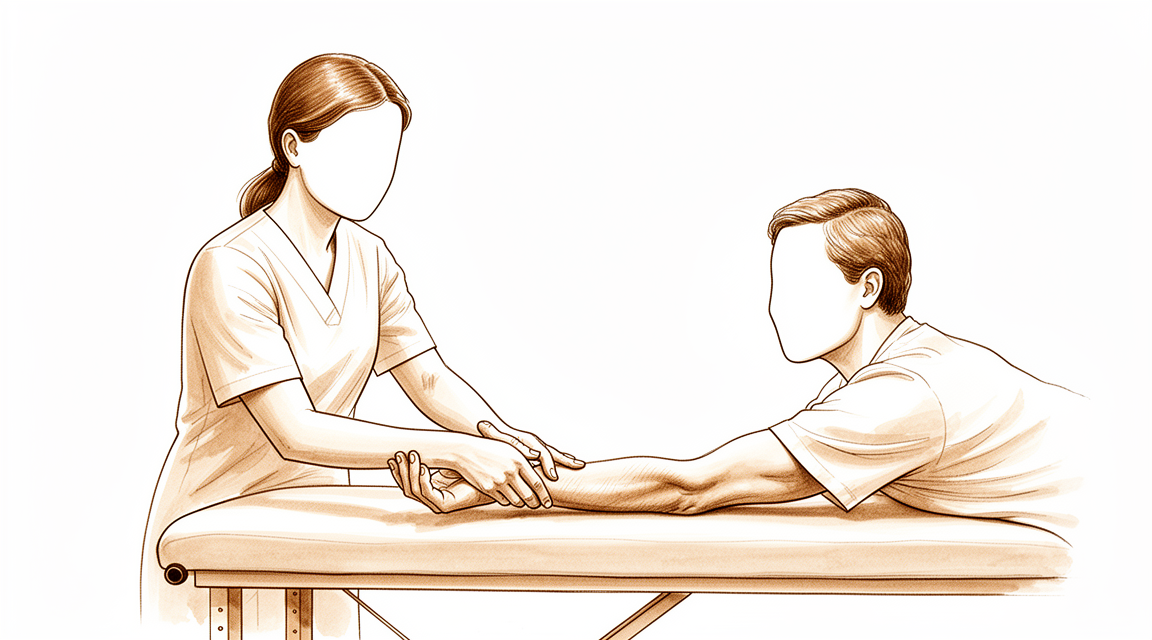
¿Por qué se ha sugerido esta operación?
El Dr. Kieran Hirpara, cirujano de extremidades superiores en el Mater Private Hospital Rockhampton, comienza por las opciones menos invasivas que se adapten a su condición. Por lo general, los pacientes son derivados a nuestra clínica por su médico de cabecera; si un fisioterapeuta le ha recomendado consultarnos, igualmente necesitará una derivación de su médico de cabecera para poder acceder al reembolso de Medicare. Nuestra evaluación, basada en su historial clínico, examen físico e imágenes cuando es necesario, permite establecer el diagnóstico.
El síndrome del túnel radial provoca dolor en la parte externa del codo y el antebrazo. El nervio radial discurre por el antebrazo, y varias estructuras a lo largo de su trayecto pueden ejercer presión sobre él; entre ellas, el borde de un músculo, una banda fibrosa o pequeños vasos sanguíneos que cruzan el nervio. Normalmente iniciamos con tratamientos no quirúrgicos como modificaciones en las actividades, fisioterapia o terapia de la mano, uso de férulas o inyecciones. La cirugía se considera únicamente cuando estos tratamientos no logran mejoría suficiente. La operación, denominada liberación del túnel radial, alivia la presión sobre el nervio en cada uno de esos puntos. La recomendamos para personas cuyo dolor persiste a pesar de haber recibido otros tratamientos. El objetivo principal es aliviar su dolor y ayudar a que su antebrazo y muñeca vuelvan a funcionar con normalidad.
Antes de la operación
Deberá abstenerse de comer y beber durante siete horas antes de la intervención. Pedimos este tiempo algo mayor que en otros centros para poder adelantar su cirugía si el programa de quirófanos lo permite. Su cirujano le indicará qué medicamentos habituales debe omitir ese día y cuáles puede tomar con normalidad. Por favor, lleve una lista por escrito de todos los fármacos que toma, incluyendo pastillas, inyecciones y cualquier gotas o cremas. Organice que alguien lo lleve a casa después de la operación, ya que no estará en condiciones de conducir. Use ropa holgada y cómoda, cuyas mangas se puedan deslizar fácilmente sobre el brazo. Algunas pruebas de imagen, como radiografías, resonancias magnéticas o ecografías, nos ayudan a planificar la intervención. Si padece otras enfermedades, es posible que necesite análisis de sangre o una consulta con el anestesista.
El día de la intervención
Llegará a la unidad de admisiones quirúrgicas del hospital, donde se le registrará y preparará para la cirugía. Allí conocerá al anestesista, el médico encargado de controlar su sueño y el dolor durante la operación. Esta intervención se realiza bajo anestesia general; usted permanecerá completamente dormido durante todo el procedimiento. En algunos pacientes también se aplica un bloqueo nervioso regional para aliviar el dolor postoperatorio; el anestesista decide al respecto ese mismo día según sus circunstancias personales. Posteriormente, será conducido al quirófano, donde se llevará a cabo la operación.
Una vez finalizada la cirugía, despertará en la sala de recuperación. Allí, las enfermeras le supervisarán mientras la anestesia va perdiendo efecto. Cuando su estado sea estable, será trasladado a una sala de hospitalización o podrá volver a casa el mismo día, dependiendo del tipo de intervención y de su evolución postoperatoria. Si regresa a casa, la persona designada para llevarle lo hará en ese momento.
Descripción del procedimiento quirúrgico
La intervención se denomina liberación del túnel radial. Se realiza mediante una sola incisión en la parte anterior del antebrazo, justo debajo del pliegue del codo. El cirujano accede a través de esta única abertura para llegar al nervio radial.
A lo largo del trayecto del nervio, varias estructuras pueden ejercer presión sobre él; entre ellas, el borde de algún músculo, una banda fibrosa o pequeños vasos sanguíneos que cruzan el nervio. El cirujano localiza cada uno de estos puntos de presión y los libera, aliviando así la compresión sobre el nervio. Se procura dejar el tejido circundante lo más intacto posible, preservando el flujo sanguíneo hacia el nervio.
Una vez liberados dichos puntos de presión, el cirujano cierra la incisión. Primero se coloca sobre la herida cerrada una malla autoadhesiva fina que mantiene unidos los bordes de la piel; posteriormente se aplica sobre dicha malla un adhesivo cutáneo líquido, el cual se solidifica para sellar por completo la zona. Este vendaje permanece en su lugar aproximadamente una o dos semanas y luego se desprende por sí solo, por lo que no es necesario retirarlo.
Después de la operación
Despertará en la sala de recuperación, rodeado de enfermeras, mientras el efecto de la anestesia desaparece. Su brazo tendrá un vendaje suave sobre la herida, y se le administrará analgesia para que se sienta cómodo. Podrá moverse en cuanto se sienta listo, y utilizará la mano para tareas ligeras de inmediato. Por lo general, se trata de una intervención ambulatoria, por lo que podrá volver a casa el mismo día; aunque en ocasiones los pacientes permanecen ingresados una noche. Alguien debe acompañarle durante las primeras 24 horas después de regresar a casa. Dejamos el vendaje puesto durante unos 10 días; por favor, no lo retire antes de ese plazo a menos que se lo indiquemos. Lo cambiamos o lo retiramos cuando venga a la consulta.
Recuperación
Al volver a casa, su brazo llevará un vendaje suave. Es normal sentir algo de dolor e hinchazón alrededor de la herida durante los primeros días. Descanse el brazo cuando pueda, manténgalo elevado mientras esté sentado y tome los analgésicos que se le hayan recetado. La mayoría de las personas notan que la molestia disminuye progresivamente con el paso de los días.
Puede utilizar la mano para tareas ligeras de inmediato. Tras esta operación no se coloca ningún yeso ni férula rutinaria, por lo que su muñeca y dedos pueden moverse libremente. Los movimientos suaves favorecen el deslizamiento del nervio y evitan la rigidez. La terapia de mano posterior a la cirugía la realizará Ruby Doolan en Extend Rehabilitation. Ruby es terapeuta especializada en mano: ella le guiará en los ejercicios y confeccionará cualquier férula que necesite. En casa, mantenga la herida seca y no toque el vendaje hasta que lo revisemos.
A medida que la hinchazón disminuya, notará que su fuerza de agarre y la potencia de su antebrazo van recuperándose poco a poco. Las actividades cotidianas como vestirse, comer y teclear suelen volver a hacerse sin problemas al principio. Levantar objetos pesados o ejercer un agarre firme vendrá después, una vez que la mano se sienta fuerte y haya desaparecido la sensibilidad dolorosa. Cuando su terapeuta considere que sus movimientos y fuerza son adecuados, podrá retomar sus actividades habituales.
Por lo general, puede conducir una vez que se haya retirado cualquier férula y el dolor haya disminuido lo suficiente para sujetar el volante y reaccionar rápidamente. Los pacientes que usan férula no deben conducir. Consulte nuestra página sobre Conducción tras cirugía de miembro superior.
La recuperación varía de una persona a otra. Su cronograma personal puede diferir; su cirujano y terapeuta le guiarán en todo momento.
Qué puede salir mal
La mayoría de los pacientes evolucionan bien, pero en ocasiones pueden presentarse problemas. Su cirujano y el equipo lo vigilarán de cerca para detectar cualquier incidencia a tiempo.
El nervio que se libera se encuentra cerca de la zona de la intervención, por lo que puede irritarse o dañarse durante la cirugía. Si esto ocurre, es posible que note debilidad al levantar la muñeca o al estirar los dedos, así como entumecimiento u hormigueo en el dorso del antebrazo, la muñeca o la mano. Algunos de estos cambios son temporales y desaparecen por sí solos en cuestión de semanas o meses. Si nota nueva debilidad o entumecimiento tras la operación, infórmelo a su cirujano en la siguiente revisión; si los síntomas aparecen de forma repentina, llame a la clínica de inmediato.
En ocasiones, al curarse, se forma tejido cicatricial alrededor del nervio. Esto puede volver a ejercer presión sobre el nervio y reactivar el dolor previo, a menudo semanas o meses después de que todo parecía haber mejorado. Si el dolor original regresa, mencione este hecho en su próxima cita. En caso necesario, se puede realizar otra cirugía para liberar dicho tejido cicatricial.
También existe la posibilidad de que la primera operación no alivie sus síntomas, o de que estos reaparezcan poco después. Esto puede suceder si se pasó por alto algún punto de presión en el nervio, o si el diagnóstico no fue del todo correcto. Si el dolor persiste o vuelve, coméntelo con su cirujano; él lo reevaluará y le explicará cuáles son los siguientes pasos, que podrían incluir tratamientos adicionales.
Cualquier intervención en el antebrazo conlleva un riesgo reducido de complicaciones en la propia herida. Esté atento a un enrojecimiento creciente que se extienda desde el corte, a la salida de líquido o pus, o a la aparición de fiebre. Si observa cualquiera de estos signos, comuníquese de inmediato con la clínica; si no puede contactarnos, acuda al servicio de urgencias.
Si en algún momento le preocupa la evolución de la curación de su brazo, llame a la clínica en lugar de esperar a su próxima visita. En la tabla de complicaciones de esta página se detallan las tasas típicas, por si desea conocer los datos exactos.
¿Cuándo deben llamarnos?
Llámenos si el dolor empeora repentinamente, o si aparecen rápidamente debilidad o entumecimiento. Llámenos también si la herida se vuelve más roja, segrega líquido o pus, o si le sube la fiebre. Acuda a urgencias si no puede contactarnos, o si pierde la sensibilidad en el brazo o no puede moverlo. Acuda de inmediato a urgencias si presenta dificultad para respirar o hinchazón en la pantorrilla. Si en algún momento le preocupa algo, llame a la clínica en lugar de esperar a su próxima cita.
¿Dónde leer más sobre esta afección?
Esta página trata sobre la intervención quirúrgica en sí. La afección que se trata, así como las evidencias sobre cuándo la cirugía resulta útil y cuándo no, se explican con mayor detalle en la página de Síndrome del túnel radial.
Evidence & references
This is the clinical evidence summary written for health professionals. It is technical, and it lists the research this page was built from. You do not need to read it to understand your treatment or to make a decision about it.
Anatomy & Pathophysiology
Bony Anatomy
- The elbow is a trocho-ginglymoid joint consisting of medial and lateral articulations that provide bony stability [3].
- The trochlea articulates with the ulna within the greater sigmoid notch to form the ulnohumeral, hinged, or trochoid portion of the elbow joint [3].
- The ulnohumeral articulation has highly congruent anatomy through almost 180° of articular contact, except for the bare area of the greater sigmoid notch which is devoid of cartilage [3].
- The coronoid process has medial and lateral facets that buttress the trochlea anteriorly [3].
- The sublime tubercle is located just distal and medial to the coronoid and serves as the attachment site for the anterior bundle of the medial ulnar collateral ligament [3].
- The medial epicondyle is larger and more posteriorly oriented than the lateral epicondyle and forms the attachment site for the origins of the flexor pronator mass [3].
- The capitellum and radial head form the radiocapitellar joint on the lateral side [3].
- The radius is held in close approximation to the ulna at the proximal radioulnar joint by the annular ligament [3].
- The area of the ulna that articulates with the margin of the radial head at the proximal radioulnar joint is known as the lesser sigmoid notch [3].
- The radial head is a concave elliptical structure covered with articular cartilage along the radiocapitellar joint and approximately 270° of the articular margin [3].
- The radial head articulates with both the capitellum and the lesser sigmoid notch [3].
- The lateral epicondyle is the origin of the lateral extensor musculature [3].
- The origin of the lateral ulnar collateral ligamentous complex is located just distal to the lateral epicondyle at the geometric center of the radiocapitellar articulation [3].
- The distal humeral articulation is angled 30° from the longitudinal axis [3].
- The anterior humeral line should pass through the center of the axis of rotation [3].
- The axis of rotation is angulated 5° to 7° in the coronal plane relative to the epicondylar axis, with the medial side more distal than the lateral side [3].
- The angulation of the distal humeral articulation accounts for the change from a valgus carrying angle to a more varus position as the elbow is flexed [3].
- There is a high correlation between the size of the radial head and capitellum on the left and right sides in the same individual [3].
- The olecranon provides a broad attachment site for the triceps posteriorly [3].
- The ulna bends approximately 8° medially at 8 cm from the tip of the olecranon [3].
- The articulation to the tip of the coronoid is approximately 30° from the long axis of the ulna in the sagittal plane [3].
- The radial head should line up with the capitellum at all arm positions on all radiographic views [4].
Ligaments and Capsule
- Elbow stability is determined by primary and secondary stabilizers [1].
- The three primary stabilizers of the elbow are the ulnohumeral articulation, the medial ulnar collateral ligament, and the lateral ulnar collateral ligament complex [1].
- Secondary stabilizers of the elbow include the radiocapitellar articulation, the common flexor tendon, the common extensor tendon, and the joint capsule [1].
- The medial ulnar collateral ligament is the primary valgus stabilizer of the elbow [4].
- The anterior bundle of the medial ulnar collateral ligament is the most important component for stability [4].
- The posterior bundle of the medial ulnar collateral ligament has the greatest change in length and becomes taut at flexion beyond 120 degrees [4].
- The lateral ulnar collateral ligament is the posterolateral stabilizer of the elbow [4].
- The joint capsule allows maximum distension at approximately 70 to 80 degrees of flexion [4].
- The anterior capsule attaches at a point approximately 6 mm distal to the tip of the coronoid [4].
- Tensile forces are present at the medial elbow and compressive forces are present at the lateral elbow [4].
- The LUCL origin center is 10.7 mm from the lateral epicondyle [2].
- The LUCL insertion is 3.3 mm from the apex of the supinator crest [2].
Muscles and Nerves
- The brachialis is the strongest elbow flexor and attaches to the coronoid 11 mm distal to the tip [4].
- The biceps brachii inserts at the ulnar margin of the radial tuberosity, with the long head inserting proximally and the short head distally [4].
- The biceps brachii is a powerful supinator of the forearm [4].
- The triceps is the primary elbow extensor and inserts on the olecranon process [4].
- The mobile wad consists of the brachioradialis, extensor carpi radialis longus, and extensor carpi radialis brevis [4].
- The flexor-pronator mass consists of the pronator teres, flexor carpi radialis, palmaris longus, flexor carpi ulnaris, and flexor digitorum superficialis [4].
- The radial nerve enters the interval between the brachialis and brachioradialis muscles in the proximal angle of the lateral approach wound [6].
- The deep branch of the radial nerve enters the supinator muscle [6].
- The common origin of the extensor muscles is attached to the lateral epicondyle [6].
- The common origin of the extensor muscles is attached to the lateral condylar fragment in lateral condyle fractures [6].
- The interval between the triceps posteriorly and the origins of the extensor carpi radialis longus and brachioradialis anteriorly is used to expose the lateral border of the humerus [6].
- The interval between the triceps posteriorly and the brachioradialis and extensor carpi radialis longus muscles anteriorly is used to expose the lateral condyle and capsule over the radial head [7].
- The extensor carpi ulnaris is separated from the anconeus distal to the radial head during the lateral J-shaped approach [7].
- The distal fibers of the anconeus are divided in line with the curved and transverse parts of the distal skin incision during the lateral J-shaped approach [7].
- The anconeus is reflected subperiosteally from the proximal ulna to dislocate and examine the joint during the lateral J-shaped approach [7].
Functional Anatomy
- The normal elbow has a range of motion from 0° to 140° from extension to flexion [1].
- The normal elbow has a range of motion of 75° in pronation and 85° in supination [1].
- A functional arc for elbow flexion and extension is 100° [1].
- A functional arc for forearm rotation is 100° [1].
Investigations
Physical Examination
- The physical examination of the elbow is directed by the history and the location of the patient's pain in the anterior, posterior, medial, or lateral aspect of the elbow [1].
- Pathologic entities associated with specific compartments aid the examiner in detecting pathologic conditions [1].
- Active and passive flexion, extension, supination, and pronation should be evaluated using a goniometer for accurate measurement [9].
- The contralateral elbow should be examined for comparison during range of motion assessment [9].
- Pain should be assessed during the mid-arc or at the terminal ends of motion [9].
- Mid-arc range of motion pain is more common with intrinsic disease and may not improve with contracture release alone [9].
- The ulnar nerve is of utmost importance in the neurovascular examination due to its anatomic proximity to the elbow [9].
- Electromyography and nerve conduction velocity studies should be performed if there is any question about neurologic dysfunction [9].
- An assessment for ulnar nerve subluxation should be performed [9].
- Subluxation of the ulnar nerve is a relative contraindication for an arthroscopic procedure secondary to possible iatrogenic nerve injury [9].
- If the elbow has less than 90° to 100° of flexion, the posterior bundle of the medial collateral ligament is contracted and must be released to restore flexion [9].
Imaging
- Plain radiographs remain the hallmark and the best screening test for elbow evaluation [1].
- Standard radiographic views include AP, lateral, and oblique views [9].
- Serial radiography is used as follow-up when heterotopic ossification is present [9].
- Primary bony landmarks assessed on radiographs include the ulnohumeral joint, coronoid process, radial head, capitellum, radiocapitellar joint, olecranon tip, coronoid/olecranon fossae, and trochlear ridge [9].
- CT is helpful when assessing for malunion architecture and the location and pattern of osteophytes and/or loose bodies [9].
- Three-dimensional CT is used to check for heterotopic ossification [9].
- CT is not necessary when the stiffness is entirely soft-tissue related [9].
- CT is beneficial if any joint incongruity or abnormal bony anatomy is present [9].
- MRI can be used to evaluate ligaments and tendons, but it is rarely indicated for elbow stiffness [9].
- AP, lateral, oblique, and axillary views of the elbow may reveal posteromedial olecranon osteophytes and/or loose bodies in valgus extension overload syndrome [11].
- CT with two-dimensional reconstruction and three-dimensional surface rendering best visualizes the pathology of valgus extension overload syndrome [11].
- MRI may be most helpful in evaluating associated injuries including partial or complete tears of the medial collateral ligament in valgus extension overload syndrome [11].
- Radiographic evaluations are essential when diagnosing an osteochondritis dissecans lesion of the elbow [12].
- Important aspects of osteochondritis dissecans lesions may be better seen with MRI [12].
- Standard AP and lateral radiographs should be obtained for the evaluation of elbow osteoarthritis [13].
- Radiographs for elbow osteoarthritis typically show osteophyte formation at the coronoid process, coronoid fossa, radial fossa, radial head, olecranon tip, and olecranon fossa [13].
- Joint spaces at the ulnohumeral joint are usually preserved in elbow osteoarthritis [13].
- Joint spaces at the radiocapitellar joint are mildly narrowed in elbow osteoarthritis [13].
- Loose bodies may be evident on radiographs, which typically underestimate the number present [13].
- CT may be useful for surgical planning of elbow osteoarthritis by allowing a detailed assessment of osteophytes and the presence of loose bodies [13].
References
[1] Orthopaedic Knowledge Update 13 Ebook Without Multimedia. Anatomy, Biomechanics, Physical Examination, and Imaging of the Elbow > Summary and Conclusions.
[2] Orthopaedic Knowledge Update 13 Ebook Without Multimedia. Anatomy, Biomechanics, Physical Examination, and Imaging of the Elbow > Annotated References.
[3] Orthopaedic Knowledge Update 13 Ebook Without Multimedia. Anatomy, Biomechanics, Physical Examination, and Imaging of the Elbow > Anatomy > Bony Anatomy.
[4] Miller S Review Of Orthopaedics. SECTION 16 PATELLAR TRACKING IN TOTAL KNEE ARTHROPLASTY > TABLE 2.3 Shoulder Spaces.
[6] Campbell S Operative Orthopaedics 4 Volume Set. LATERAL APPROACHES.
[7] Campbell S Operative Orthopaedics 4 Volume Set. LATERAL J-SHAPED APPROACH TO THE ELBOW.
[9] Aaos Comprehensive Orthopaedic Review 3. Elbow Stiffness* > IV. Evaluation.
[11] Aaos Comprehensive Orthopaedic Review 3. Elbow Injuries in the Athlete* > III. Valgus Extension Overload Syndrome and Posterior Impingement.
[12] Orthopaedic Knowledge Update. Osteochondritis Dissecans of the Knee and Elbow* > Summary.
[13] Aaos Comprehensive Orthopaedic Review 3. Arthritis and Arthroplasty of the Elbow > I. Osteoarthritis.