Patients › Shoulder
Hombro congelado
Frozen shoulder (adhesive capsulitis) causes pain and stiffness, progressing through freezing, frozen, and thawing phases.
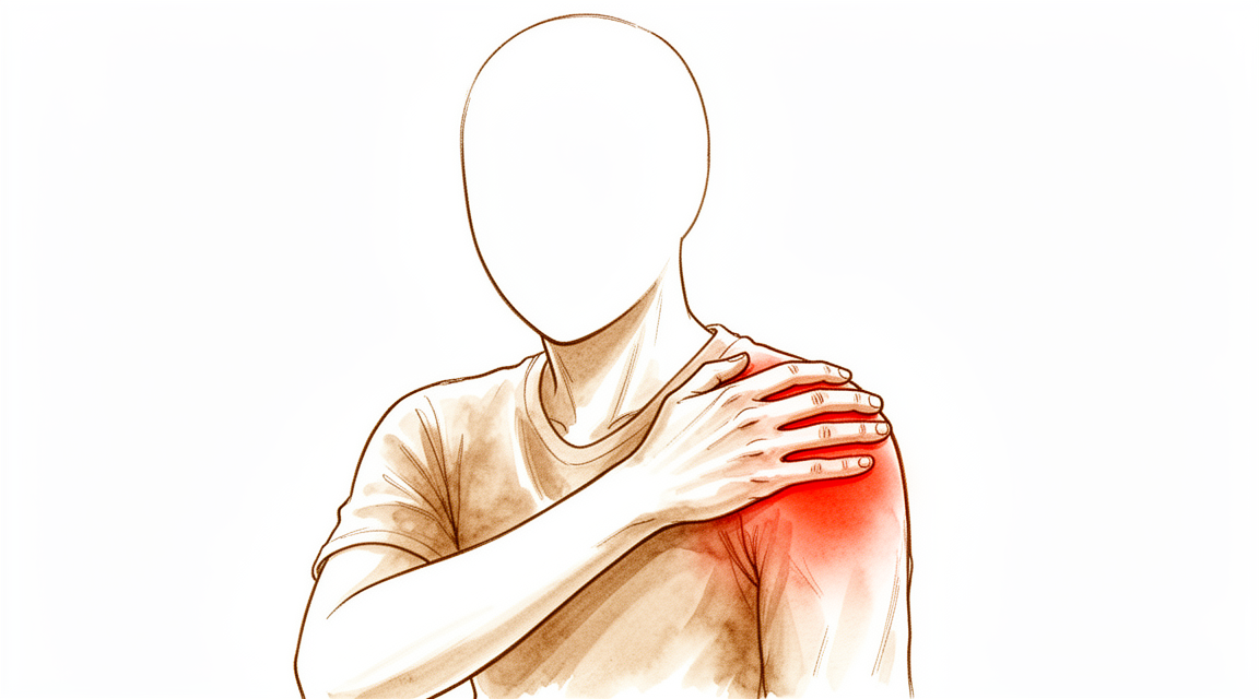
Qué está sintiendo
El hombro congelado suele comenzar como un dolor profundo y sordo que se intensifica a lo largo de semanas, en lugar de aparecer de repente. Para la mayoría de las personas, el dolor es lo peor al principio, y suele ser más intenso por la noche. Acostarse sobre ese lado se vuelve imposible, y el sueño interrumpido es uno de los aspectos más agotadores de esta afección.
Luego, el hombro comienza a quedarse rígido. Intenta alcanzar el cinturón de seguridad, un estante alto o el bolsillo, y el brazo simplemente no se mueve. Lo que diferencia al hombro congelado de la mayoría de los problemas de hombro es que la rigidez no se debe únicamente a que el paciente evite el dolor: el hombro realmente no se mueve, incluso si otra persona intenta levantarle el brazo. La pérdida de la capacidad de girar la mano hacia afuera —como al abrir una puerta o meter el brazo en una manga— es el signo más característico de todo el cuadro.
La mayoría de los afectados tienen entre 40 y 60 años. Algunos recuerdan haber sufrido un golpe leve previamente; muchos, en cambio, no tienen explicación alguna. Es más frecuente en personas con diabetes o trastornos tiroideos; además, si ya ha afectado a un hombro, existe una probabilidad moderada de que el otro hombro también se vea afectado posteriormente.
¿Qué está ocurriendo realmente?
La articulación del hombro se encuentra dentro de una envoltura de tejido resistente llamada cápsula. Normalmente, dicha envoltura es laxa y elástica, lo que permite que el hombro se mueva más que cualquier otra articulación del cuerpo. En el hombro congelado, la cápsula se inflama, luego se engrosa y contrae, reduciéndose de tamaño. El espacio dentro de la articulación puede terminar siendo menos de la mitad de su tamaño normal.
A pesar de que antiguamente se le llamaba “capsulitis adhesiva”, en realidad nada queda adherido dentro de la articulación; no existen adherencias similares a pegamento. Lo que ocurre es que el tejido se ha tensado, lo cual constituye un problema distinto; por eso algunos cirujanos sostienen que esta afección debería denominarse simplemente contractura del hombro, es decir, un estrechamiento del tejido.
Aún no se comprende bien cuál es la causa exacta; hay que reconocerlo con sinceridad. Existen pruebas sólidas de que no se trata únicamente de un problema local del hombro. Los estudios sobre el riesgo hereditario demuestran que la diabetes tipo 1 realmente provoca el hombro congelado, en lugar de estar simplemente asociada a él; además, esta relación se debe a los niveles de azúcar en sangre y no al peso corporal ni a la carga mecánica. Los cambios tisulares son sorprendentemente similares a los de la enfermedad de Dupuytren en la mano, motivo por el cual ambas afecciones suelen aparecer en la misma persona.
Siendo honestos sobre cuánto tiempo dura el proceso
Es muy probable que haya leído que el hombro congelado es una condición autolimitada que se resuelve por completo en unos dieciocho meses a dos años. Queremos ser transparentes con usted: esa afirmación se basa en una confianza mayor de la que realmente respaldan las evidencias científicas.
La idea de que la enfermedad pasa ordenadamente por tres fases —“congelación”, “estado congelado” y “descongelación”— proviene de un estudio pequeño publicado en 1975, en el cual se mantenían los brazos de los pacientes en cabestrillos hasta nueve meses. No sorprende que los hombros tratados de esa manera se volvieran rígidos y luego recuperaran cierta movilidad una vez que se les permitió moverlos. Cuando los investigadores revisaron todos los estudios disponibles, no hallaron evidencia sólida de que los hombros no tratados pasen de forma fiable por esas fases y vuelvan a la normalidad. De hecho, ningún estudio ha demostrado que las personas recuperen un rango de movimiento verdaderamente normal sin tratamiento.
Lo que sí demuestran las evidencias es algo más útil: la mayor parte de la mejora que usted experimentará suele ocurrir al principio; después, el progreso se ralentiza y se estabiliza. Esa estabilización puede ser permanente. Según el estudio que se consulte, entre un tercio y la mitad de las personas siguen experimentando cierto dolor o rigidez varios años después; aunque para la mayoría es leve y no les impide hacer lo que desean.
Nada de esto significa que usted no vaya a mejorar; la mayoría de las personas mejoran considerablemente. Lo que sí implica es que el consejo de “solo espere, todo se resolverá por sí solo” no es neutral, y que existe un argumento razonable para tratar el dolor de manera activa en lugar de simplemente soportarlo.
¿Qué podemos hacer al respecto?
En el Mater Private Hospital Rockhampton, el Dr. Kieran Hirpara le explicará las opciones disponibles según cuál síntoma le moleste más: el dolor o la rigidez, en lugar de intentar clasificarlo en una etapa numérica determinada.
La inyección de esteroides en la articulación es el tratamiento con mayor respaldo científico para la fase dolorosa. Numerosos estudios demuestran que es el único tratamiento que genera de forma fiable una mejora perceptible en el dolor y la funcionalidad, y que los efectos se notan en las primeras semanas. Su limitación es que los beneficios tienden a desaparecer al cabo de unos tres meses. Este tratamiento le brinda alivio, le permite dormir y seguir trabajando con el hombro; sin embargo, no corrige la rigidez.
Los ejercicios y la fisioterapia también son útiles, siempre con expectativas realistas. Un programa sencillo de ejercicios en casa puede aportar mejoras medibles. Lo que quizás le sorprenda es que la intensidad con la que los realiza influye poco: un ensayo que comparó estiramientos enérgicos hasta el límite de la amplitud articular con movimientos suaves y sin dolor solo halló pequeñas diferencias, ligeramente a favor del enfoque más enérgico en algunos movimientos, y otro estudio observó que las personas que se ejercitaron suavemente dentro de sus límites de dolor obtuvieron mejores resultados; además, afortunadamente, no hay evidencia de que forzar los ejercicios cause daños. Por tanto, haga los ejercicios dentro de sus posibilidades. No existe evidencia de que soportar un dolor intenso acelere la recuperación.
La hidrodilatación, que consiste en inyectar líquido para estirar la cápsula desde el interior, es un procedimiento ampliamente utilizado y de bajo riesgo. Sin embargo, la evidencia científica al respecto es contradictoria: una revisión halló solo efectos pequeños tanto en el dolor como en la movilidad, y otras hallaron una mejora inicial en algunos movimientos, en comparación con una inyección de esteroides sola, que no se mantuvo. Es una opción razonable, aunque no la promovemos excesivamente.
La manipulación bajo anestesia implica mover el hombro a través de toda su amplitud de movimiento mientras el paciente está dormido, con el fin de estirar la cápsula rígida. En el mayor ensayo clínico que comparó los principales tratamientos, esta opción resultó ser la de mejor relación coste-beneficio en general.
La liberación capsular por vía mínimamente invasiva consiste en dividir quirúrgicamente la cápsula rígida bajo visión directa, en lugar de estirarla hasta que se rompa. Su principal ventaja es que suele ser el tratamiento más duradero: en los estudios publicados, la recurrencia de la rigidez tras esta intervención es poco frecuente, mientras que tras la manipulación un porcentaje considerable de pacientes vuelven a presentar rigidez y requieren un segundo procedimiento. En el gran ensayo británico, los pacientes sometidos a liberación fueron los que menor probabilidad tuvieron de necesitar tratamiento adicional. Al ser una cirugía, conlleva un riesgo ligeramente mayor que las demás opciones y tiene un costo más elevado; además, es normal sentir más dolor durante las primeras semanas que quienes se someten a manipulación; esa diferencia inicial se iguala alrededor del sexto mes.
Cabe destacar que, en dicho ensayo, al cabo de un año los resultados de los tres tratamientos fueron muy similares. Esto es una excelente noticia, pues significa que la elección del tratamiento puede basarse en sus preferencias, su estado de salud general y sus circunstancias personales, en lugar de depender de que una opción sea claramente superior a las demás.
Cómo suele plantear el tratamiento el Dr. Hirpara. No se le pedirá que aguante el dolor durante un número fijo de meses antes de recibir cualquier tratamiento. En la práctica, suele recomendar primero la inyección de esteroides, ya que para muchos pacientes reduce el dolor lo suficiente para poder seguir con su vida mientras el hombro se recupera. Si la inyección no le brinda el alivio deseado, el Dr. Hirpara está dispuesto a proponerle la liberación capsular sin necesidad de que haya soportado el dolor durante nueve o doce meses. Dado que la afirmación de que “el problema se resuelve por sí solo” carece de fundamento, no tiene sentido pasar un año sufriendo para comprobar cómo evolucionará su caso.
Qué esperar
Si le aplican una inyección, puede esperar que el dolor disminuya durante la primera o segunda semana. Aproveche ese período para mantener el hombro en movimiento.
Si se somete a una manipulación o a una liberación capsular, la mayor parte del rango de movilidad que recuperará volverá rápidamente —generalmente dentro de las primeras cuatro a seis semanas— y, por lo general, no habrá cambios significativos después de unos tres meses. Es fundamental comenzar a mover el hombro desde el principio; la fisioterapia suele iniciarse uno o dos días después del procedimiento. La mayoría de las personas vuelven a realizar trabajos de oficina en el plazo de una a tres semanas.
Si padece diabetes, es razonable esperar una recuperación más lenta y un poco menos completa que la media; también debe saber que una inyección de esteroides puede alterar sus niveles de azúcar en sangre durante unos días. Infórmenos si le cuesta controlar la diabetes, ya que eso influye en nuestras recomendaciones.
Independientemente del procedimiento que elija, la recuperación se mide en meses, no en semanas. Lo más útil que puede hacer es seguir utilizando el brazo dentro de los límites que le resulten cómodos y protegerlo durante el sueño.
¿Cuándo consultar a un profesional?
Acuda a su médico de cabecera si el dolor de hombro le impide dormir por las noches o si no mejora tras unas pocas semanas de reposo. Si el hombro se vuelve realmente difícil de mover, y no solo le duele, pregúntele específicamente acerca de la rigidez articular del hombro.
Solicite una evaluación especializada cuanto antes si no puede girar la mano hacia afuera, si padece diabetes, si el dolor del hombro le impide trabajar o conducir, o si es tan intenso que le impide dormir. No hay ninguna ventaja en esperar mucho tiempo para que se evalúe el dolor.
Busque atención médica inmediata si el hombro se vuelve caliente, rojo e hinchado; si se siente mal o tiene fiebre; si presenta debilidad o entumecimiento significativo en el brazo; o si el dolor comenzó tras una caída o lesión. Estos síntomas no son propios de la rigidez articular del hombro, sino de una condición que requiere evaluación urgente.
En profundidad
Esta sección va un paso más allá de lo necesario para que usted tome sus propias decisiones de tratamiento. Se incluye aquí porque el hombro congelado es una de las afecciones en las que lo que se le dice a los pacientes y lo que realmente demuestran las investigaciones han ido divergiendo; además, hay personas que prefieren conocer el razonamiento detrás de las recomendaciones en lugar de simplemente aceptarlas sin cuestionarlas.
¿Qué es lo que realmente está fallando en la articulación?
La cápsula articular es una envoltura de colágeno que rodea la articulación. En el hombro congelado, esta cápsula se inflama y posteriormente fibrosa; las células llamadas fibroblastos se multiplican y generan nuevo colágeno denso; algunas de ellas se transforman en miofibroblastos, capaces de contraerse activamente. Esta es la diferencia clave con respecto a una cicatrización normal: el tejido no solo se engrosa, sino que también se tensa. El volumen articular puede reducirse de los 10–15 mL normales a 3–4 mL.
El mismo comportamiento celular se observa en la enfermedad de Dupuytren en la mano; por eso ambas afecciones aparecen en las mismas personas. De hecho, un cirujano de hombro ha sostenido que esta condición debería denominarse simplemente contractura del hombro en lugar de “capsulitis” [1]. A pesar del antiguo nombre “capsulitis adhesiva”, los cirujanos que examinan el interior de la articulación no encuentran ninguna adherencia.
¿Por qué la teoría de las “tres fases” carece de fundamento?
En todas partes se afirma que el hombro congelado pasa por tres fases: la de congelamiento, la de rigidez y la de descongelamiento. Este modelo se propuso en 1975. Al investigar su origen, se descubrió que el autor confundió las referencias de dos artículos previos; además, y esto es más importante, sus pacientes llevaban el brazo en cabestrillo durante hasta nueve meses. Un hombro inmovilizado durante tanto tiempo se vuelve rígido y, al permitirle el movimiento, recupera su flexibilidad. Por tanto, dichas “fases” podrían describir en parte el tratamiento y no la propia enfermedad.
Una revisión sistemática realizada para comprobar este modelo no encontró evidencia alguna que lo respalde; de hecho, los resultados fueron contrarios a lo predicho: la mayor parte de la mejoría se produce al principio y luego disminuye, en lugar de acumularse hasta un descongelamiento tardío [2]. Tampoco los profesionales médicos logran ponerse de acuerdo sobre en qué fase se encuentra un hombro determinado; los propios autores de la definición estándar admitieron por escrito que los datos necesarios para confirmarla “no están disponibles”.
La estadística de recuperación y por qué existen dos respuestas muy distintas
La cifra que se cita con frecuencia es que el 94 % de las personas se recuperan sin tratamiento. Esta cifra proviene de un estudio real, pero solo incluye a los pacientes que no recibieron tratamiento y que regresaron para el seguimiento. La mayoría del grupo original optó por el tratamiento o simplemente abandonó el estudio. Si se contabilizan todos los participantes, los mismos datos arrojan un porcentaje de aproximadamente 26 % [2]. El término “recuperado” significaba que la movilidad del hombro alcanzaba un rango de 10 grados respecto al otro hombro; además, en algunos de esos pacientes el otro hombro también presentaba rigidez.
El mayor estudio de seguimiento a largo plazo reveló que el 41 % de los pacientes aún presentaban síntomas, en promedio, cuatro años después del inicio del estudio. Lo más relevante es que la mejoría clínica prácticamente se detiene después de unos tres años, en lugar de continuar indefinidamente [4].
Se trata de una afección que afecta a todo el cuerpo, no solo al hombro
Un gran estudio genético identificó cinco regiones del ADN asociadas al hombro congelado y, mediante una técnica capaz de distinguir entre causa y coincidencia, demostró que la diabetes tipo 1 realmente la provoca, en lugar de simplemente acompañarla. Una vez que se tuvo en cuenta la diabetes, la relación entre el sobrepeso y esta afección desapareció. Esto indica que el nivel de azúcar en sangre, y no la carga mecánica, es el factor determinante [5].
¿Qué dice realmente la evidencia sobre la fisioterapia?
Esta es la parte que más conviene conocer si usted está recibiendo o practicando fisioterapia. Una revisión Cochrane agrupó 32 ensayos con 1,836 pacientes [6]. Destacan tres hallazgos.
En comparación con una inyección de esteroides, la fisioterapia manual arroja peores resultados al principio. En el ensayo comparativo, seis semanas de movilización pasiva y ejercicios supervisados produjeron una mejora del dolor 26 puntos menor y una mejora funcional 25 puntos menor a las siete semanas respecto a una sola inyección. En cuanto a si los pacientes consideraban que habían mejorado o se habían recuperado, el porcentaje fue de 46 % para la fisioterapia frente a 77 % para la inyección; es decir, aproximadamente una persona más se beneficiaba por cada cuatro que recibían la inyección. A los seis o doce meses, esa diferencia se había reducido y dejó de ser relevante.
El grado de intensidad, la frecuencia y la técnica empleada parecen influir poco. Un ensayo con 100 pacientes comparó la movilización firme hasta el límite de rango articular con una movilización suave sin provocar dolor: ambos grupos mejoraron, y la técnica más firme obtuvo resultados estadísticamente mejores en algunos movimientos (elevar el brazo hacia un lado a los tres y doce meses, girarlo hacia afuera a los doce meses), pero los autores consideraron que las diferencias eran pequeñas [18]. Un estudio no aleatorizado con 77 personas halló lo contrario: a los dos años, el 89 % de quienes se ejercitaron suavemente dentro de sus límites de dolor tenía una puntuación del hombro normal o casi normal, frente al 63 % de quienes recibieron estiramientos intensivos [19]. Otro ensayo con 120 pacientes varió la frecuencia de la movilización hasta el límite articular (más de dos veces por semana, una vez por semana, menos de una vez por semana); las diferencias en la abducción fueron inferiores a 8 grados, estadísticamente indistinguibles de cero. Las comparaciones directas entre técnicas —deslizamiento anterior versus posterior, PNF frente a ejercicios convencionales, mancuernas frente a ejercicios sin pesas— resultaron casi todas no significativas. Afortunadamente, no hubo indicios de que la movilización hasta el límite articular cause daño alguno.
La mayor laguna metodológica: nunca se ha realizado ningún ensayo que compare la terapia manual más ejercicios frente a la ausencia total de tratamiento. Así, aunque podemos afirmar que la fisioterapia no es claramente superior a una inyección y que los detalles de su aplicación no parecen modificar mucho los resultados, nadie ha demostrado cuánta mejora se habría producido de todos modos sin ningún tipo de intervención.
Dos matices importantes. El único ensayo de alta calidad incluido en la revisión evaluó la fisioterapia después de una distensión articular; no influyó en el dolor, la función ni la calidad de vida, pero sí aumentó el porcentaje de pacientes que se declararon recuperados y generó unos 13 grados adicionales de abducción a las seis semanas, efecto que desapareció a los seis meses. En el gran ensayo británico, el grupo de fisioterapia se describió como recibiendo estiramientos manuales dentro del rango rígido; sin embargo, los registros muestran que la movilización manual se aplicó únicamente a 11–27% paciente y el estiramiento supervisado a un solo caso. Lo que realmente se administró, y que a un año arrojó resultados similares a la cirugía, fue educación del paciente, una inyección, movimientos suaves supervisados y un programa de ejercicios en casa.
¿Cuánto tiempo tarda en mejorar, y importa el tipo de liberación?
Tiempo de recuperación. Tras una liberación capsular, el tiempo mediano que tarda el paciente en superar el umbral de “esta es una mejora real” es de aproximadamente un mes, y el umbral de “mi hombro me resulta aceptable” se alcanza en unos cuatro meses [14]. El tiempo fuera del trabajo es, en promedio, de ocho semanas; sin embargo, la variabilidad es grande: un cuarto de los pacientes regresa al trabajo en cuatro semanas, y tres cuartos, en trece semanas [15].
La manipulación restaura el movimiento más rápidamente. Ese es el verdadero compromiso frente a la mayor durabadad que ofrece la liberación capsular: la manipulación recupera antes el rango de movimiento; la liberación no logra igualar esos resultados hasta pasados unos seis meses, momento a partir del cual ambas técnicas resultan indistinguibles en todos los parámetros evaluados. Un pequeño estudio comparativo incluso determinó que la manipulación por sí sola producía la mejor rotación externa [16].
¿Cuánta parte de la cápsula se debe liberar? Es una pregunta válida, y la respuesta es “menos de lo que se podría pensar”. En una revisión que analizó 18 estudios, 629 pacientes y 811 hombros, se compararon tres técnicas: liberar únicamente la parte frontal e inferior de la cápsula, añadir también la parte posterior, o realizar una liberación completa de 360 grados. Los resultados indicaron que las liberaciones menos extensas generaban mejores puntuaciones funcionales y de dolor; además, la liberación posterior aporta una ganancia temprana en rotación interna que no persiste a largo plazo (aunque sí mejora la flexión de forma duradera); por último, una liberación completa “podría no aportar beneficios adicionales”. Las tasas de complicaciones no variaron entre las tres técnicas [17]. Dos ensayos aleatorizados sobre la extensión posterior mostraron el mismo patrón: recuperación inicial más rápida, pero sin diferencias a los seis meses. Por tanto, una intervención más extensa no equivale necesariamente a mejores resultados.
Realizar ambas técnicas conjuntamente no es la solución. En el único estudio que comparó las tres opciones, añadir manipulación a la liberación capsular produjo la mayor tasa de pérdida de rotación externa: 18 %, frente al 7 % en casos de manipulación sola y al 2 % en liberación sola [16].
¿Qué descubrió realmente el mayor ensayo terapéutico?
En un ensayo realizado en el Reino Unido, se asignó al azar a poco más de 500 pacientes a recibir fisioterapia estructurada junto con una inyección, manipulación bajo anestesia o cirugía artroscópica. Al cabo de un año, los resultados de los tres grupos difirieron apenas unos pocos puntos entre sí; esa diferencia es menor que lo que un paciente podría percibir [7]. Esta es una información verdaderamente útil: significa que la elección del tratamiento puede basarse en lo que mejor se adapte a cada persona y a sus circunstancias, en lugar de asumir que una opción sea claramente superior a las demás.
Donde sí existen diferencias entre los tres tratamientos es en la frecuencia con la que los pacientes necesitan tratamiento adicional posteriormente. En dicho ensayo, se requirió tratamiento complementario en el 4 % tras la liberación del tejido, el 7 % tras la manipulación y el 15 % tras la fisioterapia [7]. La manipulación resultó ser la opción más rentable en cuanto al costo por unidad de beneficio; la liberación del tejido fue la más costosa y generó la mayoría de las complicaciones graves registradas en el estudio, aunque estas fueron poco frecuentes en términos absolutos.
Manipulación o liberación: el problema de las recurrencias
Aquí es donde ambas intervenciones realmente se diferencian; este es el motivo por el cual el cirujano podría recomendarle una liberación aunque los resultados generales parezcan similares.
La manipulación consiste en estirar la cápsula hasta que se rompe; sin embargo, esa rotura no siempre ocurre donde se desearía. En el estudio más amplio, se siguieron 792 hombros durante 17 años: el 17,8 % necesitó una segunda manipulación; en pacientes con diabetes tipo 1, ese porcentaje subió al 37,9 % [9]. El seguimiento más prolongado, con un promedio de 13 años, reveló que el 19,2 % requirió una nueva manipulación y el 31,3 % experimentó alguna recurrencia en algún momento [10]. Dos factores atenúan esto: casi todas las recurrencias ocurren durante el primer año (después de cinco años, la tasa es inferior al 2 %); además, una segunda manipulación tiene resultados prácticamente iguales a la primera [9][10].
La liberación implica dividir la cápsula de forma deliberada, bajo visión directa. Las reintervenciones son mucho menos frecuentes: en un estudio con un seguimiento promedio de cinco años, ninguno de los 32 pacientes necesitó una nueva operación; solo hubo una recurrencia en alguien que no completó su rehabilitación [11]. El metaanálisis que agrupó ocho estudios y 768 pacientes consideró ambas técnicas comparables en cuanto al dolor, la función y el rango de movimiento (una pequeña ventaja de la liberación en el dolor a largo plazo no alcanzó una magnitud clínicamente importante), con una mayor tasa de complicaciones graves tras la liberación. Señaló una tasa mayor, aunque no estadísticamente significativa, de nuevas inyecciones de esteroides en el grupo sometido a manipulación, y sugirió que, tras un seguimiento prolongado, “el grupo sometido a manipulación podría presentar más recurrencias” [8].
Dos advertencias importantes. Primero, los principales estudios sobre recurrencias tras manipulación provienen, en su mayoría, de la práctica de un único cirujano, por lo que no constituyen confirmaciones independientes entre sí. Segundo, la liberación tampoco logra una recuperación total: un estudio halló que, dos a seis años después, la puntuación media del hombro seguía siendo de aproximadamente 77 sobre 100 [12]. En otro grupo de pacientes sin diabetes, aunque casi todos mejoraron de forma significativa, 11–22% aún no consideraban su hombro aceptable a los seis meses [13].
En resumen: para lograr que el hombro recupere movilidad y comodidad, ambas técnicas son prácticamente equivalentes, y la manipulación resulta la opción más económica. No obstante, para mantener esos resultados, la evidencia favorece la liberación. Esta diferencia resulta especialmente relevante si padece diabetes, si el hombro está muy rígido o si ya se ha sometido a una manipulación que no tuvo éxito.
Referencias
Advanced reading: the deeper science (optional)
[1] Bunker T. Es hora de un nuevo nombre para el hombro congelado: contractura del hombro. Shoulder Elbow. 2009;1(1):4-9. https://doi.org/10.1111/j.1758-5740.2009.00007.x [2] Wong CK, Levine WN, Deo K, Kesting RS, Mercer EA, Schram GA, et al. Historia natural del hombro congelado: ¿realidad o ficción? Una revisión sistemática. Physiotherapy. 2017;103(1):40-7. https://doi.org/10.1016/j.physio.2016.05.009 [3] Vastamäki H, Kettunen J, Vastamäki M. Historia natural del hombro congelado idiopático: estudio de seguimiento de 2 a 27 años. Clin Orthop Relat Res. 2012;470(4):1133-43. https://doi.org/10.1007/s11999-011-2176-4 [4] Hand C, Clipsham K, Rees JL, Carr AJ. Resultados a largo plazo del hombro congelado. J Shoulder Elbow Surg. 2008;17(2):231-6. https://doi.org/10.1016/j.jse.2007.05.009 [5] Green HD, Jones A, Evans JP, Wood AR, Beaumont RN, Tyrrell J, et al. Un estudio de asociación genómica amplia identifica 5 loci relacionados con el hombro congelado e implica a la diabetes como factor de riesgo causal. PLoS Genet. 2021;17(6):e1009577. https://doi.org/10.1371/journal.pgen.1009577 [6] Page MJ, Green S, Kramer S, Johnston RV, McBain B, Chau M, et al. Terapia manual y ejercicios para la capsulitis adhesiva (hombro congelado). Cochrane Database Syst Rev. 2014;2014(8):CD011275. https://doi.org/10.1002/14651858.CD011275 [7] Brealey S, Northgraves M, Kottam L, Keding A, Corbacho B, Goodchild L, et al. Tratamientos quirúrgicos comparados con fisioterapia estructurada temprana en atención secundaria para adultos con hombro congelado primario: el ensayo aleatorizado de tres brazos UK FROST. Health Technol Assess. 2020;24(71):1-162. https://doi.org/10.3310/hta24710 [8] Zhao Y, Yang T, Feng C, Li L, Pang L, Zhao S. Liberación capsular artroscópica versus manipulación bajo anestesia para el hombro congelado refractario: una revisión sistemática con metanálisis. Orthop Surg. 2024;16(7):1517-29. https://doi.org/10.1111/os.14077 [9] Woods DA, Loganathan K. Recurrencia del hombro congelado tras manipulación bajo anestesia (MUA). Bone Joint J. 2017;99-B(6):812-7. https://doi.org/10.1302/0301-620X.99B6.BJJ-2016-1133.R1 [10] Fairclough A, Waters C, Davies T, Dacombe P, Woods D. Resultados a largo plazo tras la manipulación bajo anestesia en pacientes con hombro congelado primario y secundario. Shoulder Elbow. 2023;15(2):173-80. https://doi.org/10.1177/17585732211070007 [11] Ranalletta M, Rossi LA, Zaidenberg EE, Bertona A, Tanoira I, Maignon GD, et al. Resultados a mediano plazo tras la liberación capsular anteroinferior artroscópica para el tratamiento de la capsulitis adhesiva idiopática. Arthroscopy. 2017;33(3):503-8. https://doi.org/10.1016/j.arthro.2016.08.024 [12] Mardani-Kivi M, Hashemi-Motlagh K, Darabipour Z. Liberación artroscópica en la capsulitis adhesiva del hombro: estudio retrospectivo con 2 a 6 años de seguimiento. Clin Shoulder Elb. 2021;24(3):172-7. https://doi.org/10.5397/cise.2021.00311 [13] Pasqualini I, Tanoira I, Hurley ET, Ranalletta M, Rossi LA. La liberación capsular artroscópica logra resultados clínicamente significativos en pacientes con capsulitis adhesiva. Arthroscopy. 2024;40(4):1081-8. https://doi.org/10.1016/j.arthro.2023.08.083 [14] Pasqualini I, Rossi LA, Oyem PC, Tanoira I, Ranalletta M. Tiempo necesario para alcanzar resultados clínicamente significativos tras la liberación capsular artroscópica. Orthop J Sports Med. 2024;12(11):23259671241275653. https://doi.org/10.1177/23259671241275653 [15] Sedlinsch A, Berndt T, Rühmann O, Lerch S. Convalecencia tras la liberación capsular artroscópica en el hombro congelado. J Orthop. 2020;20:374-9. https://doi.org/10.1016/j.jor.2020.06.013 [16] Schoch B, Huttman D, Syed UA, et al. Tratamiento quirúrgico de la capsulitis adhesiva: comparación entre manipulación, liberación capsular y ambas técnicas. Cureus. 2020;12(7):e9032. https://doi.org/10.7759/cureus.9032 [17] Sivasubramanian H, Chua CXK, Lim SY, Manohara R, Ng ZWD, Prem Kumar V, et al. Liberación capsular artroscópica para tratar el hombro congelado idiopático: ¿cuánta liberación es necesaria? Orthop Traumatol Surg Res. 2021;107(1):102766. https://doi.org/10.1016/j.otsr.2020.102766 [18] Vermeulen HM, Rozing PM, Obermann WR, le Cessie S, Vliet Vlieland TPM. Comparación de técnicas de movilización de alto grado y de bajo grado en el tratamiento de la capsulitis adhesiva del hombro: ensayo controlado aleatorizado. Phys Ther. 2006;86(3):355-68. https://pubmed.ncbi.nlm.nih.gov/16506872/ [19] Diercks RL, Stevens M. Descongelación suave del hombro congelado: estudio prospectivo de negligencia supervisada frente a fisioterapia intensiva en setenta y siete pacientes con síndrome de hombro congelado seguidos durante dos años. J Shoulder Elbow Surg. 2004;13:499-502. https://doi.org/10.1016/j.jse.2004.03.002
Evidence & references
This is the clinical evidence summary written for health professionals. It is technical, and it lists the research this page was built from. You do not need to read it to understand your treatment or to make a decision about it.
Overview
- Frozen shoulder management guidelines provide evidence-based guidance and identify key areas for future research [2].
- Magnetic resonance imaging findings in frozen shoulder should not replace clinical judgments regarding prognosis and treatment decisions [7].
- Clinicians should monitor frozen shoulder patients with diabetes more closely and offer further treatment if pain or lack of function persist long-term [8].
- Different treatment strategies for frozen shoulder may be appropriate depending on the location [9].
- A strict definition of recalcitrant idiopathic frozen shoulder is needed to prevent unnecessary interventions, as current data should not be interpreted as a plea for surgery in patients with a mean symptom duration of 4 months [10].
- The variety of participants, methods, interventions, and outcomes across trials provides limited new evidence to inform the non-surgical management and treatment of frozen shoulder [14].
- Patients with a poor outcome or recurrent symptoms after manipulation under anaesthetic (MUA) should be offered a further MUA, with an expectation of a good outcome and a low complication rate [17].
- Early surgical intervention might shorten the overall duration of symptoms in frozen shoulder and is not associated with inferior clinical outcomes compared with late surgical intervention [19].
- There is insufficient evidence to reliably recommend a single treatment approach for frozen shoulder [32].
- Further treatment was indicated in 41% of patients who could not tolerate more than 20 mL of injection during hydrodilatation [34].
- Recurrence of frozen shoulder was more common in primary (33%) versus secondary (16%) cases [34].
- Arthroscopic capsular release is a suitable option for refractory primary frozen shoulder syndrome, leading to faster and long-lasting recovery [35].
- Manipulation under anaesthetic (MUA) and arthroscopic capsular release (ACR) are both good treatment options for primary frozen shoulder [70].
- MUA is simpler but carries risks of serious complications, whereas ACR may be safer if performed by experienced surgeons and is convenient for patients with combined rotator cuff tears [70].
Anatomy & Pathophysiology
- Primary (idiopathic) frozen shoulder consists of three phases: pain, stiffness, and thawing [3].
- Secondary frozen shoulders may not exhibit all three phases and may not follow the exact chronology of primary frozen shoulder [3].
- The pain phase of primary frozen shoulder involves a gradual onset of diffuse shoulder pain that is progressive over weeks to months [3].
- Pain in the initial phase is usually worse at night and exacerbated by lying on the affected side [3].
- Reduced arm use due to pain leads to stiffness [3].
- The stiffness phase begins when patients restrict movement to seek pain relief and usually lasts 4 to 12 months [3].
- During the stiffness phase, patients experience difficulty with activities of daily living, such as men accessing back pockets or women fastening brassieres [3].
- A dull ache is present nearly all time during the stiffness phase, especially at night, often accompanied by sharp pain during range of motion at or near new endpoints [3].
- The thawing phase lasts for weeks or months, during which motion increases and pain diminishes [3].
- Without treatment, motion return in the thawing phase is gradual and may never objectively return to normal, although most patients subjectively feel near normal [3].
- Frozen shoulder is characterized by progressive pain and stiffness that usually resolves spontaneously after about 18 months [4].
- Histological features of frozen shoulder are reminiscent of Dupuytren’s disease, with active fibroblastic and myofibroblastic proliferation in the rotator interval, anterior capsule, and coracohumeral ligament [4].
- Conditions particularly associated with frozen shoulder include diabetes, Dupuytren’s disease, hyperlipidaemia, hyperthyroidism, cardiac disease, and hemiplegia [4].
- The etiology of frozen shoulder is still not known and understanding of the pathogenesis is limited [15].
- Frozen shoulder is a poorly understood condition that typically involves substantial pain, movement restriction, and considerable morbidity [12].
- The pathophysiology of frozen shoulder is complex, involving various pathophysiologic mechanisms [62].
- A systematic review summarizes the tissue pathophysiology of primary frozen shoulder [25].
- Primary frozen shoulder is defined by total elevation restricted to 135° or less [20].
- In primary frozen shoulder, motion restriction is localized to the humero-scapular joint [20].
- Primary frozen shoulder excludes cases with post-traumatic conditions, rheumatoid arthritis, osteoarthritis, hemiplegia, or other obvious changes explaining the range of motion decrease [20].
- Secondary frozen shoulder involves decreased range of motion following a traumatic lesion, including soft tissue injury, intra- and juxtaarticular fractures, and other upper limb fractures [20].
- Symptomatic subjects with frozen shoulder demonstrate substantial kinematic deficits during humeral range of motion [44].
- Patients with frozen shoulder present with altered shoulder muscle activity and kinematics [78].
- Pathomechanics of frozen shoulder are characterized by glenohumeral motion limitations, high tension in the anteroinferior glenohumeral capsule, and altered scapular motion [75].
- The scapulohumeral rhythm (SHR) of the affected shoulder is inversely related to the severity of limitation of shoulder range of motion, suggesting a compensatory pattern [80].
- The anatomical structure of passive shoulder restraints has no impact on the difference in passive joint position sense values between external and internal rotation in frozen shoulder [59].
- The thickness of the inferior glenohumeral joint capsule in the 80° scapular plane elevated arm position is a highly reliable and valid method for assessment [79].
- Imaging is an essential tool for evaluation of patients with shoulder pain to understand the extent of injury [37].
- X-rays in frozen shoulder are normal, and their main role is to exclude other causes of pain and stiffness [4].
- Post-traumatic stiffness may persist for some months after severe shoulder injury but is maximal at the start and gradually lessens, unlike the pattern of frozen shoulder [4].
- Disuse stiffness can occur if the arm is nursed overcautiously, such as following a wrist fracture, but lacks the characteristic pain pattern of frozen shoulder [4].
- Complex regional pain syndrome may follow acute trauma or be seen in patients with myocardial infarction or stroke, presenting features similar to frozen shoulder [4].
- Rheumatoid arthritis and osteoarthritis can affect the shoulder and develop bilaterally, with diagnosis usually obvious on X-ray [4].
- Shoulder stiffness is a typical outcome after bone or soft tissue injuries around the shoulder, including contusions, subluxations, dislocations, acromioclavicular joint injuries, clavicle and scapula fractures, and proximal humerus fractures in the elderly [61].
- Repetitive, low-level trauma can cause localized contractures leading to motion loss in specific patterns [61].
- Isolated posterior capsular contracture is the most commonly described localized contracture causing motion loss [61].
- Surgical procedures such as anterior or posterior capsulorrhaphy, inferior capsular shift, and rotator cuff surgery can result in limitation of motion [61].
Classification
- Frozen shoulder is a specific, painful, and debilitating condition affecting patients mainly in middle age [6].
- Most patients clinically diagnosed with primary frozen shoulder had undiagnosed systemic abnormalities and/or intra-articular pathologies [5].
- 18F-FDG PET/CT is clinically relevant in diagnostically challenging cases, such as distinguishing the first phase of frozen shoulder from subacromial impingement [11].
- Health professionals manage frozen shoulders differently for different phases of the condition [16].
- Primary frozen shoulders are defined by total elevation restricted to 135° or less [20].
- In primary frozen shoulder, the restriction of motion is localized to the humero-scapular joint [20].
- Primary frozen shoulder is diagnosed when no findings in case history, clinical examination, or radiological examination explain the decrease in range of motion [20].
- Cases with post-traumatic conditions, rheumatoid arthritis, osteoarthritis, hemiplegia, and other obvious changes are excluded from primary frozen shoulder classification [20].
- Secondary frozen shoulder is characterized by decreased range of motion following a traumatic lesion [20].
- Associated injuries in secondary frozen shoulder include soft tissue injury to the shoulder region, intra- and juxtaarticular fractures, and other fractures of the upper limb [20].
- Frozen shoulder is classified into three consecutive stages according to Reeves [20].
- Stage 1 of frozen shoulder is characterized by pain [20].
- Stage 1 of frozen shoulder has a duration of 10 to 36 weeks [20].
- In Stage 1 of frozen shoulder, there is no difference between men and women, no difference between affected dominant and nondominant shoulders, and no correlation with age [20].
- In the early stages of frozen shoulder, there is a full range of movement under anesthesia [20].
- Classification of frozen shoulder was found to be controversial [38].
- There are lower rates of agreement among Japan Shoulder Society (JSS) members than American Shoulder and Elbow Surgeons (ASES) members for the definition of primary frozen shoulder [43].
- There are lower rates of agreement among JSS members than ASES members for the classification of primary and secondary frozen shoulder [43].
- There are lower rates of agreement among JSS members than ASES members for the divisions of secondary frozen shoulder [43].
- There is disagreement among shoulder specialists regarding terminology for frozen shoulder [57].
Clinical Presentation
- Frozen shoulder is a specific, painful, and debilitating condition that primarily affects patients in middle age [6].
- Frozen shoulder is a common disease that causes significant morbidity [21].
- Frozen shoulder is characterized by severe shoulder pain and functional restriction [24].
- Frozen shoulder has considerable economic impact [28].
- Frozen shoulder typically involves substantial pain, movement restriction, and considerable morbidity [12].
- Frozen shoulder affects approximately 4% of the general population [24].
- Frozen shoulder affects up to 59% of patients with diabetes mellitus [24].
- The disease duration of frozen shoulder varies between 1 and 3 years [24].
- The clinical course of primary (idiopathic) frozen shoulder consists of three phases: pain, stiffness, and thawing [3].
- Primary frozen shoulder is characterized by progressive pain and stiffness which usually resolves spontaneously after about 18 months [4].
- In Phase I (Pain), patients experience a gradual onset of diffuse shoulder pain that is progressive over weeks to months [3].
- The pain in Phase I is usually worse at night and exacerbated by lying on the affected side [3].
- In Phase II (Stiffness), patients restrict movement to seek pain relief, leading to stiffness that usually lasts 4 to 12 months [3].
- During Phase II, patients have difficulty with activities of daily living, such as men reaching back pockets or women fastening brassieres [3].
- In Phase II, a dull ache is present nearly all the time, especially at night, often accompanied by sharp pain during range of motion at or near new endpoints [3].
- In Phase III (Thawing), motion increases and pain diminishes over weeks or months [3].
- Without treatment, motion return in Phase III is gradual and may never objectively return to normal, though patients often subjectively feel near normal [3].
- The natural history of frozen shoulder involves pain that increases in severity, prevents sleeping on the affected side, and subsides after several months [4].
- As pain subsides in frozen shoulder, stiffness becomes more prominent and persists for another 6–12 months if untreated [4].
- Movement is gradually regained in frozen shoulder but may not return to normal [4].
- Patients with frozen shoulder are typically aged 40–60 years [4].
- Patients with frozen shoulder may give a history of trauma, often trivial, followed by pain [4].
- Physical examination of frozen shoulder usually reveals slight muscle wasting and tenderness [4].
- Movements in frozen shoulder are always limited, and the shoulder may be extremely stiff in severe cases [4].
- X-rays in frozen shoulder are normal; their main role is to exclude other causes of pain and stiffness [4].
- Post-traumatic stiffness is characterized by stiffness without much pain that is maximal at the start and gradually lessens, unlike the pattern of frozen shoulder [4].
- Disuse stiffness occurs if the arm is nursed overcautiously and lacks the characteristic pain pattern of frozen shoulder [4].
- Complex regional pain syndrome may follow acute trauma or occur in patients with myocardial infarction or stroke, presenting features similar to frozen shoulder [4].
- Rheumatoid arthritis and osteoarthritis can affect the shoulder bilaterally and are usually obvious on X-ray [4].
- Primary frozen shoulder excludes cases with post-traumatic conditions, rheumatoid arthritis, osteoarthritis, hemiplegia, or other obvious changes in history or examination [20].
- Secondary frozen shoulder involves decreased range of motion following a traumatic lesion, such as soft tissue injury or fractures of the upper limb [20].
- Stage 1 (pain) of frozen shoulder lasts 10 to 36 weeks [20].
- In Stage 1 of frozen shoulder, there is no difference in presentation between men and women, affected dominant and nondominant shoulders, or correlation with age [20].
- Frozen shoulder following COVID-19 vaccination may present with clinical features similar to idiopathic frozen shoulder [39].
- Frozen shoulder can occur after COVID-19 vaccination [41].
- Frozen shoulder is particularly associated with diabetes, Dupuytren’s disease, hyperlipidaemia, hyperthyroidism, cardiac disease, and hemiplegia [4].
- MR findings in frozen shoulder should not replace clinical judgments regarding further prognosis and treatment decisions [7].
- Unusual stiffness and pain in the shoulder of a young female patient suggests a wide range of disease entities, including infection [23].
- Misdiagnosing shoulder tumors as frozen shoulder syndrome is likely to cause a significant delay in making a correct diagnosis [40].
- A detailed clinical history and examination is critical in the assessment of a painful, stiff shoulder to avoid misdiagnosis of conditions like malignant shoulder girdle tumours [1].
Investigations
- A detailed clinical history and examination is critical in the assessment of a painful, stiff shoulder [1].
- The term frozen shoulder should be reserved for a well-defined disorder characterized by progressive pain and stiffness which usually resolves spontaneously after about 18 months [4].
- The histological features of frozen shoulder are reminiscent of Dupuytren’s disease, with active fibroblastic and myofibroblastic proliferation in the rotator interval, anterior capsule and coracohumeral ligament [4].
- Patients may give a history of trauma, often trivial, followed by pain that gradually increases in severity and often prevents sleeping on the affected side [4].
- Pain begins to subside after several months, but stiffness becomes more problematic; untreated stiffness persists for another 6–12 months before movement is gradually regained, which may not return to normal [4].
- Clinical examination usually reveals slight muscle wasting and tenderness, with always limited movements and extreme stiffness in severe cases [4].
- X-rays are normal in frozen shoulder, and their main role is to exclude other causes of pain and stiffness [4].
- Disuse stiffness may occur if the arm is nursed overcautiously, such as following a wrist fracture, and lacks the characteristic pain pattern of frozen shoulder [4].
- Complex regional pain syndrome may follow acute trauma or be seen in patients with myocardial infarction or stroke, and its features can be similar to those of frozen shoulder [4].
- Both rheumatoid arthritis and osteoarthritis can affect the shoulder, with diagnosis usually obvious on X-ray, and rheumatoid arthritis may present with characteristic generalized symptoms and signs [4].
- Unusual stiffness and pain in the shoulder of a young female patient suggests a wide range of disease entities, from simple frozen shoulder to infection [23].
- Routine use of shoulder MRI scans in patients with frozen shoulder but without suspicion of additional pathology may not be indicated [31].
- Imaging is an essential tool for evaluation of patients with shoulder pain, and understanding the extent of an injury with imaging is key to successful management [37].
- Physicians should re-examine frozen shoulder patients with repeated plain radiographs and further imaging, especially MRI, if conservative therapy fails [56].
- MR arthrography reveals characteristic findings in patients with frozen shoulder [58].
- T2 signal hyperintensity and axillary capsule thickening are characteristic of the early stages of frozen shoulder, although MRI alone cannot completely define the disease stage [71].
- The burning sign is an abnormal finding that appears in dynamic MRI of severe frozen shoulder [73].
- Dynamic MRI semiquantitatively demonstrated a reduction in abnormal blood flow and improvement in clinical results after manipulation under cervical nerve root block (MUC) in patients with frozen shoulder [77].
- Clinical improvement in patients with frozen shoulder was associated with a decrease in the coefficient of enhancement (CE) in the glenohumeral synovium following intraarticular injections of hyaluronate [81].
- There may be a causal relationship between hypothyroidism and frozen shoulder [85].
- Patients who underwent image-guided (ultrasound) injections had statistically significant greater improvement in shoulder pain and function at 6 weeks after injection compared to blind injections [86].
- MR imaging of patients with severe frozen shoulder after MUC showed 29 capsule tears, 4 labrum tears, and 15 bone bruises of the humeral head [87].
Treatment
Non-Operative Management
- Treatment for frozen shoulder aims to improve pain and function through a shared decision-making process, utilizing a step-up approach from conservative measures to invasive treatments if symptoms persist [28].
- Health professionals manage frozen shoulders differently depending on the phase of the condition [16].
- There is limited evidence of the effectiveness of different forms of treatment used for frozen shoulder, with many studies carrying a moderate to high risk of bias and omitting details of symptom duration or condition phase [74].
- The variety of participants, methods, interventions, and outcomes across trials provides limited new evidence to inform non-surgical management [14].
- Conservative treatment is effective for the treatment of frozen shoulder regardless of the severity of symptoms [45].
- Intra-articular steroid injection is effective and safe for frozen shoulder, relieving pain, improving functional performance, and increasing range of motion [48].
- Multisite corticosteroid injection therapy is more effective than single intra-articular injection in terms of pain relief, restoration of motion, and functional status for primary frozen shoulder [46].
- Both multisite and single glenohumeral injections of corticosteroid are effective in patients with primary frozen shoulder [49].
- Ultrasound-guided administration of corticosteroid injections into the glenohumeral joint increases the likelihood of successful injection but does not improve clinical outcomes at 12 weeks compared with blind administration [52].
- Ultrasound-guided multisite injection is a nonsurgical treatment technique for frozen shoulder that emphasizes correct injection sites [72].
- Hydrodilatation has emerged as a potential nonsurgical option in the management of frozen shoulder, but its role has yet to be fully clarified [60].
- Management of frozen shoulder stage II to III using hydrodistension and a guided exercise programme by physiotherapists in primary care is an effective non-operative treatment strategy [66].
- Recurrence of frozen shoulder after hydrodilatation was more common in primary (33%) versus secondary (16%) frozen shoulder [34].
- A central nervous system-focused treatment approach is being evaluated in a randomized clinical trial to compare effectiveness against standard medical and physical therapy care [42].
- The comparative effectiveness of low-level laser therapy versus muscle energy technique is being evaluated in a randomized controlled trial to determine the optimal treatment approach for frozen shoulder related to diabetes [47].
Operative and Procedural Management
- Secondary frozen shoulder may be more recalcitrant to conventional conservative treatment [64].
- The study on arthroscopic capsular release timing adds information for shared decision-making but should not be a plea for surgery for patients with a mean duration of symptoms of 4 months; a strict definition of recalcitrant idiopathic frozen shoulder is needed to prevent unnecessary interventions [10].
- The arthroscopic 360° release is an effective and safe treatment modality for severe or recalcitrant frozen shoulder [53].
- A combination of limited capsular release and manipulation under anaesthesia (MUA) for primary frozen shoulder is a safe and effective procedure resulting in marked improvement in pain, function, and range of motion [55].
- The mid-term outcomes of transcatheter arterial micro embolization (TAME) for frozen shoulders resistant to conservative treatments are encouraging and warrant further evaluation [50].
- Manipulation under anesthesia versus physiotherapy treatment in stage two of a frozen shoulder is being evaluated in a randomized controlled trial to provide evidence on the best treatment strategy [51].
- Different treatment strategies for frozen shoulder may be appropriate depending on the location of pathology [9].
Complications
- Frozen shoulder is a painful and debilitating condition [6].
- Frozen shoulder causes significant morbidity [21].
- The natural history of primary frozen shoulder consists of three phases: pain, stiffness, and thawing [3].
- Primary frozen shoulder usually resolves spontaneously after about 18 months [4].
- Without treatment, motion return in primary frozen shoulder is gradual and may never objectively return to normal [3].
- In the long term, 41% of patients with frozen shoulder report some ongoing symptoms [30].
- Postoperative frozen shoulder is a serious complication after shoulder surgery with an incidence of 11% [54].
- Preoperative frozen shoulder negatively affects functional outcomes, including range of motion, at 6 months and 1 year postoperatively following arthroscopic rotator cuff repair [33].
- Preoperative frozen shoulder positively affects rotator cuff healing [33].
- Patients with diabetes should be monitored more closely for frozen shoulder, as they may experience persistent pain or lack of function long-term [8].
- An age between 46 and 60 years is a statistically significant risk factor for developing frozen shoulder after simple arthroscopic shoulder procedures [29].
- A previous history of contralateral frozen shoulder is a statistically significant risk factor for developing frozen shoulder after simple arthroscopic shoulder procedures [29].
- Most patients clinically diagnosed with primary frozen shoulder have undiagnosed systemic abnormalities and/or intra-articular pathologies [5].
- Recurrence of frozen shoulder after manipulation under anaesthetic (MUA) can occur, but late recurrence is uncommon [22].
- Patients with poor outcomes or recurrent symptoms after MUA can be offered a further MUA with an expectation of good outcome and low complication rate [17].
Recovery
- Phase I (Pain) involves a gradual onset of diffuse shoulder pain that is progressive over weeks to months [3].
- Pain in Phase I is usually worse at night and exacerbated by lying on the affected side [3].
- Phase II (Stiffness) usually lasts 4 to 12 months [3].
- During Phase II, patients restrict movement to seek pain relief, leading to difficulty with activities of daily living such as reaching back pockets or fastening brassieres [3].
- Phase II is accompanied by a dull ache present nearly all the time, especially at night, and sharp pain during range of motion at or near new endpoints [3].
- Phase III (Thawing) lasts for weeks or months, during which motion increases and pain diminishes [3].
- Without treatment, motion return in Phase III is gradual in most patients [3].
- Objective motion may never return to normal, although most patients subjectively feel near normal due to compensation or adjustment in activities of daily living [3].
- Frozen shoulder affects approximately 4% of the general population and up to 59% in patients with diabetes mellitus [24].
- In the long term, 59% of patients with frozen shoulder have normal or near normal shoulders [30].
- 94% of patients with spontaneous frozen shoulder recover to normal levels of function and motion without treatment [69].
- Idiopathic frozen shoulder is a self-limiting condition in which symptoms subside and full shoulder movement returns within a maximum of two years from the onset of symptoms [88].
- Long-term outcome after manipulation under anaesthetic (MUA) for frozen shoulder is favourable with late recurrence being uncommon [22].
- Patients with poor outcome or recurrent symptoms after MUA should be offered a further MUA with the expectation of a good outcome and a low complication rate [17].
- Arthroscopic capsular release leads to a faster and long-lasting recovery in patients with refractory primary frozen shoulder syndrome [35].
- The long-term results of arthroscopic capsular release in frozen shoulder were confirmed in 255 patients [36].
- MUA in stage 2 frozen shoulder results in a faster recovery of range of motion and improved functional outcome compared to physiotherapy alone in the short term [68].
- MUA in stage 2 frozen shoulder can be considered safe compared to physiotherapy alone in the short term [68].
- Early surgical intervention might shorten the overall duration of symptoms in frozen shoulder [19].
- Early surgical intervention is not associated with inferior clinical outcomes when compared with late surgical intervention [19].
- Timing has a significant influence on the outcome of manipulation for frozen shoulders [90].
- Diabetes is a poor prognostic factor for the conservative treatment of frozen shoulder [82].
- Male sex is a poor prognostic factor for the conservative treatment of frozen shoulder [82].
- Simultaneous bilateral involvement is a poor prognostic factor for the conservative treatment of frozen shoulder [82].
- Subsequent bilateral involvement is a poor prognostic factor for the conservative treatment of frozen shoulder [82].
- A longer duration of symptoms recorded at the first visit is a poor prognostic factor for the conservative treatment of frozen shoulder [82].
- Preoperative frozen shoulder negatively affected most functional outcomes, including range of motion, at 6 months and 1 year postoperatively after arthroscopic rotator cuff repair [33].
- Preoperative frozen shoulder positively affected rotator cuff healing after arthroscopic rotator cuff repair [33].
Key Evidence
- [L4] A detailed clinical history and examination is critical in the assessment of a painful, stiff shoulder. [1] (10.1186/1477-7800-2-2)
- [L1] This updated guideline provides evidence-based guidance for managing frozen shoulder and identifies key areas for future research. [2] (10.1177/17585732251335955)
- [L4] Most patients clinically diagnosed with primary frozen shoulder had undiagnosed systemic abnormalities and/or intra-articular pathologies. [5] (10.5397/cise.2018.21.2.82)
- [Paper] Frozen shoulder is a specific, painful and debilitating condition effecting patients mainly in middle age. [6] (10.1016/j.maturitas.2014.02.009)
- [L4] MR findings in frozen shoulder should not replace clinical judgments regarding further prognosis and treatment decisions. [7] (10.1007/s00167-015-3887-y)
- [L2] If high-quality studies can confirm the findings of this review, then clinicians should monitor frozen shoulder patients with diabetes more closely and offer further treatment if pain or lack of function persist long-term. [8] (10.1016/j.arrct.2021.100141)
- [L5] Different treatment strategies for frozen shoulder may be appropriate, depending on the location. [9] (10.1016/j.jse.2018.03.010)
- [L5] The study adds information for shared decision-making but should not be a plea for surgery for patients with a mean duration of symptoms of 4 months; a strict definition of recalcitrant idiopathic frozen shoulder is needed to prevent unnecessary interventions. [10] (10.1177/2325967120903710)
- [L2] This is clinically relevant in diagnostically challenging cases, for instance in the first phase of frozen shoulder, which can be difficult to distinguish from subacromial impingement. [11] (10.1007/s00167-020-05937-2)
- [L5] Frozen shoulder is a poorly understood condition that typically involves substantial pain, movement restriction, and considerable morbidity. [12] (10.1016/j.math.2014.07.006)
- [L1] The variety of participants included/excluded in trials and the variety of methods, interventions and outcomes used across the trials provided limited new evidence to inform the non-surgical management and treatment of people with frozen shoulder. [14] (10.2340/16501977-2578)
- [L4] The etiology of frozen shoulder is still not known and our understanding of the pathogenesis is limited. [15] (10.3109/03009749009096786)
- [L4] Health professionals manage frozen shoulders differently for different phases of the condition. [16] (10.1111/j.1758-5740.2010.00073.x)
- [L4] Patients with a poor outcome or recurrent symptoms of a frozen shoulder after a MUA should be offered a further MUA with the expectation of a good outcome and a low complication rate. [17] (10.1302/0301-620x.99b6.bjj-2016-1133.r1)
- [L3] Early surgical intervention might shorten the overall duration of symptoms in frozen shoulder and is not associated with inferior clinical outcomes when compared with late surgical intervention. [19] (10.1016/j.jse.2020.07.023)
- [L4] Frozen shoulder is a common disease which causes significant morbidity. [21] (10.5312/wjo.v6.i2.263)
- [L3] Long-term outcome after MUA for frozen shoulder is favourable with late recurrence being uncommon. [22] (10.1177/17585732211070007)
- [L5] Unusual stiffness and pain in the shoulder of a young female patients suggests a wide range of disease entities, from simple frozen shoulder to (albeit rarely) infection. [23] (10.1016/j.radcr.2020.08.006)
- [L1] This systematic review presents a summary of what is currently known about the tissue pathophysiology of primary frozen shoulder. [25] (10.1186/s12891-016-1190-9)
- [L4] Frozen shoulder is a painful and debilitating condition with considerable economic impact; treatment aims to improve pain and function through a shared decision-making process, with a step-up approach from conservative measures to invasive treatments if symptoms persist. [28] (10.1177/1758573215601779)
- [L3] An age of between 46 and 60 years and a previous history of contralateral frozen shoulder were statistically significant risk factors. [29] (10.1302/0301-620x.97b7.35387)
- [L3] In the long term, 59% of patients had normal or near normal shoulders and 41% reported some ongoing symptoms. [30] (10.1016/j.jse.2007.05.009)
- [L4] Therefore, routine use of shoulder MRI scans in patients with FS but without suspicion of an additional pathology may not be indicated. [31] (10.1016/j.jseint.2022.05.009)
- [L1] There is insufficient evidence to reliably recommend a treatment approach for frozen shoulder. [32] (10.1136/bmj.i4162)
- [L3] Preoperative frozen shoulder positively affected rotator cuff healing but negatively affected most functional outcomes, including ROM, at 6 months and 1 year postoperatively. [33] (10.1177/2325967120934449)
- [L4] Further treatment was indicated in 41% of patients who could not tolerate more than 20 mL of injection, and recurrence was more common in primary (33%) versus secondary (16%) frozen shoulder. [34] (10.1177/17585732221124914)
- [Paper] In patients with refractory primary frozen shoulder syndrome, arthroscopic capsular release emerges as a suitable option that leads to a faster and long-lasting recovery. [35] (10.1016/j.eats.2015.06.004)
- [L4] The long-term results of arthroscopic capsular release in frozen shoulder were confirmed in 255 patients. [36] (10.1186/s13018-018-0758-5)
- [L4] Imaging is an essential tool for evaluation of patients with shoulder pain; understanding the extent of an injury with imaging is key to successful management. [37] (10.1016/j.csm.2013.03.009)
- [L4] However, classification of frozen shoulder was found to be controversial. [38] (10.4055/cios.2020.12.1.60)
- [L4] Frozen shoulder following COVID-19 vaccination may present with clinical features similar to those of idiopathic frozen shoulder. [39] (10.1016/j.xrrt.2023.09.013)
- [L2] Misdiagnosing shoulder tumors as frozen shoulder syndrome is likely to cause a significant delay in making a correct diagnosis. [40] (10.1016/j.jse.2009.05.010)
- [L4] Frozen shoulder can occur after COVID-19 vaccination, and musculoskeletal specialists should be aware of this diagnosis to identify and treat such patients early. [41] (10.1016/j.jseint.2022.02.013)
- [Paper] The trial aims to compare the effectiveness of a CNS-directed treatment program versus standard medical and physical therapy care on outcomes in participants with frozen shoulder. [42] (10.1186/s13063-019-3585-z)
- [L4] The survey shows lower rates of agreement among the JSS members than the ASES members for the definition of primary frozen shoulder, the classification of primary and secondary frozen shoulder, and the divisions of secondary frozen shoulder. [43] (10.1016/j.jos.2018.12.012)
- [L4] Symptomatic subjects demonstrated substantial kinematic deficits during humeral range of motion. [44] (10.1016/s0003-9993(03)00359-9)
- [L3] Conservative treatment is effective for the treatment of frozen shoulder. [45] (10.1016/j.asmr.2025.101149)
- [Commentary] Multisite corticosteroid injection therapy is more effective in terms of pain relief, restoration of motion, and functional status than single intra-articular injection for the treatment of primary frozen shoulder. [46] (10.1016/j.arthro.2021.02.028)
- [L2] The findings of the study may provide evidence on the efficacy of these interventions and most likely, the optimal treatment approach for frozen shoulder related to diabetes, which may guide clinical practice. [47] (10.1186/s13018-024-04735-7)
- [L1] Intra-articular steroid injection is effective and safe for frozen shoulder, relieving pain, improving functional performance, and increasing range of motion. [48] (10.1177/0363546516669944)
- [L1] Both treatments were effective in patients with primary frozen shoulder. [49] (10.1016/j.arthro.2021.01.069)
- [Abstract] The mid-term outcomes of TAME for frozen shoulders that were resistant to conservative treatments are encouraging and warrant further evaluation. [50] (10.1016/j.jse.2016.11.031)
- [L2] Successful completion of this trial will provide evidence on the best treatment strategy for patients with a stage two frozen shoulder. [51] (10.1186/s12891-017-1763-2)
- [L1] In patients with frozen shoulder, US-guided administration of corticosteroid injections into the glenohumeral joint increased the likelihood of successful injection but did not improve clinical outcomes at 12 weeks compared with blind administration. [52] (10.2106/jbjs.21.01007)
- [L4] The arthroscopic 360° release is an effective and safe treatment modality for severe or recalcitrant frozen shoulder. [53] (10.1016/j.jseint.2024.07.006)
- [L2] Postoperative frozen shoulder is a serious complication after shoulder surgery, with an incidence of 11%. [54] (10.1007/s00402-016-2589-3)
- [L4] A combination of limited capsular release and MUA for the treatment of primary frozen shoulder is a safe and effective procedure resulting in marked improvement in pain, function and range of motion. [55] (10.1177/1758573215578590)
- [L4] Physicians should re-examine frozen shoulder patients with repeated plain radiographs and further imaging, especially MRI, if conservative therapy fails. [56] (10.1016/j.jse.2011.07.026)
- [L4] This survey summarized the trend in prevalent practices regarding frozen shoulder among shoulder specialists and senior shoulder surgeons of SESI, revealing strong consensus on imaging and nonoperative management but disagreement on terminology and specific surgical complications. [57] (10.1177/23259671221118834)
- [L4] MR arthrography reveals characteristic findings in patients with frozen shoulder. [58] (10.1148/radiol.2332031219)
- [L3] The anatomical structure of passive shoulder restraints has no impact on the difference in passive joint position sense values between external and internal rotation. [59] (10.1186/s12891-016-0971-5)
- [L5] Hydrodilatation has emerged as a potential nonsurgical option in the management of frozen shoulder, but its role has yet to be fully clarified. [60] (10.1302/2058-5241.2.160061)
- [L5] This scoping review outlines the complexity of the pathophysiology of frozen shoulder and provides a comprehensive overview of pathophysiologic mechanisms. [62] (10.1186/s40634-020-00307-w)
- [L4] Secondary frozen shoulder may be more recalcitrant to conventional conservative treatment. [64] (10.1016/j.jor.2015.01.030)
- [L4] This service evaluation demonstrates that management of frozen shoulder stage II to III, as conducted by physiotherapists in a primary care setting utilizing hydrodistension and a guided exercise programme, represents an effective non-operative treatment strategy. [66] (10.1177/1758573217701063)
- [L1] MUA in stage 2 frozen shoulder can be considered safe and results in a faster recovery of range of motion and improved functional outcome compared to physiotherapy alone in the short term. [68] (10.1016/j.jseint.2023.11.004)
- [L4] We found 94% of patients with spontaneous frozen shoulder recovered to normal levels of function and motion without treatment. [69] (10.1007/s11999-011-2176-4)
- [L5] MUA and ACR are good treatment options for primary frozen shoulder; MUA is simpler but carries risks of serious complications, while ACR may be safer if performed by experienced surgeons and is convenient for patients with combined rotator cuff tears. [70] (10.5397/cise.2020.00311)
- [L5] T2 signal hyperintensity and axillary capsule thickening are characteristic of the early stages of frozen shoulder, although MRI alone cannot completely define the disease stage. [71] (10.1016/j.xrrt.2024.05.002)
- [L5] This technique is presented for the nonsurgical treatment of frozen shoulder, emphasizing correct injection sites. [72] (10.1016/j.eats.2022.06.020)
- [L4] The burning sign is an abnormal finding that appears in dynamic MRI of severe frozen shoulder. [73] (10.1016/j.jse.2016.06.003)
- [L2] There is limited evidence of the effectiveness of different forms of treatment used for frozen shoulder, with many studies evaluating treatment effects carrying a moderate to high risk of bias and omitting details of the duration of symptoms or the phase of the condition. [74] (10.1111/j.1758-5740.2010.00067.x)
- [L5] Pathomechanics of the frozen shoulder characterised by glenohumeral motion limitations should be considered complicated, as confirmed by high tension in the anteroinferior glenohumeral capsule and altered scapular motion. [75] (10.1016/j.jseint.2025.04.003)
- [L4] Dynamic MRI semiquantitatively demonstrated a reduction in abnormal blood flow and improvement in clinical results after MUC in patients with frozen shoulder. [77] (10.1016/j.jseint.2021.12.007)
- [L4] Patients with frozen shoulder presented with altered shoulder muscle activity and kinematics, and one-session of heat and manual muscle release showed beneficial effects on shoulder muscle performance, kinematics, mobility, and pain. [78] (10.1186/s12891-017-1867-8)
- [L3] The thickness of the inferior glenohumeral joint capsule in the 80° scapular plane elevated arm position was shown to be a highly reliable and valid method. [79] (10.1016/j.jseint.2024.06.004)
- [L4] SHR of the affected shoulder is inversely related to severity of limitation of shoulder range of motion, which suggests a compensatory pattern. [80] (10.1016/j.jbiomech.2007.09.004)
- [L4] Clinical improvement in patients with frozen shoulder was associated with a decrease in the coefficient of enhancement (CE) in the glenohumeral synovium. [81] (10.1007/s00776-004-0766-7)
- [L2] Diabetes, male sex, simultaneous bilateral involvement, subsequent bilateral involvement, and a longer duration of symptoms recorded at the first visit were identified as poor prognostic factors for the conservative treatment of frozen shoulder. [82] (10.1016/j.jos.2019.03.011)
- [L2] Our MR analysis suggests that there may be a causal relationship between hypothyroidism and frozen shoulder. [85] (10.1186/s12891-024-07826-y)
- [L1] Patients who underwent image-guided (ultrasound) injections had statistically significant greater improvement in shoulder pain and function at 6 weeks after injection compared to blind injections. [86] (10.1186/1471-2474-12-137)
- [L4] MR imaging of patients with severe frozen shoulder after MUC showed 29 capsule tears, 4 labrum tears, and 15 bone bruises of the humeral head. [87] (10.1016/j.jse.2015.06.019)
- [L4] In the great majority of patients idiopathic frozen shoulder is a self-limiting condition, in which symptoms subside and full shoulder movement returns within a maximum of two years from the onset of symptoms. [88] (10.2106/00004623-197860040-00030)
- [L4] In the great majority of patients idiopathic frozen shoulder is a self-limiting condition, in which symptoms subside and full shoulder movement returns within a maximum of two years from the onset of symptoms. [89] (10.2106/00004623-197860040-00029)
- [L4] Timing has a significant influence on the outcome of manipulation for frozen shoulders. [90] (10.1016/j.jos.2020.11.002)
References
[1] Lessons learnt from the painful shoulder; a case series of malignant shoulder girdle tumours misdiagnosed as frozen shoulder. International Seminars in Surgical Oncology. 2005. DOI: 10.1186/1477-7800-2-2
[2] British Elbow and Shoulder Society patient care pathway: Frozen shoulder. Shoulder & Elbow. 2025. DOI: 10.1177/17585732251335955
[3] Campbell S Operative Orthopaedics 4 Volume Set. ANTERIOR CRUCIATE LIGAMENT RECONSTRUCTION WITH BONE-PATELLAR TENDON-BONE GRAFT > PRIMARY FROZEN SHOULDER.
[4] Apley And Solomon S Concise System Of Orthopaedics And Trauma. ADHESIVE CAPSULITIS (FROZEN SHOULDER).
[5] Is the Frozen Shoulder Classification a Reliable Assessment?. Clinics in Shoulder and Elbow. 2018. DOI: 10.5397/cise.2018.21.2.82
[6] Frozen shoulder – A stiff problem that requires a flexible approach. Maturitas. 2014. DOI: 10.1016/j.maturitas.2014.02.009
[7] Correlations of magnetic resonance imaging findings with clinical symptom severity and prognosis of frozen shoulder. Knee Surgery, Sports Traumatology, Arthroscopy. 2015. DOI: 10.1007/s00167-015-3887-y
[8] Diabetes as a Prognostic Factor in Frozen Shoulder: A Systematic Review. Archives of Rehabilitation Research and Clinical Translation. 2021. DOI: 10.1016/j.arrct.2021.100141
[9] Comparative proteome analysis of the capsule from patients with frozen shoulder. Journal of Shoulder and Elbow Surgery. 2018. DOI: 10.1016/j.jse.2018.03.010
[10] What Is the Right Timing for Arthroscopic Capsular Release of a Frozen Shoulder? Letter to the Editor. Orthopaedic Journal of Sports Medicine. 2020. DOI: 10.1177/2325967120903710
[11] The role of 18F‐FDG PET/CT in the diagnosis of frozen shoulder. Knee Surgery, Sports Traumatology, Arthroscopy. 2020. DOI: 10.1007/s00167-020-05937-2
[12] Frozen shoulder contracture syndrome – Aetiology, diagnosis and management. Manual Therapy. 2015. DOI: 10.1016/j.math.2014.07.006
[14] Clinical effectiveness of non-surgical interventions for primary frozen shoulder: A systematic review. Journal of Rehabilitation Medicine. 2019. DOI: 10.2340/16501977-2578
[15] Frozen Shoulder: Current Concepts. Scandinavian Journal of Rheumatology. 1990. DOI: 10.3109/03009749009096786
[16] Managing Idiopathic Frozen Shoulder: A Survey of Health Professionals' Current Practice and Research Priorities. Shoulder & Elbow. 2010. DOI: 10.1111/j.1758-5740.2010.00073.x
[17] Recurrence of frozen shoulder after manipulation under anaesthetic (MUA). The Bone & Joint Journal. 2017. DOI: 10.1302/0301-620x.99b6.bjj-2016-1133.r1
[19] Does the timing of surgical intervention impact the clinical outcomes and overall duration of symptoms in frozen shoulder?. Journal of Shoulder and Elbow Surgery. 2021. DOI: 10.1016/j.jse.2020.07.023
[20] Classifications And Scores Of The Shoulder. Classifications of frozen shoulder.
[21] Frozen shoulder: A systematic review of therapeutic options. World Journal of Orthopedics. 2015. DOI: 10.5312/wjo.v6.i2.263
[22] Long-Term Outcomes Following Manipulation Under Anaesthetic for Patients with Primary and Secondary Frozen Shoulder. Shoulder & Elbow. 2021. DOI: 10.1177/17585732211070007
[23] Subscapularis pyomyositis presenting as shoulder stiffness mistaken as frozen shoulder in young female: a case report. Radiology Case Reports. 2020. DOI: 10.1016/j.radcr.2020.08.006
[24] Frozen Shoulder. 2024.
[25] The pathophysiology associated with primary (idiopathic) frozen shoulder: A systematic review. BMC Musculoskeletal Disorders. 2016. DOI: 10.1186/s12891-016-1190-9
[28] Frozen Shoulder. Shoulder & Elbow. 2015. DOI: 10.1177/1758573215601779
[29] Frozen shoulder after simple arthroscopic shoulder procedures. The Bone & Joint Journal. 2015. DOI: 10.1302/0301-620x.97b7.35387
[30] Long-term outcome of frozen shoulder. Journal of Shoulder and Elbow Surgery. 2008. DOI: 10.1016/j.jse.2007.05.009
[31] Is routine magnetic resonance imaging necessary in patients with clinically diagnosed frozen shoulder? Utility of magnetic resonance imaging in frozen shoulder. JSES International. 2022. DOI: 10.1016/j.jseint.2022.05.009
[32] What is the most effective treatment for frozen shoulder?. BMJ. 2016. DOI: 10.1136/bmj.i4162
[33] Effect of Preoperative Frozen Shoulder on Clinical Outcomes After Arthroscopic Rotator Cuff Repair. Orthopaedic Journal of Sports Medicine. 2020. DOI: 10.1177/2325967120934449
[34] Recurrence of the frozen shoulder after hydrodilatation, what is the true incidence?. Shoulder & Elbow. 2022. DOI: 10.1177/17585732221124914
[35] Primary Frozen Shoulder Syndrome: Arthroscopic Capsular Release. Arthroscopy Techniques. 2015. DOI: 10.1016/j.eats.2015.06.004
[36] Clinical outcome of arthroscopic capsular release for frozen shoulder: essential technical points in 255 patients. Journal of Orthopaedic Surgery and Research. 2018. DOI: 10.1186/s13018-018-0758-5
[37] Imaging of the Shoulder with Arthroscopic Correlation. Clinics in Sports Medicine. 2013. DOI: 10.1016/j.csm.2013.03.009
[38] Definition, Diagnosis, Treatment, and Prognosis of Frozen Shoulder: A Consensus Survey of Shoulder Specialists. Clinics in Orthopedic Surgery. 2020. DOI: 10.4055/cios.2020.12.1.60
[39] Frozen shoulder after COVID-19 vaccination versus idiopathic frozen shoulder: similar clinical features and functional improvement at 1-year follow-up. JSES Reviews, Reports, and Techniques. 2024. DOI: 10.1016/j.xrrt.2023.09.013
[40] Tumors masked as frozen shoulders: A retrospective analysis. Journal of Shoulder and Elbow Surgery. 2010. DOI: 10.1016/j.jse.2009.05.010
[41] Frozen shoulder after COVID-19 vaccination. JSES International. 2022. DOI: 10.1016/j.jseint.2022.02.013
[42] A central nervous system-focused treatment approach for people with frozen shoulder: protocol for a randomized clinical trial. Trials. 2019. DOI: 10.1186/s13063-019-3585-z
[43] Representative survey of frozen shoulder questionnaire responses from the Japan Shoulder Society: What are the appropriate diagnostic terms for primary idiopathic frozen shoulder, stiff shoulder or frozen shoulder?. Journal of Orthopaedic Science. 2019. DOI: 10.1016/j.jos.2018.12.012
[44] Shoulder kinematics in subjects with frozen shoulder11No commercial party having a direct financial interest in the results of the research supporting this article has or will confer a benefit upon the author(s) or upon any organization with which the author(s) is/are associated.. Archives of Physical Medicine and Rehabilitation. 2003. DOI: 10.1016/s0003-9993(03)00359-9
[45] Conservative Treatment for Frozen Shoulder Is Effective Regardless of the Severity of Symptoms. Arthroscopy, Sports Medicine, and Rehabilitation. 2025. DOI: 10.1016/j.asmr.2025.101149
[46] Editorial Commentary: Corticosteroid Injections and Physical Therapy Are Effective First-Line Treatments for Frozen Shoulder. Arthroscopy: The Journal of Arthroscopic & Related Surgery. 2021. DOI: 10.1016/j.arthro.2021.02.028
[47] Comparative effectiveness of low-level laser therapy versus muscle energy technique among diabetic patients with frozen shoulder: a study protocol for a parallel group randomised controlled trial. Journal of Orthopaedic Surgery and Research. 2024. DOI: 10.1186/s13018-024-04735-7
[48] Intra-articular Steroid Injection for Frozen Shoulder: A Systematic Review and Meta-analysis of Randomized Controlled Trials With Trial Sequential Analysis. The American Journal of Sports Medicine. 2016. DOI: 10.1177/0363546516669944
[49] A Multisite Injection Is More Effective Than a Single Glenohumeral Injection of Corticosteroid in the Treatment of Primary Frozen Shoulder: A Randomized Controlled Trial. Arthroscopy: The Journal of Arthroscopic & Related Surgery. 2021. DOI: 10.1016/j.arthro.2021.01.069
[50] Mid-term outcomes of prospective clinical trial of transcatheter arterial micro embolization (TAME) for resistant frozen shoulder. Journal of Shoulder and Elbow Surgery. 2017. DOI: 10.1016/j.jse.2016.11.031
[51] Manipulation under anesthesia versus physiotherapy treatment in stage two of a frozen shoulder: a study protocol for a randomized controlled trial. BMC Musculoskeletal Disorders. 2017. DOI: 10.1186/s12891-017-1763-2
[52] In Frozen Shoulder, US-Guided Versus Blind Administration of Intra-Articular Corticosteroid Injections Increased Accuracy of Injections but Did Not Improve Clinical Outcomes at 12 Weeks. Journal of Bone and Joint Surgery. 2021. DOI: 10.2106/jbjs.21.01007
[53] The clinical course and outcomes following arthroscopic frozen shoulder 360° release. JSES International. 2024. DOI: 10.1016/j.jseint.2024.07.006
[54] Incidence and prognostic factors for postoperative frozen shoulder after shoulder surgery: a prospective cohort study. Archives of Orthopaedic and Trauma Surgery. 2017. DOI: 10.1007/s00402-016-2589-3
[55] Limited capsular release and controlled manipulation under anaesthesia for the treatment of frozen shoulder. Shoulder & Elbow. 2015. DOI: 10.1177/1758573215578590
[56] Glenohumeral joint tuberculosis that mimics frozen shoulder: a retrospective analysis. Journal of Shoulder and Elbow Surgery. 2012. DOI: 10.1016/j.jse.2011.07.026
[57] Trends in Practice Among Shoulder Specialists in the Management of Frozen Shoulder: A Consensus Survey. Orthopaedic Journal of Sports Medicine. 2022. DOI: 10.1177/23259671221118834
[58] Frozen Shoulder: MR Arthrographic Findings. Radiology. 2004. DOI: 10.1148/radiol.2332031219
[59] Shoulder proprioception – lessons we learned from idiopathic frozen shoulder. BMC Musculoskeletal Disorders. 2016. DOI: 10.1186/s12891-016-0971-5
[60] Indications for hydrodilatation for frozen shoulder. EFORT Open Reviews. 2017. DOI: 10.1302/2058-5241.2.160061
[61] Rockwood And Matsen S The Shoulder. Arthroscopic Management of Prearthritic and Arthritic Conditions of the Shoulder and the Postarthroplasty Shoulder > Secondary Frozen Shoulder.
[62] The puzzling pathophysiology of frozen shoulders – a scoping review. Journal of Experimental Orthopaedics. 2020. DOI: 10.1186/s40634-020-00307-w
[64] The management of secondary frozen shoulder after anterior shoulder dislocation – The results of manipulation under anaesthesia and injection. Journal of Orthopaedics. 2016. DOI: 10.1016/j.jor.2015.01.030
[66] The effectiveness of ultrasound guided hydrodistension and physiotherapy in the treatment of frozen shoulder/adhesive capsulitis in primary care: a single centre service evaluation. Shoulder & Elbow. 2017. DOI: 10.1177/1758573217701063
[68] Improved range of motion after manipulation under anesthesia versus physiotherapy for stage two frozen shoulder: a randomized controlled trial. JSES International. 2024. DOI: 10.1016/j.jseint.2023.11.004
[69] The Natural History of Idiopathic Frozen Shoulder: A 2- to 27-year Followup Study. Clinical Orthopaedics & Related Research. 2012. DOI: 10.1007/s11999-011-2176-4
[70] Arthroscopic capsular release versus manipulation under anesthesia for primary frozen shoulder. Clinics in Shoulder and Elbow. 2020. DOI: 10.5397/cise.2020.00311
[71] Can magnetic resonance imaging distinguish clinical stages of frozen shoulder? A state-of-the-art review. JSES Reviews, Reports, and Techniques. 2024. DOI: 10.1016/j.xrrt.2024.05.002
[72] Ultrasound Guıded Multisıte Injectıon Technıque in the Treatment of Frozen Shoulder. Arthroscopy Techniques. 2022. DOI: 10.1016/j.eats.2022.06.020
[73] Characteristics of dynamic magnetic resonance imaging of idiopathic severe frozen shoulder. Journal of Shoulder and Elbow Surgery. 2017. DOI: 10.1016/j.jse.2016.06.003
[74] An Overview of Factors Relevant to Undertaking Research and Reviews on the Effectiveness of Treatment for Frozen Shoulder. Shoulder & Elbow. 2010. DOI: 10.1111/j.1758-5740.2010.00067.x
[75] Pathomechanics of glenohumeral capsule and scapula in idiopathic frozen shoulder: a study using a three-dimensional finite element model. JSES International. 2025. DOI: 10.1016/j.jseint.2025.04.003
[77] Reduction of abnormal blood flow in frozen shoulder after shoulder manipulation under ultrasound-guided cervical nerve root block: semiquantitative analysis using dynamic magnetic resonance imaging. JSES International. 2022. DOI: 10.1016/j.jseint.2021.12.007
[78] The immediate effect of muscle release intervention on muscle activity and shoulder kinematics in patients with frozen shoulder: a cross-sectional, exploratory study. BMC Musculoskeletal Disorders. 2017. DOI: 10.1186/s12891-017-1867-8
[79] Inferior glenohumeral joint capsule thickness in frozen shoulder via ultrasonography. JSES International. 2024. DOI: 10.1016/j.jseint.2024.06.004
[80] Three-dimensional scapular kinematics and scapulohumeral rhythm in patients with glenohumeral osteoarthritis or frozen shoulder. Journal of Biomechanics. 2008. DOI: 10.1016/j.jbiomech.2007.09.004
[81] Synovial response to intraarticular injections of hyaluronate in frozen shoulder: a quantitative assessment with dynamic magnetic resonance imaging. Journal of Orthopaedic Science. 2004. DOI: 10.1007/s00776-004-0766-7
[82] Is frozen shoulder completely resolved at 2 years after the onset of disease?. Journal of Orthopaedic Science. 2020. DOI: 10.1016/j.jos.2019.03.011
[85] Causal associations of hypothyroidism with frozen shoulder: a two-sample bidirectional Mendelian randomization study. BMC Musculoskeletal Disorders. 2024. DOI: 10.1186/s12891-024-07826-y
[86] Image-guided versus blind corticosteroid injections in adults with shoulder pain: A systematic review. BMC Musculoskeletal Disorders. 2011. DOI: 10.1186/1471-2474-12-137
[87] Magnetic resonance imaging and short-term clinical results of severe frozen shoulder treated with manipulation under ultrasound-guided cervical nerve root block. Journal of Shoulder and Elbow Surgery. 2016. DOI: 10.1016/j.jse.2015.06.019
[88] Brief Note The Natural History of 'Idiopathic' Frozen Shoulder. The Journal of Bone & Joint Surgery. 1978. DOI: 10.2106/00004623-197860040-00030
[89] The natural history of 'idiopathic' frozen shoulder.. The Journal of Bone & Joint Surgery. 1978. DOI: 10.2106/00004623-197860040-00029
[90] Does the timing of shoulder manipulation under ultrasound-guided cervical nerve root block for frozen shoulder affect the clinical outcome?. Journal of Orthopaedic Science. 2022. DOI: 10.1016/j.jos.2020.11.002