Patients › Shoulder
Osteoartritis de la articulación acromioclavicular
AC joint osteoarthritis causes localized shoulder pain with cross-body movements; treatment ranges from activity modification to surgery.
Qué está sintiendo
El dolor se localiza justo en la parte superior del hombro, en la pequeña articulación donde la clavícula se une a la parte más alta del omóplato. A menudo se extiende hacia la parte frontal del hombro o sube hacia el músculo que va desde el cuello hasta el hombro. Levantar cualquier objeto pesado lo desencadena, y acostarse sobre ese lado por la noche puede despertarle o dificultar que encuentre una postura cómoda.
Al presionar directamente sobre ese punto, normalmente se encuentra la zona sensible. Llevar el brazo a través del pecho, o levantarlo hasta arriba y hacia un lado, suele doler en la articulación. Las tareas diarias que cargan la parte superior del hombro se vuelven incómodas: llevar las bolsas de la compra colgando a un lado, levantar una sartén pesada para ponerla en la cocina, alcanzar un estante alto o tirar del cinturón de seguridad para cruzarlo sobre el cuerpo.
El dolor suele empeorar después de la actividad más que durante ella, de modo que un día de levantar cargas o de trabajo por encima de la cabeza puede dejarle dolorido esa noche y a la mañana siguiente. El dolor nocturno en el lado afectado es frecuente, y algunas personas se dan cuenta de que tienen que dormir boca arriba o sobre el otro lado.
Hay algo que conviene saber: la artritis de esta articulación aparece en los estudios de imagen de muchas personas que no sienten nada en absoluto. La mayoría de las articulaciones de este tipo que parecen desgastadas en una radiografía no dan problemas, y un hombro dolorido con una articulación de aspecto desgastado no siempre significa que la articulación sea la causa. Por eso su cirujano puede proponerle una inyección de un medicamento anestésico en la propia articulación. Si el dolor se calma durante un rato después, es muy probable que la articulación sea el origen de su problema. Si no se calma, probablemente el desgaste que se ve en el estudio de imagen no es lo que le duele, y tratarlo difícilmente le ayudaría.
Si el dolor de hombro no mejora con el paso de las semanas, empeora, le despierta por la noche o le impide usar el brazo o trabajar, acuda a su médico de cabecera o solicite una evaluación especializada.
¿Qué está ocurriendo realmente?
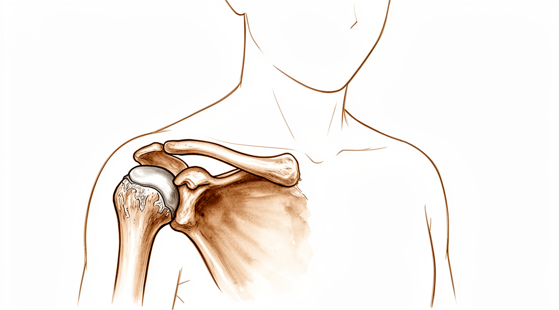
El hombro tiene una pequeña articulación donde el extremo externo de la clavícula se une a la punta del omóplato. Es la articulación AC, abreviatura de articulación acromioclavicular. Es una articulación móvil, no una unión rígida. Se desplaza unos milímetros a medida que el omóplato gira y se desliza durante el uso normal del brazo, y permite que el omóplato y la clavícula funcionen como una sola pieza conectada. Unos ligamentos resistentes la mantienen unida, con la ayuda de los músculos que cubren la parte superior del hombro.
Dentro de la articulación hay un fino disco amortiguador, algo parecido a una junta entre dos piezas de hueso. A partir del inicio de la mediana edad, este disco empieza a desgastarse, y las superficies articulares que hay detrás se desgastan con él. Eso es la artritis por desgaste, y en esta articulación es muy frecuente. El desgaste suele comenzar de forma silenciosa entre los veinte y los treinta años y avanza poco a poco con los años. Levantar cargas pesadas o de forma repetitiva, sobre todo el trabajo por encima de la cabeza, lo acelera. También puede acelerarlo un antiguo esguince o una separación de la articulación por una caída, incluso una leve que pareció curarse.
El problema es que una articulación de aspecto desgastado no siempre duele. Muchas personas tienen desgaste en sus estudios de imagen y ningún síntoma, y la mayoría de las articulaciones que parecen desgastadas no dan problemas. Así pues, el dolor que siente procede de la propia articulación solo cuando el desgaste, las superficies en carne viva o la inflamación que las rodea irritan realmente los tejidos de esa zona. Por eso sus síntomas se localizan justo en la parte superior del hombro y empeoran después de cargarlo.
Una forma menos frecuente de este problema se llama osteólisis distal de la clavícula. En ella, el propio extremo externo de la clavícula se deteriora, con inflamación, pequeñas fracturas en el hueso y pérdida ósea. Se observa sobre todo en hombres jóvenes que levantan cargas pesadas o de forma repetitiva, y provoca el mismo tipo de artritis en la articulación.
Si el desgaste se vuelve grave, pueden formarse espolones óseos cerca de la articulación que presionan sobre el tendón que pasa por debajo del techo del hombro, lo que agrava el problema.
Qué podemos hacer al respecto
El Dr. Kieran Hirpara, cirujano de extremidades superiores en el Mater Private Hospital Rockhampton, comienza con las opciones menos invasivas que se adapten a su condición. Por lo general, los pacientes son derivados a nuestra clínica por su médico de cabecera; si un fisioterapeuta le ha sugerido que nos consulte, igualmente necesitará una derivación de su médico de cabecera para poder acceder al reembolso de Medicare. Confirmamos lo que ocurre mediante una historia clínica cuidadosa, una exploración y estudios de imagen cuando son necesarios. En un problema de larga evolución como este, normalmente probamos primero el tratamiento no quirúrgico y solo consideramos la cirugía cuando este no ha producido una mejoría suficiente.
Los primeros pasos son medidas que puede tomar usted mismo. Dejar la articulación en reposo significa reducir el levantamiento de cargas pesadas y el trabajo por encima de la cabeza que cargan la parte superior del hombro. El hielo puede calmar un brote de dolor después de un día ajetreado. La fisioterapia busca calmar la articulación irritada y desarrollar la fuerza alrededor del hombro para que las tareas diarias la sometan a menos esfuerzo. Dé a estos cambios un tiempo razonable antes de juzgarlos, ya que el dolor suele empeorar después de la actividad más que durante ella.
Si el autocuidado no es suficiente, los analgésicos y los antiinflamatorios pueden ayudar. Los antiinflamatorios son tabletas que calman la inflamación de la articulación además de aliviar el dolor. También utilizamos inyecciones de cortisona, un potente medicamento antiinflamatorio, aplicadas en la propia articulación. Una inyección puede cumplir dos funciones: ayuda a confirmar que esta articulación es el origen de su dolor y puede aliviar el dolor como tratamiento. Aproximadamente 47 de cada 100 personas que reciben una inyección siguen teniendo un buen resultado un año después.
La cirugía se plantea cuando el dolor persiste a pesar de estos tratamientos. La operación extirpa unos milímetros de hueso del extremo externo de la clavícula, la mitad de la articulación desgastada que se encuentra en el lado de la clavícula. Al eliminar esa superficie desgastada, los dos extremos óseos en carne viva dejan de rozar entre sí. Puede realizarse a través de pequeñas incisiones con una cámara, o a través de una pequeña incisión abierta. Si ha tenido una separación previa de esta articulación con inestabilidad persistente, lo hablaremos detenidamente con usted, porque eso cambia lo bien que suele funcionar esta operación. Si la cirugía es adecuada para usted es una decisión que tomamos juntos, sopesando lo que quiere que su hombro pueda hacer frente a lo que implica la operación.
Qué esperar
Para la mayoría de las personas, el desgaste de esta articulación que aparece en un estudio de imagen pero no causa dolor simplemente sigue sin dar problemas. A lo largo de siete años, el 90 % de las articulaciones de este tipo que al principio no dolían nunca llegaron a dar síntomas. Así que, si su articulación parece desgastada pero no le duele, hay muchas probabilidades de que siga sin darle problemas.
Cuando la articulación sí duele, la evolución varía. Algunas personas notan que el dolor se calma con reposo, reduciendo el levantamiento de cargas pesadas y con fisioterapia. Otras notan que va y viene: empeora después de un día ajetreado y se alivia cuando vuelve a descansar. Sin tratamiento, el dolor tiende a persistir o a reaparecer cada vez que carga la parte superior del hombro, porque el desgaste de la articulación no se revierte por sí solo.
Si el tratamiento no quirúrgico no proporciona suficiente alivio, una inyección de cortisona en la articulación ayuda a algunas personas. Aproximadamente 47 de cada 100 personas que reciben una inyección siguen teniendo un buen resultado un año después. Eso significa que, en aproximadamente la mitad de las personas, el alivio desaparece en el plazo de un año, y puede ser necesario repetir la inyección o plantear otras opciones.
Si el dolor persiste a pesar de estos tratamientos, extirpar unos milímetros de hueso del extremo externo de la clavícula es una opción que su cirujano puede comentar con usted. El objetivo es que las dos superficies desgastadas y en carne viva dejen de rozar entre sí. La recuperación de esa operación lleva de semanas a meses, guiada por su fisioterapeuta, y la mayoría de las personas nota que el dolor en la parte superior del hombro se alivia a medida que la articulación se asienta.
Lo que puede esperar de forma realista en los próximos meses depende del camino que elija. Con un autocuidado sensato y fisioterapia, muchas personas vuelven a sus actividades normales con el dolor reducido a un nivel con el que pueden convivir. Si la articulación sigue empeorando a pesar de haber probado estas medidas durante un tiempo razonable, conviene volver a su médico de cabecera o solicitar una evaluación especializada en lugar de seguir aguantando el dolor.
¿Cuándo consultar a un especialista?
Se trata de un problema de desgaste, no de una urgencia, por lo que con esta afección no hay signos de peligro que requieran atención el mismo día. Lo importante es si el dolor se calma. Acuda a su médico de cabecera si el dolor en la parte superior del hombro no mejora después de unas semanas de evitar levantar cargas pesadas, o si sigue despertándole cuando se acuesta sobre ese lado. Solicite una evaluación especializada si el dolor empeora con el paso de las semanas, le impide trabajar o usar el brazo con normalidad, o si levantar el brazo y llevarlo a través del cuerpo siguen siendo dolorosos a pesar de haber reducido la actividad. Si su hombro se pone caliente, rojo, hinchado y doloroso, sobre todo con fiebre, eso no es artritis y necesita atención el mismo día en un servicio de urgencias.
En mayor profundidad
Advanced reading: the deeper science (optional)
Esta sección va más allá de lo necesario para que usted tome sus propias decisiones de tratamiento. La artritis de la articulación acromioclavicular merece ser leída con mayor atención por una razón desagradable: es extremadamente frecuente en las imágenes diagnósticas, se le atribuye con frecuencia el dolor en el hombro, y la evidencia de que su tratamiento resulta beneficioso es más escasa que en casi cualquier otro tema tratado en este sitio.
Las evidencias no demuestran que nada funcione
Una revisión sistemática sobre el tratamiento de la artrosis primaria de la articulación acromioclavicular agrupó a 1,902 pacientes y llegó a una conclusión que rara vez se expresa de forma tan clara: los estudios presentaban variaciones en cuanto a indicaciones, intervenciones y calidad; no aportaron evidencia de que ni las intervenciones no quirúrgicas ni las quirúrgicas sean eficaces [1].
Esto no equivale a afirmar que nada sirve de ayuda. Significa simplemente que no se han realizado ensayos con un nivel de rigor suficiente para demostrarlo. La excisión distal de la clavícula es una operación de larga tradición, fundamentada en el razonamiento mecánico de que al eliminar la superficie articular desgastada se elimina el dolor; sin embargo, “ser una técnica de larga tradición y tener sentido desde el punto de vista mecánico” no constituye evidencia científica. Es importante conocer esta distinción a la hora de tomar decisiones terapéuticas.
Añadir este procedimiento a otra operación no mejora dicha operación
La prueba más directa proviene de pacientes a quienes se realizó otro procedimiento al mismo tiempo. Entre 208 pacientes con roturas del manguito rotador, la resección distal de la clavícula no generó mejores puntuaciones de resultado clínico ni mayor amplitud de movimiento [2].
Esto es relevante porque esa es precisamente la situación en la que con mayor frecuencia se realiza la resección: el cirujano ya está dentro de la articulación del hombro, la articulación acromioclavicular parece degenerada en las imágenes, y la extracción de unos pocos milímetros de hueso es un procedimiento rápido. La evidencia indica que el paciente no obtiene beneficios perceptibles. Si se propone este procedimiento como complemento a la reparación del manguito rotador, es razonable plantear esta duda.
La técnica no es la cuestión relevante
Se han comparado repetidamente la resección abierta y la resección artroscópica; los resultados son consistentes: resultados funcionales y clínicos similares con cualquiera de los dos métodos en 319 pacientes [3]. Asimismo, una comparación previa con 429 pacientes tampoco arrojó una clara ventaja para ninguno de los métodos [4].
Cuando dos técnicas muy distintas producen el mismo resultado, la conclusión lógica es que la técnica no es lo que determina el desenlace; lo que realmente importa es la selección del paciente.
¿Por qué es tan difícil hacer una selección diagnóstica en este caso?
La articulación acromioclavicular se degenera en casi todas las personas con la edad. Se trata de una articulación pequeña que soporta una carga elevada; su disco articular es delgado y se desgasta con facilidad. Por ello, encontrar signos de degeneración de dicha articulación en un estudio por imagen es algo esperado a partir de la mediana edad, más que un verdadero diagnóstico.
Existe una correlación estructural medible: en personas con síntomas de degeneración de la articulación acromioclavicular, tanto la extremidad distal de la clavícula como el acromion presentan un aumento de tamaño; en cambio, en personas asintomáticas esa relación entre ambas estructuras permanece inalterada [5]. Este dato indica que la degeneración sintomática y la degeneración incidental difieren a nivel estructural; sin embargo, se trata de una observación a nivel grupal, no de una prueba que pueda aplicarse a cada caso individual.
En la práctica, esto explica por qué la inyección diagnóstica en la articulación tiene tanta importancia en estos casos. Si el anestésico local, administrado con precisión en la articulación acromioclavicular, elimina el dolor, es muy probable que dicha articulación sea la fuente del problema. Si, por el contrario, el dolor persiste, la degeneración observada en el estudio por imagen probablemente sea un hallazgo incidental; en ese caso, resecarla no aportaría beneficios, tal como advierten los datos clínicos mencionados anteriormente.
Referencias
[1] Welch M, Rankin S, How Saw Keng M, Woods D. Revisión sistemática del tratamiento de la artrosis primaria de la articulación acromioclavicular. Shoulder Elbow. 2023;16(2):129-44. https://doi.org/10.1177/17585732231157090
[2] Wang J, Ma J, Zhu S, Jia H, Ma X. ¿Reduce la resección de la clavícula distal el dolor o mejora la función del hombro en pacientes con rotura del manguito rotador? Un metaanálisis. Clin Orthop Relat Res. 2018;476(12):2402-14. https://doi.org/10.1097/CORR.0000000000000424
[3] Hohmann E, Tetsworth K, Glatt V. Resección abierta versus artroscópica de la articulación acromioclavicular: revisión sistemática y metaanálisis. Arch Orthop Trauma Surg. 2019;139(5):685-94. https://doi.org/10.1007/s00402-019-03114-w
[4] Pensak M, Grumet RC, Slabaugh MA, Bach BR. Resección abierta versus artroscópica de la clavícula distal. Arthroscopy. 2010;26(5):697-704. https://doi.org/10.1016/j.arthro.2009.12.007
[5] Bulkmans K, Peeters I, De Wilde L, Van Tongel A. Relación entre el acromion y la clavícula distal en articulaciones acromioclaviculares normales y con degeneración sintomática. Arch Orthop Trauma Surg. 2019;140(4):465-72. https://doi.org/10.1007/s00402-019-03258-9
Evidence & references
This is the clinical evidence summary written for health professionals. It is technical, and it lists the research this page was built from. You do not need to read it to understand your treatment or to make a decision about it.
Overview
- Asymptomatic acromioclavicular osteoarthritis remained asymptomatic in 90% of cases over a seven-year course [1].
- Treatment choice for acromioclavicular dislocation should not be influenced by the potential development of acromioclavicular osteoarthritis [2].
- Further characterisation is needed to identify patients in whom mild arthroscopic findings of acromioclavicular joint osteoarthritis are clinically significant and warrant resection [3].
- Persistent pain and osteoarthritis progression remain concerns following revision of failed open anterior stabilization of the shoulder [4].
- Both open and arthroscopic resection arthroplasty techniques provide predictable pain relief for symptomatic acromioclavicular osteoarthritis [5].
- Symptomatic acromioclavicular joint osteoarthritis occurred in 15.9% of patients after total anatomic shoulder replacement with follow-up of up to 12 years [6].
- Untreated acromioclavicular joint osteoarthritis, symptomatic or not, encountered during arthroscopic rotator cuff repair is associated with a low percentage of failure [8].
- Biplanar reconstruction with pectoralis minor tendon and coracoacromial ligament transfer aims to preserve the lateral portion of the clavicle whenever possible, limiting resection exclusively to cases of acromioclavicular joint osteoarthritis or irreducible joints [9].
- Radiographic acromioclavicular joint osteoarthritis is common in patients undergoing reverse shoulder arthroplasty [10].
- Clinicians should consider overreduction of the acromioclavicular joint because it may lead to favorable radiological results [11].
- A distinction between symptomatic and asymptomatic radiographic acromioclavicular osteoarthritis is unnecessary, as all patients were equally satisfied with the outcome following preoperative acromioclavicular injection [12].
- Open and arthroscopic acromioclavicular joint reconstruction techniques have no differences in loss of reduction, the complication rate, and the revision rate [13].
- Arthroscopic debridement of the acromioclavicular joint can produce a favorable result in young patients presenting with isolated acromioclavicular joint-related pain but with normal imaging [15].
- Limited distal clavicle excision of patients with acromioclavicular joint osteoarthritis resistant to conservative treatment reduced pain and improved shoulder function at midterm follow-up [17].
- All patients undergoing free tissue graft reconstruction of the acromioclavicular joint are satisfied with their surgery and demonstrate good reduction of the acromioclavicular joint on radiographs, although follow-up periods are less than 1 year [21].
- Additional research is needed to determine the main cause of pain and compare clinical outcomes of intra-articular versus extra-articular injections for isolated acromioclavicular osteoarthritis [23].
- Nanoscopic distal clavicle resection offers advantages including decreased morbidity, less risk to the acromioclavicular joint ligaments, and decreased loss and need for fluid [44].
Anatomy & Pathophysiology
Bony Anatomy and Joint Structure
- The acromioclavicular (AC) joint is a diarthrodial joint that supports the shoulder girdle through the clavicular “strut” [36].
- The AC joint serves as a primary link between the axial skeleton and the upper extremity [122].
- The AC joint is movable in all planes and is not a rigid structure [122].
- The normal AC joint is capable of translating 4 to 6 mm in the anterior, posterior, and superior planes under 70-N loads [122].
- The AC joint accommodates rotary motion of 5° to 8° during scapulothoracic motion and 40° to 45° with shoulder abduction and elevation [122].
- The scapula and clavicle are linked into a single segment referred to as the “claviscapula” [112].
Ligamentous Anatomy and Stability
- The AC and coracoclavicular (CC) ligaments are the static stabilizers of the AC joint, whereas the deltoid and trapezius muscles are the dynamic stabilizers [122].
- The horizontal plane stability of the clavicle is provided by the AC ligaments, specifically the posterior and superior portions [25].
- The AC and CC ligaments play a role in torque transduction and horizontal and vertical stability [112].
- Injury to the AC joint results in progressive disruption of the ligamentous support, beginning with the capsular ligaments and progressing to the CC ligaments [30].
Pathophysiology of Degeneration
- AC joint osteoarthritis is more common with advanced age following degeneration of the intra-articular disk [25].
- Arthritic deterioration of the AC joint starts in early middle age [25].
- AC joint osteoarthritis is more common in patients engaged in repetitive overhead or lifting activities [25].
- Previous low-grade AC joint separations can result in painful arthritis [25].
- The radiographic severity of AC joint arthritis does not always correlate with patient symptoms [25].
- Asymptomatic AC joint degeneration is frequent and does not always correlate with the presence of symptoms [36].
- Degeneration of the AC joint is classified as primary osteoarthritis, posttraumatic osteoarthritis, or distal clavicle osteolysis [123].
- The process of AC joint degeneration begins in the second decade of life and can progress to complete obliteration of the joint [123].
- Complete obliteration of the AC joint can lead to subclavicular spurring, which contributes to subacromial impingement of the supraspinatus [123].
- Asymptomatic AC osteoarthritis remained asymptomatic in 90% of cases over a seven-year course [1].
- Patients with loss of immediate postoperative reduction after AC joint dislocation more often developed radiologic and symptomatic AC osteoarthritis than those in whom the AC joint was anatomically restored [7].
- Kinematic changes associated with AC joint dislocation could be a potential source of pain and dysfunction in the shoulder [59].
- Scapular and clavicular kinematics are affected in AC separation models [60].
- Decoupling the claviscapular segment has deleterious effects on the functional integrity of the AC joint complex within scapulohumeral rhythm [112].
Distal Clavicle Osteolysis
- Distal clavicle osteolysis is characterized by localized hyperemia of the distal clavicle, resulting in inflammation, bone resorption, microfractures, and secondary arthritis of the AC joint [25].
- Distal clavicle osteolysis is more common in males and seen in younger patients [25].
- Distal clavicle osteolysis is associated with heavy lifting or repetitive motions [25].
- Osteolysis of the distal clavicle was first reported in conjunction with acute trauma [121].
- Intraarticular distal clavicle fractures and AC separations can lead to osteolysis [121].
- Other causes of distal clavicle osteolysis include rheumatoid arthritis, hyperparathyroidism, and repetitive microtrauma [121].
- The most common cause of distal clavicle osteolysis is repetitive microtrauma [121].
- Radiographic findings indicative of distal clavicle osteolysis include loss of subchondral bone detail, cystic reabsorption of the distal clavicle, and generalized osteopenia of the distal clavicle [121].
Classification
- Asymptomatic acromioclavicular osteoarthritis diagnosed by MRI remained asymptomatic in 90% of cases over a seven-year course [1].
- Treatment choice for acromioclavicular dislocation should not be influenced by the potential development of acromioclavicular osteoarthritis based on available evidence [2].
- Patients with loss of immediate postoperative reduction following arthroscopically assisted reduction of acute acromioclavicular joint dislocation more often developed radiologic and symptomatic acromioclavicular osteoarthritis than those in whom the joint was anatomically restored [7].
- Untreated acromioclavicular joint osteoarthritis, whether symptomatic or not, encountered during arthroscopic rotator cuff repair is associated with a low percentage of failure [8].
- Biplanar reconstruction with pectoralis minor tendon and coracoacromial ligament transfer aims to preserve the lateral portion of the clavicle, limiting resection exclusively to cases of acromioclavicular joint osteoarthritis or irreducible joints [9].
- Open and arthroscopic acromioclavicular joint reconstruction techniques have no differences in loss of reduction, complication rate, and revision rate based on available literature [13].
- A study evaluated three groups of patients: 84 acromioclavicular joints without clinical or radiological pathology, 39 patients with signs of osteoarthritis on CT without symptoms, and 30 patients with acromioclavicular joint pain [14].
- In the study of acromion-clavicle relationships, Group 2 included patients with signs of osteoarthritis on CT (joint space narrowing, osteophytes, and/or bone cyst) who were asymptomatic based on physical examination and negative cross-body adduction test [14].
- In the study of acromion-clavicle relationships, Group 3 included patients with acromioclavicular joint pain confirmed by tenderness and temporary pain relief with intra-articular injection, who were treated with open distal clavicle resection after failure of non-operative treatment [14].
- The occurrence of acromioclavicular joint osteoarthrosis was surprisingly frequent in a study of shoulder pain [18].
- Anatomic techniques that address both coracoclavicular ligaments and the acromioclavicular capsule are recommended to restore horizontal and vertical stability while allowing physiological rotation [19].
- The acromial center line to dorsal clavicle radiographic measurement and use of the Alexander view provides a more realistic appreciation of true acromioclavicular joint displacement, especially in defining watershed cases (IIIA/IIB/IV) [22].
- Osteolytic changes seemed to be associated with incongruity of the acromioclavicular joint but did not correlate with clinical results in a review of surgical treatment for acromioclavicular dislocation [24].
- A procedure for exact anatomical acromioclavicular joint reconstruction using acromioclavicular and coracoclavicular FiberWire cerclage shows good objective results with high Constant and Rowe scores for shoulder function [27].
- Preoperative radiographs were evaluated to determine whether there was any osteolysis of the distal clavicle in a study of preoperative factors associated with subsequent distal clavicle resection after rotator cuff repair [31].
- Preoperative radiographs were graded for severity of arthritis per the Petersson classification by two attending orthopaedic surgeons blinded to each other's grade in a study of preoperative factors associated with subsequent distal clavicle resection after rotator cuff repair [31].
- Type V acromioclavicular dislocations may be given a trial of conservative therapy [35].
- Non-operative reduction and stabilization of high-grade acromioclavicular joint separations seems to be a valuable treatment option [45].
- Evaluating the integrity of the capsuloligamentous structures stabilizing the acromioclavicular joint is reproducible and gives additional information on the severity of the injury, which might also influence the treatment decision [49].
- Severe chronic symptomatic acromioclavicular joint separations (Rockwood types III through V) can be repaired entirely by arthroscopy safely and effectively by transferring the coracoacromial ligament with a bone block in the distal clavicle [50].
- The treatment of Type III acromioclavicular joint separation has remained controversial, with many studies demonstrating good results with nonoperative management and most series failing to document the superiority of either operative or nonoperative management [51].
- Surgical management of acromioclavicular joint injuries is associated with a relatively high complication profile and common loss of reduction, though the revision surgery rate is low [53].
- Arthroscopically assisted treatment of acute acromioclavicular joint dislocation provides good clinical results and few complications, though the rate of recurrence and postoperative loss of reduction requires better definition of indications and improvement of surgical implants and technique [55].
- In every coracoclavicular loop repair, regardless of hole placement, complete acromioclavicular joint congruity is not restored [86].
- Given that more than 100 surgical stabilization techniques have been proposed for acromioclavicular joint injuries, a deeper understanding of the underlying anatomy integrated with biomechanical evidence may help guide the development of a standardized, evidence-based approach to treatment [88].
- Early and delayed surgical interventions of high-grade acromioclavicular joint dislocation provide equivalent clinical scores when combined coracoclavicular and acromioclavicular joint fixation is used for stabilization [95].
- Acromioclavicular joint osteoarthritis was graded using the Kellgren-Lawrence classification system in a retrospective cohort study of concomitant open distal clavicle excision after reverse total shoulder arthroplasty [101].
Clinical Presentation
History and Symptoms
- Patients report activity-related pain localized to the AC joint [25].
- Pain may radiate anteriorly or along the trapezius [25].
- Pain is reported with heavy lifting or when sleeping on the affected side [25].
- AC joint osteoarthrosis is a frequent cause of shoulder pain [18].
- In patients with rheumatoid arthritis, clinical symptoms and radiologic appearances are often poor indicators of the source of pain in the shoulder joint complex [64].
Physical Examination
- Point tenderness is seen at the AC joint [25].
- Horizontal stability of the AC joint should be assessed [25].
- Pain at the AC joint with terminal elevation and cross-body motion is often seen [25].
- The adduction sign, where pain is produced in the AC joint during maximal adduction of the internally rotated arm, was positive in all 20 patients with AC joint arthritis in one study [63].
- An average decrease of 5 cm in the distance from the ipsilateral antecubital fossa to the contralateral anterior shoulder was noted during cross-body adduction in patients with AC joint arthritis [63].
- Selective injection of anesthetic into the AC joint can confirm the diagnosis [25].
- Patients with ACJ pain showed clinical signs including AC joint tenderness to palpation and temporary pain relief with intra-articular injection [14].
Imaging
- An AP view and/or a Zanca view of the shoulder provides good visualization of the AC joint [25].
- Osteophyte formation, sclerotic reaction, and bone cysts are commonly seen on radiographs of the AC joint [25].
- Bone and joint edema on MRI correlate with AC joint pain [25].
- The radiographic severity of arthritis does not always correlate with patient symptoms [25].
- Radiographic ACJ osteoarthritis is common in patients undergoing reverse shoulder arthroplasty [10].
- Asymptomatic AC-OA remained asymptomatic in 90% of patients over a seven-year course [1].
- A distinction between symptomatic and asymptomatic radiographic AC OA may be unnecessary, as all patients were equally satisfied with the outcome in one study [12].
- Osteolytic changes seemed to be associated with incongruity of the AC joint but did not correlate with clinical results [24].
Investigations
Clinical Evaluation
- Patients with AC joint osteoarthritis report activity-related pain localized to the AC joint, with occasional radiation anteriorly or along the trapezius [25].
- Pain with heavy lifting or when sleeping on the affected side is reported in patients with AC joint osteoarthritis [25].
- Physical examination of the AC joint reveals point tenderness at the joint [25].
- Horizontal stability of the AC joint should be assessed during physical examination [25].
- Pain at the AC joint with terminal elevation and cross-body motion is often seen in patients with AC joint osteoarthritis [25].
- Selective injection of anesthetic into the AC joint can confirm the diagnosis of symptomatic AC osteoarthritis [25].
- A distinction between symptomatic and asymptomatic radiographic AC OA is unnecessary, as all patients were equally satisfied with the outcome following preoperative injection [12].
Imaging
- Patients with edema on MRI were more likely to present pain than patients without edema [92].
- Subchondral bone edema on histologic examination was more frequent in patients with pain [92].
- The AC-DC measurement and use of the Alexander view provides a more realistic appreciation of true AC joint displacement, especially in defining watershed cases (ie, IIIA/IIB/IV) [22].
- Preoperative radiographs are evaluated to determine whether there is any osteolysis of the distal clavicle [31].
- The Petersson classification is used to grade the severity of arthritis on preoperative radiographs [31].
- In a study of AC joint osteoarthrosis, the occurrence was surprisingly frequent [18].
Treatment
Non-Operative Management
- Rest, ice, and NSAIDs are used initially for AC joint osteoarthritis [25].
- Corticosteroid injections can be used for diagnostic or therapeutic purposes in AC joint osteoarthritis [25].
- AC injections offer a 1-year success rate of 47% [33].
- Conservative and surgical treatments are both effective in acromioclavicular joint osteoarthritis management [38].
- A distinction between symptomatic and asymptomatic radiographic AC OA is unnecessary, as all patients were equally satisfied with the outcome following injection [12].
- Additional research is needed to determine the main cause of pain and compare clinical outcomes of intra-articular versus extra-articular injections [23].
Operative Management: Distal Clavicle Resection
- Surgical indications for AC joint osteoarthritis include persistent pain and failure of nonsurgical treatment [25].
- Both open and arthroscopic resection arthroplasty techniques provide predictable pain relief for symptomatic AC osteoarthritis [5].
- Arthroscopic distal clavicle excision provides reliable pain relief in >90% of patients in the absence of instability [25].
- Previous traumatic instability is associated with persistent pain in 30% to 40% of cases following distal clavicle excision [25].
- One systematic review showed slightly better results with arthroscopic excision than with open distal clavicle excision [25].
- Direct comparison studies have shown similar or better results with arthroscopic excision than with open techniques [25].
- Biomechanical evidence suggests a resection of 5 mm is needed to prevent contact between the clavicle and the acromion in the absence of instability [25].
- Care should be taken to preserve the posterior and superior AC ligaments during arthroscopic distal clavicle excision [25].
- Between 5 and 10 mm of the distal clavicle should be resected during an open Mumford procedure [25].
- Meticulous repair of the deltotrapezial fascia is important during open distal clavicle resection [25].
- Limited distal clavicle excision of patients with AC joint osteoarthritis resistant to conservative treatment reduced pain and improved shoulder function at midterm follow-up [17].
- Arthroscopic debridement of the AC joint can produce a favorable result in young patients presenting with isolated AC joint-related pain but with normal imaging [15].
- The nanoscopic distal clavicle resection technique offers advantages including decreased morbidity, less risk to the AC joint ligaments, and decreased loss and need for fluid [44].
- Patients with AC joint instability have poor results after distal clavicle resection [36].
- The optimal amount of distal clavicle resection remains elusive [36].
Operative Management: Concomitant Procedures and Specific Populations
- Preventive arthroscopic distal clavicle resection is not recommended in patients with radiological but asymptomatic ACJ arthritis and rotator cuff tears [34].
- Untreated ACJ osteoarthritis, symptomatic or not, encountered during arthroscopic rotator cuff repair is associated with a low percentage of failure [8].
- Further characterisation of patients in whom mild arthroscopic findings of OA of AC joint are clinically significant and warrant resection is needed [3].
- Symptomatic ACJ OA occurred in 15.9% of patients after total anatomic shoulder replacement with follow-up of up to 12 years [6].
- Osteoarthritis is associated with poorer final clinical outcomes after rotator cuff repair, and an unhealed or re-torn cuff increases the risk of osteoarthritis [26].
Operative Management: Reconstruction and Stabilization
- Open and arthroscopic AC joint reconstruction techniques have no differences in loss of reduction, the complication rate, and the revision rate based on the available literature [13].
- Treatment choice for AC dislocation should not be influenced by the potential development of AC OA [2].
- Achieving and maintaining satisfactory joint reduction is a factor of good prognosis, both radiologically and functionally, in AC dislocation treatment [7].
- Patients with loss of immediate postoperative reduction more often developed radiologic and symptomatic AC osteoarthritis than those in whom the AC joint was anatomically restored [7].
- Clinicians should consider overreduction of the AC joint because it may lead to favorable radiological results [11].
- Ensuring good initial recovery of the coracoclavicular distance and precise placement of the AC joints was important in maintaining the proper AC position at the final follow-up [46].
- Combined stabilization of the AC capsule and CC ligaments demonstrated the greatest capacity to restore native stability against translational and rotational loads [81].
- The biplanar reconstruction procedure aims to preserve the lateral portion of the clavicle whenever possible, limiting resection exclusively to cases of AC joint osteoarthritis or irreducible joints [9].
- Some persistent pain and osteoarthritis progression remain concerns following revision of failed open anterior stabilization of the shoulder [4].
Complications
Natural History and Progression
- Patients who experienced loss of immediate postoperative reduction after acute acromioclavicular dislocation more often developed radiologic and symptomatic acromioclavicular osteoarthritis than those in whom the joint was anatomically restored [7].
- Osteoarthritis is associated with poorer final clinical outcomes following rotator cuff repair [26].
- An unhealed or re-torn rotator cuff increases the risk of developing osteoarthritis [26].
- Severe acromioclavicular joint osteoarthritis is associated with acromial stress fractures after reverse shoulder arthroplasty [10].
Surgical Complications and Outcomes
- Treatment of acromioclavicular dislocation by synthetic ligament reconstruction carries a risk of significant early osteolysis [56].
- Complications such as anterior shoulder pain, acromioclavicular joint asymmetry, and activity-related weakness are common sequelae of coracoclavicular ligament reconstruction [43].
- Three patients with old acromioclavicular separations had prominence of the distal clavicle or symptoms due to residual hypermobility of the clavicle despite concurrent coracoclavicular stabilization [114, 115, 116, 117].
- A patient who did not undergo anterior acromioplasty despite subacromial impingement had unchanged impingement symptoms after distal clavicle resection [114, 115, 116, 117].
- Open and arthroscopic resection arthroplasty techniques for symptomatic acromioclavicular osteoarthritis each have a unique set of potential complications that may be minimized with improved understanding of anatomy, biomechanics, and meticulous surgical technique [5].
Recovery
Natural History and Prognosis
- Asymptomatic acromioclavicular osteoarthritis diagnosed by MRI remained asymptomatic in 90% of patients over a seven-year course [1].
- Osteolytic changes associated with incongruity of the acromioclavicular joint did not correlate with clinical results in a review of 39 patients [24].
- An unhealed or re-torn rotator cuff increases the risk of osteoarthritis following rotator cuff repair [26].
Impact of Joint Reduction and Stability
- Patients with loss of immediate postoperative reduction after arthroscopically assisted reduction of acute acromioclavicular joint dislocation more often developed radiologic and symptomatic acromioclavicular osteoarthritis than those in whom the joint was anatomically restored [7].
- Achieving and maintaining satisfactory joint reduction is a factor of good prognosis, both radiologically and functionally [7].
- Ensuring good initial recovery of the coracoclavicular distance and precise placement of the acromioclavicular joints was important in maintaining the proper acromioclavicular position at final follow-up [46].
Surgical Considerations Regarding Osteoarthritis
- Further characterisation of patients in whom mild arthroscopic findings of acromioclavicular joint osteoarthritis are clinically significant and warrant resection is needed [3].
- Biplanar reconstruction for chronic acromioclavicular joint dislocations aims to preserve the lateral portion of the clavicle, limiting resection exclusively to cases of acromioclavicular joint osteoarthritis or irreducible joints [9].
- The average angle from the central portion of the acromioclavicular joint anteriorly for a novel anterior portal for arthroscopic distal clavicle excision was 6 degrees, with a range of 0 to 15 degrees [29].
Non-Operative Management and Diagnostic Implications
- Acromioclavicular injections offer a 1-year success rate of 47% in patients with acromioclavicular osteoarthritis [33].
Key Evidence
- [L2] Asymptomatic AC-OA remained asymptomatic in 90% over 7 years. [1] (10.1016/j.jse.2019.04.004)
- [L4] Based on the available evidence, treatment choice for AC dislocation should not be influenced by the potential development of AC OA. [2] (10.2106/jbjs.rvw.24.00085)
- [L2] Further characterisation of patients in whom mild arthroscopic findings of OA of AC joint are clinically significant and warrant resection is needed. [3] (10.1007/s00167-014-3114-2)
- [L4] Some persistent pain and osteoarthritis progression remain concerns. [4] (10.1016/j.arthro.2009.04.073)
- [L5] Both open and arthroscopic resection arthroplasty techniques provide predictable pain relief for symptomatic AC osteoarthritis, though each has a unique set of potential complications that may be minimized with improved understanding of anatomy, biomechanics, and meticulous surgical technique. [5] (10.1177/0363546513485359)
- [L4] Symptomatic ACJ OA occurred in 15.9% of patients after total anatomic shoulder replacement with follow-up of up to 12 years. [6] (10.1177/17585732221114796)
- [L4] [7] (10.1016/j.otsr.2017.11.001)
- [L2] Untreated ACJ osteoarthritis, symptomatic or not, encountered during arthroscopic RCR is associated with a low percentage of failure. [8] (10.1007/s00167-020-06098-y)
- [L5] The procedure aims to preserve the lateral portion of the clavicle whenever possible, limiting resection exclusively to cases of AC joint osteoarthritis or irreducible joints. [9] (10.1016/j.eats.2024.103104)
- [L3] Radiographic ACJ osteoarthritis is common in patients undergoing RSA. [10] (10.1016/j.jseint.2021.11.008)
- [L3] Clinicians should consider overreduction of the AC joint because it may lead to favorable radiological results. [11] (10.1177/0363546519862850)
- [L4] The outcomes of this study seem to suggest that a distinction between symptomatic and asymptomatic radiographic AC OA is unnecessary, as all patients were equally satisfied with the outcome. [12] (10.5397/cise.2023.00073)
- [L1] Open and arthroscopic AC joint reconstruction techniques have no differences in loss of reduction, the complication rate, and the revision rate based on the available literature. [13] (10.1177/0363546518795147)
- [L3] [14] (10.1007/s00402-019-03258-9)
- [L4] Arthroscopic debridement of the AC joint can produce a favorable result in this difficult group of young patients presenting with isolated AC joint-related pain but with normal imaging. [15] (10.1097/bte.0b013e31816276cb)
- [L4] Limited distal clavicle excision of patients with AC joint osteoarthritis resistant to conservative treatment reduced pain and improved shoulder function at midterm follow-up. [17] (10.1016/j.otsr.2016.01.008)
- [L3] In this study the occurrence of AC joint osteoarthrosis was surprisingly frequent. [18] (10.1016/s1058-2746(96)80122-4)
- [L5] Anatomic techniques that address both coracoclavicular ligaments and the AC capsule are recommended to restore horizontal and vertical stability while allowing physiological rotation. [19] (10.1016/j.arthro.2019.01.038)
- [L4] Although the follow-up periods are less than 1 year, all patients are satisfied with their surgery and demonstrate good reduction of the AC joint on radiographs. [21] (10.1097/bte.0b013e31818a5dd9)
- [L4] The AC-DC measurement and use of the Alexander view provides the clinician with a more realistic appreciation of true AC joint displacement, especially in defining watershed cases (ie, IIIA/IIB/IV) and may better inform the decision-making process regarding management options and recommendations. [22] (10.1016/j.jse.2019.12.014)
- [L4] Additional research is needed to determine the main cause of pain and compare clinical outcomes of intra-articular versus extra-articular injections. [23] (10.5397/cise.2023.00311)
- [L4] Osteolytic changes seemed to be associated with incongruity of the AC joint, but did not correlate with clinical results. [24] (10.1016/0020-1383(83)90092-x)
- [L4] Osteoarthritis is associated with poorer final clinical outcomes, and an unhealed or re-torn cuff increases the risk of osteoarthritis. [26] (10.1016/j.otsr.2017.03.007)
- [L4] This procedure for an exact anatomical AC joint reconstruction shows good objective results with a high Constant and Rowe scores for shoulder function. [27] (10.1055/s-2008-1038577)
- [L4] The average angle from the central portion of the AC joint anteriorly was 6 degrees (range, 0 to 15 degrees). [29] (10.1097/bte.0000000000000033)
- [L3] [31] (10.1177/2325967119844295)
- [L4] AC injections offer a 1-year success rate of 47%. [33] (10.5397/cise.2023.00031)
- [L1] Preventive arthroscopic DCR is not recommended in patients with radiological but asymptomatic ACJ arthritis. [34] (10.1177/0363546514547254)
- [L4] This suggests that Type V AC dislocations may be given a trial of conservative therapy. [35] (10.1177/2325967115s00017)
- [L5] [36] (10.5435/00124635-199905000-00004)
- [L4] Conservative and surgical treatments are both effective in acromioclavicular joint osteoarthritis management. [38] (10.1007/s00167-020-06377-8)
- [L4] Most patients were ultimately treated surgically, although complications such as anterior shoulder pain, AC joint asymmetry, and activity-related weakness were common sequelae resulting in physical limitations and separation from military service. [43] (10.1177/03635465211036713)
- [L5] The technique offers advantages including decreased morbidity, less risk to the AC joint ligaments, and decreased loss and need for fluid. [44] (10.1016/j.eats.2021.12.006)
- [L4] Non-operative reduction and stabilization of high-grade AC joint separations seems to be a valuable treatment option. [45] (10.1007/s00402-020-03630-0)
- [L4] Ensuring good initial recovery of the CCD and precise placement of the AC joints was important in maintaining the proper AC position at the final follow-up. [46] (10.1016/j.arthro.2018.07.007)
- [L1] Evaluating the integrity of the capsuloligamentous structures stabilizing the AC joint is reproducible and gives additional information on the severity of the injury, which might also influence the treatment decision. [49] (10.1016/j.jse.2020.10.026)
- [L4] Severe chronic symptomatic AC joint separations (Rockwood types III through V) can be repaired entirely by arthroscopy safely and effectively by transferring the coracoacromial ligament with a bone block in the distal clavicle. [50] (10.1016/j.arthro.2009.08.008)
- [L5] [51] (10.1097/00132589-200603000-00004)
- [L4] Surgical management of AC joint injuries is associated with a relatively high complication profile and common loss of reduction, though the revision surgery rate is low. [53] (10.1016/j.jse.2023.03.019)
- [L4] [55] (10.1016/j.otsr.2010.10.004)
- [L4] Treatment of ACD by synthetic ligament reconstruction gave satisfactory results, notably in terms of recovery of strength, but evolution is not risk-free with onset of significant early osteolysis. [56] (10.1016/j.otsr.2010.06.004)
- [L5] The kinematic changes could be a potential source of pain and dysfunction in the shoulder with AC joint dislocation. [59] (10.1177/0363546512458571)
- [L5] Scapular and clavicular kinematics were affected in AC separation models. [60] (10.1016/j.jse.2013.01.004)
- [L4] [63] (10.1177/036354659302100113)
- [L4] Clinical symptoms and radiologic appearances are often poor indicators of the source of pain in the shoulder joint complex in patients with rheumatoid arthritis. [64] (10.1016/s1058-2746(09)80111-0)
- [L5] Combined stabilization of the AC capsule and CC ligaments demonstrated the greatest capacity to restore native stability against translational and rotational loads. [81] (10.1177/0363546518807908)
- [L5] In every CC loop repair, regardless of hole placement, complete AC joint congruity is not restored. [86] (10.1016/s1058-2746(03)00050-8)
- [L5] Given that more than 100 surgical stabilization techniques have been proposed for AC-joint injuries, a deeper understanding of the underlying anatomy, integrated with biomechanical evidence, may help to guide the development of a standardized, evidence-based approach to the treatment of AC-joint instability. [88] (10.2106/jbjs.25.01392)
- [L4] Patients with edema on MRI were more likely to present pain than patients without edema, and subchondral bone edema on histologic examination was more frequent in patients with pain. [92] (10.1016/j.jseint.2020.03.007)
- [L3] Early and delayed surgical interventions of high-grade AC joint dislocation provide equivalent clinical scores when combined CC and AC joint fixation is used for stabilization. [95] (10.1016/j.jse.2020.06.026)
- [L3] [101] (10.5397/cise.2023.00465)
- [L5] [112] (10.5435/jaaos-d-24-00360)
- [L4] [114] (10.1016/s1058-2746(96)80521-0)
- [L4] [115] (10.1016/s1058-2746(96)80520-9)
- [L4] [116] (10.1016/s1058-2746(96)80523-4)
- [L3] [117] (10.1016/s1058-2746(96)80522-2)
- [L4] [121] (10.1016/s1058-2746(10)80007-2)
- [L4] [122] (10.1302/2058-5241.3.170027)
- [L5] [123] (10.1016/s0278-5919(02)00102-3)
References
[1] Seven-year course of asymptomatic acromioclavicular osteoarthritis diagnosed by MRI. Journal of Shoulder and Elbow Surgery. 2019. DOI: 10.1016/j.jse.2019.04.004
[2] Choice of Acromioclavicular Dislocation Treatment Should Not Be Influenced by Risk of Development of Acromioclavicular Osteoarthritis. JBJS Reviews. 2024. DOI: 10.2106/jbjs.rvw.24.00085
[3] Outcome of distal clavicle resection in patients with acromioclavicular joint osteoarthritis and full‐thickness rotator cuff tear. Knee Surgery, Sports Traumatology, Arthroscopy. 2014. DOI: 10.1007/s00167-014-3114-2
[4] The Role of Arthroscopy in Revision of Failed Open Anterior Stabilization of the Shoulder. Arthroscopy. 2009. DOI: 10.1016/j.arthro.2009.04.073
[5] Degenerative Joint Disease of the Acromioclavicular Joint. The American Journal of Sports Medicine. 2013. DOI: 10.1177/0363546513485359
[6] The incidence and treatment of symptomatic acromioclavicular joint osteoarthritis following total shoulder arthroplasty. Shoulder & Elbow. 2022. DOI: 10.1177/17585732221114796
[7] Arthroscopically assisted reduction of acute acromioclavicular joint dislocation using a single double-button device: Medium-term clinical and radiological outcomes. Orthopaedics & Traumatology: Surgery & Research. 2018. DOI: 10.1016/j.otsr.2017.11.001
[8] Acromioclavicular joint arthritis is not an indication for routine distal clavicle excision in arthroscopic rotator cuff repair. Knee Surgery, Sports Traumatology, Arthroscopy. 2020. DOI: 10.1007/s00167-020-06098-y
[9] Biplanar Reconstruction With Pectoralis Minor Tendon and Coracoacromial Ligament Transfer for Chronic Acromioclavicular Joint Dislocations. Arthroscopy Techniques. 2024. DOI: 10.1016/j.eats.2024.103104
[10] Severe acromioclavicular joint osteoarthritis is associated with acromial stress fractures after reverse shoulder arthroplasty. JSES International. 2022. DOI: 10.1016/j.jseint.2021.11.008
[11] Factors Predicting the Outcome After Arthroscopically Assisted Stabilization of Acute High-Grade Acromioclavicular Joint Dislocations. The American Journal of Sports Medicine. 2019. DOI: 10.1177/0363546519862850
[12] Diagnostic value of a preoperative acromioclavicular injection for symptomatic acromioclavicular osteoarthritis: a retrospective study of cross-sectional midterm outcomes. Clinics in Shoulder and Elbow. 2024. DOI: 10.5397/cise.2023.00073
[13] Current Concepts in the Operative Management of Acromioclavicular Dislocations: A Systematic Review and Meta-analysis of Operative Techniques. The American Journal of Sports Medicine. 2018. DOI: 10.1177/0363546518795147
[14] The relationship of the acromion to the distal clavicle in normal and symptomatic degenerated acromioclavicular joints. Archives of Orthopaedic and Trauma Surgery. 2019. DOI: 10.1007/s00402-019-03258-9
[15] Acromioclavicular Joint Arthroscopy and Debridement. Techniques in Shoulder & Elbow Surgery. 2008. DOI: 10.1097/bte.0b013e31816276cb
[17] Limited distal clavicle excision of acromioclavicular joint osteoarthritis. Orthopaedics & Traumatology: Surgery & Research. 2016. DOI: 10.1016/j.otsr.2016.01.008
[18] Osteoarthrosis of the acromioclavicular joint, an underestimated cause of shoulder pain. Journal of Shoulder and Elbow Surgery. 1996. DOI: 10.1016/s1058-2746(96)80122-4
[19] Editorial Commentary: Why We Have To Respect The Anatomy In Acromioclavicular Joint Surgery And Why Clinical Shoulder Scores Might Not Give Us The Information We Need!. Arthroscopy. 2019. DOI: 10.1016/j.arthro.2019.01.038
[21] Free Tissue Graft Reconstruction of the Acromioclavicular Joint. Techniques in Shoulder & Elbow Surgery. 2008. DOI: 10.1097/bte.0b013e31818a5dd9
[22] Improved identification of unstable acromioclavicular joint injuries in a clinical population using the acromial center line to dorsal clavicle radiographic measurement. Journal of Shoulder and Elbow Surgery. 2020. DOI: 10.1016/j.jse.2019.12.014
[23] Isolated acromioclavicular osteoarthritis and steroid injection. Clinics in Shoulder and Elbow. 2023. DOI: 10.5397/cise.2023.00311
[24] Surgical treatment of acromioclavicular dislocation: A review of 39 patients. Injury. 1983. DOI: 10.1016/0020-1383(83)90092-x
[25] Aaos Comprehensive Orthopaedic Review 3. Disorders of the Acromioclavicular Joint > III. Atraumatic and Degenerative Conditions of the AC Joint.
[26] Osteoarthritis after rotator cuff repair: A 10-year follow-up study. Orthopaedics & Traumatology: Surgery & Research. 2017. DOI: 10.1016/j.otsr.2017.03.007
[27] Versorgung der Schultereckgelenksprengung mittels akromioklavikulärer und korakoklavikulärer FiberWire®-Cerclage. Zeitschrift für Orthopädie und Unfallchirurgie. 2008. DOI: 10.1055/s-2008-1038577
[29] A Novel and Reproducible Anterior Portal for Arthroscopic Distal Clavicle Excision. Techniques in Shoulder & Elbow Surgery. 2014. DOI: 10.1097/bte.0000000000000033
[30] Aaos Comprehensive Orthopaedic Review 3. Disorders of the Acromioclavicular Joint > II. Traumatic Conditions of the AC Joint.
[31] Preoperative Factors Associated With Subsequent Distal Clavicle Resection After Rotator Cuff Repair. Orthopaedic Journal of Sports Medicine. 2019. DOI: 10.1177/2325967119844295
[33] Mid- to long-term success rate and functional outcomes of acromioclavicular injections in patients with acromioclavicular osteoarthritis. Clinics in Shoulder and Elbow. 2023. DOI: 10.5397/cise.2023.00031
[34] Is Arthroscopic Distal Clavicle Resection Necessary for Patients With Radiological Acromioclavicular Joint Arthritis and Rotator Cuff Tears?. The American Journal of Sports Medicine. 2014. DOI: 10.1177/0363546514547254
[35] Successful Conservative Therapy in Rockwood Type V Acromioclavicular Dislocations. Orthopaedic Journal of Sports Medicine. 2015. DOI: 10.1177/2325967115s00017
[36] Painful Conditions of the Acromioclavicular Joint. Journal of the American Academy of Orthopaedic Surgeons. 1999. DOI: 10.5435/00124635-199905000-00004
[38] No differences between conservative and surgical management of acromioclavicular joint osteoarthritis: a scoping review. Knee Surgery, Sports Traumatology, Arthroscopy. 2021. DOI: 10.1007/s00167-020-06377-8
[43] Coracoid or Clavicle Fractures Associated With Coracoclavicular Ligament Reconstruction. The American Journal of Sports Medicine. 2021. DOI: 10.1177/03635465211036713
[44] Nanoscopic Distal Clavicle Resection. Arthroscopy Techniques. 2022. DOI: 10.1016/j.eats.2021.12.006
[45] Can an acute high-grade acromioclavicular joint separation be reduced and stabilized without surgery? A surgeon’s experience. Archives of Orthopaedic and Trauma Surgery. 2020. DOI: 10.1007/s00402-020-03630-0
[46] Arthroscopic Coracoclavicular Fixation Using Multiple Low‐Profile Devices in Acute Acromioclavicular Joint Dislocation. Arthroscopy. 2018. DOI: 10.1016/j.arthro.2018.07.007
[49] The ligamentous injury pattern in acute acromioclavicular dislocations and its impact on clinical and radiographic parameters. Journal of Shoulder and Elbow Surgery. 2021. DOI: 10.1016/j.jse.2020.10.026
[50] All‐Arthroscopic Weaver‐Dunn‐Chuinard Procedure With Double‐Button Fixation for Chronic Acromioclavicular Joint Dislocation. Arthroscopy. 2009. DOI: 10.1016/j.arthro.2009.08.008
[51] Primary and Revision Acromioclavicular Joint Reconstruction. Techniques in Shoulder and Elbow Surgery. 2006. DOI: 10.1097/00132589-200603000-00004
[53] Complications after operative treatment of high-grade acromioclavicular injuries. Journal of Shoulder and Elbow Surgery. 2023. DOI: 10.1016/j.jse.2023.03.019
[55] Endoscopically assisted reconstruction of acute acromioclavicular joint dislocation using a synthetic ligament. Outcomes at 12 months. Orthopaedics & Traumatology: Surgery & Research. 2011. DOI: 10.1016/j.otsr.2010.10.004
[56] Acute grade III and IV acromioclavicular dislocations: Outcomes and pitfalls of reconstruction procedures using a synthetic ligament. Orthopaedics & Traumatology: Surgery & Research. 2010. DOI: 10.1016/j.otsr.2010.06.004
[59] The Function of the Acromioclavicular and Coracoclavicular Ligaments in Shoulder Motion. The American Journal of Sports Medicine. 2012. DOI: 10.1177/0363546512458571
[60] Acromioclavicular joint ligamentous system contributing to clavicular strut function: a cadaveric study. Journal of Shoulder and Elbow Surgery. 2013. DOI: 10.1016/j.jse.2013.01.004
[63] Arthroscopic resection of the acromioclavicular joint. The American Journal of Sports Medicine. 1993. DOI: 10.1177/036354659302100113
[64] The source of shoulder pain in rheumatoid arthritis: Usefulness of local anesthetic injections. Journal of Shoulder and Elbow Surgery. 1994. DOI: 10.1016/s1058-2746(09)80111-0
[81] Primary Stability of an Acromioclavicular Joint Repair Is Affected by the Type of Additional Reconstruction of the Acromioclavicular Capsule. The American Journal of Sports Medicine. 2018. DOI: 10.1177/0363546518807908
[86] A cadaveric study examining acromioclavicular joint congruity after different methods of coracoclavicular loop repair. Journal of Shoulder and Elbow Surgery. 2003. DOI: 10.1016/s1058-2746(03)00050-8
[88] Anatomical Nuance and Relevance: Reflections on the Trapezius and Acromioclavicular Joint Stability. Journal of Bone and Joint Surgery. 2026. DOI: 10.2106/jbjs.25.01392
[92] Histologic and magnetic resonance image evaluation in acromioclavicular joint osteoarthritis. JSES International. 2020. DOI: 10.1016/j.jseint.2020.03.007
[95] Early and delayed acromioclavicular joint reconstruction provide equivalent outcomes. Journal of Shoulder and Elbow Surgery. 2021. DOI: 10.1016/j.jse.2020.06.026
[101] Concomitant open distal clavicle excision is associated with greater improvement in range of motion without increased risk of acromial stress fracture after reverse total shoulder arthroplasty: a retrospective cohort study. Clinics in Shoulder and Elbow. 2023. DOI: 10.5397/cise.2023.00465
[112] Effect of Acromioclavicular Joint Injuries on the Acromioclavicular Joint Complex and Scapulohumeral Rhythm: A Functional and Mechanical Perspective. Journal of the American Academy of Orthopaedic Surgeons. 2025. DOI: 10.5435/jaaos-d-24-00360
[114] Arthroscopic surgery for the throwing injury of the shoulder department of orthopaedic surgery school of medicine, fukuoka university. Journal of Shoulder and Elbow Surgery. 1996. DOI: 10.1016/s1058-2746(96)80521-0
[115] The usefulness of distal clavicle resection for acromioclavicular joint disease. Journal of Shoulder and Elbow Surgery. 1996. DOI: 10.1016/s1058-2746(96)80520-9
[116] Open reduction and internal fixation for fractures of the proximal humerus by rush pin and wire. Journal of Shoulder and Elbow Surgery. 1996. DOI: 10.1016/s1058-2746(96)80523-4
[117] Natural history of rotator cuff tear using the evaluation of MR imaging. Journal of Shoulder and Elbow Surgery. 1996. DOI: 10.1016/s1058-2746(96)80522-2
[121] Arthroscopic resection of the distal clavicle with a superior approach. Journal of Shoulder and Elbow Surgery. 1995. DOI: 10.1016/s1058-2746(10)80007-2
[122] Acromioclavicular joint injuries: diagnosis, classification and ligamentoplasty procedures. EFORT Open Reviews. 2018. DOI: 10.1302/2058-5241.3.170027
[123] Complications of the treatment of the acromioclavicular and sternoclavicular joint injuries, including instability. Clinics in Sports Medicine. 2003. DOI: 10.1016/s0278-5919(02)00102-3