Patients › Wrist
Reducción y fijación interna del radio distal
Open reduction and volar plate fixation of the distal radius — operation, recovery, rehabilitation.
¿Por qué se ha recomendado esta operación?
El Dr. Kieran Hirpara, cirujano de extremidades superiores en el Mater Private Hospital Rockhampton, adapta el tratamiento a su lesión específica. Por lo general, los pacientes son derivados a nuestra clínica por su médico de cabecera; si un fisioterapeuta le ha sugerido que nos consulte, igualmente necesitará una derivación de su médico de cabecera para poder acceder al reembolso de Medicare. En su consulta, tomamos su historia clínica, examinamos su muñeca y solicitamos estudios de imagen si es necesario. Esa evaluación nos permite determinar si la fractura es inestable o si los fragmentos óseos han desplazado hacia el interior de la articulación; en esos casos, normalmente se recomienda la cirugía.
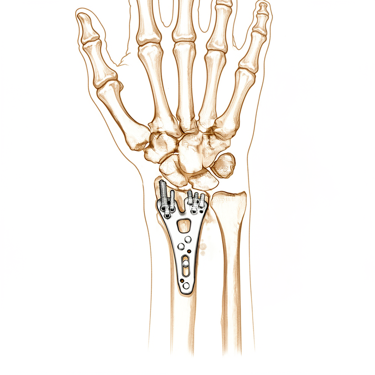
La operación se denomina reducción abierta y fijación interna. En términos sencillos, significa colocar el hueso fracturado en su posición normal y mantenerlo allí mediante una pequeña placa metálica y tornillos. Para la mayoría de las fracturas de muñeca de este tipo, el método habitual es colocar la placa en la cara palmar de la muñeca. En fracturas menos graves, primero se suele intentar el tratamiento no quirúrgico, como el uso de un yeso o férula. No obstante, cuando el hueso es inestable o se ha desplazado, puede ser necesario proceder a la cirugía de inmediato, pues el yeso podría no ser suficiente para mantener los fragmentos en su sitio.
El objetivo es que la muñeca sane en una posición adecuada, de modo que usted pueda mover la mano y volver a sus actividades cotidianas lo antes posible.
Antes de la operación
El día de la operación, deberá abstenerse de comer y beber durante siete horas antes de la hora programada para la operación. Pedimos que sean siete horas en lugar de seis para poder adelantar la intervención si el programa quirúrgico avanza antes de lo previsto. Su cirujano le indicará qué medicamentos habituales debe omitir ese día y cuáles debe tomar como de costumbre. Lleve una lista por escrito de todos los fármacos que toma, incluyendo pastillas, gotas y cualquier producto de la farmacia. Organice que alguien lo lleve a casa después de la operación, ya que no podrá conducir usted mismo. Use ropa holgada y cómoda, cuya manga sea fácil de quitar.
Para planificar la operación, utilizamos radiografías tomadas desde varios ángulos. En ocasiones, también es necesario realizar una tomografía computarizada (una radiografía detallada) o una resonancia magnética (un estudio que muestra los tejidos blandos, como los ligamentos). Si padece otras enfermedades, es posible que necesite análisis de sangre o una consulta con el anestesista antes del día de la intervención.
El día de la intervención
Llegará a la unidad de admisiones quirúrgicas del hospital, donde se le registrará y preparará para la cirugía. Allí conocerá al anestesista. Esta operación se realiza bajo anestesia general; usted estará completamente dormido durante todo el procedimiento. En algunos pacientes también se aplica un bloqueo nervioso regional para aliviar el dolor postoperatorio; el anestesista decide al respecto ese mismo día, según sus circunstancias individuales.
Posteriormente, será llevado al quirófano, donde se lleva a cabo la operación. Después, despertará en la sala de recuperación, donde las enfermeras lo vigilarán mientras la anestesia va desapareciendo. Una vez que su estado sea estable, será trasladado a una sala de hospitalización o podrá regresar a su casa, según el tipo de intervención y su recuperación.
Descripción del procedimiento quirúrgico
La operación se realiza mediante una incisión en la cara palmar de la muñeca. A través de esta abertura, el cirujano vuelve a colocar los fragmentos óseos rotos en su posición normal. Luego se moldea una pequeña placa metálica para adaptarla al hueso y se fija con tornillos. La placa se sitúa en la parte frontal del hueso de la muñeca; entre ella y los tendones que mueven el pulgar y los dedos hay una capa de tejido.
En ocasiones, es necesario examinar más detenidamente la superficie articular. En esos casos, el cirujano puede utilizar un instrumento delgado dotado de una cámara diminuta para observar el interior de la articulación mientras se alinean los fragmentos óseos. Esto permite confirmar que la superficie articular es lisa y uniforme antes de fijar la placa.
Una vez que el hueso queda firmemente sujeto, el cirujano verifica su posición mediante radiografías tomadas durante la intervención. Posteriormente, se cierran los bordes de la incisión con puntos de sutura y se coloca un vendaje sobre la zona.
La placa y los tornillos permanecen en la muñeca de forma permanente; mantienen el hueso estable mientras cicatriza, actuando como una férula interna. En ciertas fracturas, se requiere soporte adicional: si el hueso se ha roto en varios fragmentos, el cirujano puede añadir una segunda placa en la parte dorsal de la muñeca o emplear un injerto óseo para rellenar los espacios y favorecer la unión del hueso.
Al salir del centro médico, el vendaje seguirá intacto. Le pedimos que lo mantenga seco y en su sitio durante unos 10 días, momento en el cual deberá volver para que revisemos la herida.
Después de la operación
Al despertar, se encontrará en la sala de recuperación, donde las enfermeras lo vigilarán de cerca mientras el efecto de la anestesia desaparece. Su muñeca estará envuelta en un vendaje suave, y le administraremos analgésicos para mantenerlo cómodo. Por lo general, podrá levantarse y caminar poco después; un miembro del equipo de enfermería le ayudará la primera vez. Dado que la anestesia puede hacer que se sienta inestable durante un tiempo, le rogamos que cuente con alguien que lo acompañe durante las primeras 24 horas después de regresar a casa. La mayoría de los pacientes permanecen una noche en el hospital tras esta operación, aunque algunos pueden volver a casa el mismo día. Dejamos el vendaje puesto durante unos 10 días; por favor, no lo retire antes de ese plazo a menos que se lo indiquemos. Lo cambiamos o lo retiramos cuando venga a la consulta.
Recuperación
Durante los primeros días, su muñeca estará adolorida e hinchada; esto es normal. El descanso, mantener la mano elevada sobre almohadas y los analgésicos que le proporcionamos aliviarán la molestia. La hinchazón suele disminuir durante las siguientes semanas, aunque puede tardar un tiempo en desaparecer por completo.
Se irá a casa con un vendaje blando en lugar de un yeso; por eso, la mayoría de las personas encuentran que la vida cotidiana es más fácil de lo que esperaban. Puede levantarse y moverse de inmediato, y utilizar la otra mano para vestirse, comer y asearse. Mantenga el vendaje seco. La terapia de mano es una parte esencial de su recuperación: consultará con Ruby Doolan en Extend Rehabilitation, nuestra terapeuta de mano, quien le guiará en los ejercicios y le confeccionará una férula si fuera necesario. Mover los dedos desde el principio ayuda a prevenir la rigidez y favorece la cicatrización.
Con el paso de las semanas, notará que la hinchazón disminuye y que recupera la movilidad. Su terapeuta le enseñará ejercicios para fortalecer el agarre y recuperar la flexión de la muñeca. Una vez que se sienta estable, podrá realizar actividades ligeras en casa; sin embargo, evite levantar objetos pesados o ejercer presión sobre la muñeca hasta que su cirujano lo autorice. No podrá conducir mientras su muñeca esté en proceso de curación; una vez retirado el vendaje y con el permiso del cirujano, podrá volver a conducir; en nuestra página sobre conducción tras una cirugía de mano y extremidad superior encontrará más información al respecto.
Cada persona cicatriza a su propio ritmo, por lo que su cronograma puede variar. Lo iremos viendo a lo largo del proceso y lo guiaremos en cada etapa.
Qué puede salir mal
La mayoría de los pacientes evolucionan bien, pero en ocasiones pueden surgir problemas. Su cirujano y el equipo lo vigilarán de cerca para detectar cualquier anomalía a tiempo.
En algunos casos, la placa metálica o los tornillos provocan irritación. Es posible sentir un borde duro bajo la piel o una sensación de roce al mover la muñeca. Si esto le molesta, mencione el tema en su próxima consulta.
El tendón encargado de estirar el pulgar puede, en ocasiones, desgarrarse o romperse. Lo notará de forma repentina: no podrá levantar el pulgar recto y la parte posterior del mismo se sentirá flácida. Esto suele ocurrir alrededor de los 3 meses después de la operación, a veces sin dolor previo. Si su pulgar deja de funcionar de esta manera, comuníquese con la clínica de inmediato.
El nervio que inerva el pulgar, el índice y el dedo medio puede irritarse. Es posible notar hormigueo, sensación de pinchazos o zonas de entumecimiento en dichos dedos. Los síntomas leves son frecuentes y suelen desaparecer solos. Indique cualquier entumecimiento que no mejore durante su cita de seguimiento.
Las infecciones de la herida son poco comunes, pero requieren atención rápida. Esté atento a un dolor profundo y palpitante que no ceda con analgésicos comunes, enrojecimiento que se extienda desde la herida, calor en la zona o secreción que atraviese el vendaje. Si observa alguno de estos signos, llame a la clínica de inmediato; si es fuera del horario laboral, acuda a urgencias.
En ocasiones, el hueso se desplaza antes de haberse consolidado, o la cicatrización es lenta. Puede sentir un nuevo crujido o chasquido en la muñeca, aumento del dolor o sensación de inestabilidad. Infórmenos en su próxima revisión para realizar una radiografía y comprobar la posición ósea.
Después de la curación, algunas muñecas permanecen rígidas o sensibles. Es posible que le resulte difícil girar la mano, extender la muñeca hacia atrás o ejercer fuerza al agarrar. La terapia ocupacional y los ejercicios indicados por su terapeuta constituyen el tratamiento principal para este caso.
Fumar y el consumo de nicotina en cualquier forma, incluidos parches y gomas de mascar, incrementan el riesgo de varios de estos problemas, como infección de la herida, cicatrización lenta e incluso necesidad de nuevas cirugías. Si le es posible, deje de fumar antes de la operación.
En la tabla de complicaciones de esta página se detallan las tasas típicas; si desea conocer los datos específicos, puede consultarla.
¿Cuándo deben llamarnos?
La mayoría de los problemas presentan señales de alerta. Llámenos si tiene fiebre, o si la herida se vuelve más roja, caliente o segrega líquido. Llámenos si el dolor sigue empeorando a pesar de tomar analgésicos comunes. Acuda a urgencias si nota hinchazón o dolor en la pantorrilla, o si experimenta dificultad respiratoria repentina, pues estos síntomas pueden indicar la presencia de un coágulo sanguíneo. Acuda a urgencias si sus dedos se entumecen y permanecen así, o si no puede moverlos en absoluto. Si su pulgar deja de levantarse en línea recta, comuníquese con la clínica de inmediato. Cuando tenga dudas, llámenos.
¿Dónde leer más sobre esta afección?
Esta página trata sobre la operación en sí. La afección que se trata con ella, incluyendo lo que demuestran las evidencias sobre cuándo la cirugía es útil y cuándo no, se explica con mayor detalle en la página de Fractura distal del radio.
Evidence & references
This is the clinical evidence summary written for health professionals. It is technical, and it lists the research this page was built from. You do not need to read it to understand your treatment or to make a decision about it.
Overview
- A network meta-analysis of randomized trials found that open reduction and internal fixation with a plate offers the best results for adult patients with a distal radius fracture in terms of early and sustained functional recovery and a reduction in fracture healing complications [7].
- In adults, early mobilization for patients with distal radius fractures treated with ORIF may have a beneficial functional effect compared to late mobilization at earlier stages [1].
- The mean differences in DASH scores at 6 weeks for early mobilization compared to late mobilization surpass the minimal clinically important difference [1].
- Substantial variations in surgical direct costs for distal radius ORIF exist, with implant choice being the predominant driver [3].
- In comminuted intra-articular distal radius fractures, placing the distal row of screws greater than 3mm from subchondral bone was associated with increased odds of worsening ulnar variance [2].
- The Lift-Off Screw (LOS) length can be calculated, and the technique can potentially be used with any distal radius periarticular locking plate with locking options in the shaft [5].
- Radial column plating of the distal radius is a safe treatment modality and a valuable adjunct in the setting of complex distal radius fractures [6].
- Patients undergoing radial column plating of the distal radius should be counseled that there is a 28% chance that hardware removal may be required [6].
- The treatment of displaced intra-articular distal radius fractures with a dorsally versus a volarly placed interlocking plate system demonstrated similar clinical results [9].
Anatomy & Pathophysiology
Bony Anatomy
- The distal radius articular surface is biconcave and consists of scaphoid and lunate facets [31].
- The distal radioulnar joint (DRUJ) articulates with the ulna at the sigmoid notch [31].
- Lister tubercle is a small dorsal prominence that serves as a landmark for the dorsal approach to the wrist [31].
- Lister tubercle is a cause of attritional rupture of the extensor pollicis longus (EPL) after a distal radius fracture [31].
- The distal radial metaphysis has thin cortex and is vulnerable to bending forces [31].
- The brachioradialis insertion on the radial styloid acts as a deforming force in distal radius fractures [31].
- In a normal wrist with neutral ulnar variance, the distal radius bears 80% of axial load [31].
- The carpus encompasses two rows of eight bones that serve as a bridge between the forearm and the hand [19].
- The proximal carpal row from radial to ulnar includes the scaphoid, lunate, and triquetrum [19].
- The distal carpal row from radial to ulnar includes the trapezium, trapezoid, capitate, and hamate [19].
- The scaphoid is a small, irregular S-shaped tubular bone located in the proximal carpal row on the radial aspect of the wrist [25].
- The scaphoid lies entirely within the wrist joint at a 45-degree plane to the longitudinal and horizontal axis of the wrist [25].
- The scaphoid articulates with the trapezium/trapezoid, radius, capitate, and lunate [25].
- The scaphoid has a surface extensively covered with articular cartilage (over 80%), resulting in reduced capacity for periosteal healing and an increased tendency for delayed union and nonunion [25].
- The scaphoid is ridged across its nonarticular dorsoradial surface, along which the critical dorsal ridge vessels traverse [25].
- The dorsal ridge of the scaphoid is the insertion point for both the dorsal component of the scapholunate and intercarpal ligaments [25].
- The radioscapocapitate ligament does not attach to the bone itself but crosses the waist, acting as a sling across it allowing it to rotate [25].
- There are no tendon attachments to the scaphoid [25].
- The scaphoid acts as a midcarpal joint “bridge” linking and synchronizing the motions of the proximal and distal carpal rows as part of the key intercalated segment [25].
- Motion of the scaphoid includes rotation proximally and gliding distally, while providing stability to the midcarpal joint [25].
Vascular Anatomy
- The blood supply of the scaphoid is largely retrograde and meagre, provided by two vascular pedicles originating from the scaphoid branches of the radial artery [25].
- The dorsal branch of the scaphoid blood supply enters via small foramina along the spiral groove and dorsal ridge, supplying 70% to 80% of the scaphoid proximally, including the proximal pole [25].
- The volar branch of the scaphoid blood supply enters via the scaphoid tubercle and supplies the remaining 20% to 30% of the distal scaphoid [25].
- The waist of the scaphoid has minimal or no perforating vasculature [25].
- No vessels perforate the proximal dorsal cartilaginous area or through the scapholunate ligament [25].
- Proximal scaphoid fractures are associated with at least temporary disruption of the interosseous blood supply to the proximal pole [25].
Ligamentous Anatomy
- The extrinsic ligaments of the carpus connect the carpal bones to the forearm bones proximally and the metacarpals distally [27].
- The extrinsic palmar radiocarpal ligaments include the transverse carpal, radioscaphocapitate (RSC), radioscapholunate (RSL), radial collateral, long radiolunate (RLT), and short radiolunate ligaments [27].
- The extrinsic ulnocarpal ligaments include the ulnotriquetral (dorsal and palmar), ulnolunate, and ulnocapitate ligaments [27].
- Strong oblique extrinsic palmar radial ligaments prevent the carpus from translating medially on the angulated slope of the distal radius through two V-shaped ligamentous bands [27].
- The proximal V-shaped ligamentous band connects the forearm to the proximal carpal row and includes the long radiolunate, radioscapholunate, ulnolunate, and ulnotriquetral ligaments [27].
- The distal V-shaped ligamentous band connects the forearm to the distal carpal row and includes the radioscaphocapitate and ulnocapitate ligaments [27].
- A V-shaped interligamentous sulcus over the capitolunate articulation, known as the space of Poirier, is an interval of capsular weakness [27].
- Maximal space of Poirier is seen when the wrist is dorsiflexed, with the space almost disappearing in palmar flexion [27].
- The lunate displaces into the carpal canal through the space of Poirier during dorsal dislocations [27].
- The arcuate ligament is found in the central third of the palmar joint capsule and is formed from the interdigitation of transverse fibers of the radioscaphocapitate, ulnocapitate, triquetrocapitate, and volar scaphotriquetral ligaments [27].
- The arcuate ligament forms a support sling for the midcarpal region, particularly the head of the capitate [27].
- The extrinsic dorsal carpal ligaments include the dorsal radiocarpal (DRC) ligament and the dorsal intercarpal ligament, which form a V-shaped configuration [27].
- The ulnodorsal capsule of the wrist is reinforced by the ulnolunate and ulnotriquetral ligaments and the floors of the fifth and sixth extensor compartments [27].
- The intrinsic ligaments connect individual carpal bones to one another and are intra-articular short fibers [27].
- The intrinsic ligaments include the palmar midcarpal ligaments (scaphotrapeziotrapezoid, scaphocapitate, triquetrocapiate, triquetrohamate), the proximal interosseous ligaments (scapholunate, lunotriquetral), and the distal interosseous ligaments (trapeziotrapezoid, trapeziocapitate, capitohamate) [27].
- The V-shaped scaphotrapezium–trapezoid ligament on the radial side of the wrist provides stability to the scaphoid–trapezium–trapezoid articulation as well as the scaphoid itself [27].
- The scaphocapitate ligament is a large robust ligament that provides midcarpal stability [27].
Pathophysiology & Biomechanics
- Distal radius fractures are the most common fractures of the upper extremity, with over 300,000 cases per year in the United States [31].
- Distal radius fractures have a bimodal distribution: young patients typically sustain high-energy trauma, while elderly patients typically sustain low-energy falls [31].
- In elderly patients, distal radius fractures are the most common upper extremity osteoporotic fracture [31].
- Normal radiographic parameters for the distal radius include an average radial height of 11 mm, radial inclination of 22 degrees, and volar tilt of 11 degrees [31].
- Acceptable radiographic deviations for distal radius fractures include less than 5 mm of shortening, less than a 5-degree change in radial inclination, and less than 10-degree dorsal angulation [31].
- Ulnar variance is assessed with the forearm in neutral rotation and compared with the contralateral side [31].
- The distal radioulnar joint (DRUJ) alignment is assessed on a true lateral radiograph [31].
- Ligamentous injuries associated with distal radius fractures include scapholunate (SL), lunotriquetral (LT), or triangular fibrocartilage complex (TFCC) injuries [31].
- Radiocarpal dislocation or “inferior arc” injury is highly unstable and difficult to reduce closed [31].
- Computed tomography (CT) is used for detail of complex intraarticular patterns, while magnetic resonance imaging (MRI) is used for occult fracture, bone contusion, and associated soft tissue injury [31].
- Placing the distal row of screws greater than 3mm from subchondral bone in comminuted intra-articular distal radius fractures is associated with increased odds of worsening ulnar variance [2].
Classification
- In adults, early mobilization for distal radius fractures treated with open reduction and internal fixation may have a beneficial functional effect compared to late mobilization at earlier stages [1].
- The mean difference in DASH scores at 6 weeks between early and late mobilization for distal radius fractures treated with ORIF surpasses the minimal clinically important difference [1].
- In comminuted intra-articular distal radius fractures, placing the distal row of screws greater than 3mm from subchondral bone is associated with increased odds of worsening ulnar variance [2].
- The Lift-Off Screw (LOS) length can be calculated for accurate sagittal tilt correction in a distal radius fracture model [5].
- The Lift-Off Screw technique can potentially be used with any distal radius periarticular locking plate that has locking options in the shaft [5].
- Open reduction and internal fixation with a plate offers the best results for adult patients with a distal radius fracture in terms of early and sustained functional recovery [7].
- Open reduction and internal fixation with a plate offers the best results for adult patients with a distal radius fracture in terms of a reduction in fracture healing complications [7].
- The lifetime risk of distal radius fracture is 15% for women and 2% for men [13].
- Most distal radius fractures are treated nonsurgically with a plaster cast after closed reduction [13].
- The number of distal radius fractures receiving surgical intervention has increased as surgical treatment has advanced and the general population ages [13].
- Surgical methods for distal radius fractures have shifted from percutaneous pinning or external fixation to open reduction and internal fixation [13].
- The benefit of immediate structural rigidity of fractures, even in osteoporotic bone, after ORIF using volar plating and screws enables early cast removal and mobilization of the hand [13].
- ORIF using volar plating and screws has become the preferred surgical approach among hand and orthopedic surgeons for distal radius fractures [13].
- Complication rates following ORIF of distal radius fractures vary from 0% to 60% [13].
- A systematic review of 55 studies including 3,911 fractures reported complication rates following ORIF of distal radius fractures varying from 0% to 60% [13].
Clinical Presentation
- The mean difference in DASH scores at 6 weeks for early mobilization compared to late mobilization in distal radius fractures treated with ORIF surpasses the minimal clinically important difference [1].
- A network meta-analysis of randomized trials indicates that open reduction and internal fixation with a plate offers the best results for adult patients with a distal radius fracture in terms of early and sustained functional recovery [7].
- Open reduction and internal fixation with a plate is associated with a reduction in fracture healing complications for adult patients with a distal radius fracture compared to other interventions in a network meta-analysis of randomized trials [7].
- The benefit of immediate structural rigidity of distal radius fractures after ORIF using volar plating and screws enables early cast removal and mobilization of the hand [13].
- Complication rates following ORIF of distal radius fractures have been reported to vary from 0% to 60% in a systematic review of 55 studies including 3,911 fractures [13].
Investigations
Imaging Protocols and Diagnostic Sensitivity
- Standard scaphoid radiographs are used for primary assessment to detect displacement and associated fractures in radiocarpal instability [29].
- Provocative stress tests may be required to demonstrate dynamic radiocarpal instability [29].
- CT may be required to better define associated bony injuries in radiocarpal instability [29].
- MRI can be used to determine the extent of ligamentous disruption in radiocarpal instability [29].
- A decrease in the ulnocarpal index may provide the only clue to diagnosis in cases of subtle ulnar translation [29].
- CT is more sensitive for diagnosing a scaphoid fracture and is useful for confirming alignment of bone fragments if surgery is planned [34].
- MRI is the definitive way to confirm or exclude a diagnosis of scaphoid fracture if the technique is available [34].
- 10–15% of scaphoid fractures are not visible on initial X-rays [28].
- If initial X-rays are normal but clinical suspicion remains, the wrist should be immobilized and re-imaged in 2 weeks or via MRI [28].
- Secondary imaging modalities are predominantly used in the assessment of scaphoid fractures and the diagnosis of intercarpal ligament injury and any associated instability [35].
- Ultrasound scanning (USS) is used for suspected carpal fractures and ligament injuries [35].
- CT (2D/3D) is used for suspected carpal fractures, fracture displacement, malunion, nonunion, and bone loss [35].
- Dynamic CT is used by some for ligament injuries [35].
- Bone scintigraphy is used for suspected carpal fractures and avulsion injuries [35].
- Arthrography ± videofluoroscopy is used for ligament injuries [35].
- MRI is used for suspected carpal fractures, avascular necrosis (AVN) of carpal bones, and ligament injuries [35].
- Wrist arthroscopy is used for suspected carpal fractures, fracture displacement, and ligament injuries [35].
- Live/video fluoroscopic evaluation of the wrist provides diagnostic clarity for dynamic instability with sensitivities reported between 86% and 95% and specificity between 80% and 97% for diagnosing scapholunate ligament injury [35].
- Ultrasound scanning (USS) provides an additional tool for the detection of carpal ligament injuries, though the technique is operator dependent [35].
Radiographic Measurements and Instability Patterns
- The lateral intrascaphoid angle is normally 30 degrees ±5 degrees on a sagittal view, with an angle greater than 35 degrees used as a cut-off for displacement [35].
- The AP intrascaphoid angle is normally 40 degrees ± 5 degrees on coronal views [35].
- The dorsal cortical angle is normally 140 degrees, with values greater than 160 degrees considered abnormal on a sagittal view [35].
- The scaphoid height-to-length ratio is normally 0.60, with values greater than 0.65 considered abnormal on a sagittal view [35].
- Intraobserver reliability for the lateral intrascaphoid angle is poor, while interobserver reliability is poor to moderate [35].
- Intraobserver reliability for the dorsal cortical angle is moderate to excellent, while interobserver reliability is moderate to excellent [35].
- Intraobserver reliability for the scaphoid height-to-length ratio is excellent, while interobserver reliability is moderate to excellent [35].
- Dorsal intercalated segmental instability (DISI) is characterized by the lunate tilting backwards and the scaphoid tilting somewhat volarwards [28].
- Volar intercalated segment instability (VISI) is characterized by the lunate and scaphoid tilting somewhat volarwards and the capitate and metacarpals lying anterior (volar) to the radius [28].
- In a normal lateral X-ray, the axes of the radius, lunate, capitate, and third metacarpal are co-linear, and the scaphoid projects at an angle of about 45 degrees to this line [28].
- DISI pattern is most commonly associated with displaced scaphoid fractures and scapholunate dissociation (SLD) [35].
- Assessment of Gilula's lines can aid in the diagnosis of perilunate dislocations [35].
Anatomical Context for Imaging Interpretation
- The proximal carpal row has no direct tendon attachments, and its movement results from bone shape, interaction with other bones, and ligament attachments [19].
- The pisiform bone is a sesamoid bone enclosed within the sheath of the flexor carpi ulnaris tendon and should not theoretically be considered within the proximal carpal row [19].
- The trapezium articulates with the first metacarpal, the trapezoid with the second, the capitate with the third, and the hamate with the fourth and fifth metacarpals [19].
- There is 30 to 40 degrees of flexion–extension and rotation at the metacarpotrapezial joint [19].
- Motion at the distal carpal row is controlled by the extrinsic wrist flexors and extensors [19].
- The ligaments of the wrist are predominantly contained within the joint capsule [19].
- Apart from the scaphocapitate ligament, carpal ligaments are not described consistently across anatomical studies [19].
Treatment
Surgical Approach and Technique
- Open reduction and internal fixation (ORIF) with a plate offers the best results for adult patients with distal radius fractures in terms of early and sustained functional recovery and a reduction in fracture healing complications [7].
- The immediate structural rigidity of fractures after ORIF using volar plating and screws enables early cast removal and mobilization of the hand [13].
- The large tenaculum clamp facilitates anatomical restoration of volar tilt and volar translation while allowing intraoperative fluoroscopy [22].
- The use of a large tenaculum clamp potentially minimizes complications such as flexor tendon abrasion or rupture [22].
- The lift-off screw technique can potentially be used with any distal radius periarticular locking plate with locking options in the shaft [5].
Implant Selection and Positioning
- Substantial variations in surgical direct costs for distal radius ORIF exist, and implant choice is the predominant driver [3].
- Placing the distal row of screws greater than 3mm from subchondral bone was associated with increased odds of worsening ulnar variance in comminuted intra-articular distal radius fractures [2].
Postoperative Management
- Early mobilization for patients with distal radius fractures treated with ORIF may have a beneficial effect compared to late mobilization at earlier stages [1].
- Mean differences in DASH scores at 6 weeks for early mobilization compared to late mobilization surpass the minimal clinically important difference [1].
Complications and Hardware
- There is a 28% chance that hardware removal may be required for patients treated with radial column plating of the distal radius [6].
Complications
- Complication rates following open reduction and internal fixation (ORIF) of distal radius fractures have been reported to vary from 0% to 60% [13].
- A systematic review of 55 studies including 3,911 fractures found no standard for evaluating what should be defined as a complication after ORIF using volar plating [13].
- In a retrospective study of 822 patients, the complication rate following volar locking plate fixation of distal radius fractures was determined as the primary aim [13].
- Radial column plating of the distal radius is associated with a 28% chance that hardware removal may be required [6].
- Open reduction and internal fixation with a plate offers a reduction in fracture healing complications compared to other treatments for adult patients with distal radius fractures [7].
- Early mobilization for patients with distal radius fractures treated with ORIF may have a beneficial functional effect compared to late mobilization, with mean differences in DASH scores at 6 weeks surpassing the minimal clinically important difference [1].
Recovery
- Early mobilization for patients with distal radius fractures treated with ORIF may have a beneficial functional effect compared to late mobilization at earlier stages [1].
Key Evidence
- [L1] Functionally, at earlier stages, early mobilization for patients with distal radius fractures treated with ORIF may have a beneficial effect compared to late mobilization, with mean differences in DASH scores at 6 weeks surpassing the minimal clinically important difference. [1] (10.1186/s13018-021-02837-0)
- [L4] In this two-center retrospective cohort of comminuted intra-articular distal radius fractures, placing the distal row of screws greater than 3mm from subchondral bone was associated with increased odds of worsening ulnar variance. [2] (10.1016/j.jhsa.2025.03.016)
- [L3] Substantial variations in surgical direct costs for distal radius ORIF exist, and implant choice is the predominant driver. [3] (10.1016/j.jhsa.2018.04.015)
- [L5] The LOS length can be calculated, and this technique can potentially be used with any distal radius periarticular locking plate with locking options in the shaft. [5] (10.1016/j.jhsa.2018.02.011)
- [L4] Radial column plating of the distal radius is a safe treatment modality and a valuable adjunct in the setting of complex distal radius fractures, but patients should be counseled that there is a 28% chance that hardware removal may be required. [6] (10.1177/1558944718760861)
- [L1] A network meta-analysis of randomized trials revealed that open reduction and internal fixation with a plate offers the best results for adult patients with a distal radius fracture, in terms of early and sustained functional recovery and a reduction in fracture healing complications. [7] (10.5435/jaaos-d-18-00424)
- [L3] The treatment of displaced intra-articular distal radius fractures with a dorsally versus a volarly placed interlocking plate system demonstrated similar clinical results. [9] (10.1177/1558944716675129)
- [L4] [13] (10.1016/j.jhsa.2022.11.012)
- [L4] The large tenaculum clamp facilitates anatomical restoration of volar tilt and volar translation while allowing intraoperative fluoroscopy, potentially minimizing complications such as flexor tendon abrasion or rupture. [22] (10.1016/j.jhsa.2018.11.017)
References
[1] In adults, early mobilization may be beneficial for distal radius fractures treated with open reduction and internal fixation: a systematic review and meta-analysis. Journal of Orthopaedic Surgery and Research. 2021. DOI: 10.1186/s13018-021-02837-0
[2] The Association Between Distal Screw and Articular Subsidence in the Open Treatment of Intra-articular Distal Radius Fractures. The Journal of Hand Surgery. 2026. DOI: 10.1016/j.jhsa.2025.03.016
[3] Evaluation of Factors Driving Cost Variation for Distal Radius Fracture Open Reduction Internal Fixation. The Journal of Hand Surgery. 2018. DOI: 10.1016/j.jhsa.2018.04.015
[5] Lift-Off Screw Results in Accurate Sagittal Tilt Correction in a Distal Radius Fracture Model. The Journal of Hand Surgery. 2018. DOI: 10.1016/j.jhsa.2018.02.011
[6] Complications of Radial Column Plating of the Distal Radius. HAND. 2018. DOI: 10.1177/1558944718760861
[7] Interventions for Distal Radius Fractures: A Network Meta-analysis of Randomized Trials. Journal of the American Academy of Orthopaedic Surgeons. 2019. DOI: 10.5435/jaaos-d-18-00424
[9] Management of Intra-Articular Distal Radius Fractures: Volar or Dorsal Locking Plate—Which Has Fewer Complications?. HAND. 2016. DOI: 10.1177/1558944716675129
[13] Complications After Volar Locking Plate Fixation of Distal Radius Fractures: A Retrospective Study of 822 Patients. The Journal of Hand Surgery. 2024. DOI: 10.1016/j.jhsa.2022.11.012
[19] Rockwood And Green S Fractures In Adults. 42: Fractures of the Distal Radius and Ulna > Pathoanatomy and Applied Anatomy Relating to Carpal Fractures and Dislocations.
[22] Use of a Large Tenaculum Clamp as a Reduction Technique for Treatment of Distal Radius Fractures. The Journal of Hand Surgery. 2019. DOI: 10.1016/j.jhsa.2018.11.017
[25] Rockwood And Green S Fractures In Adults. 42: Fractures of the Distal Radius and Ulna > Pathoanatomy and Applied Anatomy Related to Scaphoid Fractures.
[27] Rockwood And Green S Fractures In Adults. 42: Fractures of the Distal Radius and Ulna > Extrinsic Ligaments.
[28] Apley And Solomon S Concise System Of Orthopaedics And Trauma. FRACTURES OF THE DISTAL RADIUS IN CHILDREN > Imaging.
[29] Rockwood And Green S Fractures In Adults. 42: Fractures of the Distal Radius and Ulna > Assessment of Radiocarpal Instability.
[31] Miller S Review Of Orthopaedics. SECTION 16 PATELLAR TRACKING IN TOTAL KNEE ARTHROPLASTY > DISTAL RADIUS FRACTURES.
[34] Apley And Solomon S Concise System Of Orthopaedics And Trauma. FRACTURES OF THE DISTAL RADIUS IN CHILDREN > FRACTURE OF THE SCAPHOID.
[35] Rockwood And Green S Fractures In Adults. 42: Fractures of the Distal Radius and Ulna > Secondary Imaging Methods.