Patients › Hand
Ganglio en la vaina del tendón flexor
A benign, fluid-filled cyst arising from the flexor tendon sheath – often painless, but can cause clicking or limited finger movement.
Qué está sintiendo
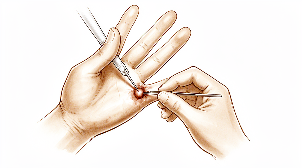
Un quiste ganglionar de la vaina del tendón flexor es un bulto pequeño y firme en la base de un dedo, en la palma de la mano. Normalmente mide solo unos pocos milímetros de diámetro, pero puede doler más de lo que cabría esperar de algo tan pequeño. El dolor aparece cuando agarra algo duro y estrecho, como un volante, un palo de golf, el manillar de una bicicleta o el asa de una bolsa de la compra. El bulto queda aplastado entre el objeto y el hueso del dedo, sin nada blando que lo amortigüe.
El dolor suele aumentar durante esas tareas de agarre o después de ellas. Actividades como llevar bolsas de la compra pesadas, sostener herramientas o escurrir la ropa lavada pueden volverse incómodas. Algunas personas notan que el dolor se calma en cuanto sueltan el objeto. El bulto en sí es firme y no blando, y se sitúa justo donde los dedos se doblan hacia la palma.
Como el dolor está ligado al agarre, puede empezar a limitar lo que hace con esa mano. Es posible que se encuentre evitando ciertas tareas o cambiando la forma de sostener las cosas para no presionar ese punto.
Si el dedo, la mano o el brazo se ponen calientes, rojos, hinchados y dolorosos, sobre todo si tiene fiebre, necesita atención ese mismo día en un servicio de urgencias. No necesita una derivación de su médico de cabecera para acudir.
¿Qué está ocurriendo realmente?
El dedo se dobla gracias a dos tendones que van desde el antebrazo, a lo largo de la cara palmar del dedo, y se anclan en el hueso del dedo. Imagínelos como cuerdas que tiran cuando usted dobla el dedo. Para mantener esas cuerdas pegadas al hueso, en lugar de que se separen de él formando un arco, discurren por dentro de un túnel formado por bandas resistentes llamadas poleas. Ese túnel está revestido por una capa fina y resbaladiza que nutre los tendones y les permite deslizarse con suavidad.
Un quiste ganglionar de la vaina del tendón flexor es un pequeño saco de líquido espeso y gelatinoso que sobresale de ese túnel. El saco se llena a partir del propio revestimiento de la vaina, por lo que se comporta como una pequeña fuga bajo presión: si se drena, vuelve a llenarse, porque la fuga sigue ahí. El saco se encuentra justo en la base del dedo, en la palma, donde el túnel está firmemente unido al hueso sin ningún acolchado blando por encima. Por eso un bulto de solo unos pocos milímetros de diámetro puede doler tanto al agarrar algo: el bulto queda pellizcado entre lo que esté sosteniendo y el hueso que tiene debajo.
Esa misma localización explica las otras cosas que quizá haya notado. Como el bulto ocupa espacio dentro de un túnel estrecho, puede estorbar al tendón cuando se desliza por él; por eso algunas personas con un bulto en este punto también notan que el dedo se engancha o hace “clic”. Los nervios del dedo discurren justo al lado de la vaina, uno a cada lado, por lo que un bulto que presione un nervio puede causar hormigueo o entumecimiento, y no solo dolor.
Conviene saber que un bulto en este punto exacto también puede deberse a otras causas, como un tendón que se engancha al deslizarse o un engrosamiento del tejido de la palma que tira del dedo hacia la palma. Su cirujano normalmente puede distinguirlas examinando la mano, y el tratamiento es diferente en cada caso.
Qué podemos hacer al respecto
El Dr. Kieran Hirpara, cirujano de extremidades superiores en el Mater Private Hospital Rockhampton, comienza con las opciones menos invasivas que se adapten a su condición. Por lo general, los pacientes son derivados a nuestra clínica por su médico de cabecera; si un fisioterapeuta le ha sugerido consultarnos, igualmente necesitará una derivación de su médico de cabecera para poder acceder al reembolso de Medicare. En su consulta, tomamos un historial clínico, examinamos su mano y solicitamos estudios de imagen, como una ecografía, si son necesarios para confirmar qué es el bulto.
Como este bulto es inofensivo y no sigue creciendo, el tratamiento depende por completo de cuánto le moleste. Un bulto que se puede palpar pero que no duele durante su agarre habitual puede no necesitar ningún tratamiento. Si una forma concreta de agarre le sigue causando dolor, cambiar la manera de sostener las cosas o reducir esa tarea durante un tiempo puede ser suficiente. La terapia de mano también puede ayudarle a encontrar formas de usar la mano que ejerzan menos presión sobre ese punto.
Drenar el bulto con una aguja, lo que se denomina aspiración, suele intentarse antes de la cirugía. Así se vacía el líquido del saco, pero como el saco se alimenta de la vaina del tendón, normalmente vuelve a llenarse. Algunos bultos también desaparecen por sí solos con el tiempo. Hablaremos con usted sobre si merece la pena intentar el drenaje en su caso.
Si estas medidas más sencillas no le han aportado suficiente alivio, puede plantearse la cirugía. La operación extirpa el bulto a través de un pequeño corte en la base del dedo, junto con el pequeño fragmento de vaina tendinosa del que nace, que es lo que evita que vuelva a aparecer. Normalmente proponemos la cirugía cuando el dolor limita una actividad concreta y repetida de su vida diaria, y tomamos esa decisión junto con usted.
Si el dedo, la mano o el brazo se ponen calientes, rojos, hinchados y dolorosos, sobre todo si tiene fiebre, acuda a un servicio de urgencias ese mismo día.
Qué esperar
La mayoría de los quistes ganglionares como este son inofensivos y no siguen creciendo. Algunos desaparecen por sí solos con el tiempo, y otros aparecen y desaparecen. Si el suyo no le duele, es posible que nunca necesite tratamiento.
Si el bulto le duele al agarrar, algunas medidas sencillas pueden ayudar. Drenarlo con una aguja vacía el saco, pero como el saco se alimenta de la vaina del tendón, normalmente vuelve a llenarse. Cambiar la forma de sostener las cosas, o reducir la tarea que lo agrava, puede ser suficiente mientras espera a ver si mejora.
Si esas medidas no han ayudado lo suficiente, la cirugía extirpa el bulto junto con el pequeño fragmento de vaina del que nace, que es lo que evita que vuelva a aparecer. La mayoría de las personas notan que agarrar resulta más cómodo una vez que ese punto deja de quedar pellizcado. Como en cualquier operación de la mano, hay riesgos que sopesar, como una zona entumecida a lo largo de un lado del dedo, rigidez, infección y una cicatriz gruesa o sensible. Su cirujano los comentará con usted antes de que tome una decisión.
La recuperación tras la cirugía lleva semanas, no días. Al principio la mano le dolerá, y el pequeño corte en la base del dedo necesitará tiempo para asentarse. La terapia de mano después de la cirugía la llevará a cabo Ruby Doolan en Extend Rehabilitation. Ruby es terapeuta especializada en mano: ella le guiará en los ejercicios y confeccionará cualquier férula que necesite mientras el dedo se cura. La mayoría de las personas vuelven a sus tareas de agarre habituales en cuestión de semanas, aunque cierta sensibilidad en la cicatriz puede durar un poco más.
Si sus síntomas no mejoran, empeoran a lo largo de las semanas, le despiertan por la noche o le impiden trabajar o usar la mano, consulte a su médico de cabecera o pida una valoración por un especialista.
¿Cuándo consultar a un especialista?
La mayoría de los bultos en la base de un dedo son quistes ganglionares inofensivos, pero algunas otras afecciones pueden tener el mismo aspecto en esa zona. Un dedo que se bloquea o hace “clic” al doblarse, o un cordón firme en la palma que tira del dedo hacia la palma, son afecciones que su cirujano puede distinguir examinando la mano. Conviene revisar cualquier bulto nuevo para saber cuál tiene usted.
Solicite una valoración por un especialista si el bulto le duele al agarrar, si nota hormigueo o entumecimiento en el dedo, o si el dedo se engancha o hace “clic”. Solicítela también si sus síntomas no mejoran, empeoran a lo largo de las semanas, le despiertan por la noche o le impiden trabajar o usar la mano.
Si el dedo, la mano o el brazo se ponen calientes, rojos, hinchados y dolorosos, sobre todo si tiene fiebre, acuda a un servicio de urgencias ese mismo día. No necesita una derivación de su médico de cabecera para acudir.
En mayor profundidad
Advanced reading: the deeper science (optional)
Esta sección profundiza más de lo necesario para que usted tome decisiones sobre su propio tratamiento. El ganglio en la vaina del tendón flexor merece ser leído con atención, ya que se trata del bulto más pequeño en cirugía de mano que, de manera constante, provoca síntomas desproporcionados; además, lo que lo une a otras estructuras determina tanto el motivo del dolor como el porqué los tratamientos sencillos no surten efecto.
Un bulto del tamaño de un guisante que duele de forma desproporcionada
Estos quistes, también denominados quistes retinaculares o gangliones retinaculares volares, se originan en la vaina fibrosa que mantiene los tendones flexores adheridos a los huesos de los dedos [1]. Se localizan en la base del dedo, en la palma de la mano; suelen medir solo unos pocos milímetros de diámetro y son firmes en lugar de blandos.
El síntoma es característico y se explica fácilmente una vez que se comprende la anatomía: dolor al agarrar objetos duros y estrechos, como un volante, un palo de golf, el manillar de una bicicleta o el asa de una bolsa de compras. El quiste queda atrapado entre el objeto y el hueso subyacente, y no hay tejido blando que lo amortigue, ya que la vaina está adherida directamente a la falange.
Por eso la relación entre el tamaño del quiste y los síntomas es tan débil. Un bulto grande y blando en el dorso de la muñeca puede ser indoloro, mientras que una lesión mucho más pequeña en la base de un dedo puede llegar a limitar seriamente la movilidad.
La misma regla que para cualquier otro ganglio
Su comportamiento depende de a qué estructura está conectado. Este quiste se origina en la vaina tendinosa y se llena a partir de ella; por lo tanto, su mecánica es idéntica a la de los ganglios de muñeca y los quistes mucosos: el saco quístico es solo la manifestación visible del problema, no su origen.
En consecuencia, puncionar o aspirar el quiste soluciona el hinchazón, pero no el origen de la fuga de líquido. Es frecuente que el quiste reaparezca tras un drenaje simple; el tratamiento definitivo consiste en extirpar el quiste junto con la porción afectada de la vaina tendinosa de la que proviene.
¿Por qué la operación parece menor de lo que parece, pero no es trivial?
La escisión es un procedimiento breve que se realiza mediante una incisión pequeña en la base del dedo, y generalmente resulta curativa. Dos características anatómicas justifican la necesidad de extremar el cuidado en esa zona.
Los nervios digitales discurren justo al lado de la vaina flexora, uno a cada lado; en la base del dedo, estos nervios se encuentran en una posición superficial y cercana a las estructuras de la línea media que se van a extirpar. La aparición de una zona entumecida a lo largo de uno de los bordes del dedo es un riesgo conocido de cualquier operación pequeña en esta zona.
En segundo lugar, solo se puede extirpar la porción redundante de la vaina. Las poleas que mantienen los tendones adheridos al hueso son estructuras que soportan carga; si se pierde una de ellas, el tendón tiende a separarse del dedo, lo que debilita la capacidad de agarre. Por ello, la escisión se limita deliberadamente al segmento de la vaina que no cumple esa función.
¿Cuándo dejarlo sin tratar?
Dado que esta lesión es benigna y no crece indefinidamente, el tratamiento se decide únicamente en función de los síntomas. Una quiste que es visible pero no causa dolor durante el agarre diario no requiere ninguna intervención. Cuando sí se trata, es porque una actividad específica y repetida resulta afectada; esto constituye un indicador más claro que la mayoría de los demás, ya que la forma de agarre que provoca el problema suele ser fácil de identificar para la persona afectada.
Los quistes relacionados en la articulación de la punta del dedo y en la muñeca se tratan en sus propias páginas; el principio común a los tres casos es que es el tallo del quiste, y no la propia bolsa, lo que determina si el problema volverá a aparecer.
Referencias
[1] Foret AL, Chhabra AB. Gangliones del retináculo volar. J Hand Surg Am. 2012;37(3):566-7. https://doi.org/10.1016/j.jhsa.2011.05.013
Evidence & references
This is the clinical evidence summary written for health professionals. It is technical, and it lists the research this page was built from. You do not need to read it to understand your treatment or to make a decision about it.
Overview
Epidemiology and Natural History
- Pediatric ganglions are more commonly found on the volar surface of the hand and wrist compared to adults [7].
- Pediatric ganglions more commonly have a tendon sheath origin compared to adults [7].
- Approximately 40% of wrist ganglion lesions decrease over the first 6 years after evaluation by a hand surgeon [25].
Diagnosis
- Ultrasound is useful for assisting in the diagnosis of suspected flexor tendon sheath ganglions [1].
- Ultrasound is useful for determining whether patients with suspected flexor tendon sheath ganglions should undergo aspiration or surgical excision [1].
- Routine wrist radiography is not cost-effective in the evaluation and treatment decision-making process for patients with a wrist ganglion due to a low prevalence of therapeutically significant findings [21].
Non-Operative Management
- A nonsurgical approach to flexor tendon sheath ganglions is both cost-effective and successful [3].
- Most ganglions recur after aspiration [25].
- Percutaneous puncture is a practical option for managing flexor tendon sheath ganglions given its low cost, lack of downtime, and low recurrence rate [5].
- No recurrences were observed after a second puncture in the cohort assessed for percutaneous puncture of flexor sheath ganglions [5].
Operative Management
- Surgical excision is a simple, safe, and effective method for treating a painful ganglion of the digital flexor tendon sheath [2].
- Surgical intervention for wrist ganglions has about a 10% recurrence rate [25].
- Surgical intervention for wrist ganglions leaves scars and has some risk for adverse events [25].
- Ultrasound-guided excision of flexor tendon sheath ganglion using a 2-mm portal under local anaesthesia was reliable and efficient without specific morbidity [4].
- Endoscopic ganglionectomy via flexor carpi radialis tendoscopy is a minimally invasive approach that offers better cosmesis and less soft tissue trauma [9].
- Endoscopic ganglionectomy via flexor carpi radialis tendoscopy allows for the management of concomitant FCR tendon pathology [9].
- Endoscopic ganglionectomy via flexor carpi radialis tendoscopy carries risks of recurrence and nerve or vessel injury [9].
- Operation-related complications after arthroscopic volar wrist ganglionectomy are associated with the anatomical location of the ganglion being distal to the bifurcation of the radial artery and concurrently penetrated up to the superficial fascia layer [60].
Pediatric Management
- There is no consensus within the literature regarding the best management of pediatric wrist ganglia [22].
- No single treatment modality confers a particular advantage or disadvantage over another for pediatric wrist ganglia [22].
Anatomy & Pathophysiology
Flexor Tendon Sheath Anatomy
- The fibroosseous tunnel, or digital flexor sheath, extends distally from the metacarpal neck to the proximal aspect of the distal phalanx [75].
- The tendinous sheath consists of annular pulleys that provide mechanical stability and cruciate pulleys that provide flexibility [75].
- The A2 and A4 pulleys are situated over the middle portion of the proximal and middle phalanges, respectively [75].
- The A2 and A4 pulleys are the most essential in maintaining the mechanical advantage of the flexor tendons [75].
- The tenosynovium lining the fibroosseous tunnel supplies both nutrition and lubrication to the poorly vascularized flexor tendons [75].
- Within the sheath, tendon vascularity is supplied via the vincula system, specifically the vinculum longus and brevis [75].
- Anatomical studies have identified two annular and one oblique pulley in the thumb [91].
- A branch from the digital nerve enters the flexor tendon sheath at the same place as the transverse branch of the digital artery [8].
- The nerve branch entering the flexor tendon sheath supplies the nerve fibres found within the vinculum [8].
- The transverse branch of the digital artery enters the edge of the sheath to reach the area of the vinculum brevis [8].
Gliding Mechanisms and Biomechanics
- In narrow crowded areas, the gliding mechanism is assured by the synovial sheath, which allows a considerable amplitude of movement [76].
- Fibrous sheaths surrounding synovial sheaths keep the tendon close to the skeleton, particularly when the tendon crosses an articular angle [76].
- The fibrous sheath assumes the role of a pulley when the tendon changes direction [76].
- Each synovial sheath has a visceral and parietal component separated by a potential synovial cavity containing a very thin layer of synovial fluid [76].
- This synovial fluid constitutes the basic gliding and nutritional mechanism for the tendon [76].
- Diffusion is a significant nutrient pathway to the flexor tendon, supporting the importance of sheath closure at the time of tendon suture [36, 37].
- At half the total tendon excursion, the force required to flex the digit through half its range is always less than a third of the force required to flex the digit fully [46].
- The force required to fully flex the digit decreased with more extensive A1 pulley release, becoming significant when more than 50% of the pulley was released [97].
Pathophysiology of Triggering and Ganglions
- Triggering results from the loss of smooth gliding of a tendon within its tendon sheath [6].
- Inflammation of the synovium lining the tendon sheath interferes with the normal gliding mechanism [6].
- Interference with the gliding mechanism is especially troublesome at a point of change in direction of a tendon [6].
- Ganglions are defined as cystic swellings closely connected to joints or tendon sheaths which contain mucinous material [141].
- The etiology of ganglions is unknown [141].
- The articular (synovial) theory has gained worldwide acceptance as a scientifically proven explanation for the formation of intraneural ganglions [17].
- Triggering of the finger can occur secondary to a partial flexor tendon tear after closed direct injury [98].
Classification
- Volar wrist ganglions may arise from a variety of locations, unlike dorsal wrist ganglions which typically arise from a specific location at the dorsal margin of the scapholunate interosseous membrane [16].
- Approximately two thirds of anterior wrist ganglions reportedly arise from the radiocarpal joint [16].
- One third of anterior wrist ganglions reportedly arise from the scaphotrapezial joint [16].
- Ganglion cysts that arise from the flexor carpi radialis (FCR) sheath are often in a slightly more medial location, either just radial or palmar to the sheath [16].
- Ganglions arising from the FCR sheath are typically less mobile than ganglions arising from the wrist joint [16].
- Ganglions arising from the first extensor compartment tend to adhere to the sheath (extensor retinaculum) [16].
- Ganglions arising from the first extensor compartment tend to be quite firm and are less mobile [16].
- In children aged <10 years, ganglion cysts present on the volar aspect of the wrist [13].
- In patients aged >10 years, ganglions resemble those in the adult population and present on the dorsal aspect of the wrist [13].
Clinical Presentation
General Characteristics
- Ganglions are the most common cause of focal masses in the hand and foot [50].
- Ganglions commonly arise from the synovium of joints, tendon sheaths, or the epineurium of nerves [50].
- Ganglions are filled with synovial fluid that may become jelly-like over time [50].
- Most patients with intraneural ganglions present with a painless mass [17].
- Some patients with intraneural ganglions present with symptoms of nerve irritation or entrapment neuropathy [17].
- In many patients with intraneural ganglions, definitive diagnosis is made only at the time of surgery [17].
Volar Wrist and Hand
- Volar wrist ganglia tend to arise in the interval between the first extensor compartment and flexor carpi radialis (FCR) tendon sheath, just proximal to the wrist flexion crease [16].
- Volar wrist ganglia typically appear to be 1 to 2 cm in size but may be significantly larger at the time of surgical dissection [16].
- Patients with volar wrist ganglions often present with complaints of a mass that has been present for a number of months or years and is typically asymptomatic [16].
- Patients with volar wrist ganglions often note discomfort when the mass is "bumped" [16].
- Volar wrist ganglia are clinically compressible, slightly mobile, nontender, and visible when transilluminated [16].
- Volar wrist ganglia are nonpulsatile but are often intimate with the radial artery, which is palpable alongside the lesion [16].
- Approximately one third of anterior wrist ganglions reportedly arise from the scaphotrapezial joint [16].
- Ganglion cysts that arise from the FCR sheath are often in a slightly more medial location, either just radial or palmar to the sheath [16].
- Ganglion cysts arising from the FCR sheath are typically less mobile than ganglions arising from the wrist joint [16].
Pediatric Population
- In children aged <10 years, ganglion cysts are generally amenable to observation with spontaneous regression [13].
- Ganglions in pediatric populations most commonly affect the dorsal wrist [47].
- Ganglions in pediatric populations demonstrate a female predilection [47].
- Pediatric ganglions of the hand have a greater rate of resolution than ganglions of the wrist [52].
Diagnostic Imaging
- Ultrasound is useful for assisting in diagnosis and determining whether patients should undergo aspiration or surgical excision for suspected flexor tendon sheath ganglions [1].
- MRI scans provide relatively good reliability in establishing the diagnosis of an occult dorsal wrist ganglion, with a sensitivity of 83% when using intra-operative findings as the standard [48].
- MRI is an excellent diagnostic modality to evaluate rapidly growing upper extremity masses and distinguish ganglions from malignant processes [33].
- The use of the 3-dimensional FSE extended echo train MRI sequence, called cube, has revolutionized the visualization of intraneural ganglions and articular branches that connect them to the joint [17].
Investigations
Imaging Modalities
- The architecture and relationship of the FDS to FDP within the flexor sheath is accurately defined using a non-invasive imaging technique and the pulley system can be clearly imaged in cross-section [58].
- Sonography-guided wrist arthroscopy provides visualization of the ganglions and ganglion stalk, as well as of the arthroscopic shaver and adjacent structures such as nerves, vessels, and tendons to perform surgery safely [124].
- Sonography-assisted arthroscopy allows clear visualization of ganglia, vessels, nerves, and tendons, enabling safe guidance of the arthroscopic shaver to the lesion without blind resection [126].
Diagnostic Limitations and Utility
- The study concludes that routinely performing wrist radiography is not cost-effective in the evaluation and treatment decision-making process for patients with a wrist ganglion due to a low prevalence of therapeutically significant findings [21].
- Radiologists are less likely to identify occult dorsal ganglion cysts on MRI if the cyst is smaller or if the requisition form does not mention a cyst [138].
Pathology and Histology
- In patients with the clinical diagnosis of wrist ganglion cyst, quality of care would not be compromised by abandoning the practice of routinely submitting surgical specimens for pathological examination after excision of the ganglion cyst [27, 31].
Treatment
Non-Operative Management
- Ultrasound imaging assists in determining whether patients with suspected flexor tendon sheath ganglions should undergo aspiration or surgical excision [1].
- Percutaneous puncture is a practical option for managing flexor tendon sheath ganglions due to its low cost, lack of downtime, and low recurrence rate [5].
- No recurrences were observed after a second percutaneous puncture in the cohort assessed for recurrence [5].
- Ganglion aspiration should be considered as a first-line intervention [55].
- Nonsurgical treatment is largely ineffective in treating ganglion cysts, though it may be considered for symptomatic relief in patients who do not want surgery [125].
- Aspiration, with or without cortisone injection, has an average success rate of 35% to 50% [122].
- Approximately 40% of lesions decrease over the first 6 years after evaluation by a hand surgeon [25].
Operative Management
- Ultrasound-guided excision of flexor tendon sheath ganglions using a 2-mm portal under local anaesthesia was reliable and efficient without specific morbidity [4].
- Open surgical excision offers a significantly lower chance of recurrence compared with aspiration in the treatment of wrist ganglions [135].
- Surgical intervention for ganglions has about a 10% recurrence rate [25].
- Surgical intervention for ganglions leaves scars and carries some risk for adverse events [25].
- In patients with a clinical diagnosis of wrist ganglion cyst, quality of care would not be compromised by abandoning the practice of routinely submitting surgical specimens for pathological examination after excision [27, 31].
- Arthroscopic resection is an effective treatment strategy for intra-articular synovial ganglions refractory to conservative measures, providing complete resolution of symptoms and functional recovery with minimal soft tissue damage [119].
- Arthroscopic ganglionectomy through an intrafocal cystic portal is a safe and efficacious option for the treatment of painful wrist ganglia [120].
- Arthroscopic ganglionectomy is a safe and reliable alternative to open resection [140].
- At 12 months follow-up, the rates of recurrence with arthroscopic dorsal ganglion excision are comparable with and not superior to those of open excision [24].
- Routine midcarpal joint exploration during arthroscopic excision of dorsal wrist ganglions appeared to reduce recurrence at 1 year without negatively impacting patient outcomes [35].
- Recurrent ganglion cysts originating from the midcarpal joint are not contraindications for arthroscopic resection [38].
- Arthroscopy allows for the simultaneous treatment of ganglions and other pathologies [29].
- Endoscopic ganglionectomy via flexor carpi radialis tendoscopy is a minimally invasive approach that offers better cosmesis, less soft tissue trauma, and the ability to manage concomitant FCR tendon pathology [9].
- There was no impairment of wrist motion and function in all patients following arthroscopic resection of palmar ganglions [51].
- The key to successful open treatment of dorsal wrist ganglions is excision of the ganglion stalk based at the scapholunate ligament and its associated dorsal-capsular attachments [122].
- Open ganglionectomy creates a defect in the capsule of 1 to 1.5 cm, which is left open [122].
- Stiffness has been reported from prolonged immobilization following open ganglionectomy [122].
- The rate of recurrence for open ganglionectomy can be as high as 40% [122].
- Indications for surgery include pain, restricted motion, and/or aesthetic complaints as a result of the ganglion cyst [113].
- Ultrasound, magnetic resonance imaging, or X-rays are performed to confirm the presence of a ganglion cyst or to rule out other conditions when surgery is indicated [113].
- The operation for dorsal wrist ganglion excision is generally performed under regional anaesthesia and a tourniquet [113].
- A dorsal incision distal to Lister’s tubercle is used, with the extensor retinaculum between the tendons of the second and fourth extensor compartments incised [113].
- A small arthrotomy is performed over the scapholunate ligament, where the dorsal wrist ganglion usually originates [113].
- Complete excision of the ganglion along with its capsule is performed with local synovectomy [113].
- The capsule is either fenestrated or sutured, depending on the surgeon [113].
- A compressive dressing is applied after skin closure and left in place for 3 days [113].
- Active mobilization of the wrist starts after the hand therapist changes the dressing [113].
- Sutures are removed 10 to 14 days postoperatively [113].
- Splints are not used on a regular basis [113].
- Patients start with a stabilizing and strength programme under the guidance of the hand therapist after 3 weeks [113].
- In pediatric wrist ganglion excision, a transverse incision is made over the ganglion to expose the cyst's wall [123].
- Fluid is aspirated and 0.3e0.5 mL of methylene blue is injected into the cavity to facilitate dye penetration into the cyst wall [123].
- The base of the ganglion is ligated with a 4e0 absorbable suture [123].
- All blue-stained ganglion tissue is thoroughly resected [123].
- The wrist is immobilized with a plaster cast for 7 to 10 days following pediatric excision [123].
- Followup examinations occur monthly for 6 months and every 3 months thereafter [123].
- Recurrence is assessed within 48 months' follow-up by palpation alone [123].
- Patients undergoing initial pediatric excision had been treated conservatively for more than 1 year [123].
- Recurrence of the wrist ganglion cyst occurred in 9% of patients in a cohort of 53 arthroscopic resections [116].
- PRWE scores were significantly higher in patients with a recurrence (30 vs. 12, p = 0.002) [116].
- Patients initially presenting with a new ganglion cyst had fewer recurrences at final follow-up compared with patients initially presenting with recurrence (4 vs. 15%) [116].
- Asymptomatic ganglion cysts were left untreated in the arthroscopic cohort [116].
- One patient experienced neuropraxia of the radial superficial nerve, which resolved spontaneously [116].
- One patient developed extensor carpi ulnaris (ECU) tendinitis, which was successfully treated with splinting [116].
- One patient experienced painful scar tissue, which was successfully removed surgically [116].
Complications
- Surgical intervention for wrist ganglions carries a recurrence rate of approximately 10% [25].
- Surgical intervention for wrist ganglions leaves scars and carries some risk for adverse events [25].
- Operation-related complications after arthroscopic volar wrist ganglionectomy are associated with the anatomical location of the cyst, specifically when it is distal to the bifurcation of the radial artery and concurrently penetrated up to the superficial fascia layer [60].
- Open surgical excision of pediatric wrist ganglions demonstrates minimal complications [13].
Recovery
Non-Operative Management
- No recurrences were observed after a second puncture in the cohort assessed for percutaneous puncture management [5].
- About 40% of lesions decrease over the first 6 years after evaluation by a hand surgeon [25].
- In a child with a wrist ganglion, if the cyst ultimately resolved, it usually did so within 18 months [20].
Operative Management
- Surgical intervention has about a 10% recurrence rate [25].
- Surgical intervention leaves scars and has some risk for adverse events [25].
Key Evidence
- [L4] Ultrasound is useful for assisting in diagnosis and determining whether patients should undergo aspiration or surgical excision for suspected flexor tendon sheath ganglions. [1] (10.1016/s0363-5023(97)80043-3)
- [L4] Surgical excision is a simple, safe, and effective method for treating a painful ganglion of the digital flexor tendon sheath. [2] (10.1007/s11552-007-9028-4)
- [L4] A nonsurgical approach to flexor tendon sheath ganglions is both cost-effective and successful. [3] (10.1053/jhsu.2002.34318)
- [L4] The ultrasound-guided excision of flexor tendon sheath ganglion, using a 2-mm portal, under local anaesthesia was reliable and efficient, without specific morbidity. [4] (10.1016/j.hansur.2018.10.116)
- [L4] Percutaneous puncture is a practical option for managing flexor tendon sheath ganglions given its low cost, lack of downtime, and low recurrence rate, with no recurrences observed after a second puncture in this cohort. [5] (10.1177/17531934221115983)
- [L5] [6] (10.1016/0266-7681(94)90139-2)
- [L4] Pediatric ganglions are more commonly found on the volar surface of the hand and wrist and more commonly have tendon sheath origin compared to adults. [7] (10.1007/s11552-008-9122-2)
- [L5] [8] (10.1016/0266-7681(91)90138-e)
- [Paper] Endoscopic ganglionectomy via flexor carpi radialis tendoscopy is a minimally invasive approach that offers better cosmesis, less soft tissue trauma, and the ability to manage concomitant FCR tendon pathology, though it carries risks of recurrence and nerve or vessel injury. [9] (10.1016/j.eats.2017.06.002)
- [L4] [13] (10.1016/j.jhsa.2021.12.015)
- [L5] [16] (10.1016/j.hcl.2004.03.015)
- [L4] [17] (10.1016/j.jhsa.2015.05.025)
- [L4] In a child with a wrist ganglion, if the cyst ultimately resolved, it usually did so within 18 months. [20] (10.1016/j.jhsa.2019.10.032)
- [L4] The study concludes that routinely performing wrist radiography is not cost-effective in the evaluation and treatment decision-making process for patients with a wrist ganglion due to a low prevalence of therapeutically significant findings. [21] (10.1007/s11552-007-9032-8)
- [L4] There is no consensus within the literature regarding the best management of pediatric wrist ganglia, and no single treatment modality confers a particular advantage or disadvantage over another. [22] (10.1177/1558944720966716)
- [L1] At 12 months follow-up, the rates of recurrence with arthroscopic dorsal ganglion excision are comparable with and not superior to those of open excision. [24] (10.1016/j.jhsa.2008.01.009)
- [L5] Current best evidence suggests that about 40% of lesions decrease over the first 6 years after evaluation by a hand surgeon, that most ganglions recur after aspiration, and that surgical intervention has about a 10% recurrence rate, leaves scars, and has some risk for adverse events. [25] (10.1016/j.jhsa.2010.11.048)
- [L3] This study suggests that, in patients with the clinical diagnosis of wrist ganglion cyst, quality of care would not be compromised by abandoning the practice of routinely submitting surgical specimens for pathological examination after excision of the ganglion cyst. [27] (10.1016/j.jhsa.2010.03.021)
- [L4] Arthroscopy allows for the simultaneous treatment of ganglions and other pathologies. [29] (10.1016/j.jhsa.2012.04.042)
- [L4] In patients with the clinical diagnosis of wrist ganglion cyst, the quality of care would not be compromised by abandoning the practice of routine submission of surgical specimens for pathological examination after excision of the ganglion cyst. [31] (10.1016/s0363-5023(10)60107-4)
- [L4] MRI is an excellent diagnostic modality to evaluate rapidly growing upper extremity masses and distinguish ganglions from malignant processes. [33] (10.1007/s11552-007-9083-x)
- [L3] Routine midcarpal joint exploration during arthroscopic excision of dorsal wrist ganglions appeared to reduce recurrence at 1 year without negatively impacting patient outcomes. [35] (10.1177/17531934251405730)
- [L5] The importance of sheath closure at the time of tendon suture is based in part on the evidence that diffusion is a significant nutrient pathway to the flexor tendon. [36] (10.1016/0266-7681(88)90077-0)
- [L5] The importance of sheath closure at the time of tendon suture is based in part on the evidence that diffusion is a significant nutrient pathway to the flexor tendon. [37] (10.1016/0266-7681_88_90077-0)
- [L4] Ganglion cysts also have a high association with certain interosseous laxities, and recurrent ganglion cysts originating from the midcarpal joint are not contraindications for arthroscopic resection. [38] (10.1016/j.jhsa.2008.11.025)
- [L5] At half the total tendon excursion, the force required to flex the digit through half its range is always less than a third of the force required to flex the digit fully. [46] (10.1016/0266-7681(90)90086-j)
- [L2] Ganglions in pediatric populations, which most commonly affect the dorsal wrist, demonstrate a female predilection. [47] (10.1016/j.jhsa.2021.02.026)
- [L3] MRI scans provide relatively good reliability in establishing the diagnosis of an occult dorsal wrist ganglion, with a sensitivity of 83% when using intra-operative findings as the standard. [48] (10.1177/1753193408092041)
- [Case_report] [50] (10.1007/s12593-014-0117-7)
- [L4] There was no impairment of wrist motion and function in all patients. [51] (10.1016/j.main.2006.07.028)
- [L4] Pediatric ganglions of the hand have a greater rate of resolution than ganglions of the wrist. [52] (10.1016/j.jhsa.2023.07.002)
- [L4] Ganglion aspiration should be considered as a first-line intervention, with surgical excision remaining an effective option for symptomatic cases. [55] (10.1177/1753193411434376)
- [L5] The architecture and relationship of the FDS to FDP within the flexor sheath is accurately defined using a non-invasive imaging technique and the pulley system can be clearly imaged in cross-section. [58] (10.1016/j.jhsb.2005.08.001)
- [L3] The operation-related complications after arthroscopic volar wrist ganglionectomy are associated with its anatomical location: distal to the bifurcation of the radial artery and concurrently penetrated up to the superficial fascia layer. [60] (10.1186/s12891-025-08766-x)
- [L5] Anatomical studies have identified two annular and one oblique pulley in the thumb. [91] (10.1016/s0363-5023(77)80101-9)
- [L5] The force required to fully flex the digit decreased with more extensive A1 pulley release, becoming significant when more than 50% of the pulley was released. [97] (10.1016/s0266-7681(98)80217-9)
- [L5] This illustrates another post-traumatic mechanical cause for triggering of the finger. [98] (10.1016/0363-5023(93)90059-c)
- [L2] [113] (10.1177/17531934231153029)
- [L4] [116] (10.1055/s-0040-1716509)
- [L4] Arthroscopic resection is an effective treatment strategy for intra-articular synovial ganglions refractory to conservative measures, providing complete resolution of symptoms and functional recovery with minimal soft tissue damage. [119] (10.1016/j.jhsg.2024.05.007)
- [L4] Arthroscopic ganglionectomy through an intrafocal cystic portal is a safe and efficacious option for the treatment of painful wrist ganglia. [120] (10.1016/j.arthro.2009.08.021)
- [L4] [122] (10.1016/s0749-0712(21)00020-2)
- [L2] [123] (10.1016/j.jhsa.2015.01.015)
- [L4] Sonography-guided wrist arthroscopy provides several advantages for surgeons, including visualization of the ganglions and ganglion stalk, as well as of the arthroscopic shaver and adjacent structures such as nerves, vessels, and tendons to perform surgery safely. [124] (10.1016/j.jhsa.2012.04.012)
- [L4] Nonsurgical treatment is largely ineffective in treating ganglion cysts, though it may be considered for symptomatic relief in patients who do not want surgery. [125] (10.1155/2013/940615)
- [Paper] Sonography-assisted arthroscopy allows clear visualization of ganglia, vessels, nerves, and tendons, enabling safe guidance of the arthroscopic shaver to the lesion without blind resection. [126] (10.1016/j.eats.2011.12.007)
- [L1] Open surgical excision offers significantly lower chance of recurrence compared with aspiration in the treatment of wrist ganglions. [135] (10.1016/j.jhsa.2014.12.014)
- [L3] Radiologists are less likely to identify occult dorsal ganglion cysts on MRI if the cyst is smaller or if the requisition form does not mention a cyst. [138] (10.1055/s-0039-1683847)
- [L4] Arthroscopic ganglionectomy is a safe and reliable alternative to open resection. [140] (10.1016/j.jhsa.2003.10.018)
- [L4] [141] (10.2106/00004623-197254070-00009)
References
[1] Treatment of flexor tendon sheath ganglions using ultrasound imaging. The Journal of Hand Surgery. 1997. DOI: 10.1016/s0363-5023(97)80043-3
[2] Flexor Tendon Sheath Ganglions: Results of Surgical Excision. HAND. 2007. DOI: 10.1007/s11552-007-9028-4
[3] Management of flexor tendon sheath ganglions: A cost analysis. The Journal of Hand Surgery. 2002. DOI: 10.1053/jhsu.2002.34318
[4] Flexor tendon sheath ganglions — A new procedure to perform excision under ultrasound-guidance. Hand Surgery and Rehabilitation. 2018. DOI: 10.1016/j.hansur.2018.10.116
[5] Percutaneous puncture of flexor sheath ganglions: an assessment of recurrence. Journal of Hand Surgery (European Volume). 2022. DOI: 10.1177/17531934221115983
[6] Extensor Tendon Sheath Stenosis Resulting in Triggering of the Little Finger. Journal of Hand Surgery. 1994. DOI: 10.1016/0266-7681(94)90139-2
[7] Pediatric Ganglion Cysts of the Hand and Wrist: An Epidemiologic Analysis. HAND. 2008. DOI: 10.1007/s11552-008-9122-2
[8] Anatomical Demonstration of the Nerve-Supply to the Flexor Tendon. Journal of Hand Surgery. 1991. DOI: 10.1016/0266-7681(91)90138-e
[9] Endoscopic Ganglionectomy of Palmar Ganglion via Flexor Carpi Radialis Tendoscopy. Arthroscopy Techniques. 2017. DOI: 10.1016/j.eats.2017.06.002
[13] Pediatric Ganglions of the Hand and Wrist: A Review of Current Literature. The Journal of Hand Surgery. 2022. DOI: 10.1016/j.jhsa.2021.12.015
[16] Ganglion cysts and other tumor related conditions of the hand and wrist. Hand Clinics. 2004. DOI: 10.1016/j.hcl.2004.03.015
[17] Intraneural Ganglions of the Hand and Wrist. The Journal of Hand Surgery. 2015. DOI: 10.1016/j.jhsa.2015.05.025
[20] Wrist Ganglia in Children: Nonsurgical Versus Surgical Treatment. The Journal of Hand Surgery. 2020. DOI: 10.1016/j.jhsa.2019.10.032
[21] The Use of Routine Wrist Radiography is Not Useful in the Evaluation of Patients with a Ganglion Cyst of the Wrist. HAND. 2007. DOI: 10.1007/s11552-007-9032-8
[22] Wrist Ganglion Cysts in Children: An Update and Review of the Literature. HAND. 2022. DOI: 10.1177/1558944720966716
[24] Arthroscopic Versus Open Dorsal Ganglion Excision: A Prospective, Randomized Comparison of Rates of Recurrence and of Residual Pain. The Journal of Hand Surgery. 2008. DOI: 10.1016/j.jhsa.2008.01.009
[25] Wrist Ganglions. The Journal of Hand Surgery. 2011. DOI: 10.1016/j.jhsa.2010.11.048
[27] Necessity of Routine Pathological Examination After Surgical Excision of Wrist Ganglions. The Journal of Hand Surgery. 2010. DOI: 10.1016/j.jhsa.2010.03.021
[29] Ganglions of the Wrist and Associated Triangular Fibrocartilage Lesions: A Prospective Study in Arthroscopically-treated Patients. The Journal of Hand Surgery. 2012. DOI: 10.1016/j.jhsa.2012.04.042
[31] Necessity of Routine Pathological Examination following Surgical Excision of Wrist Ganglions. The Journal of Hand Surgery. 2010. DOI: 10.1016/s0363-5023(10)60107-4
[33] Compression Neuropathy of the Radial Nerve Due to Ganglion Cysts. HAND. 2008. DOI: 10.1007/s11552-007-9083-x
[35] Arthroscopic resection of dorsal wrist ganglions with or without midcarpal exploration. Journal of Hand Surgery (European Volume). 2025. DOI: 10.1177/17531934251405730
[36] Flexor tendon healing. The Journal of Hand Surgery: Journal of the British Society for Surgery of the Hand. 1988. DOI: 10.1016/0266-7681(88)90077-0
[37] Flexor Tendon Healing. Journal of Hand Surgery. 1988. DOI: 10.1016/0266-7681_88_90077-0
[38] Prospective Outcomes and Associations of Wrist Ganglion Cysts Resected Arthroscopically. The Journal of Hand Surgery. 2009. DOI: 10.1016/j.jhsa.2008.11.025
[46] The Mechanical Effect of Partial Resection of the Digital Fibrous Flexor Sheath. Journal of Hand Surgery. 1990. DOI: 10.1016/0266-7681(90)90086-j
[47] Clinical Presentation and Characteristics of Hand and Wrist Ganglion Cysts in Children. The Journal of Hand Surgery. 2021. DOI: 10.1016/j.jhsa.2021.02.026
[48] Magnetic Resonance Imaging in the Diagnosis of Occult Dorsal Wrist Ganglions. Journal of Hand Surgery (European Volume). 2008. DOI: 10.1177/1753193408092041
[50] Intraneural Ganglion of Digital Nerve of Thumb: A Case Report and Review of Literature. Journal of Hand and Microsurgery. 2015. DOI: 10.1007/s12593-014-0117-7
[51] Résection des kystes synoviaux palmaires par arthroscopie. Chirurgie de la Main. 2006. DOI: 10.1016/j.main.2006.07.028
[52] Natural History of Pediatric Hand and Wrist Ganglion Cysts: Longitudinal Follow-Up of a Prospective, Dual-Center Cohort. The Journal of Hand Surgery. 2023. DOI: 10.1016/j.jhsa.2023.07.002
[55] Patient outcomes following wrist ganglion excision surgery. Journal of Hand Surgery (European Volume). 2012. DOI: 10.1177/1753193411434376
[58] Mri ‘Magic Angle’ Imaging of Finger Tendons. Journal of Hand Surgery. 2006. DOI: 10.1016/j.jhsb.2005.08.001
[60] Anatomical location of volar wrist ganglion in preoperative MRI is a risk factor for operation-related complications after arthroscopic ganglionectomy. BMC Musculoskeletal Disorders. 2025. DOI: 10.1186/s12891-025-08766-x
[75] A Lange Medical Book Current Diagnosis Treatment In Orthopedics Fifth Edition. 9Hand Surgery > FLEXOR TENDON INJURY.
[76] Exam Of The Hand Wrist 2Ed. 1.3 MOVEMENTS OF THE HAND AND WRIST > Gliding mechanisms.
[91] Anatomy of the flexor tendon sheath and pulleys of the thumb. The Journal of Hand Surgery. 1977. DOI: 10.1016/s0363-5023(77)80101-9
[97] The Effect of the Extent of A1 Pulley Release on the Force Required to Flex the Digits. Journal of Hand Surgery. 1998. DOI: 10.1016/s0266-7681(98)80217-9
[98] Triggering of the finger secondary to partial flexor tendon tear after closed direct injury. The Journal of Hand Surgery. 1993. DOI: 10.1016/0363-5023(93)90059-c
[113] Factors associated with self-reported pain and hand function following dorsal wrist ganglion excision. Journal of Hand Surgery (European Volume). 2023. DOI: 10.1177/17531934231153029
[116] Patient-Related Outcomes of Arthroscopic Resection of Ganglion Cysts of the Wrist. Journal of Wrist Surgery. 2020. DOI: 10.1055/s-0040-1716509
[119] Intra-articular Synovial Ganglion of the Wrist. Journal of Hand Surgery Global Online. 2024. DOI: 10.1016/j.jhsg.2024.05.007
[120] Arthroscopic Ganglionectomy Through an Intrafocal Cystic Portal for Wrist Ganglia. Arthroscopy. 2010. DOI: 10.1016/j.arthro.2009.08.021
[122] ARTHROSCOPIC RESECTION OF DORSAL GANGLION OF THE WRIST. Hand Clinics. 1995. DOI: 10.1016/s0749-0712(21)00020-2
[123] Visualization of the Wrist Ganglion Capsule by Methylene Blue Staining as an Aid for Complete Resection in Children. The Journal of Hand Surgery. 2015. DOI: 10.1016/j.jhsa.2015.01.015
[124] Sonography-guided Arthroscopy for Wrist Ganglion. The Journal of Hand Surgery. 2012. DOI: 10.1016/j.jhsa.2012.04.012
[125] Treatment of Ganglion Cysts. ISRN Orthopedics. 2013. DOI: 10.1155/2013/940615
[126] Sonography‐Assisted Arthroscopic Resection of Volar Wrist Ganglia: A New Technique. Arthroscopy Techniques. 2012. DOI: 10.1016/j.eats.2011.12.007
[135] Wrist Ganglion Treatment: Systematic Review and Meta-Analysis. The Journal of Hand Surgery. 2015. DOI: 10.1016/j.jhsa.2014.12.014
[138] Radiologist Identification of Occult Dorsal Wrist Ganglion Cysts on MRI. Journal of Wrist Surgery. 2019. DOI: 10.1055/s-0039-1683847
[140] Arthroscopic resection in the management of dorsal wrist ganglions: results with a minimum 2-year follow-up period. The Journal of Hand Surgery. 2004. DOI: 10.1016/j.jhsa.2003.10.018
[141] Ganglions of the Wrist and Hand. The Journal of Bone & Joint Surgery. 1972. DOI: 10.2106/00004623-197254070-00009