Clinicians › Hand
Flexor sheath ganglion excision
Surgeon-side topic for flexor sheath ganglion excision. Backed by 121 articles from the corpus, retrieved via combined MeSH + title-text matching.
For patients: a plain-language version of this topic is available. See the patient guide.
Overview
Wrist ganglions are common cystic lesions, with approximately 40% decreasing in size over the first six years following evaluation by a hand surgeon [2]. While aspiration is considered a first-line intervention, most ganglions recur after this procedure [2]. Performing at least one aspiration prior to surgical excision improves the cost-effectiveness of dorsal wrist ganglion treatment, as patient preferences may preclude routinely performing two aspirations [31]. Surgical excision remains an effective option for symptomatic cases, offering a significantly lower chance of recurrence compared with aspiration [11]. However, open surgical intervention carries a recurrence rate of about 10%, leaves scars, and presents some risk for adverse events [2].
Arthroscopic ganglion excision has emerged as a viable alternative, with advances in technique allowing recurrence rates similar to those of open management [1]. Recurrence and complication rates after arthroscopic resection appear comparable to open resections [22], and outcomes at four years of follow-up support the use of arthroscopy for dorsal wrist ganglions [23]. Patients experience significant increases in function and decreases in pain within six weeks after arthroscopic resection [22]. Arthroscopy also allows for the simultaneous treatment of ganglions and other pathologies [3]. Despite these benefits, surgical costs from arthroscopic excision are significantly higher than those of open excision [40].
Operative considerations include the advice to excise the articular nerve branch to avoid recurrences when a ganglion communicates with the distal radioulnar joint via an articular branch [7]. Complications after arthroscopic volar wrist ganglionectomy are associated with the anatomical location of the ganglion, specifically when it is distal to the bifurcation of the radial artery and concurrently penetrated up to the superficial fascia layer [16]. Patients whose occupation or activities require forceful wrist extension should be counseled on the considerable risk of residual pain and functional limitations following open dorsal wrist ganglion excision [5]. Hand surgeons remain divided regarding the need to immobilize the wrist after dorsal wrist ganglion excision [15].
Anatomy & Pathophysiology
General Pathophysiology
Current best evidence indicates that approximately 40% of ganglion lesions decrease in size over the first 6 years following evaluation by a hand surgeon [2]. Surgical intervention carries an approximately 10% recurrence rate, results in scarring, and presents a risk for adverse events [2]. Patients with wrist hyperlaxity demonstrate a predisposition to developing ganglions [54]. Cystic soft tissue tumours of the dorsal wrist comprise two distinct histological subtypes [6]. At recurrence, both histological tissue types coexist, with equal recurrence rates observed in both initial synovial and ganglion groups [6]. Nerve tumors are rare and frequently overlooked in the differential diagnosis of upper extremity masses [19]. Inappropriate surgery for nerve tumors can cause irreversible functional loss, necessitating strict tumor surgery techniques and careful preoperative planning [19]. Treatment of benign bony and soft tissue tumors in the hand is often based on small series or experience with similar lesions elsewhere in the body [37].
Pediatric Pathophysiology
Ganglions in children aged >10 years resemble adult ganglions, occurring mainly on the dorsal aspect [4]. Open excision surgery for ganglions in this age group has a recurrence rate ranging from 2.8% to 35% [4]. There is no consensus within the literature regarding the best management of pediatric wrist ganglia [13]. No single treatment modality confers a particular advantage or disadvantage over another for pediatric wrist ganglia [13]. Ganglions in pediatric populations, which most commonly affect the dorsal wrist, demonstrate a female predilection [21]. Pediatric ganglions of the hand have a greater rate of resolution than ganglions of the wrist [26].
Intraneural Ganglions
Involvement of the peripheral nerves of the upper extremity by intraneural ganglion is rare [17]. The ulnar nerve is the most commonly involved nerve in the upper extremity by intraneural ganglion [17]. Most patients with intraneural ganglions are aged 30 to 50 years, though occurrence in children has also been reported [17]. Most patients present with a painless mass [17], while some present with symptoms of nerve irritation or entrapment neuropathy [17]. In many patients, definitive diagnosis is made only at the time of surgery [17]. Recent advances in MRI technology have provided better visualization of intraneural ganglions and articular connections to the cyst [17]. The use of the 3-dimensional FSE extended echo train MRI sequence, called cube, has revolutionized the visualization of intraneural ganglions and the articular branches that connect them to the joint [17]. The articular (synovial) theory has gained worldwide acceptance as a scientifically proven explanation for the formation of intraneural ganglions [17]. Intraneural ganglions are nonneoplastic, mucinous cysts found within the epineurium of peripheral nerves of the hand [24]. The majority of intraneural ganglions reported in the literature involve the common peroneal nerves [24]. The pathogenesis of intraneural ganglions remains controversial [24]. Historically, the majority of investigators have supported the de novo theory of formation, which proposes that intraneural ganglions are the result of mucoid degeneration of fibrotic tissue after repetitive microtrauma [24]. Spinner et al. proposed a unifying articular (synovial) theory as the basis for the pathogenesis of intraneural cysts [24]. Spinner et al. believe that a joint connection via an articular branch is present in all intraneural ganglions [24]. Excision of the articular nerve branch is advised to avoid recurrences of intraneural ganglions [7].
Clinical Presentation & Differential Diagnosis
Trigger digit, Dupuytren's disease, and ganglion cysts are three common disorders treated by hand surgeons that can all present as a mass at the flexor crease in the distal palm [25]. Trigger digit is caused by pathological inflammatory changes in the retinacular tendon sheath and on the peritendinous tissue it encloses [25]. This condition leads to hypertrophy of one or both these structures, which progressively restricts the motion of the flexor tendon in its sheath [25]. Clinically, trigger digit is manifested by symptoms of locking and clicking, most commonly occurring at the A1 pulley of the ring finger [25]. Lack of full motion due to painful triggering or locking can lead to secondary contracture of the proximal interphalangeal (PIP) joint [25]. Dupuytren's is a disease of the palmar fascia presenting as a formation of fibromatous nodules and cords that can result in flexion contracture and decreased hand function [25]. The most common lumps encountered in the hand and wrist region are ganglion cysts [25]. Surgeons should maintain a high index of suspicion for ganglion cysts in presentations of atraumatic, progressive, and painful elbow contracture [12]. This is the first reported case of triggering pathology at the wrist to be treated with interventional radiological measures rather than open surgery [32]. This case highlights the importance of considering uncommon aetiologies in patients with atypical symptoms of carpal tunnel syndrome [41]. Carpal tunnel syndrome caused by a space occupying lesion is rare and more complicated than idiopathic carpal tunnel syndrome [91].
Risk Factors & Prognostic Factors
Dominant side, female sex, and age of 24 years or less are considered to be the most influential risk factors for recurrence after arthroscopic excision of dorsal wrist ganglia [20]. Worse hand function was associated with recurrence following prior surgery, worse baseline hand function and lower treatment credibility [38]. Be wary of offering surgery to young, less educated patients with dorsal volar or wrist ganglia as the surgery is often unrewarding for both the surgeon and the patient [35].
Anatomical Context
The hand is both an organ designed to obtain information and an organ of execution [44]. The hand functions efficiently only if the proximal joints of the limb are stable and yet mobile [44]. The combined movements of the wrist and forearm place the hand in a position for grasping [44]. For gripping, the wrist is usually in flexion when close to the trunk and in extension when placed at a distance [44]. The hand consists of 19 bones, 17 articulations, and 19 muscles situated entirely within the hand, and about the same number of tendons activated by the forearm muscles [44]. The skeleton of the hand and wrist consists of 27 bones, of which 19 are long bones [48]. The skeleton is divided into five rays, each ray making up a polyarticulated chain comprising the metacarpals and phalanges [48]. The radioulno-carpal articulation has two axes of movement to which is added a third—pronation and supination from the forearm [48]. The wrist has three axes of movement, permitting the hand to be positioned in any spatial configuration [48].
The extrinsic extensors run through six different fibroosseous retinacular compartments at the wrist level [47]. * First compartment: Contains the abductor pollicis longus and the extensor pollicis brevis [47]. * Second compartment: Contains the extensor carpi radialis longus and the extensor carpi radialis brevis [47]. * Third compartment: Contains the extensor pollicis longus, which runs longitudinally down the forearm through the third compartment and turns abruptly radialward about Lister tubercle [47]. * Fourth compartment: Contains the extensor indicis proprius lying deep to the four tendons of the extensor digitorum communis [47]. * Fifth compartment: Contains the extensor digiti quinti [47]. * Sixth compartment: The extensor carpi ulnaris tendon runs through this compartment and inserts at the base of the little finger metacarpal [47].
The extrinsic finger flexors are the flexor digitorum profundus and the flexor digitorum superficialis [47]. The flexor digitorum profundus inserts on the proximal volar aspect of the distal phalanx, flexing the distal interphalangeal joint as well as the proximal interphalangeal and metacarpophalangeal joints [47]. The flexor digitorum superficialis acts as a flexor of the proximal interphalangeal and metacarpophalangeal joints [47]. The extrinsic flexors of the finger consist of the flexor digitorum profundus and the flexor digitorum superficialis [64]. The flexor digitorum profundus originates from the proximal ulna and the interosseous membrane [64]. The tenosynovial sheath of the flexor pollicis longus is continuous with the radial bursa [64]. The tenosynovial sheath to the little finger is continuous with the ulnar digital bursa [64]. In some patients, these two bursae communicate, allowing a so-called horseshoe abscess to spread between the thumb and little finger if infection occurs in the flexor tendon sheath of either one of these digits [64].
As the flexor tendons pass distal to the metacarpal neck, they enter the fibroosseous tunnel, or digital flexor sheath [64]. The fibroosseous tunnel extends distally to the proximal aspect of the distal phalanx [64]. The tendinous sheath consists of annular pulleys, which provide mechanical stability, and cruciate pulleys, which provide flexibility [64]. * Annular Pulleys: The first, third, and fifth annular pulleys (A1, A3, and A5) are located over the metacarpophalangeal, proximal interphalangeal, and distal interphalangeal joints, respectively [64]. The second and fourth pulleys (A2 and A4) are situated over the middle portion of the proximal and middle phalanges [64]. The A2 and A4 pulleys are the most essential in maintaining the mechanical advantage of the flexor tendons [64]. * Vascularity & Nutrition: The tenosynovium that lines the fibroosseous tunnel supplies both nutrition and lubrication to the poorly vascularized flexor tendons [64]. Within the sheath, tendon vascularity is supplied via the vincula system: the vinculum longus and brevis [64].
The gliding mechanism represented by the synovial sheaths is much more developed on the palmar aspect [65]. There are three digitopalmar synovial sheaths for the flexor tendons of the index finger, long finger, and ring finger [65]. On the dorsal aspect, the extensor synovial tendon sheaths are present only at the level of the wrist [65]. Each synovial sheath has a visceral and parietal component separated by a potential synovial cavity containing a very thin layer of synovial fluid [65].
The superficial palmar fascia lies in a coronal plane beneath the palmar subcutaneous tissue [67]. The digital neurovascular structures are surrounded by a diffuse network of thin transverse oblique fibers [67]. Fibers dorsal to the neurovascular bundle are collectively called Cleland ligament, and those palmar to the neurovascular bundle are called Grayson ligament [67]. The dorsal skin owes its suppleness and mobility to its histological structure [66]. Flexion of the fingers produces a significant lengthening of the dorsal skin [66]. In the middle finger, the distance between the wrist and the ungual fold shows an average increase of 3 cm as the finger goes from extension to full flexion [66]. The dorsal and palmar areas of skin are independent because of a system of adhesions that anchors their common boundary to the underlying plane [66].
Classification
Histological: The coexistence of both histological tissue types has been demonstrated at recurrence [6]. Recurrence rates are equal in both initial synovial and ganglion groups [6].
Anatomical: The unifying articular (synovial) theory offers a logical, anatomical explanation for all cases of intraneural ganglia [82]. The ulnar nerve is the most commonly involved nerve in the upper extremity for intraneural ganglions [17].
Other Considerations: Ganglions in pediatric populations most commonly affect the dorsal wrist [21]. Ganglions in pediatric populations demonstrate a female predilection [21]. Ganglions in children aged >10 years occur mainly on the dorsal aspect [4].
Clinical Presentation
General Presentation and Differential Diagnosis
A mass at the flexor crease in the distal palm may represent a trigger digit, Dupuytren's disease, or a ganglion cyst [25]. A simple clinical exam method exists to assist in distinguishing between these three entities [25]. Ultrasonography and electromyographic studies are helpful in confirming the diagnosis of ganglions [30]. Patients with atypical symptoms of carpal tunnel syndrome should be evaluated for uncommon aetiologies such as interosseous ganglions of the lunate [41]. Bilateral plexiform neurofibromas of the posterior interosseous nerve can mimic dorsal wrist ganglions [73]. Inappropriate surgery for nerve tumors can result in irreversible loss of function [19].
Pediatric Presentation
Ganglions in pediatric populations most commonly affect the dorsal wrist and demonstrate a female predilection [21]. In a child with a wrist ganglion, if the cyst ultimately resolved, it usually did so within 18 months [28].
Intraneural and Nerve-Related Presentation
Intraneural ganglions of the hand are rare and remain poorly understood [24]. Most patients with intraneural ganglions are aged 30 to 50 years, though occurrence in children has been reported [17]. An intraneural ganglion arising from the pisotriquetral joint can penetrate the ulnar nerve at Guyon's canal [24]. Intraneural ganglions should be considered in the differential diagnosis of any neoplasms causing compression neuropathy [24]. Suprascapular nerve denervation can occur secondary to attenuation by a ganglionic cyst [9].
Volar and Specific Anatomical Presentations
A volar wrist ganglion can present as trigger finger [32]. An adventitial cyst of the radial artery with a wrist joint connection can present with progressive weakness and thenar atrophy [27]. A complex volar radial ganglion can abut the radial artery and cause median nerve compression [27]. The anatomical location of a volar wrist ganglion on preoperative MRI is a risk factor for operation-related complications after arthroscopic ganglionectomy [16]. Operation-related complications after arthroscopic volar wrist ganglionectomy are associated with the cyst being distal to the bifurcation of the radial artery and concurrently penetrated up to the superficial fascia layer [16].
Investigations
Clinical Examination: A careful physical examination is essential to direct care and determine the need for future testing [45]. Patients often have difficulty accurately describing their symptoms and may incorrectly attribute pathology to a perceived deficit [45]. A simple clinical exam method can assist in distinguishing between trigger digit, Dupuytren's disease, and flexor sheath ganglion cysts [25].
MRI: High-resolution MRI techniques have the potential to improve patient outcomes by allowing better preoperative planning and more accurate surgical intervention [17]. The 3-dimensional FSE extended echo train MRI sequence, called cube, has revolutionized the visualization of intraneural ganglions and the articular branches that connect them to the joint [17]. Magnetic resonance imaging of the wrist without contrast can evaluate the relationship of a wrist mass to the carpal tunnel and identify denervation atrophy in associated muscles [27].
Treatment
Non-Operative
Approximately 40% of wrist ganglion lesions decrease over the first 6 years after evaluation by a hand surgeon [2]. In children with wrist ganglions, if the cyst resolves spontaneously, it usually does so within 18 months [28]. Ganglion aspiration should be considered as a first-line intervention for wrist ganglions [10], and performing at least one aspiration before surgical excision improves the cost-effectiveness of dorsal wrist ganglion treatment [31]. There is no consensus in the literature regarding the best management of pediatric wrist ganglia, and no single treatment modality confers a particular advantage or disadvantage over another [13]. Operative treatment did not result in less pain intensity or disability, or higher satisfaction compared with non-operative treatment for wrist ganglia [86].
Operative
Indications: Surgical intervention for wrist ganglions has an approximately 10% recurrence rate [2]. Patients whose occupation or activities require forceful wrist extension should be counseled on the considerable risk of residual pain and functional limitations following open dorsal wrist ganglion excision [5]. Dominant side, female sex, and age of 24 years or less are considered the most influential risk factors for recurrence after arthroscopic excision of dorsal wrist ganglia [20].
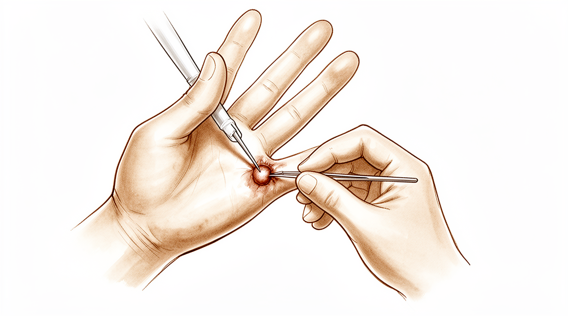
Surgical Approach / Technique: Open excision involves removing the ganglion and its pedicle, a strategy confirmed by ultrasonography and electromyographic studies [30]. In pediatric wrist ganglion excision, marginal excision of the ganglion and its base is used in cases with unclear boundaries [29], with the base ligated using a 4-0 absorbable suture [29]. The WALANT (Wide-Awake Local Anesthesia No Tourniquet) technique can be used in pediatric patients undergoing ganglion removal with similar benefits previously demonstrated in the adult population, provided appropriate patient selection is made [50]. Treatment of intratendinous ganglions should include preserving the tendon, which may be weakened by the ganglion [43]. Excision of the articular nerve branch is advised to avoid recurrences in cases of ganglions within the ulnar nerve communicating with the distal radioulnar joint [7].
Arthroscopic excision of dorsal wrist ganglions involves excising the area of the ganglion’s implantation in the scapholunate ligament, leaving a 1-cm² hole in the dorsal capsule [49]. Care must be taken to preserve the dorsal component of the scapholunate ligament as well as the extensor tendons [49]. Immediate disappearance of the ganglion mass should be confirmed during arthroscopic excision, especially for multi-lobulated cysts, to avoid early recurrence due to inadequate resection [49]. Dye-assist arthroscopic inside-out ganglionectomy is safe and uncomplicated [53]. The outcomes, recurrence, and complications rates after 4 years of follow-up support the use of arthroscopy as a treatment for dorsal wrist ganglion [23]. The described technique for periglenoidal ganglion cysts combines the advantages of open and arthroscopic surgery, allowing one to address the underlying intra-articular pathology, completely evacuate the ganglion cyst, and protect the suprascapular nerve [18].
Post-Operative Care: In pediatric wrist ganglion excision, the wrist is immobilized with a plaster cast for 7 to 10 days [29].
Other Considerations: Surgical excision of wrist ganglions leaves scars and carries some risk for adverse events [2]. A synovial fistula is a rare complication of recurrent dorsal wrist ganglion excision, potentially caused by extensive capsular excision and steroid injection [34]. Subsequent excision of a ganglion and external neurolysis resulted in complete return of nerve function, muscle bulk, and strength in a case of suprascapular nerve denervation secondary to attenuation by a ganglionic cyst [9].
Complications
Recurrence
Surgical intervention for wrist ganglions carries a recurrence rate of approximately 10% [2]. Open surgical excision offers a significantly lower chance of recurrence compared with aspiration [11]. Advances in surgical techniques have allowed arthroscopic ganglion excision to achieve recurrence rates similar to those of open management [1]. Reported recurrence rates for arthroscopic resection range from 8.5% to 30% [71], while rates for surgical excision range from 1% to 50% [71]. In contrast, recurrence rates for aspiration range from 60% to 95% [71]. Pediatric wrist ganglion cysts have a reported low recurrence rate of 5.3% [36].
Dominant side, female sex, and age of 24 years or less are the most influential risk factors for recurrence after arthroscopic excision of dorsal wrist ganglia [20]. Inadequate resection of multi-lobulated cysts during arthroscopic excision can lead to early recurrence [49]. Excision of the articular nerve branch is advised to avoid recurrences in cases of intraneural ganglions [7]. The coexistence of both synovial and ganglion tissue types at recurrence occurs with equal recurrence rates in both initial synovial and ganglion groups [6].
Nerve and Vascular Injury
Radial artery damage, neuropraxia, neuroma formation, and paresthesia are among the most common complications reported in pooled populations of open and arthroscopic excisions [75]. Extensor tendon lacerations are a reported complication of arthroscopic excision of dorsal wrist ganglions [14]. Operation-related complications after arthroscopic volar wrist ganglionectomy are associated with anatomical location distal to the bifurcation of the radial artery and concurrent penetration up to the superficial fascia layer [16]. Patients whose occupation or activities require forceful wrist extension should be counseled on the considerable risk of residual pain and functional limitations after open dorsal wrist ganglion excision [5].
General Surgical Complications
The pooled complication rate for open excision is 5.6% (median 2.8%) [75], while the pooled complication rate for arthroscopic excision is 4% (median 3.6%) [75]. Joint stiffness, wound infection, hematoma requiring surgical drainage, hypertrophic scar, and extensor tenosynovitis are among the most common complications reported in pooled populations of open and arthroscopic excisions [75]. Surgical intervention for wrist ganglions leaves scars [2] and carries some risk for adverse events [2]. Dye-assist arthroscopic inside-out ganglionectomy is described as safe and uncomplicated [53].
Recovery
Postoperative Outcomes and Function: In cases of suprascapular nerve denervation secondary to attenuation by a ganglionic cyst, subsequent excision of the ganglion and external neurolysis resulted in complete return of nerve function, muscle bulk, and strength [9]. Similarly, in a case of ulnar nerve double crush by entrapment of a peri-cubital tunnel ganglion cyst and cubital tunnel, the patient experienced a complete recovery with no functional limitations [80].
Recurrence: Surgical intervention carries an approximately 10% recurrence rate [2]. Open surgical excision offers a significantly lower chance of recurrence compared with aspiration in the treatment of wrist ganglions [11]. In cases of a ganglion within the ulnar nerve communicating with the distal radioulnar joint, excision of the articular nerve branch is advised to avoid recurrences [7]. A study demonstrated equal recurrence rates in both initial synovial and ganglion groups of cystic soft tissue tumours of the dorsal aspect of the wrist [6].
Postoperative Care and Management: Performing at least 1 aspiration before surgical excision improves the cost-effectiveness of dorsal wrist ganglions treatment [31]. Treatment of the intratendinous ganglion should include preserving the tendon, which may be weakened by the ganglion [43].
Key Evidence
- [L4] Advances in surgical techniques have allowed surgeons to conduct arthroscopic ganglion excision with recurrence rates similar to those of open management. [1] (10.5435/jaaos-d-22-00105)
- [L5] Current best evidence suggests that about 40% of lesions decrease over the first 6 years after evaluation by a hand surgeon, that most ganglions recur after aspiration, and that surgical intervention has about a 10% recurrence rate, leaves scars, and has some risk for adverse events. [2] (10.1016/j.jhsa.2010.11.048)
- [L4] Arthroscopy allows for the simultaneous treatment of ganglions and other pathologies. [3] (10.1016/j.jhsa.2012.04.042)
- [L4] Ganglions in children aged >10 years resemble adult ganglions, occurring mainly on the dorsal aspect and are amenable to open excision surgery, which has a recurrence rate of 2.8% to 35%. [4] (10.1016/j.jhsa.2021.12.015)
- [L4] Patients whose occupation or activities require forceful wrist extension should be counseled on the considerable risk of residual pain and functional limitations that may occur after open dorsal wrist ganglion excision. [5] (10.1016/j.jhsa.2015.05.030)
- [L4] The study demonstrated two histologically distinct tissue types at primary surgery and the coexistence of both tissue types at recurrence, with equal recurrence rates in both initial synovial and ganglion groups. [6] (10.1177/17531934241251721)
- [Case_report] Excision of the articular nerve branch is advised to avoid recurrences. [7] (10.1016/j.jhsa.2011.08.008)
- [L3] This study suggests that, in patients with the clinical diagnosis of wrist ganglion cyst, quality of care would not be compromised by abandoning the practice of routinely submitting surgical specimens for pathological examination after excision of the ganglion cyst. [8] (10.1016/j.jhsa.2010.03.021)
- [L4] Subsequent excision of the ganglion and external neurolysis resulted in complete return of nerve function, muscle bulk, and strength. [9] (10.2106/00004623-198668040-00025)
- [L4] Ganglion aspiration should be considered as a first-line intervention, with surgical excision remaining an effective option for symptomatic cases. [10] (10.1177/1753193411434376)
- [L1] Open surgical excision offers significantly lower chance of recurrence compared with aspiration in the treatment of wrist ganglions. [11] (10.1016/j.jhsa.2014.12.014)
- [L4] Surgeons should maintain a high index of suspicion for ganglion cysts in such presentations. [12] (10.1016/j.jhsa.2020.06.005)
- [L4] There is no consensus within the literature regarding the best management of pediatric wrist ganglia, and no single treatment modality confers a particular advantage or disadvantage over another. [13] (10.1177/1558944720966716)
- [L3] [14] (10.1177/15589447211003184)
- [L2] The systematic review and survey of Canadian hand surgeons reveal that hand surgeons are divided regarding the need to immobilize the wrist after dorsal wrist ganglion excision. [15] (10.1177/15589447211014631)
- [L3] The operation-related complications after arthroscopic volar wrist ganglionectomy are associated with its anatomical location: distal to the bifurcation of the radial artery and concurrently penetrated up to the superficial fascia layer. [16] (10.1186/s12891-025-08766-x)
- [L4] [17] (10.1016/j.jhsa.2015.05.025)
- [L4] The described technique combines the advantages of open and arthroscopic surgery, allowing one to address the underlying intra-articular pathology, completely evacuate the ganglion cyst, and protect the suprascapular nerve. [18] (10.1016/j.arthro.2005.10.022)
- [L5] Nerve tumors are rare and often overlooked in the differential diagnosis of upper extremity masses; inappropriate surgery can result in irreversible loss of function, necessitating strict tumor surgery techniques and careful preoperative planning. [19] (10.1016/j.hcl.2004.03.003)
- [L4] Dominant side, female sex, and age of 24 years or less are considered to be the most influential risk factors for recurrence after arthroscopic excision of dorsal wrist ganglia. [20] (10.1016/j.arthro.2013.04.002)
- [L2] Ganglions in pediatric populations, which most commonly affect the dorsal wrist, demonstrate a female predilection. [21] (10.1016/j.jhsa.2021.02.026)
- [L4] Patients experienced significant increases in function and decreases in pain within 6 weeks after arthroscopic ganglion cyst resection, and the recurrence and complication rates appear to be comparable to open resections. [22] (10.1016/j.jhsa.2008.11.025)
- [L4] The outcomes, recurrence, and complications rates after 4 years of follow-up presented in this study support the use of arthroscopy as a treatment for dorsal wrist ganglion. [23] (10.1177/1558944717743601)
- [Case_report] [24] (10.1016/j.jhsa.2010.08.033)
- [L4] [25] (10.1177/15589447221109644)
- [L4] Pediatric ganglions of the hand have a greater rate of resolution than ganglions of the wrist. [26] (10.1016/j.jhsa.2023.07.002)
- [L4] [27] (10.1016/j.jhsa.2006.10.001)
- [L4] In a child with a wrist ganglion, if the cyst ultimately resolved, it usually did so within 18 months. [28] (10.1016/j.jhsa.2019.10.032)
- [L2] [29] (10.1016/j.jhsa.2015.01.015)
- [L2] As patient preferences may preclude routinely performing 2 aspirations, performing at least 1 aspiration before surgical excision improves the cost-effectiveness of dorsal wrist ganglions treatment. [31] (10.1016/j.jhsa.2022.09.002)
- [L4] This is the first reported case of triggering pathology at the wrist to be treated with interventional radiological measures rather than open surgery and demonstrates the efficacy of the technique. [32] (10.1177/1753193412453699)
- [Case_report] A synovial fistula is a rare complication of recurrent dorsal wrist ganglion excision, potentially caused by extensive capsular excision and steroid injection. [34] (10.1016/j.jhsa.2012.02.015)
- [Commentary] Be wary of offering surgery to young, less educated patients with dorsal volar or wrist ganglia as the surgery is often unrewarding for both the surgeon and the patient. [35] (10.1177/1753193412466985)
- [L4] This retrospective review reports a low recurrence rate of 5.3% for pediatric wrist ganglion cysts, indicating potential merit in the surgeon's operative and postoperative techniques. [36] (10.1177/1558944717751195)
- [L5] Treatment of benign bony and soft tissue tumors in the hand is often based on small series or experience with similar lesions elsewhere in the body. [37] (10.1016/j.jhsa.2010.08.015)
- [L2] Worse hand function was associated with recurrence following prior surgery, worse baseline hand function and lower treatment credibility. [38] (10.1177/17531934231153029)
- [L4] Surgical costs from arthroscopic ganglion excision are significantly more than open excision. [40] (10.1016/j.jhsa.2017.05.012)
- [L4] This case highlights the importance of considering uncommon aetiologies in patients with atypical symptoms of carpal tunnel syndrome. [41] (10.1177/17531934241227809)
- [Case_report] Treatment of the intratendinous ganglion should include preserving the tendon, which may be weakened by the ganglion. [43] (10.1177/1753193412453428)
- [L4] [49] (10.1016/j.arthro.2010.05.008)
- [L4] The WALANT technique can be used in pediatric patients undergoing ganglion removal with similar benefits previously demonstrated in the adult population, provided appropriate patient selection is made. [50] (10.1016/j.jhsa.2025.05.010)
- [L4] Dye-assist arthroscopic inside-out ganglionectomy is safe and uncomplicated, worth of clinical promotion. [53] (10.1186/s12891-024-07797-0)
- [L3] Patients with wrist hyperlaxity have a predisposition to developing ganglions, a finding corroborated by independent investigations using similar prospective cohort designs. [54] (10.1016/j.jhsa.2013.11.025)
- [L3] [71] (10.1177/1558944720921477)
- [L4] [73] (10.1016/j.jhsa.2019.10.016)
- [L2] [75] (10.1177/1753193417734428)
- [Case_report] The patient was followed up for 1 month and experienced a complete recovery with no functional limitations. [80] (10.1186/s12891-025-08526-x)
- [L5] The unifying articular (synovial) theory offers a logical, anatomical explanation for all cases of intraneural ganglia, and disconnecting the articular branch connection is effective treatment. [82] (10.1016/j.jhsa.2016.04.004)
- [L3] Operative treatment did not result in less pain intensity or disability, or higher satisfaction compared with non-operative treatment. [86] (10.1177/1753193412442468)
- [L4] Carpal tunnel syndrome caused by a space occupying lesion is rare and more complicated than idiopathic carpal tunnel syndrome. [91] (10.1177/1753193411414352)
See Also
- Arthroscopy
- Tumors
- Neuropathy
- Dupuytren's Disease
- Trigger Finger
- Flexor Sheath Ganglion
References
[1] Ganglions in the Hand and Wrist: Advances in 2 Decades. Journal of the American Academy of Orthopaedic Surgeons. 2023. DOI: 10.5435/jaaos-d-22-00105
[2] Wrist Ganglions. The Journal of Hand Surgery. 2011. DOI: 10.1016/j.jhsa.2010.11.048
[3] Ganglions of the Wrist and Associated Triangular Fibrocartilage Lesions: A Prospective Study in Arthroscopically-treated Patients. The Journal of Hand Surgery. 2012. DOI: 10.1016/j.jhsa.2012.04.042
[4] Pediatric Ganglions of the Hand and Wrist: A Review of Current Literature. The Journal of Hand Surgery. 2022. DOI: 10.1016/j.jhsa.2021.12.015
[5] Outcomes of Open Dorsal Wrist Ganglion Excision in Active-Duty Military Personnel. The Journal of Hand Surgery. 2015. DOI: 10.1016/j.jhsa.2015.05.030
[6] Cystic soft tissue tumours of the dorsal aspect of the wrist have two distinct histological subtypes. Journal of Hand Surgery (European Volume). 2024. DOI: 10.1177/17531934241251721
[7] A Ganglion Within the Ulnar Nerve and Communication With the Distal Radioulnar Joint via an Articular Branch: Case Report. The Journal of Hand Surgery. 2011. DOI: 10.1016/j.jhsa.2011.08.008
[8] Necessity of Routine Pathological Examination After Surgical Excision of Wrist Ganglions. The Journal of Hand Surgery. 2010. DOI: 10.1016/j.jhsa.2010.03.021
[9] Suprascapular nerve denervation secondary to attenuation by a ganglionic cyst.. The Journal of Bone & Joint Surgery. 1986. DOI: 10.2106/00004623-198668040-00025
[10] Patient outcomes following wrist ganglion excision surgery. Journal of Hand Surgery (European Volume). 2012. DOI: 10.1177/1753193411434376
[11] Wrist Ganglion Treatment: Systematic Review and Meta-Analysis. The Journal of Hand Surgery. 2015. DOI: 10.1016/j.jhsa.2014.12.014
[12] Atraumatic, Progressive, and Painful Elbow Contracture From a Ganglion Cyst. The Journal of Hand Surgery. 2021. DOI: 10.1016/j.jhsa.2020.06.005
[13] Wrist Ganglion Cysts in Children: An Update and Review of the Literature. HAND. 2020. DOI: 10.1177/1558944720966716
[14] Recurrence Rates of Dorsal Wrist Ganglion Cysts After Arthroscopic Versus Open Surgical Excision: A Retrospective Comparison. HAND. 2021. DOI: 10.1177/15589447211003184
[15] Immobilization of the Wrist After Dorsal Wrist Ganglion Excision: A Systematic Review and Survey of Current Practice. HAND. 2021. DOI: 10.1177/15589447211014631
[16] Anatomical location of volar wrist ganglion in preoperative MRI is a risk factor for operation-related complications after arthroscopic ganglionectomy. BMC Musculoskeletal Disorders. 2025. DOI: 10.1186/s12891-025-08766-x
[17] Intraneural Ganglions of the Hand and Wrist. The Journal of Hand Surgery. 2015. DOI: 10.1016/j.jhsa.2015.05.025
[18] Combined Intra‐ and Extra‐articular Arthroscopic Treatment of Entrapment Neuropathy of the Infraspinatus Branches of the Suprascapular Nerve Caused by a Periglenoidal Ganglion Cyst. Arthroscopy. 2006. DOI: 10.1016/j.arthro.2005.10.022
[19] Nerve tumors of the hand and upper extremity. Hand Clinics. 2004. DOI: 10.1016/j.hcl.2004.03.003
[20] Arthroscopic Excision of Dorsal Wrist Ganglion: Factors Related to Recurrence and Postoperative Residual Pain. Arthroscopy. 2013. DOI: 10.1016/j.arthro.2013.04.002
[21] Clinical Presentation and Characteristics of Hand and Wrist Ganglion Cysts in Children. The Journal of Hand Surgery. 2021. DOI: 10.1016/j.jhsa.2021.02.026
[22] Prospective Outcomes and Associations of Wrist Ganglion Cysts Resected Arthroscopically. The Journal of Hand Surgery. 2009. DOI: 10.1016/j.jhsa.2008.11.025
[23] Arthroscopic Resection of Dorsal Wrist Ganglion: Results and Rate of Recurrence Over a Minimum Follow-up of 4 Years. HAND. 2017. DOI: 10.1177/1558944717743601
[24] An Ulnar Intraneural Ganglion Arising From the Pisotriquetral Joint: Case Report. The Journal of Hand Surgery. 2011. DOI: 10.1016/j.jhsa.2010.08.033
[25] A Simple Physical Exam Maneuver to Distinguish Trigger Digit, Dupuytren’s Nodule, and Flexor Sheath Ganglion. HAND. 2022. DOI: 10.1177/15589447221109644
[26] Natural History of Pediatric Hand and Wrist Ganglion Cysts: Longitudinal Follow-Up of a Prospective, Dual-Center Cohort. The Journal of Hand Surgery. 2023. DOI: 10.1016/j.jhsa.2023.07.002
[27] Adventitial Cyst of the Radial Artery With a Wrist Joint Connection. The Journal of Hand Surgery. 2007. DOI: 10.1016/j.jhsa.2006.10.001
[28] Wrist Ganglia in Children: Nonsurgical Versus Surgical Treatment. The Journal of Hand Surgery. 2020. DOI: 10.1016/j.jhsa.2019.10.032
[29] Visualization of the Wrist Ganglion Capsule by Methylene Blue Staining as an Aid for Complete Resection in Children. The Journal of Hand Surgery. 2015. DOI: 10.1016/j.jhsa.2015.01.015
[30] A REPORT OF THREE CASES*. 1991.
[31] Minimizing Costs for Dorsal Wrist Ganglion Treatment: A Cost-Minimization Analysis. The Journal of Hand Surgery. 2023. DOI: 10.1016/j.jhsa.2022.09.002
[32] Volar wrist ganglion presenting as trigger finger. Journal of Hand Surgery (European Volume). 2012. DOI: 10.1177/1753193412453699
[34] Synovial Fistula as a Complication of Recurrent Dorsal Wrist Ganglion Excision: Case Report. The Journal of Hand Surgery. 2012. DOI: 10.1016/j.jhsa.2012.02.015
[35] Commentary on Peters et al. Ganglions of the hand and wrist: determinants of treatment choice. Journal of Hand Surgery (European Volume). 2013. DOI: 10.1177/1753193412466985
[36] Pediatric Ganglion Cysts: A Retrospective Review. HAND. 2018. DOI: 10.1177/1558944717751195
[37] Benign Bony and Soft Tissue Tumors of the Hand. The Journal of Hand Surgery. 2010. DOI: 10.1016/j.jhsa.2010.08.015
[38] Factors associated with self-reported pain and hand function following dorsal wrist ganglion excision. Journal of Hand Surgery (European Volume). 2023. DOI: 10.1177/17531934231153029
[40] Cost Minimization Analysis of Ganglion Cyst Excision. The Journal of Hand Surgery. 2017. DOI: 10.1016/j.jhsa.2017.05.012
[41] Carpal tunnel syndrome caused by an interosseous ganglion of the lunate. Journal of Hand Surgery (European Volume). 2024. DOI: 10.1177/17531934241227809
[43] Tenosynovitis of the extensor pollicis longus tendon caused by an intratendinous ganglion: a case report. Journal of Hand Surgery (European Volume). 2012. DOI: 10.1177/1753193412453428
[44] Exam Of The Hand Wrist 2Ed. INTRODUCTION.
[45] Orthopaedic Knowledge Update 13 Ebook Without Multimedia. Anatomy, Evaluation, Clinical Examination, and Imaging > Evaluation and Clinical Examination: Current Concepts.
[47] A Lange Medical Book Current Diagnosis Treatment In Orthopedics Fifth Edition. 9Hand Surgery > Image DISORDERS OF THE MUSCULATURE OF THE HAND.
[48] Exam Of The Hand Wrist 2Ed. 1.1 SKELETON OF THE HAND > The osseous skeleton.
[49] Arthroscopic Resection of Dorsal Wrist Ganglia: 114 Cases With Minimum Follow‐Up of 2 Years. Arthroscopy. 2010. DOI: 10.1016/j.arthro.2010.05.008
[50] Impact of Wide-Awake Local Anesthesia No Tourniquet for Ganglion Excision in Pediatric Patients. The Journal of Hand Surgery. 2026. DOI: 10.1016/j.jhsa.2025.05.010
[53] Outcome evaluation of dye-assist arthroscopic inside-out ganglionectomy: a retrospective study of 29 patients. BMC Musculoskeletal Disorders. 2024. DOI: 10.1186/s12891-024-07797-0
[54] Increased Prevalence of Ganglion Formation Among Patients With Wrist Hyperlaxity. The Journal of Hand Surgery. 2014. DOI: 10.1016/j.jhsa.2013.11.025
[64] A Lange Medical Book Current Diagnosis Treatment In Orthopedics Fifth Edition. 9Hand Surgery > FLEXOR TENDON INJURY.
[65] Exam Of The Hand Wrist 2Ed. 1.3 MOVEMENTS OF THE HAND AND WRIST > Gliding mechanisms.
[66] Exam Of The Hand Wrist 2Ed. The dorsal skin.
[67] Green S Operative Hand Surgery. PERTINENT ANATOMY.
[71] Factors Impacting Recurrence Rate After Open Ganglion Cyst Excision. HAND. 2020. DOI: 10.1177/1558944720921477
[73] Bilateral Plexiform Neurofibromas of the Posterior Interosseous Nerve Mimicking Dorsal Wrist Ganglions. The Journal of Hand Surgery. 2020. DOI: 10.1016/j.jhsa.2019.10.016
[75] Arthroscopic versus open excision of dorsal ganglion cysts: a systematic review. Journal of Hand Surgery (European Volume). 2017. DOI: 10.1177/1753193417734428
[80] Ulnar nerve double crush by entrapment of a peri-cubital tunnel ganglion cyst and cubital tunnel: a case report. BMC Musculoskeletal Disorders. 2025. DOI: 10.1186/s12891-025-08526-x
[82] The Unifying Articular (Synovial) Origin for Intraneural Ganglion Cysts: Moving Beyond a Theory. The Journal of Hand Surgery. 2016. DOI: 10.1016/j.jhsa.2016.04.004
[86] Ganglions of the hand and wrist: determinants of treatment choice. Journal of Hand Surgery (European Volume). 2012. DOI: 10.1177/1753193412442468
[91] Unusual causes of carpal tunnel syndrome: space occupying lesions. Journal of Hand Surgery (European Volume). 2011. DOI: 10.1177/1753193411414352