Clinicians › Shoulder
EnFix Biological Scaffold
For patients: a plain-language version of this topic is available. See the patient guide.
Overview
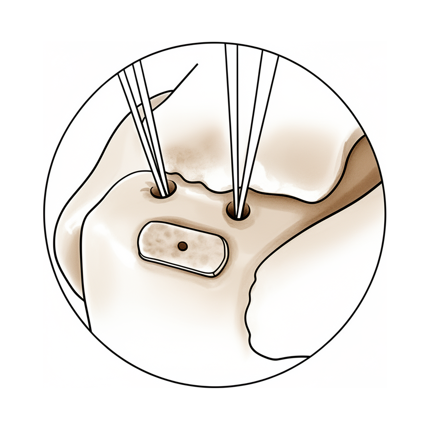
Scaffold devices, including extracellular matrix or synthetic grafts, offer the potential to mechanically and biologically augment repair sites [1]. EnFix implants are procedure-specific allogenic tissue products designed to enhance enthesis healing in rotator cuff repair [8]. These implants utilize patented Demineralized Bone Fiber technology to provide osteoinductive and osteoconductive properties [8]. By promoting enthesis reformation and Sharpey's fiber formation, EnFix implants enhance healing at the tendon-bone interface [15]. This approach represents a significant advance over traditional overlay augmentation products [15]. In preclinical studies, inlay scaffold positioning has been shown to enhance the healing biology of the enthesis while providing histologic similarities to its native 4-zone configuration [12]. This technique allows for optimal placement of the scaffold to maximize its healing properties and achieve full footprint coverage [7].
The scaffold is intended to resorb over a period of 3 to 4 months [3]. Aside from the biologic scaffold, this technique requires minimal extra implant costs except for 2 additional sutures [5]. Although clinical studies are required to evaluate the results of this technique, it offers a unique combination of biologic and inlay graft augmentation [14]. The one-year follow-up evaluation will assess the long-term maintenance of good functional results and the possible benefit of adding orthobiological stimuli to the repair [13].
Current evidence remains limited by small patient cohorts, heterogeneous scaffold designs, and insufficient long-term outcome data [2]. In animal models, treatment with scaffold alone improved defect stability at 16 weeks compared to nontreatment, but did not achieve bone union or restoration of mechanical function [4]. Isolated scaffolds did not heal any defects, whereas other treatments led to healing in 7/10 (BMP-7), 10/10 (BMP-7 scaffold), and 9/10 (BMP-7 scaffold systemic bisphosphonate) rats [11].
Anatomy & Pathophysiology
Bony Anatomy
The proximal humerus comprises the humeral head, greater tuberosity, lesser tuberosity, and humeral shaft [40]. The articular head is spherical with a diameter of 37 to 57 mm [40] and an arc of approximately 160 degrees covered by articular cartilage [52]. The radius of curvature of the humeral head is approximately 25 mm [52], while the glenoid articular surface radius of curvature is 2 to 3 mm larger [52]. The most superior portion of the articular surface averages 8 mm above the greater tuberosity [40], with the superior margin normally superior to the top of the greater tuberosity by 8 to 10 mm [52]. Humeral version averages 29.8 degrees, with a range of 10 to 55 degrees [40]. The humeral head is inclined approximately 130 degrees with respect to the humeral shaft [40], averaging 41° of inclination [43]. The average neck-shaft angle is 45 degrees, with a range of 30 to 50 degrees [52]. The anatomic neck is located at the junction of the articular surface and the tuberosities [40], while the surgical neck represents an indistinct region below the tuberosities but above the humeral shaft [40].
The greater tuberosity serves as the attachment site for the supraspinatus, infraspinatus, and teres minor tendons [40]. The lesser tuberosity serves as the attachment site for the subscapularis tendon [40]. The distance from the lateral base of the coracoid process to the lateral margin of the greater tuberosity is called the lateral humeral offset [52]. The humeral head height is approximately 5.6 cm above the superior border of the pectoralis major tendon [54]. The transverse humeral ligament is an important stabilizer of the biceps tendon [54].
The glenoid is a convex structure of shallow depth shaped like an inverted pear [40]. The glenoid cavity is a shallow socket, approximately one third the size of the humeral head [41]. The glenoid labrum increases the depth of the socket by 50% around the humeral head [53]. The glenoid articular surface and the labrum combine to create a socket that is approximately 9 mm deep in the superoinferior direction and 5 mm deep in the anteroposterior direction [53]. Adding the glenoid labrum increases the glenoid surface to 75% of the humeral head vertically and 57% horizontally [53]. The glenoid averages 5° of retroversion in relation to the axis of the scapular body [43] and is retroverted approximately 5 degrees relative to the scapular body [54]. The humeral head averages 19° of retroversion [43] and is retroverted 30 degrees relative to the transepicondylar axis of the humerus [54].
The scapula is attached to the axial skeleton by the acromioclavicular and sternoclavicular joints [42]. The glenoid is connected with the flat body of the scapula by the scapular neck [42]. The coracoid process curves forwards from the superior surface of the scapular neck [42]. The acromion is a flattened bony process that curves forwards from the scapular spine [42]. The lateral pillar connects the inferior border of the glenoid with the inferior angle of the scapula [42]. The spinal pillar arises from the central part of the glenoid and continues medially to become part of the base of the scapular spine [42]. The lateral pillar and spinal pillar form the basic load-bearing structure of the scapular body [42]. The weakest bone in the scapula is located primarily in the central part of the infraspinous fossa [42]. The weakest area of the circumference of the biomechanical body of the scapula is the spinomedial angle [42]. The scapula spans second through seventh ribs and serves as an attachment for 17 muscles [54]. The scapula is anteverted on the chest wall approximately 30 degrees relative to the body [54].
Soft Tissue Anatomy
The rotator cuff consists of the subscapularis, supraspinatus, infraspinatus, and teres minor muscles [41]. These muscles serve as depressors of the humeral head to allow the deltoid to efficiently abduct the humerus [41]. The infraspinatus and teres minor are external rotators, while the subscapularis is an internal rotator of the humerus [41]. The rotator cuff is a sheet of conjoined tendons closely applied over the shoulder capsule and inserting mainly into the greater tuberosity of the humerus [48]. The subscapularis is inserted into the lesser tuberosity [48].
The acromion, the coracoacromial ligament, and the coracoid process form the coracoacromial arch [40]. The rotator cuff, subacromial bursa, and subdeltoid bursa pass underneath the coracoacromial arch [40]. The coracoacromial arch is formed by the acromion process posterosuperiorly, the coracoid process anteriorly, and the coracoacromial ligament joining them [48]. The subacromial bursa separates the tendons from the coracoacromial arch and allows them to glide [48]. The subscapular bursa lies between the subscapularis tendon and the neck of the scapula [44]. The subscapular bursa communicates with the joint cavity between the superior and middle glenohumeral ligaments [44].
The rotator interval is defined medially by the base of the coracoid, superiorly by the supraspinatus tendon, and inferiorly by the subscapularis tendon [43]. The rotator interval contains the coracohumeral ligament, the superior glenohumeral ligament, and the intra-articular portion of the long head of the biceps tendon [43]. The tendons of the infraspinatus and supraspinatus muscles join approximately 15 mm proximal to their insertion [53]. The infraspinatus and teres minor fuse near their musculotendinous junctions [53]. The supraspinatus and subscapularis tendons join as a sheath that surrounds the biceps tendon at the entrance of the bicipital groove [53].
The coracohumeral ligament is a thick band of fibrous tissue extending from the coracoid process along the surface of the capsule to the tuberosities between the supraspinatus and subscapularis tendons [53]. The superior glenohumeral ligament is the primary restraint to inferior humeral subluxation in 0 degrees of abduction [53]. The middle glenohumeral ligament limits external rotation when the arm is in the lower and middle ranges of abduction [53]. The inferior glenohumeral ligament is composed of an anterior band, a posterior band, and a thinner intervening axillary pouch [53]. The anteroinferior glenohumeral ligament complex is the main stabilizer to anterior and posterior stresses when the shoulder is abducted 45 degrees or more [53].
The superior shoulder suspensory complex provides a stable connection between the scapula and the axial skeleton [43]. It is composed of the glenoid, the coracoid process, the coracoclavicular ligaments, the distal clavicle, the AC joint, and the acromion [43]. The coracoclavicular ligaments are the primary stabilizers to superior translation of the distal clavicle [43]. The posterior SC joint capsule and ligaments are the primary stabilizers to anterior and posterior translation of the medial clavicle [43]. The superior and posterior AC ligaments are the primary stabilizers to anterior and posterior translation of the clavicle [43].
The humeroscapular motion interface lies between the inner structures of the proximal humerus, rotator cuff, coracohumeral ligament, and biceps tendon sheath and the superficial layer of the acromion, deltoid, coracoacromial ligament, coracoid process, and the conjoined tendon [46]. The axillary nerve is a terminal branch coming off the posterior cord of the brachial plexus just proximal to the coracoid process [46]. The axillary nerve passes beneath the conjoined tendon anterior to the subscapularis 3 to 5 mm medial to the musculotendinous junction [46]. The axillary nerve enters the quadrilateral space posteriorly [46]. The anterior and middle deltoid muscle receives sole innervation from the anterior branch of the axillary nerve [46]. Posterior deltoid muscle innervation varies, with supply only from the anterior branch in 2.3% of cases, from the posterior branch in 8.5%, and from both branches in 89.1% [46]. The posterior branch of the axillary nerve branches to supply the teres minor muscle and then terminates as the superior lateral brachial cutaneous nerve [46].
Vascular Anatomy
The proximal humerus receives its blood supply from the anterior and posterior humeral circumflex branches from the third division of the axillary artery [40]. The anterior and posterior humeral circumflex arteries provide a rich blood supply to the proximal humerus [47]. The anterior humeral circumflex artery arises from the axillary artery at the inferior border of the subscapularis [40]. The anterior humeral circumflex artery provides vascular inflow to the humeral head by way of its terminal anterolateral branch known as the artery of Laing [40]. The ascending branch of the anterior humeral circumflex artery courses parallel to the lateral aspect of the long head biceps tendon and enters the humeral head at the interface of the bicipital groove and greater tuberosity [40]. The anterolateral ascending branch of the anterior humeral circumflex artery provides the primary blood supply to the humeral head [43]. The terminal intraosseous portion of the artery enters at the proximal aspect of the intertubercular groove as the arcuate artery [43].
The posterior humeral circumflex artery travels with the axillary nerve, enters the quadrilateral space posteriorly, and anastomoses with a branch of the anterior circumflex to supply the posterior cuff [40]. Sixty-four percent of the humeral head blood supply arises from the posterior humeral circumflex artery [47]. The axillary nerve circles the humeral neck just inferior to the glenohumeral joint as it courses posteriorly [47].
The primary blood supply to the clavicle is periosteal, and no nutrient artery is present [43]. The primary blood supply to the clavicle is periosteal, and there is no nutrient blood supply [49].
Pathophysiology & Biomechanics
Stability and function of the glenohumeral joint is provided by the interaction of the glenohumeral joint that promote a near global range of motion and purposeful function [40]. External loads that are transferred to the shoulder girdle are initially offset by joint surface anatomy, joint volume, atmospheric pressure, and joint fluid cohesion and adhesion [40]. Moderate and large loads are counterbalanced by the deltoid and rotator cuff and by the capsulolabral and bone structures, respectively [40]. The glenohumeral joint depends on the static and dynamic stabilizers for movement and stability, especially the rotator cuff [52]. The rotator cuff stabilizes the glenohumeral joint while allowing greater freedom of motion and fixes the fulcrum of the upper extremity against which the deltoid can contract and elevate the humerus [52]. The rotator cuff stabilizes the joint via joint compression [43].
The bony anatomy contributes little to stability and has been compared with a golf ball on a tee [53]. Most of the stability of the shoulder is provided by the surrounding muscles and ligaments due to very little bony constraint [53]. The glenoid labrum provides concavity and up to 50% of marginal glenoid socket depth [43]. The glenoid labrum deepens the socket 50% and provides a bumper to translation [54]. Laxity of the rotator interval results in inferior laxity, and contracture of the interval is seen with adhesive capsulitis [43].
The coracohumeral ligament restricts external rotation in adduction and is a static restraint to inferior and posterior translation in adduction and external rotation [43]. The coracohumeral ligament restrains inferior translation and external rotation of the adducted arm [54]. The superior glenohumeral ligament is a primary static restraint against anterior translation with the arm at the side [43]. The superior glenohumeral ligament restrains external rotation and inferior translation of the adducted or slightly abducted arm [54]. The middle glenohumeral ligament is a primary static restraint against anterior translation with the arm in external rotation and 45° of abduction [43]. The middle glenohumeral ligament restrains anterior translation with the arm abducted to 45 degrees [54]. The anterior band of the inferior glenohumeral ligament is a primary static restraint against anterior-inferior dislocation of the glenohumeral joint in 90° of abduction and external rotation [43]. The inferior glenohumeral ligament, anterior band restrains anterior and inferior translation with the arm externally rotated and abducted to 90 degrees [54]. The posterior band of the inferior glenohumeral ligament is a primary static restraint against posterior-inferior translation in internal rotation and adduction [43]. The inferior glenohumeral ligament, posterior band restrains posterior and inferior translation with the arm internally rotated and abducted to 90 degrees [54]. The coracoacromial ligament contributes to anterosuperior stability in rotator cuff deficiency and should be preserved with irreparable cuff tears to prevent anterosuperior escape [54].
The sternoclavicular joint is the only true joint connecting the upper extremity with the axial skeleton [54]. The posterior sternoclavicular ligament is the strongest and primary restraint to anteroposterior instability of the sternoclavicular joint [54]. The AC ligaments prevent anteroposterior displacement of the clavicle [54]. The coracoclavicular ligaments prevent superior displacement of the distal clavicle [54]. The trapezoid ligament is located approximately 25 mm from the AC joint [54]. The conoid ligament is located approximately 45 mm from the AC joint [54].
The scapula is suspended by muscles alone and has shifted caudally from the cervical position in lower animals [50]. Broadening of the infraspinatus fossa has resulted in a change in the vector of muscle pull from the axillary border of the scapula to the glenoid fossa [50]. This adaptation allows the infraspinatus and teres minor muscles to be more effective in their roles as depressors and external rotators of the humeral head [50]. The acromion has enlarged over time, reflecting the increasing role of the deltoid muscle in shoulder function [50]. The broader attachment of the deltoid on the acromion and its more distal insertion on the humerus have increased its mechanical advantage in shoulder motion [50]. The coracoid process has undergone an increase in size over time [50]. With the shoulder in 90 degrees of abduction, the coracoid extension over the glenohumeral joint can mechanically limit anterior translation of the humerus relative to the glenoid [50].
The proximal humerus is primarily cartilaginous at birth [47]. The ossification center for the humeral head is usually present at birth [47]. The greater tuberosity ossification center appears by 1 to 3 years of age [47]. The lesser tuberosity ossification center appears by 5 years of age [47]. The proximal humeral ossification center fuses by 5 to 7 years of age to form the humeral head [47]. The proximal humeral physis closes by 14 to 17 years of age in girls and by 16 to 18 years in boys [47]. Humeral retroversion averages 65 degrees in infants and young children and gradually decreases, approaching adult values by 11 years of age [47]. Eighty percent of subsequent growth of the humerus comes from the proximal humeral physis [47].
Classification
Product Classification: EnFix implants are classified as procedure-specific allogenic tissue products [8]. These implants utilize patented Demineralized Bone Fiber (DBF) technology [8]. The product provides both osteoinductive and osteoconductive properties [8]. The primary design intent is to enhance enthesis healing in rotator cuff repair [8].
Biological Mechanism: DBF is a form of demineralized bone matrix (DBM) that contains bone morphogenetic proteins (BMPs) and other growth factors known to facilitate healing [26]. BMPs stimulate the differentiation of mesenchymal stem cells (MSCs) into osteoblasts and chondrocytes, thereby promoting osteoinduction [26]. The osteoconductive property of DBF enables cells necessary for tissue repair to migrate to the area and provides a framework for new tissue formation at the enthesis [26].
Enthesis Reformation: EnFix implants promote enthesis reformation and Sharpey's fiber formation [15]. The recreation of the four-zone fibrocartilaginous structure of the native enthesis is achieved through DBF augmentation [26]. The porous structure of the DBF functions as a scaffold, facilitating collagen deposition, neovascularisation, and infiltration of host cells [26].
Clinical Presentation
Radiologic Assessment: At 6 months postoperatively, radiologic evaluation demonstrated complete healing in the augmented group [26]. This group also exhibited a trend towards more robust tendon quality and greater surface area of tendon encroachment onto bone compared to the non-augmented group [26]. Neither the augmented nor non-augmented group experienced any re-tears during the 6-month postoperative period [26].
Functional Outcomes: Improvements in pain, function, active range of motion, and strength were comparable between the augmented and non-augmented cohorts [26]. Early results indicated significantly better ASES scores [26], Constant scores [26], and UCLA scores [26] in the group with DBF augmentation. A trend of achieving greater rotator cuff relative strengths was also observed in the DBF augmentation group [26].
Biologic Mechanism: Bone collagen is the primary constituent of DBF, which contains bone morphogenetic proteins (BMPs) and other growth factors known to facilitate healing [26]. BMPs are primarily responsible for stimulating the differentiation of mesenchymal stem cells (MSCs) into osteoblasts and chondrocytes, thereby promoting osteoinduction [26]. The porous structure of the DBF functions as a scaffold that facilitates collagen deposition, neovascularisation, and infiltration of host cells [26].
Preclinical and Scaffold Evidence: Inlay scaffold positioning in preclinical studies has been shown to enhance the healing biology of the enthesis and provides histologic similarities to the native 4-zone configuration of the enthesis [12]. Bioinductive collagen scaffolds may improve pain, function, and structural outcomes in selected rotator cuff tears [27]. However, evidence for these scaffolds remains limited, heterogeneous, and predominantly non-randomized [27].
Inlay Implant Literature: The clinical literature for interpositional scaffolds is scarce, with only 2 studies in humans reporting on the use of inlay positioned implants [68]. Both human studies on inlay positioned implants reported a lower failure rate than that currently reported for onlay-augmented repairs and observed improved functional outcomes [68]. Neither human investigation presented a control group for direct comparisons between treatment arms, so results should be interpreted with caution due to the lack of control groups [68]. Further clinical studies are needed to fully ascertain the clinical effect of the observed histologic enhancement from interpositional demineralized bone fiber scaffolds [68].
Long-term Follow-up: The one-year follow-up evaluation will assess the long-term maintenance of good functional results for decellularized human dermis scaffold augmentation and the possible benefit of adding orthobiological stimuli to the repair [13].
Investigations
General Principles: Shoulder imaging serves to establish diagnosis, determine pathoanatomical severity, assist surgical planning, and illustrate the condition to the patient [32]. The shoulder is a three-dimensional structure that cannot be represented by a single planar view, and critical relationships such as humeral head centering change with arm position [60]. Pathology may involve numerous bones and soft tissues, while overlying structures and metallic implants can complicate imaging [60]. Unless a specific research protocol dictates otherwise, clinicians should resist the temptation to "overimage" and obtain only the scans necessary for patient care [32].
Plain Radiography: Standardized plain films are almost always sufficient for shoulder evaluation, and proper radiographic technique is as important as surgical technique [32]. The standard series includes orthogonal views: a true anteroposterior (AP) view in the scapular plane, an AP view, an axillary view, and a scapular Y view [65]. The true AP view in the scapular plane is taken with the x-ray beam perpendicular to the scapula and the arm in neutral rotation with slight abduction, visualizing the anterior greater tuberosity in profile [65]. The standard AP view is taken with the arm in internal rotation to visualize the posterior greater tuberosity and lesser tuberosity in profile, with the beam perpendicular to the coronal plane [65]. This view demonstrates the superoinferior position of the humeral head, osteophytes, joint space narrowing, medial displacement relative to the lateral acromial line, bone quality, loose bodies, and humeral head collapse or deformity [32].
The axillary view is taken with the arm in the functional position of elevation in the plane of the scapula, oriented so that both the spinoglenoid notch and scapular neck are visible [32]. Referred to as the "truth view," it demonstrates glenohumeral relationships in elevation, a perspective not captured by CT scans taken with the arm adducted [32]. It reveals humeral anatomy, glenoid bone amount, shape, version, and the relationship of the humeral head to the glenoid fossa [32]. Joint space narrowing and posterior subluxation or "functional decentering" are most evident on this view compared to images with the arm at the side [32]. Posterior subluxation can be measured by the position of the humeral head center relative to the scapular plane, the glenoid face, or the point of contact of the articular surfaces [32]. The point of contact reflects the centering of the net humeral joint reaction force; malcentering leads to posterior instability, posterior glenoid wear, and "rocking horse" loosening of prosthetic glenoid components [32]. The axillary view is also necessary for evaluating glenohumeral instability, determining humeral head position, detecting occult locked posterior dislocation in patients with limited passive external rotation, and evaluating glenoid morphology in osteoarthritis [65]. It provides good visualization of the coracoid process, acromion, and distal clavicle [65].
The scapular Y view visualizes the coracoacromial arch and can reveal coracoacromial spurs associated with rotator cuff pathology [65]. It is a reliable alternative for evaluating glenohumeral subluxation and dislocation, and can show scapular body abnormalities such as osteochondroma or fracture, as well as acromial shape [65]. Normal acromiohumeral distance is 7 to 14 mm, and the glenohumeral joint space width should be symmetric superiorly and inferiorly [65]. The coracoclavicular distance is normally 1.1 to 1.3 cm [65]. Neer classified acromial morphology as type I (flat), type II (curved), and type III (hooked) [65]. Type III morphology correlates with rotator cuff disease, though no direct causal relationship has been demonstrated, and the classification shows relatively poor interobserver reliability [65].
For proximal humerus fractures, the recommended series is the Neer trauma series, consisting of an AP view, a lateral view in the scapular plane (tangential Y-view), and a Velpeau modified axillary view [59]. This combination allows evaluation of the shoulder joint in three separate perpendicular planes [59]. The axillary view is important for evaluating the glenoid articular surface and the relationship of the humeral head anteriorly and posteriorly [59]. Conventional radiographs are appropriate for patients presenting with shoulder pain with any history of trauma, dislocation, night pain, or chronic shoulder pain [65]. Arthritis, calcific tendinitis, and osteolysis of the distal clavicle can be observed on plain radiograph [64].
Computed Tomography (CT): CT scans may offer a few degrees of increased precision in measuring glenoid version, but there is no conviction that this increased precision improves surgical quality or clinical outcome [32]. CT is helpful for planning fracture surgery and shoulder joint replacement [57]. It is frequently used to evaluate shoulder fractures, assess bony lesions in recurrent instability cases, or for preoperative templating for shoulder arthritis [64]. CT with three-dimensional reconstructions is the advanced imaging study of choice for determining the extent of glenoid bone loss in shoulder instability [65]. Three-dimensional sagittal CT scans allow for optimal evaluation of displaced glenoid fractures [65]. On occasion, CT scanning may be necessary for detailing bony anatomy in proximal humerus fractures [59].
Magnetic Resonance Imaging (MRI): MRI is useful for identifying osteonecrosis of the humeral head or bone tumors [57]. It can identify labral and rotator cuff tears, although accuracy is enhanced by combining the scan with arthrography [57]. MRI is the modality of choice for evaluating the rotator cuff, biceps, and subacromial/subdeltoid bursa [64]. T1-weighted MRI can reveal Hill-Sachs lesions and is often used with magnetic resonance (MR) arthrograms to provide a more detailed picture of joint surfaces [64]. T2-weighted MRI provides better visualization of full-thickness rotator cuff tears [64]. Traditional MRI is utilized for soft tissue evaluation with high contrast and spatial resolution [62]. MR accuracy in identifying labral and rotator cuff tears ranges from 70% to 100% [62]. Multi-planar imaging allows detailed evaluation of the glenoid, labrum, joint capsule, and rotator cuff in different planes [62]. In a meta-analysis of 4,667 shoulders, MRA had greater diagnostic test accuracy for glenoid labral lesions than MRI, with MRA sensitivity of 88% and specificity of 93% versus MRI sensitivity of 76% and specificity of 87% [62].
With standard MRI or MRA, the shoulder is routinely positioned in neutral or partial external rotation, but alternative positions can increase sensitivity for detecting labroligamentous injuries [62]. Abduction and external rotation (ABER) of the arm is an alternative position utilized to increase sensitivity and specificity for detecting anteroinferior labroligamentous injury [62]. Limited range of motion or pain may prohibit patients from performing the ABER provocative maneuver [62]. MRAs can demonstrate a patulous capsule on coronal, sagittal, and axial imaging in patients with multidirectional instability [62]. MRAs can be helpful in evaluating lesions of the rotator interval and other associated findings that may affect the eventual surgical plan [62]. The presence of glenoid dysplasia, increased capsular cross-sectional area, and increased glenoid retroversion have been found to be associated with increased posterior labral tears and symptomatic instability [62]. Glenoid retroversion was significantly increased in patients with symptomatic posterior labral tears, but there was no significant association between instability and increased humeral head subluxation [62]. The diagnosis of multidirectional instability is a clinical one, and the need for expensive and/or invasive imaging should be weighed against the information gained [62].
Magnetic Resonance Arthrography (MRA): MR arthrography refers to MRI of a joint injected with an intra-articular contrast agent such as diluted gadolinium or saline solution [62]. The contrast material is injected prior to MRI by fluoroscopic or ultrasound guidance under strict aseptic technique [62]. By distending the joint capsule, the cartilage, ligaments, and labrum are outlined with contrast, increasing sensitivity for detecting tears and other lesions [62]. In the acute dislocation setting, a joint effusion with distension of the joint may outline structures similarly, making the arthrogram unnecessary [62]. MRA has proven utility by increasing both sensitivity and specificity in detecting injuries to the capsulolabral–ligamentous complex compared to traditional MRI [62]. MR arthrography is considered the benchmark for evaluation of labral tears and is rarely indicated for evaluation of rotator cuff pathology [64]. Arthrography involves injection of contrast agent in conjunction with either an MRI or CT scan, enhancing imaging of the joint to enable better identification of normal structures and pathology involving the joint surfaces [64]. When MRI or MR arthrography is contraindicated (e.g., pacemaker, vascular clips), CT arthrography is indicated [64].
Ultrasonography: Ultrasonography is a simple and accurate test for identifying rotator cuff tears and calcific tendinitis [57]. It can be useful in guiding injections or barbotage (aspirating calcific deposits in the rotator cuff) [57]. Ultrasonography is a low-cost alternative to MRI and arthrography for evaluating both skeletal and soft-tissue structures of the shoulder [64]. It can provide immediate, real-time visualization of the rotator cuff, biceps tendon, and calcific deposits [64]. Ultrasonography can be used to measure the subacromial space and detect atrophy of rotator cuff muscles [64]. As a result of providing images in real-time, ultrasonography can evaluate impingement in various positions and motions [64]. It is highly operator dependent and is not as useful for evaluating labral tears or rotator cuff tears that are very small or larger than 3 cm [64]. The most commonly performed joint examination using ultrasonography is the shoulder examination [55]. Accuracy of rotator cuff ultrasonography depends on the skill of the scanner operator and an awareness of pitfalls that are encountered [55]. There are 10 common pitfalls of rotator cuff ultrasonography that can reduce overdiagnosis or underdiagnosis of rotator cuff pathology [55]. In a study of 147 patients with rotator cuff calcific tendinitis treated with ultrasonography-guided lavage, 70% of shoulders showed significant reduction of symptoms [55]. Calcifications that were softer and middle-sized (12 to 17 mm) had more significant improvement in ultrasonography-guided lavage for calcific tendinitis [55]. Better results occurred in patients aged 30 to 40 years undergoing ultrasonography-guided lavage for calcific tendinitis [55]. Ultrasonography can pick up on partial tears that may need treatment but have normal physical examination findings due to the ability to move the tendon with direct visualization [55].
Arthroscopy: Arthroscopy is useful for diagnosing and treating subacromial impingement, intra-articular lesions, detachment of the glenoid labrum, and rotator cuff tears [57]. Evaluation of the integrity of the rotator cuff may be difficult in the acute setting, and ultrasound, MRI, arthrogram, or arthroscopy may be valuable in making this diagnosis [59].
Treatment
Non-Operative
The provided evidence does not detail specific conservative management protocols such as weight loss, physical therapy, NSAIDs, or injections.
Operative
Indications: EnFix is indicated for rotator cuff repair to enhance healing at the tendon-bone interface [15]. It also serves as a biologically enhanced alternative for proximal biceps tenodesis, where it may optimize enthesis healing and reduce the risk of failure [34].
Surgical Approach / Technique: The arthroscopic slide technique for bioinductive collagen scaffold augmentation requires minimal extra implant costs aside from the scaffold, specifically 2 additional sutures [5]. The interpositional nanofiber scaffold technique allows for optimal placement of the scaffold to maximize its healing properties and full footprint coverage [7]. The EnFix TAC technique offers a reproducible and biologically enhanced alternative for proximal biceps tenodesis [34].
Implant Selection: EnFix utilizes patented Demineralized Bone Fiber (DBF) technology to provide osteoinductive and osteoconductive properties [8]. DBF is a form of demineralized bone matrix that provides a structural scaffold and promotes healing and integration at the enthesis following rotator cuff repair [26]. BMPs in DBF stimulate the differentiation of mesenchymal stem cells into osteoblasts and chondrocytes, thereby promoting osteoinduction [26]. The osteoconductive property of DBF enables cells necessary for tissue repair to migrate to the area and provides a framework for new tissue formation at the enthesis [26]. Collagen deposition, neovascularisation, and infiltration of host cells occurs through the porous structure of the DBF [26]. EnFix implants enhance healing at the tendon-bone interface by promoting enthesis reformation and Sharpey's fiber formation [15]. EnFix offers a significant advance over traditional overlay augmentation products [15].
Adjuncts: The EnFix TAC allograft requires adherence to donor screening, aseptic technique, and traceability requirements [33].
Other Considerations: Radiologic assessment at 6 months postoperatively demonstrated complete healing in the augmented group using the Enfix RC implant [26]. A trend towards more robust tendon quality was observed in the augmented group compared to the non-augmented group at 6 months [26]. A trend towards greater surface area of tendon encroachment onto bone was observed in the augmented group compared to the non-augmented group at 6 months [26]. Improvements in pain, function, active ROM, and strength were comparable between the augmented and non-augmented cohorts [26]. Promising early results of significantly better ASES, Constant, and UCLA scores were observed in the group with DBF augmentation [26]. A trend of achieving greater rotator cuff relative strengths was observed in the group with DBF augmentation [26].
Current evidence for 3D printed scaffolds remains limited by small patient cohorts, heterogeneous scaffold designs, and insufficient long-term outcome data [2]. Bioinductive collagen scaffolds may improve pain, function, and structural outcomes in selected rotator cuff tears [27]. The one-year follow-up evaluation for decellularized human dermis scaffold augmentation will assess the long-term maintenance of good functional results [13]. The one-year follow-up evaluation for decellularized human dermis scaffold augmentation will assess the possible benefit of adding orthobiological stimuli to the repair [13]. Clinical studies are required to evaluate the results of the enthesis interpositional allograft and minced autologous subacromial bursa technique [14]. The enthesis interpositional allograft and minced autologous subacromial bursa technique offers a unique combination of biologic and inlay graft augmentation [14].
Complications
Other Considerations: Preclinical data indicates that the delivery of 2.0 mg rhBMP-2 with a synthetic matrix resulted in the formation of abnormal bony masses at defect sites, suggesting this dose exceeded the therapeutic range [19]. In a rat femoral critical size defect model, isolated scaffolds failed to heal any defects, whereas treatments combining BMP-7 and scaffolds achieved healing in 7/10, 10/10, and 9/10 rats respectively [11].
Recovery
Light activity (weeks): The available evidence does not specify a defined week range for light activity, desk work, driving, or light activities of daily living.
Full activity (months): The available evidence does not specify a defined month range for the return to manual work, sport, or full range of motion and strength.
Complete recovery / outcome plateau (months): The nanofiber scaffold is intended to resorb over a period of 3 to 4 months [3]. In a rabbit model, extracellular matrix formation and spatial alignment of collagen fibers were delayed and not complete by 24 weeks after supraspinatus tendon attachment [78]. In a rat model, treatment with scaffold alone improved defect stability at 16 weeks compared to nontreatment but did not achieve bone union or restoration of mechanical function [4].
Rehabilitation protocol: The available evidence does not specify a rehabilitation protocol, including physical therapy phasing, immobilisation duration, weight-bearing or range of motion progression, or sling or brace removal timing.
Functional milestones: The available evidence does not provide validated patient-reported outcome measure trajectories or specific outcome-measure benchmarks such as Constant, ASES, or WOMAC scores.
Other Considerations: EnFix implants are designed to enhance enthesis healing in rotator cuff repair by utilizing patented Demineralized Bone Fiber technology to provide osteoinductive and osteoconductive properties [8]. In preclinical studies, inlay scaffold positioning has been shown to enhance the healing biology of the enthesis while providing histologic similarities to its native 4-zone configuration [12]. In a rabbit model, delay of supraspinatus repair by up to 12 weeks did not impair enthesis formation [39]. In a goat model, trends of increased strength of early rotator cuff fixation were observed at 3 weeks and decreased strength at later time points, most pronounced at 12 weeks, but these changes did not reach statistical significance at any time point [20]. One-year follow-up evaluation is planned to assess the long-term maintenance of good functional results and the possible benefit of adding orthobiological stimuli to the repair [13]. In a rat model, isolated scaffolds did not heal any critical-sized femoral defects, whereas treatments involving BMP-7 and scaffolds led to healing in 7/10, 10/10, and 9/10 rats respectively [11]. Current evidence for 3D printed scaffolds in segmental long bone defects remains limited by small patient cohorts, heterogeneous scaffold designs, and insufficient long-term outcome data [2].
Key Evidence
- [Paper] Scaffold devices, in the form of extracellular matrix or synthetic grafts, hold promise in their abilities to mechanically and/or biologically augment repair sites. [1] (10.1097/bte.0000000000000122)
- [Paper] Despite encouraging preclinical results and emerging early clinical applications, current evidence remains limited by small patient cohorts, heterogeneous scaffold designs, and insufficient long-term outcome data. [2] (10.1016/j.injury.2026.113758)
- [L5] The scaffold is intended to resorb over a period of 3 to 4 months. [3] (10.1016/j.eats.2022.08.061)
- [Paper] Treatment with scaffold alone improved defect stability at 16 weeks compared to nontreatment, but did not achieve bone union or restoration of mechanical function. [4] (10.1002/jor.20372)
- [L5] Aside from the biologic scaffold, this technique requires minimal extra implant costs except for 2 additional sutures. [5] (10.1002/atn2.70286)
- [L5] This technique allows for optimal placement of the scaffold to maximize its healing properties and full footprint coverage. [7] (10.1016/j.eats.2025.103476)
- [Paper] Isolated scaffolds (Group A) did not heal any defects, whereas the other treatments led to healing in 7/10 (BMP-7), 10/10 (BMP-7 scaffold), and 9/10 (BMP-7 scaffold systemic bisphosphonate) rats. [11] (10.1002/jor.22815)
- [L1] Inlay scaffold positioning in preclinical studies has been shown to enhance the healing biology of the enthesis while providing histologic similarities to its native 4-zone configuration. [12] (10.1016/j.arthro.2024.06.048)
- [L1] The one-year follow-up evaluation will assess the long-term maintenance of good functional results and the possible benefit of adding orthobiological stimuli to the repair. [13] (10.1016/j.jseint.2025.101543)
- [L5] Although clinical studies are required to evaluate the results of this technique, it offers a unique combination of biologic and inlay graft augmentation. [14] (10.1002/atn2.70273)
- [Paper] Abnormal boney masses formed at defect sites receiving 2.0 mg rhBMP2 delivered with the synthetic matrix, suggesting this dose was well above the therapeutic range. [19] (10.1002/jor.22243)
- [Paper] The study demonstrated trends of increased strength of early rotator cuff fixation at 3 weeks and decreased strength at later time points, which were most pronounced at 12 weeks, but did not reach statistical significance at any time point. [20] (10.1016/j.jse.2005.11.009)
- [L4] BCS may improve pain, function, and structural outcomes in selected rotator cuff tears; however, evidence remains limited, heterogeneous, and predominantly non-randomized. [27] (10.5397/cise.2025.01326)
- [L5] This method offers a reproducible and biologically enhanced alternative for proximal biceps tenodesis that may optimize enthesis healing and reduce the risk of failure. [34] (10.1002/atn2.70167)
- [L3] These results support both a trial of conservative treatment after a rotator cuff tear and a positive outcome of rotator cuff repair even if delayed by up to 12 weeks. [39] (10.1002/jor.20031)
- [L5] [68] (10.1016/j.eats.2024.103133)
- [Paper] Extracellular matrix formation as well as spatial alignment of collagen fibers were delayed and not complete by 24 weeks. [78] (10.1016/j.orthres.2005.02.015)
See Also
- Rotator Cuff Repair
- Rotator Cuff
- Fractures
- Calcific Tendinitis
- Shoulder Arthritis
- Shoulder Instability
- Biceps Tenodesis
References
[1] An Update on Scaffold Devices for Rotator Cuff Repair. Techniques in Shoulder & Elbow Surgery. 2017. DOI: 10.1097/bte.0000000000000122
[2] 3D printed scaffolds for reconstruction of segmental long bone defects: A mini narrative review. Injury. 2026. DOI: 10.1016/j.injury.2026.113758
[3] Rotator Cuff Repair Augmented With Interpositional Nanofiber Scaffold. Arthroscopy Techniques. 2022. DOI: 10.1016/j.eats.2022.08.061
[4] Quantitative assessment of scaffold and growth factor‐mediated repair of critically sized bone defects. Journal of Orthopaedic Research. 2007. DOI: 10.1002/jor.20372
[5] Augmentation of Rotator Cuff Repair With a Bioinductive Collagen Scaffold: The Arthroscopic Slide Technique. Arthroscopy Techniques. 2026. DOI: 10.1002/atn2.70286
[7] Bioaugmentation of Rotator Cuff Repair With an Interpositional Nanofiber Scaffold. Arthroscopy Techniques. 2025. DOI: 10.1016/j.eats.2025.103476
[8] EnFix White Paper — Demineralized Bone Fiber Scaffold for Rotator Cuff Repair. 2024.
[11] The masquelet induced membrane technique with BMP and a synthetic scaffold can heal a rat femoral critical size defect. Journal of Orthopaedic Research. 2015. DOI: 10.1002/jor.22815
[12] Inlay Scaffold Augmentation of Rotator Cuff Repairs Enhances Histologic Resemblance to Native Enthesis in Animal Studies: A Systematic Review. Arthroscopy. 2024. DOI: 10.1016/j.arthro.2024.06.048
[13] Efficacy of the augmentation of the rotator cuff primary repair with decellularized human dermis scaffold: preliminary results. JSES International. 2026. DOI: 10.1016/j.jseint.2025.101543
[14] Augmentation of Arthroscopic Rotator Cuff Repair Using Enthesis Interpositional Allograft and Minced Autologous Subacromial Bursa. Arthroscopy Techniques. 2026. DOI: 10.1002/atn2.70273
[15] EnFix Shoulder Brochure — Enthesis Failure Syndrome. n.d..
[19] Synthetic hydrogel scaffold is an effective vehicle for delivery of INFUSE (rhBMP2) to critical‐sized calvaria bone defects in rats. Journal of Orthopaedic Research. 2012. DOI: 10.1002/jor.22243
[20] Biomechanical evaluation of a rotator cuff defect model augmented with a bioresorbable scaffold in goats. Journal of Shoulder and Elbow Surgery. 2006. DOI: 10.1016/j.jse.2005.11.009
[26] EnFix White Paper — Clinical Evidence for DBF Scaffold (Gupta). 2025.
[27] Bioinductive collagen scaffolds for partial- and full-thickness rotator cuff repair: a systematic review. Clinics in Shoulder and Elbow. 2026. DOI: 10.5397/cise.2025.01326
[32] Rockwood And Matsen S The Shoulder. Arthroscopic Management of Prearthritic and Arthritic Conditions of the Shoulder and the Postarthroplasty Shoulder > Radiographic Evaluation.
[33] EnFix TAC Instructions for Use. n.d..
[34] Mini‐Open Biceps Tenodesis Using an Onlay Technique With Enthesis Growth Augment. Arthroscopy Techniques. 2026. DOI: 10.1002/atn2.70167
[39] Delay of supraspinatus repair by up to 12 weeks does not impair enthesis formation: A quantitative histologic study in rabbits. Journal of Orthopaedic Research. 2005. DOI: 10.1002/jor.20031
[40] Rockwood And Matsen S The Shoulder. Shoulder and Elbow Specialty Clinic Workers’ Survey > ANATOMY.
[41] A Lange Medical Book Current Diagnosis Treatment In Orthopedics Fifth Edition. 2Musculoskeletal Trauma Surgery > SHOULDER AND ARM INJURIES.
[42] Rockwood And Green S Fractures In Adults. 29: Principles of Nonunion and Bone Defect Treatment > Applied Anatomy Related to Scapular Fractures.
[43] Aaos Comprehensive Orthopaedic Review 3. Anatomy of the Shoulder, Arm, and Elbow > I. Shoulder.
[44] Rockwood And Matsen S The Shoulder. Developmental Anatomy of the Shoulder and Anatomy of the Glenohumeral Joint > Bursae.
[46] Rockwood And Matsen S The Shoulder. Developmental Anatomy of the Shoulder and Anatomy of the Glenohumeral Joint > EDITOR COMMENTARY.
[47] Rockwood And Matsen S The Shoulder. Fractures, Dislocations, and Acquired Problems of the Shoulder in Children > FRACTURES OF THE PROXIMAL HUMERUS.
[48] Apley And Solomon S Concise System Of Orthopaedics And Trauma. DISORDERS OF THE ROTATOR CUFF.
[49] Aaos Comprehensive Orthopaedic Review 3. Fractures of the Clavicle, Scapula, and Glenoid > I. Clavicular Fractures.
[50] Rockwood And Matsen S The Shoulder. Developmental Anatomy of the Shoulder and Anatomy of the Glenohumeral Joint > Development of Individual Regions.
[52] Campbell S Operative Orthopaedics 4 Volume Set. RECONSTRUCTIVE PROCEDURES OF THE SHOULDER AND ELBOW IN ADULTS > ANATOMY AND BIOMECHANICS.
[53] Campbell S Operative Orthopaedics 4 Volume Set. ANTERIOR CRUCIATE LIGAMENT RECONSTRUCTION WITH BONE-PATELLAR TENDON-BONE GRAFT > SHOULDER INJURIES > ANATOMY AND BIOMECHANICS.
[54] Miller S Review Of Orthopaedics. Genetics of musculoskeletal conditions and abnormalities are summarized in Table 1.27 > UPPER EXTREMITY > SHOULDER.
[55] Orthopaedic Knowledge Update Sports Medicine 6. Diagnostic Ultrasonography and Ultrasonography-Guided Procedures > Annotated References.
[57] Apley And Solomon S Concise System Of Orthopaedics And Trauma. INVESTIGATION.
[59] A Lange Medical Book Current Diagnosis Treatment In Orthopedics Fifth Edition. 2Musculoskeletal Trauma Surgery > FRACTURES AND DISLOCATIONS AROUND THE SHOULDER.
[60] Rockwood And Matsen S The Shoulder. Developmental Anatomy of the Shoulder and Anatomy of the Glenohumeral Joint > SENIOR EDITOR COMMENTARY.
[62] Rockwood And Green S Fractures In Adults. 29: Principles of Nonunion and Bone Defect Treatment > Magnetic Resonance Imaging and Arthrography.
[64] Orthopaedic Knowledge Update 13 Ebook Without Multimedia. Shoulder Anatomy and Biomechanics, Clinical Evaluation, Imaging > Clinical Evaluation > Imaging.
[65] Aaos Comprehensive Orthopaedic Review 3. Imaging of the Shoulder and Elbow > I. Shoulder.
[68] Arthroscopic Transosseous‐Equivalent Double‐Row Rotator Cuff Repair Augmentation With Interpositional Demineralized Bone Fiber Implant. Arthroscopy Techniques. 2024. DOI: 10.1016/j.eats.2024.103133
[78] Formation of a new enthesis after attachment of the supraspinatus tendon: A quantitative histologic study in rabbits. Journal of Orthopaedic Research. 2005. DOI: 10.1016/j.orthres.2005.02.015