Clinicians › Recovery
Preparing for Surgery
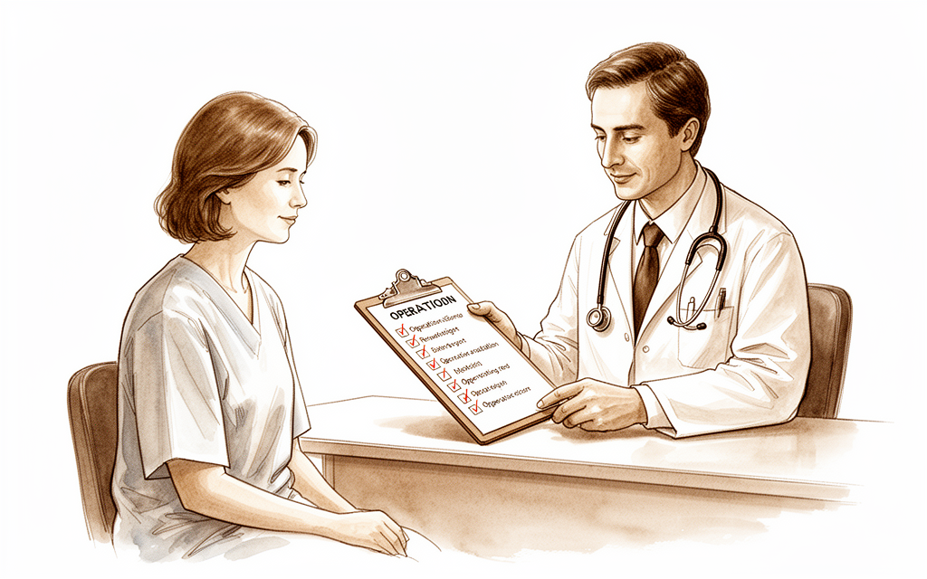
General checklist for the days before upper-limb surgery — fasting, what to bring, medications.
For patients: a plain-language version of this topic is available. See the patient guide.
Overview
Preoperative preparation for elective arthroplasty and joint surgery extends beyond clinical optimization to include structured educational interventions that influence patient expectations and postoperative recovery. Formal preoperative educational programs can help lower a patient's length of stay following hip or knee arthroplasty [3], while preoperative educational classes can modify patients' expectations of recovery from total hip or total knee arthroplasty [5]. Specific modalities, such as preoperative video counseling, have resulted in significantly decreased opioid consumption within the first week after total knee arthroplasty [9]. Although some indication of favourable outcomes exists following preoperative opioid education for elective hand surgery, the number of studies was small, evidence quality was poor, and data were limited [1]. A pilot randomized controlled trial is currently being conducted to evaluate the feasibility and satisfaction of a preoperative rehabilitation and education program (PREPS) for individuals undergoing shoulder replacement [2].
The efficacy of specific counseling tools varies by context and patient population. Preoperative counseling with haptic 3D hip models does not appear to favorably affect patient-reported understanding or satisfaction regarding femoroacetabular impingement when compared with the use of CT imaging alone [8]. Similarly, underserved patients' knowledge about total joint arthroplasty increased only modestly after taking a preoperative class during a surgical mission trip [6]. In the digital sphere, most content on orthobiologics found on YouTube is produced by independent users, with minimal contributions from verified health organizations [10].
Operational protocols and communication strategies further shape the preoperative experience. Enhanced office and staff protocols that are proactive rather than reactive can provide a safe and successful outpatient experience by anticipating potential postoperative pitfalls associated with same-day discharge [4]. Maximizing opportunities to improve communication, learning from others, and treating patients as more than their radiographic findings can help improve patient outcomes and trust [7].
Anatomy & Pathophysiology
Peripheral Nerve Structure and Injury Classification
Peripheral nerves comprise a mixture of myelinated and unmyelinated axons, with motor, sensory, and sympathetic fibers often traveling together in a single nerve [62]. Axons are grouped in bundles termed fascicles, which are surrounded by perineurium [62]. The fine connective tissue between axons within a fascicle is called endoneurium [62], while the epineurium holds the fascicles together as a nerve [62]. The mesoneurium, the connective tissue surrounding the epineurium, facilitates longitudinal gliding of the nerve [62]. Nerves are classified as monofascicular, oligofascicular, or polyfascicular depending on the number of fascicles [62]. The relationship between fascicles changes along the longitudinal course of the nerve, with the degree of fascicular change decreasing distally [62].
Injury classification is defined by the extent of structural disruption: * Neurapraxia: A conduction block that occurs without axonal disruption, with recovery usually complete within days to a few months [62]. * Axonotmesis: An injury in which axonal disruption occurs, with the endoneurial tube remaining in continuity [62]. * Neurotmesis: Transection of the nerve, where regenerating axons cannot find a suitable path and recovery does not occur unless the nerve is repaired [62].
In neurotmesis, all connective tissue layers of the nerve are affected as well as the axons, and Wallerian degeneration occurs [21]. Neurotmesis is usually caused by sharp laceration or high-energy traction injury that ruptures the nerve [21]. Other possible causes of neurotmesis include intra-neural injection of drugs and ischemia [21]. Recovery from neurotmesis is only possible by axonal regeneration after surgical repair of the nerve, with function returning in a proximal to distal pattern [21]. The quality of functional recovery after neurotmesis is never normal, partly because of the failure of correct "rewiring" [21]. Regenerating nerve fibers may connect with different muscle and sensory organs from those they previously innervated, resulting in impairment of function [21]. Outcome after neurotmesis repair is affected by the extent of the zone of injury, with poorer results after repair of a nerve ruptured by severe traction compared with repair of a sharp laceration [21]. Clinical features and neurophysiologic findings may be the same for axonotmesis and neurotmesis, yet there is a major difference in prognosis and management [21].
Pathophysiology of Nerve Injury and Regeneration
After a nerve is injured, the somatosensory cortex reorganizes so the area represented by the injured nerve diminishes [62]. The cell body of the lacerated axon increases in size, production of materials for repair of the cytoskeleton increases, and production of neurotransmitters decreases [62]. In the axon distal to the laceration, Schwann cells phagocytose the axon, allowing the surrounding myelin tube to collapse [62]. Within 24 hours of injury, axonal sprouting occurs from the proximal stump [62]. Multiple axons in a fascicle form a regenerating unit, and the number of axons in the unit decreases with time [62]. Longitudinal growth of the regenerating nerve depends on the ability of the axons to adhere to trophic factors in the basal lamina of the Schwann cell [62].
At the motor endplate, muscle fibers atrophy, and the sensitivity and number of acetylcholine receptors increase as their location expands from pits to the entire length of the muscle fiber [62]. If the muscle fiber is reinnervated, both old and new motor endplates become active [62]. The recovery of strength is greatest after primary nerve repair, less vigorous after repair with nerve grafting, and weakest after direct implantation of the nerve end into muscle [62]. Muscle reinnervation occurs only if the axon reaches the muscle within a year [62]. Sensory receptors may be effectively reinnervated years after injury [62].
Axons must exit the proximal nerve face by a phasic, staggered outgrowth, with very little significant progress achieved in humans until some 2 to 3 weeks have passed [59]. Increasing numbers of neurites follow pathfinder fibers out from the nerve face and must cross the nerve repair site [59]. Neurites seek contact guidance, neurotropic direction, and support from Schwann cells [59]. Successful fibers reach the distal nerve face and enter endoneurial tubes that railroad them to distal targets [59]. Around 50% of fibers will fail to cross the repair, forming a neuroma in continuity instead [59]. Many fibers entering the distal nerve are effectively lost due to poor type specificity, such as motor axons entering an endoneurial tube previously occupied by a sensory axon [59]. Other fibers are misdirected to an inappropriate part of the body due to poor topographical specificity [59]. Axons grow at an average of 1 mm/day, but growth may slow or peter out distally [59].
Axons exhibit a high degree of spontaneous activity and mechanosensitivity, accounting in part for the ubiquity of episodic lancinating pain [59]. The physiologic basis of the Hoffman-Tinel sign is related to the spontaneous activity and mechanosensitivity of axons [59]. After a brachial plexus injury, it may take over 12 to 18 months for axons to re-innervate distal nerve segments, muscles, or skin [59]. Denervation atrophy and denervation plasticity within the CNS inexorably march toward irreversibility during the period of axonal regeneration [59]. If sensory re-innervation is successful, the CNS must undergo re-innervation plasticity to optimize cortical interpretation of the resulting typically poorly spatially defined sensory input [59]. Sensory recovery is highly associated with significant reductions in neurogenic pain [59]. Regenerated axons remain abnormal with reduced fiber diameters and hence conduction velocity [59]. Denervation changes in muscle are partially reversed when regenerating axons connect with muscle fibers, with increased diameter and muscle bulk [59]. The number of axons re-innervating a muscle is reduced as a result of neuronal loss, and hence fewer but larger motor units will be formed [59]. Neurobiologic changes pertaining to quality of nerve regeneration after a repair exhibit profound deterioration when the delay from injury to repair is extended beyond 1 to 2 months [59].
Compression neuropathy can be considered as a progressive metabolic failure of axonal conduction, followed by disruption of the anatomical structures that physiologically enable axonal conduction [27]. Neuronal death is a fundamental issue that requires timely nerve repair and/or pharmacologic intervention [27]. The repair site environment is not adequately conducive to bridging by neurite growth, with the result that many axons are lost [27]. Nerve regeneration is far too slow for optimal salvage of much of the distal nerve, target muscles, and higher-order sensory organs from irreversible denervation atrophy [27]. Plasticity is initially disadvantageous during denervation and subsequently inadequate to make best use of what re-innervation occurs [27]. Neurobiologic determinants of regeneration are time-dependent and show profound worsening after some 1 to 2 months delay from injury to nerve repair [27].
Clinical Signs of Neural Regeneration
Recovery of neural function occurs from proximal to distal [47]. The signs of sensory recovery precede those of voluntary motor activity [47]. Tinel's sign is the first detectable clinical sign of recovery, where percutaneous percussion of the nerve trunk distal to the lesion produces a "pins and needles" sensation distally [47]. Tinel distinguished between peripheral paresthesia, a sign of axonal regeneration, and local pain, which indicates irritation of the nerve [47]. The "pins and needles" sensation resulting from percussion is caused by regeneration of the sensory axons, which are very sensitive to pressure [47]. Tinel's sign signifies a favorable prognosis and enables one to follow the progress of the regenerating nerve [47]. Only percussion of the tactile fibers triggers the pins and needles sensation, not those transmitting pain, heat, and cold [47].
Axonal regrowth usually occurs at a rate of 1–2 mm per day after nerve suturing [47]. Tinel's sign is absent in the early stages following injury or nerve suturing, appearing only four to six weeks after the injury [47]. Tinel's sign may be difficult to elicit if the nerve lies deep to a large mass of muscle [47]. Tinel's sign cannot be demonstrated when the lesion is proximal to the posterior root ganglion [47]. A false positive result for Tinel's sign is elicited when sensory fibers grow into motor sheaths [47]. Tinel's test has no quantitative value and can be positive with only a few fibers regenerating [47]. Steady distal progression of Tinel's sign suggests a good prognosis, even though the sign gives little information concerning the functional quality of reinnervation [47]. Interrupted progress of Tinel's sign must be regarded as alarming and, if persistent, confirms the need for surgical exploration [47].
Sensory recovery progresses in time and space according to successive stages: perception of pain and temperature, perception of low frequency vibratory stimuli and moving tact sense, perception of static tact sense and high frequency vibratory stimuli, and two point discrimination [73]. Perception of pain and temperature occurs first because small caliber pain fibers regenerate more rapidly [73]. Two point discrimination is the last form of sensation to develop [73]. Motor recovery is always slower than sensory recovery [73]. The first sign of motor recovery is regression of the atrophy in the territory normally supplied by the injured nerve [73]. A weak contraction can be detected in the first muscle supplied by the nerve distal to the lesion, but it is not powerful enough to produce movement or to overcome gravity [73]. Early signs of neural regeneration are of limited prognostic value and offer no guarantee of functional recovery [73]. Regeneration can be halted at any stage, and there is frequently a marked difference between motor recovery and sensory recovery [73]. Sensation returns first to the proximal margin of the anesthetic zone [73]. Early electrodiagnosis is of limited value after a peripheral nerve lesion, as the process of Wallerian degeneration can be recorded only from four to five weeks after the nerve division [73].
Sensory and Motor Deficits
After severance of a peripheral nerve, only a small area of complete sensory loss is found, known as the autonomous zone or isolated zone of supply for that nerve [49]. A larger area of tactile and thermal anesthesia corresponds more closely to the gross anatomic distribution of the nerve and is known as the intermediate zone [49]. When a nerve is intact and adjacent nerves are blocked or sectioned, an area of sensibility exceeds the gross anatomic distribution of the nerve, known as the maximal zone [49]. The autonomous zone becomes smaller during the first few days or weeks after injury, long before regeneration is possible [49]. This decrease in the area of sensory loss might be interpreted by an inexperienced surgeon as evidence of regeneration or of incomplete injury [49].
In injury to the median and ulnar nerves, pinprick is the first perception to return, followed by 30 cycles/s vibratory stimulus, and then moving touch [49]. The perception of constant touch and the perception of a 256 cycles/s vibratory stimulus are the last to return [49]. The early return of pain perception results from the faster regeneration of the small-diameter pain fibers [49]. The larger-diameter touch fibers regenerate more slowly [49]. The return of moving touch perception before the return of constant touch is explained by differential maturation of the respective receptors, rather than by the diameter of the nerve fibers alone [49]. Two-point discrimination has been shown to directly correlate with return of hand function and object identification [49].
The British Medical Research Council established a six-level grading scale for sensory return, ranging from S0 (absence of sensibility) to S4 (complete recovery) [49]. Motor recovery is graded from M0 (no contraction) to M5 (complete recovery) [49]. In the hand, proximal muscles are defined as extrinsic muscles and distal muscles are defined as intrinsic muscles [49]. Electrodiagnostic studies essentially can detect only two types of pathophysiology of the peripheral nerve system fibers: axon loss (axonotmetic lesion) and focal demyelination (neuropraxic lesion) [74]. The severity of conduction slowing has no correlation with the severity of clinical symptoms, such as weakness or static large-fiber sensory loss [74]. If substantial weakness or static large-fiber sensory loss is present, substantial amounts of either conduction block, axon loss, or a combination of both must be present [74]. High axonotmetic lesions may take 1 to 2 years for maximal recovery, whereas even lesions at the wrist may take 6 to 9 months for maximal recovery of nerve function [74]. An advancing Tinel's sign can be monitored and is a good prognostic sign [74].
Preoperative Considerations and Comorbidities
Obesity has been identified as an impactful comorbid condition serving as an independent risk factor for morbidity and mortality after trauma [15]. Obesity can represent concomitant risk factors including diabetes mellitus, hyperlipidemia, heart disease, and hypertension that have been demonstrated to impact trauma outcomes negatively [15]. Critical airway management, intravenous access, ultrasonography, plain radiography and CT, and volume resuscitation are directly negatively affected by obesity [15]. Surgical exposures, patient positioning, early mobilization, and post-hospitalization rehabilitation are negatively affected by obesity [15]. Longer surgical times and more complications have been reported in obese patients treated for fracture [15]. Obesity-induced inflammatory stress compounded by trauma-induced inflammatory stress increases morbidity and mortality after major trauma [15].
Undiagnosed diabetes and trauma-induced hyperglycemia are frequent among trauma patients and have been shown to be associated with infections and other complications [15]. Elevations in perioperative blood glucose greater than 220 mg/dL were associated with a sevenfold increase in infections in orthopedic trauma patients, none of whom had a diagnosis of diabetes [15]. In known diabetic patients, injury and surgery are disruptive to normal glycemic control, leading to greater risk for early infections and other complications and for longer hospital stay [15]. Pre-existing cardiovascular diseases pose a unique modifier in the ability to recover from multisystem trauma [15]. Patients with pre-existing cardiac disease are at higher risk of cardiac failure and myocardial infarction [15]. Cardiovascular disease is associated with an active state of inflammation, whereby within the context of traumatic insult, morbidity and mortality are worsened [15]. Microvascular disease significantly affects end-tissue perfusion, with higher incidences of wound dehiscence and infections described in patients with pre-existing vascular disease [15]. Many patients with cardiovascular disease are on therapeutic anticoagulation, increasing the risk of death by uncontrolled exsanguination as well as risks of other bleeding complications, including wound infections [15].
Hyperglycemia has been shown to be an independent risk factor for multiple complications in both the general surgery and orthopedic surgery patient [25]. Following a traumatic injury, the balance of endogenous hormones shifts, favoring an environment of increased blood glucose levels, partially caused by increased circulating cortisol and a relative decrease in insulin sensitivity [25]. When hyperglycemia results from this process in patients without a known history of diabetes, it is termed "stress-induced hyperglycemia" [25]. With preoperative fasting considered, blood sugar should be checked every 4 to 6 hours in the perioperative period [25].
The assessment of patient comorbidities is an essential component in the preoperative workup of any patient [36]. Comorbidities can be classified into chronic-stable, chronic-unstable, and newly diagnosed [36]. The Charlson Comorbidity Index was developed in 1987 as a novel method of classifying prognostic comorbidity for longitudinal studies [36]. The Charlson Comorbidity Index can be used to predict the risk of 1-year, 5-year, and 10-year mortality rates of patients with various conditions [36]. The American College of Surgeons developed the National Surgical Quality Improvement Program (NSQIP) risk calculator to provide patient-specific perioperative risk outcome estimates to guide surgical decision making and informed consent [36]. The NSQIP risk calculator uses 20 medical and surgical risk factors combined with the planned surgical procedure to predict 18 different outcomes following surgery [36]. Outcomes predicted by the NSQIP risk calculator include death, serious complications such as myocardial infarction, sepsis, organ failure, and venous thromboembolic disease, and care site complications such as discharge to a skilled nursing facility or readmission to the hospital [36].
Anesthetic complications are higher in the obese pediatric population, with an associated higher-than-normal incidence of baseline hypertension, asthma, and sleep apnea [34]. Preoperative evaluation must assess the cardiopulmonary systems to adequately address the altered physiology in obese patients [34].
Classification
Other Considerations: The provided evidence does not contain data for specific fracture or injury classification systems (e.g., Schatzker, AO/OTA, Gustilo–Anderson). Instead, the available literature addresses preoperative educational and protocol frameworks. Preoperative opioid education showed some indication of favourable outcomes following elective hand surgery, but the supporting studies were small, of poor evidence quality, and had limited data [1]. A pilot randomized controlled trial protocol was developed to evaluate the feasibility and satisfaction of a preoperative rehabilitation and education program (PREPS) for individuals undergoing shoulder replacement [2]. A formal preoperative educational program can lower a patient's length of stay following hip or knee arthroplasty [3]. Proactive office and staff protocols can provide a safe and successful outpatient experience by anticipating potential postoperative pitfalls associated with the 'ripple effects' of same-day discharge [4]. Patients who received preoperative video-based opioid counseling had significantly decreased opioid consumption within the first week after total knee arthroplasty [9]. Most YouTube content on orthobiologics is produced by independent users, with minimal contributions from verified health organizations [10].
Clinical Presentation
Preoperative Education and Expectations
Preoperative educational classes can modify patients' expectations of recovery from total hip or knee arthroplasty [5]. In a surgical mission trip context, underserved patients' knowledge about total joint arthroplasty increased only modestly after taking a preoperative class [6]. Preoperative video-based opioid counseling is associated with significantly decreased opioid consumption within the first week after total knee arthroplasty [9]. While there was some indication of favourable outcomes following preoperative opioid education, the number of studies was small and evidence quality was poor [1]. Most online content regarding orthobiologics is produced by independent users, with minimal contributions from verified health organizations [10].
History and Physical Examination Principles
The clinical evaluation is the beginning of the doctor-patient relationship, with the goal of carrying out an evaluation that leads to a reasonable management plan rather than just a diagnosis [79]. The four factors determining treatment outcome are the patient, the shoulder problem experienced, the procedure to treat the patient and problem, and the physician rendering the treatment [79]. A "no touch" approach to physical examination involves asking patients to demonstrate difficult actions and describe what they feel is happening before the examiner touches the patient [79]. If a problem is not apparent on history, physical examination, and plain radiographs, or if the patient does not appear to be an excellent surgical candidate, nonoperative management is likely recommended [79].
The history and physical exam are important in the evaluation of the patient, with every office visit constituting a history and physical exam [29]. Regulations require that a chief complaint be specified, which determines the direction for the rest of the history and physical [29]. The social history and past medical history are important because they change billing codes without necessarily affecting outcome or success of care [29]. The physical exam must cover the essentials necessary for diagnosis, and frequently the confirmation of the diagnosis is based on physical exam [29]. Considerations such as skin condition and blood supply must be documented as part of the surgical evaluation [29].
Imaging and Laboratory Evaluation
The most important point in imaging and laboratory exams is to use the most cost-effective examination possible while keeping patient safety, satisfaction, and convenience in mind [29]. Roentgenography is still the most cost-effective and most important initial diagnostic test in the orthopedist’s armamentarium [29]. Almost every patient should have a radiograph prior to going to a more sophisticated imaging study [29]. MRI is sometimes too revealing and should be reserved for clarifying a particular problem [29]. Frequently in orthopedics, a bony lesion can be localized with a radiograph or bone scan, which then provides a focus for the MRI [29].
MRI is useful for some bony lesions, such as osteonecrosis, tumors, fatigue fractures, and osteomyelitis [29]. MRI is helpful in some soft-tissue problems, such as knee meniscus tears and shoulder rotator cuff tears [29]. Distortion of the magnetic field by metallic implants may limit the usefulness of MRI studies of conditions such as total knee or hip replacement, or fracture fixation devices [29]. MRI should not be used when the diagnosis can be made with a less expensive test [29]. The use of MRI in knee studies in patients older than 45 years should always be preceded by plain films of the knee [29].
Metastatic Disease and Pathologic Fractures
A thorough history and physical examination with appropriate imaging studies often leads to the correct diagnosis, particularly in the case of widespread metastatic bone disease [42]. A solitary bone lesion in a patient with or without a history of cancer should be biopsied to obtain an accurate diagnosis, as presuming it is a metastasis may lead to the wrong operation and compromise life or limb if it is a primary sarcoma [42]. In individuals with recent and known metastatic disease to bone, a new biopsy of bone is not necessary [42]. If a patient has a remote history of cancer or no known metastasis to bone, a biopsy should be performed to confirm that the lesion is not a primary sarcoma [42]. A needle biopsy is usually definitive when differentiating a carcinoma from a sarcoma [42]. Specific immunohistochemical staining may allow determination of the primary site of origin of a carcinoma, most commonly from the lung, breast, thyroid, kidney, or prostate [42].
When a pathologic fracture occurs through a lytic lesion, biopsy can be complicated due to bleeding and early fracture callus [42]. The fracture should be stabilized initially with traction or a cast to allow preliminary staging studies to be completed when a pathologic fracture complicates biopsy [42]. Care should be taken to not place traction pins through more distal lesions when stabilizing a pathologic fracture for biopsy [42]. If a needle biopsy is nondiagnostic or unable to be done, a careful incisional biopsy should be performed using oncologic principles so as not to preclude subsequent definitive surgical treatment [42]. Tissue for biopsy should be obtained from a site near but unaffected by the fracture when possible [42]. The biopsy should be as small as possible, in a longitudinal fashion in line with the extremity, and performed with excellent hemostasis [42]. Tissues contaminated by a postbiopsy hematoma must be considered contaminated by tumor [42]. Cultures should always be sent at the time of biopsy to rule out infection, which can appear radiographically similar to a tumor [42]. If a definitive diagnosis of metastatic disease can be made on an intraoperative frozen section, surgical treatment of the pathologic fracture can be performed at the same operative setting [42]. If the frozen section is nondiagnostic, it is best to wait for the permanent sections before definitively treating the tumor and fracture [42].
The goals of surgical treatment in a patient with an impending pathologic fracture are to alleviate pain, reduce narcotic utilization, restore skeletal stability, and regain functional independence [11]. The decision to proceed with operative intervention for an impending pathologic fracture is multifactorial and must be individualized [11]. Factors included in the decision making for impending pathologic fracture treatment are life expectancy, patient comorbidities, extent of the disease, tumor histology, anticipated future oncologic treatments, and degree of pain [11]. Patients with a life expectancy of less than 6 weeks may not gain significant benefit from major reconstructive surgery for impending pathologic fractures [11]. An accurate prognosis is not always possible, and the decision of whether to proceed with surgery should be discussed with the multidisciplinary team, the patient, and the patient's family [11].
Patients treated by prophylactic stabilization of an impending fracture have shorter hospitalization (average 2 days) compared to those treated after an actual fracture [11]. Patients treated by prophylactic stabilization of an impending fracture are more likely to be discharged to home (40%) compared to those treated after an actual fracture [11]. Patients treated by prophylactic stabilization of an impending fracture experience more immediate pain relief compared to those treated after an actual fracture [11]. Patients treated by prophylactic stabilization of an impending fracture undergo faster and less complicated surgery compared to those treated after an actual fracture [11]. Patients treated by prophylactic stabilization of an impending fracture have less blood loss compared to those treated after an actual fracture [11]. Patients treated by prophylactic stabilization of an impending fracture have a quicker return to premorbid function compared to those treated after an actual fracture [11]. Patients treated by prophylactic stabilization of an impending fracture have improved survival compared to those treated after an actual fracture [11]. Patients treated by prophylactic stabilization of an impending fracture have fewer hardware complications compared to those treated after an actual fracture [11]. Elective stabilization allows the medical oncologist and surgeon to coordinate operative treatment and systemic chemotherapy [11].
Fracture risk is greatest during surgical positioning, preparation, and draping when treating patients with impending pathologic fractures [11]. When patients are anesthetized, they cannot protect the affected extremity and must rely on the surgical team to proceed carefully [11]. Low-energy fractures will occur after very minor trauma or a twisting movement in patients with impending pathologic fractures [11]. If a pathologic fracture occurs, damage to the surrounding soft tissues is typically less than in traumatic fractures in healthy bone [11].
Hand and Wrist Examination
Clinical evaluation of the injured or dysfunctional hand and wrist can be a daunting task because painless and full hand function requires seamless integration of joints, muscles, and nerves [26]. Patients often have difficulty accurately describing their symptoms and may incorrectly attribute pathology to a perceived deficit, whether real or imagined [26]. The task of the astute clinician is to combine the patient history with a careful physical examination to pinpoint or at least narrow the scope of possible pathologic processes [26]. Diagnostic tests such as imaging and serum laboratory studies are useful in this determination but can be expensive, time consuming, and often nonspecific [26]. A careful physical examination is essential to direct care and future testing if indicated [26]. A systematic method to approaching the physical examination of the hand and wrist is essential due to the many structures in a small space [26]. Some clinicians may prefer to organize their examination by anatomic location or region of the hand, while others may choose to proceed by organ system or pathology [26].
General Examination Considerations
It is important for orthopaedic surgeons to be familiar with the numerous musculoskeletal and neuromuscular examinations detailed in Chapter 3 [35]. If the nature of the patient’s medical condition is unclear, the physician may have to perform a comprehensive examination to arrive at a differential diagnosis [35]. In most cases, the orthopaedic surgeon does not have the time or the need to perform an all-encompassing examination on every patient seen in the clinical setting [35]. The pediatric orthopaedic examination must be tailored to the child’s age, level of cooperation, and chief complaint [35]. The two most common types of examinations performed are the screening examination and focused examination [35]. Screening examinations are performed as part of a comprehensive or abbreviated examination to detect disorders that may be asymptomatic but could cause significant morbidity or mortality if undiagnosed and untreated [35]. Focused examinations concentrate on specific abnormalities for which the patient has been referred or on the chief presenting complaint [35]. The examiner should make the clinical assessment as orderly and organized as possible to avoid neglecting any essential parts of the examination [35]. An uncooperative child, the presence of multiple family members, and limited time provide an impetus to perform the examination as expeditiously as possible while still maintaining good rapport with the patient and parents [35].
Knee Examination
To diagnose the cause of a patient’s knee pain, it is imperative to obtain a detailed history [67]. Elements of a detailed history include onset, quality, duration, tempo, and location of symptoms, modifying factors, ability to bear weight, and history of trauma [67]. A detailed physical examination of the knee includes multiple elements: inspection, palpation, gait assessment, range of motion testing, stability testing, neurovascular assessment, a hip examination, and special tests aimed at the diagnosis of particular pathologies [67].
Inspection of the knee can reveal skin abnormalities, evidence of trauma, malalignment, and swelling [67]. Inspection of the patient’s gait may reveal abnormalities that suggest either intra-articular or extra-articular causes [67]. Palpation of the knee with a particular focus on points of tenderness can alert the diagnostician to focal pathologies [67]. Palpation of the peripatellar tissue can reveal the presence of effusion and/or synovitis [67]. Determination of overall knee alignment (ie, varus, valgus, or neutral alignment) is important as an adjunct to the diagnostic process [67]. Knee alignment should be assessed in both supine and standing positions, as bearing weight may change the knee’s alignment dynamically [67].
Range of motion testing can be divided into two parts: active and passive [67]. Active range of motion refers to the patient’s ability to move his or her own knee through range of motion [67]. Passive range of motion refers to the examiner’s ability to move the patient’s knee through range of motion [67]. Flexion contractures and hyperextension should be noted during range of motion testing [67]. Blocks to motion can be pain-related or mechanical [67]. When active and passive ranges of motion differ, the diagnostician must differentiate between pain-related, mechanical, or neuromuscular causes [67]. Hip range of motion should be examined and may reveal resultant knee pain, indicating the possibility of referred pain from intra-articular hip pathology [67].
Stability testing of the knee can reveal ligamentous competency or deficiency [67]. Basic varus and valgus stability testing should be performed at 0° and 30° of flexion [67]. Firm end points indicate ligament competence, while pronounced laxity can indicate ligament deficiency [67]. Testing at 30° of flexion isolates the MCL and LCL best, as testing in full extension also engages some secondary stabilizers [67]. ACL and PCL competence can be tested using special tests [67]. The Lachman test involves flexing the knee to 30°, holding the femur firmly, and translating the tibia anteriorly on the femur [67]. A positive Lachman test is indicated by no firm end point and significant translation [67]. The posterior drawer test involves flexing the knee to 90° with the patient supine, stabilizing the distal tibia, and translating the tibia posteriorly on the femur [67]. A positive posterior drawer test is indicated by no firm end point and significant translation [67]. The J-sign involves bringing the knee from full extension into flexion [67]. A positive J-sign is indicated by a visible patellar shift from lateral (subluxated) to medial (relocated) in a J-shaped path [67]. The McMurray test for lateral meniscus tear involves flexing the knee, internally rotating the tibia, extending the knee, and applying pressure to the lateral joint line [67]. The McMurray test for medial meniscus tear involves flexing the knee, externally rotating the tibia, extending the knee, and applying pressure to the medial joint line [67]. A positive McMurray test is indicated by pain or click with the maneuver [67]. The dial test for PLC deficiency involves placing the patient prone with the knee flexed to 30° and externally rotating both tibiae [67]. The dial test for PLC plus PCL deficiency involves placing the patient prone with the knee flexed to 90° and externally rotating both tibiae [67]. A positive dial test is indicated by greater than 10° difference from the contralateral side [67].
Hip and Pelvic Examination
The complexity of the hip and pelvic region can make accurate diagnosis of painful conditions difficult [80]. A thorough understanding of normal anatomy and biomechanics is necessary to identify pathology and determine the appropriate course of treatment [80]. Because many hip conditions present with similar symptoms, a comprehensive clinical examination is required to determine a differential diagnosis [80]. Findings from imaging studies should complement clinical examination findings to provide the most accurate diagnosis [80]. The hip is a complex multiaxial joint capable of producing large forces and moving the thigh through large ranges of motion [80]. Any alteration to joint morphology or function can place the hip at risk for pathology [80]. A thorough history is essential to differentiating between common causes of hip pain [80]. Clinical examination tests and imaging findings should be used to confirm a suspected clinical diagnosis [80].
Cervical Spine Examination
Evaluation of cervical degenerative disorders starts with a careful history and physical examination [82]. Questions that may provide clues to the cause of a patient’s symptoms include whether the patient has pain, loss of sensation, or weakness [82]. The primary location of the symptoms is also important, such as whether the pain is in the neck or radiates into the arms [82]. Important factors include when the symptoms occurred, how long the symptoms were present, and if there was antecedent trauma [82]. It is important to consider factors that alleviate and exacerbate the symptoms [82].
Patients with disk degeneration may complain of axial neck pain that is often chronic and insidious in nature [82]. Sometimes there is an inciting event that worsens the pain in patients with disk degeneration [82]. The neurologic examination will be benign in most cases of disk degeneration [82]. Patients with a cervical disk herniation will complain of a sudden and acute pain that is very intense [82]. Depending on the level of the herniation, patients will have loss of sensation and motor weakness in a distribution that is consistent with the nerve root affected [82]. Myelopathy presents with a variety of subtle neurologic findings [82]. Characteristic signs and symptoms of myelopathy can present insidiously and include the loss of manual dexterity in the hands, weakness, stiffness, urinary symptoms, spasticity in their extremities, and gait disturbance including a stiff or spastic gait [82]. Patients with myelopathy demonstrate a wide-based gait and report a history of loss of balance and falls [82]. Sensory findings in myelopathy often include proprioceptive loss, and patients may report that they have difficulty with buttons, a change in their handwriting, or
Investigations
Other Considerations: Preoperative education serves as a key component of the investigative and preparatory phase. A formal preoperative educational program can help to lower a patient's length of stay for hip or knee arthroplasty [3]. For total knee arthroplasty, patients who received preoperative video counseling had significantly decreased opioid consumption within the first week after surgery [9]. Preoperative opioid education showed some indication of favourable outcomes following elective hand surgery, though the number of studies was small, evidence quality was poor, and data were limited [1].
Treatment
Preoperative Education and Counseling
Preoperative education remains a critical component of surgical preparation, though its efficacy varies by setting. In surgical mission trip contexts, underserved patients demonstrated only modest increases in knowledge about total joint arthroplasty after attending a preoperative class [6]. For shoulder replacement, the PREPS (Preoperative Rehabilitation and Education Program for Surgery) pilot randomized controlled trial is currently evaluating the feasibility and patient satisfaction of this structured program [2].
Preoperative Opioid Management
Early intervention regarding opioid use is associated with improved cessation outcomes. Orthopedic trauma patients educated about a cessation plan early in their injury course were more successful at early discontinuation of opioids, although this effect was not necessarily sustained long-term [46]. Expectations about long-term opioid use represent one of the strongest predictors of whether patients will continue using these medications one year after initiation [46]. While some indication of favourable outcomes exists following preoperative opioid education for elective hand surgery, the evidence base remains limited by a small number of studies, poor quality, and restricted data [1].
Preoperative Planning and Optimization
Fracture and Trauma Planning: The preoperative plan for surgical repair of a fracture must include patient-specific factors, including optimization and soft tissue elements that may impact outcomes more significantly than the technical components of fracture reconstruction [13]. For complex fracture patterns, planning includes templating, which can involve a demonstration of the fracture itself and specific fixation methods [13]. In younger patients with femoral neck fractures, understanding and addressing the pathoanatomy of vertical femoral neck fractures is mandatory to minimize risks for treatment failure [16]. These injuries differ from typical osteoporotic fractures, contributing to a specific set of clinical problems including nonunion, failed fixation, osteonecrosis, and malunion [16]. Quality reduction and stable fixation are vital elements in managing these at-risk fractures [16]. Although clinical and laboratory studies of this injury are lacking, recent efforts have focused on improving mechanical testing models and assembling large, multicenter clinical studies to evaluate treatment and outcomes [16]. Fracture management has evolved to improve implant mechanics and biology, with several new implants and ideas on implant application imminent, though their success remains to be determined [16].
Periprosthetic Fractures: Preoperative medical optimization is of paramount importance for periprosthetic fractures, requiring a thorough patient history including endocrine, musculoskeletal, and oncologic histories [58]. Identification and review of any previous associated surgeries, including complications, should be done before surgery to improve planning [58]. Knowledge of the prior surgical approach along with the type of currently implanted hardware improves preoperative planning [58]. In the octogenarian population, an intimate knowledge of the patient’s code status, functional activity level, and goals of care are important discussion points [58]. The surgeon should be prepared to perform fracture fixation, revision, or both [58]. If cement is present, removal instruments should be available, especially if the implant is being revised [58]. It is always prudent to consider concomitant infection despite the presence of trauma, maintaining a low threshold for an associated workup [58]. Critical assessment of bone quality and the expected amount of bone loss should be completed preoperatively [58]. A preoperative list of implants and fixation devices should be determined [58]. Careful planning combined with medical optimization and goals consistent with early mobilization can lead to a consistently reproducible desired clinical outcome [58].
Pathologic Fractures: For impending pathologic fractures, the decision to proceed with operative intervention is multifactorial and must be individualized based on life expectancy, comorbidities, extent of disease, tumor histology, anticipated future oncologic treatments, and degree of pain [11]. The goals of surgical treatment are to alleviate pain, reduce narcotic utilization, restore skeletal stability, and regain functional independence [11]. Fracture risk is greatest during surgical positioning, preparation, and draping because anesthetized patients cannot protect the affected extremity [11]. If a pathologic fracture occurs during surgery, damage to the surrounding soft tissues is typically less than in traumatic fractures in healthy bone [11]. Patients treated by prophylactic stabilization of an impending fracture versus those treated after an actual fracture have shorter hospitalization (average 2 days) [11]. They are more likely to be discharged to home (40%) [11]. They also experience more immediate pain relief [11], faster and less complicated surgery [11], less blood loss [11], quicker return to premorbid function [11], improved survival [11], and fewer hardware complications [11]. Elective stabilization allows the medical oncologist and surgeon to coordinate operative treatment and systemic chemotherapy [11]. Low-energy fractures will occur after very minor trauma or a twisting movement [11]. The decision of whether to proceed with surgery should be discussed with the multidisciplinary team, the patient, and the patient's family [11]. An accurate prognosis is not always possible [11].
Pathologic Fracture Management Algorithm: The management algorithm distinguishes between known malignancy history, remote history of cancer, and no known malignancy history [11]. For patients with a known malignancy history, site-specific axial imaging with contrast, radiographs, bone scan, CT of chest, abdomen, and pelvis with IV and PO contrast is indicated [11]. Biopsy is a step in the management algorithm for patients with a known malignancy history or under current treatment for malignancy [11]. For patients with a remote history of cancer, site-specific axial imaging with contrast, radiographs, and bone scan is indicated [11]. For patients with no known malignancy history, site-specific axial imaging with contrast and radiographs is indicated [11]. Resectable disease or single site is a factor in the management algorithm [11]. Multiple lesions is a factor in the management algorithm [11]. Treatment based on location, disease pathology, and patient characteristics is the final step in the management algorithm [11].
Spine Tumors: For patients with spine tumors, proper diagnosis with a biopsy is the critical first step in devising proper treatment [22]. Proper care requires a multidisciplinary team approach with continued follow-up and optimization of the care as the patient’s needs can be dynamic and needs to be individualized [22].
Hip Fractures: Preoperative traction should not be routinely used for hip fracture patients [56]. An echocardiogram during the preoperative evaluation for hip fractures increases the time to surgery [56]. It is recommended to operate early for hip fractures despite recent antiplatelet drug use [56]. Preinjury cognitive function and mobility predict postoperative functional outcome in elderly patients with hip fractures [56]. Comanagement care teams should be used in the care of hip fracture patients to decrease complications and improve outcomes [56]. Venous thromboembolism prophylaxis should be used [56]. Definitive treatment in 24 hours is associated with a reduced 30-day and 1-year mortality [56]. Multimodal analgesia incorporating preoperative nerve block is recommended [56]. Tranexamic acid should be administered to reduce blood loss and transfusions [56]. The goal of treatment is to allow early weight bearing to minimize complications [56]. For intracapsular femoral neck fractures, rapid preoperative medical optimization is a general principle, with mortality risk reduced if surgery occurs within 48 hours [56].
Knee Dislocations: For emergent knee dislocations, preoperative planning involves early discussion with the orthopedic trauma team, general surgery trauma team, and vascular surgery team so that all surgical personnel are aware of the emergent situation and the surgical plan [54]. For open knee dislocations, preoperative planning requires having 9 to 12 L of normal saline with low-pressure pulsatile lavage available and ensuring a negative pressure wound therapy device is readily available [54].
Pediatric Hand Reconstruction: The decision-making process must consider the child’s variable and often limited ability to cooperate in the postoperative period, the risk associated with anesthesia in early life, the size of the structures, absence of critical parts, the need for growth, and occasionally the reduced life span of some patients with particular congenital syndromes [12]. Surgeons must carefully consider the probable responses of the child, the parent, and the tissues of the child’s hand to the planned operation [12]. Surgeons must realistically evaluate what they can actually deliver in the operating room and postoperative period when planning pediatric hand reconstruction [12]. A realistic time schedule should be agreed upon at the beginning of pediatric hand reconstruction to provide comfort to both the surgeon and the family [12]. Failure to take preoperative photographs, measure and record joint angles and grip strength, and perform functional tests before treatment renders an honest, accurate assessment of the result at the end of treatment impossible in pediatric hand surgery [12].
Pediatric Limb Lengthening: The team should discuss various options, likely problems, and outcomes with the patient and family before beginning treatment [53]. It is imperative that the child and family have considered their goal of the treatment and choose the medical intervention that is most suitable to them [53]. Age-appropriate education for the child is key because the child will be the active participant in all aspects of the treatment [53]. A plan for pain management should be established based on the child and family’s previous responses to pain and anxiety before the surgical procedure [53]. Social support, the role of caretakers, and who will be responsible for the child’s follow-up and daily care should be established before the surgical procedure [53]. A decision not to proceed may be made when there is a demonstrated inability to adhere to care and exercises, significant psychological disorders, family disharmony, and/or poor understanding of treatment [53].
Rotator Cuff Tears: Clinical decision-making is complex and lacks consensus among orthopedic surgeons [14]. Neither the clinical practice guidelines set out by the American Academy of Orthopaedic Surgeons nor the Cochrane systematic reviews provide guidance on the management of rotator cuff tears [14]. Patients with rotator cuff tears can generally be divided into three main categories based on the potential risk of nonoperative treatments and the proposed benefits of surgical intervention: those needing urgent or early operative repair, those that can benefit from a trial of conservative treatment, and those that may be best suited for nonoperative treatment [14]. As understanding of the natural history of rotator cuff disease improves, the complex decision-making process will continue to be refined over time [14]. General guidelines regarding operative indications and the importance of surgical timing can be generated based on current understanding of the natural history of rotator cuff disease and the results of operative repair in a multitude of age groups and tear characteristics [14].
Fracture-Related Infections: For early fracture-related infections, the timing of presentation affects the nature and extent of intervention, and host status, fixation construct, and soft-tissue envelope condition must be considered [60]. In early fracture-related infection before bony union, débridement with implant retention may be possible if the biofilm has not been established [60]. For frail patients or those with hip fractures or complex periarticular fractures, débridement and lavage with retention of implants, antibiotic suppressive therapy, and ultimately removal of hardware after union may be appropriate for early fracture-related infection [60].
Osteomyelitis: For surgical debridement of osteomyelitis, treating comorbidities and nutritional support should be initiated in the preoperative period where possible [55]. The operating surgeon should carefully consider the risks and benefits of radical debridement in the presence of uncertainty regarding bone condition, ideally with informed consent before the operation [55]. For patients with many comorbidities who may not survive extensive surgical stress for osteomyelitis treatment, less aggressive options such as long-term suppressive antibiotic treatment should be considered [55].
Complications
Complications are an inevitable but hopefully rare consequence of surgery [70]. The possibility of complications should be addressed during the informed consent process, striking a balance between describing meaningful risks and providing an all-inclusive list of possible problems [70]. Directly admitting mistakes that have caused complications prevents malpractice suits more effectively than evasion or avoiding discussion with the patient or the patient’s family [70].
Other Considerations: Overall complication rates for arthroscopy are reported to be between 1% and 4.7% [71]. Salzler et al. found 4305 complications reported in approximately 92,565 arthroscopic knee procedures, a 4.7% complication rate [71]. The most commonly reported complications for arthroscopy include return to the operating room and superficial infections [71]. The complication rate for arthroscopy is proportional to experience, operating time, tourniquet time, procedure complexity, multi-ligament and posterior cruciate ligament (PCL) injuries, number of procedures, and meniscal repairs [71]. Procedure complexity was correlated with an increase in adverse outcomes in arthroscopic knee procedures [71]. PCL reconstruction had the highest complication rate among arthroscopic knee procedures, which was 20% [71]. Comorbidities and smoking increased the risk of complications after arthroscopic meniscectomy [71].
Obesity is an independent risk factor for morbidity and mortality after trauma [15]. Patients with pre-existing cardiovascular disease are at higher risk of cardiac failure and myocardial infraction following multisystem trauma [15]. Higher incidences of wound dehiscence and infections have been described in patients with pre-existing vascular disease [15]. Patients on therapeutic anticoagulation are at increased risk of death by uncontrolled exsanguination as well as risks of other bleeding complications, including wound infections [15].
The WHO Surgical Safety Checklist is a 19-item checklist that has proven to reduce morbidity and mortality [76]. The Joint Commission Universal Protocol is a safety surgical checklist required for hospitals to use to obtain accreditation [76]. The Joint Commission Universal Protocol includes conducting a preprocedure verification process, marking the procedure site, and performing a time-out [76]. Preoperative functional status as measured in METs is critical in the preoperative medical and cardiac evaluation for geriatric patients with orthopaedic trauma [87]. Medical comanagement reduces time to surgery, postoperative complications, and length of stay in patients with hip fracture [87].
Recovery
Other Considerations: Preoperative preparation significantly influences postoperative recovery trajectories and patient satisfaction. A formal preoperative educational program can help to lower a patient's length of stay following hip or knee arthroplasty [3]. Similarly, patients' preoperative expectations of their recovery from total hip arthroplasty or total knee arthroplasty can be modified by preoperative educational classes [5]. Preoperative opioid education showed some indication of favourable outcomes following elective hand surgery, but the number of studies was small, the evidence quality was poor, and data were limited [1]. For shoulder replacement, a pilot randomized controlled trial is planned to evaluate the feasibility and satisfaction of a preoperative rehabilitation and education program (PREPS) for individuals undergoing shoulder replacement [2]. In outpatient settings, proactive office and staff protocols can provide a safe and successful outpatient experience by anticipating potential postoperative pitfalls associated with the unique 'ripple effects' of same-day discharge [4]. Regarding specific counseling modalities, preoperative counseling with haptic 3D hip models does not appear to favorably affect patient-reported understanding or satisfaction with regard to femoroacetabular impingement when compared with the use of CT imaging alone [8].
Key Evidence
- [L1] There was some indication of favourable outcomes following preoperative opioid education; however, the number of studies were small, the evidence quality was poor, and data were limited. [1] (10.1177/17589983241301449)
- [L2] This pilot randomized controlled trial will evaluate the feasibility and satisfaction of the PREPS program for individuals undergoing a shoulder replacement. [2] (10.1177/17589983251345393)
- [L3] A formal preoperative educational program can indeed help to lower a patient's length of stay. [3] (10.1016/j.arth.2009.03.012)
- [L5] Enhanced office and staff protocols that are proactive rather than reactive can provide a safe and successful outpatient experience by anticipating potential postoperative pitfalls associated with the unique 'ripple effects' of same-day discharge. [4] (10.1016/j.arth.2019.01.001)
- [L1] Patients' preoperative expectations of their recovery from THA or TKA can be modified by preoperative educational classes. [5] (10.1007/s11999-007-0052-z)
- [L4] On this surgical mission trip, underserved patients' knowledge about total joint arthroplasty increased only modestly after taking a preoperative class. [6] (10.1016/j.arth.2020.04.084)
- [L5] The author emphasizes that maximizing opportunities to improve communication, learning from others, and treating patients as more than their radiographic findings can help improve patient outcomes and trust. [7] (10.2106/jbjs.24.01274)
- [L2] Preoperative counseling with haptic 3D hip models does not appear to favorably affect patient-reported understanding or satisfaction with regard to FAI when compared with the use of CT imaging alone. [8] (10.1177/2325967118794645)
- [L1] This study found significantly decreased opioid consumption within the first week after TKA in patients who received preoperative video counseling. [9] (10.1016/j.arth.2024.02.027)
- [L4] Most content is produced by independent users, with minimal contributions from verified health organizations. [10] (10.1016/j.arthro.2025.03.062)
References
[1] The effect of preoperative interventions on postoperative outcomes following elective hand surgery: A systematic review. Hand Therapy. 2024. DOI: 10.1177/17589983241301449
[2] Preoperative rehabilitation and education program for surgery (PREPS): A pilot randomized control trial protocol. Hand Therapy. 2025. DOI: 10.1177/17589983251345393
[3] Patient Education Before Hip or Knee Arthroplasty Lowers Length of Stay. The Journal of Arthroplasty. 2010. DOI: 10.1016/j.arth.2009.03.012
[4] Considerations for Office and Staff Protocols for Outpatient Joint Replacement. The Journal of Arthroplasty. 2019. DOI: 10.1016/j.arth.2019.01.001
[5] Randomized Trials to Modify Patients' Preoperative Expectations of Hip and Knee Arthroplasties. Clinical Orthopaedics & Related Research. 2008. DOI: 10.1007/s11999-007-0052-z
[6] Preoperative Patient Education Class During an Orthopedic Mission Trip: Effects on Knowledge, Anxiety, and Informed Consent. The Journal of Arthroplasty. 2020. DOI: 10.1016/j.arth.2020.04.084
[7] What’s Important: Treat the Patient Instead of the Disease. Journal of Bone and Joint Surgery. 2025. DOI: 10.2106/jbjs.24.01274
[8] Patient-Specific 3-Dimensional Modeling and Its Use for Preoperative Counseling of Patients Undergoing Hip Arthroscopy. Orthopaedic Journal of Sports Medicine. 2018. DOI: 10.1177/2325967118794645
[9] The Efficacy of Preoperative Video-Based Opioid Counseling on Postoperative Opioid Consumption After Total Knee Arthroplasty: A Prospective Randomized Controlled Trial. The Journal of Arthroplasty. 2024. DOI: 10.1016/j.arth.2024.02.027
[10] YouTube Is an Inconsistent Source of Information on Orthobiologics: Implications for Content Quality, Reliability, Comprehensiveness, and Patient Decision Making. Arthroscopy. 2025. DOI: 10.1016/j.arthro.2025.03.062
[11] Rockwood And Green S Fractures In Adults. 21: Psychosocial Aspects of Recovery After Trauma > Authors' Preferred Treatment for Pathologic Fractures.
[12] Tachdjian S Pediatric Orthopaedics From The Texas Scottish Rite Hospital For Children E Book. Evaluation of the Child for Reconstruction.
[13] Rockwood And Green S Fractures In Adults. 11: Principles and Biomechanics of Internal Fixation > Templating in Preoperative Planning.
[14] Rockwood And Matsen S The Shoulder. Fractures, Dislocations, and Acquired Problems of the Shoulder in Children > Indications for Rotator Cuff Repair.
[15] Rockwood And Green S Fractures In Adults. 13: Management of the Multiply Injured Patient > Patient Challenges in Care.
[16] Orthopaedic Knowledge Update Trauma. Femoral Neck Fractures in the Younger Patient > Summary.
[21] Rockwood And Green S Fractures In Adults. 19: Principles of Nerve Injuries and Their Management > Neurotmesis.
[22] Orthopaedic Knowledge Update 13 Ebook Without Multimedia. Concepts in Primary Benign, Primary Malignant, and Metastatic Tumors of the Spine > Summary.
[25] Rockwood And Green S Fractures In Adults. 21: Psychosocial Aspects of Recovery After Trauma > Perioperative Glucose Management.
[26] Orthopaedic Knowledge Update 13 Ebook Without Multimedia. Anatomy, Evaluation, Clinical Examination, and Imaging > Evaluation and Clinical Examination: Current Concepts.
[27] Rockwood And Green S Fractures In Adults. 19: Principles of Nerve Injuries and Their Management > Summary.
[29] A Lange Medical Book Current Diagnosis Treatment In Orthopedics Fifth Edition. 1General Considerations in Orthopedic Surgery > DIAGNOSTIC WORKUP.
[34] Rockwood And Green S Fractures In Adults. 21: Psychosocial Aspects of Recovery After Trauma > Anesthesia Considerations.
[35] Tachdjian S Pediatric Orthopaedics From The Texas Scottish Rite Hospital For Children E Book. Chapter Contents.
[36] Orthopaedic Knowledge Update Trauma. Perioperative and Postoperative Considerations in the Geriatric Patient > Preoperative Considerations > Evaluation of Comorbidities.
[42] Rockwood And Green S Fractures In Adults. 21: Psychosocial Aspects of Recovery After Trauma > Biopsy.
[46] Rockwood And Green S Fractures In Adults. 21: Psychosocial Aspects of Recovery After Trauma > Pain Management During Recovery.
[47] Exam Of The Hand Wrist 2Ed. 4.4 SIGNS OF NEURAL REGENERATION > The clinical signs of neural regeneration.
[49] Campbell S Operative Orthopaedics 4 Volume Set. PERIPHERAL NERVE INJURIES OF THE UPPER AND LOWER EXTREMITIES > EFFECTS OF PERIPHERAL NERVE INJURIES > SENSORY.
[53] Tachdjian S Pediatric Orthopaedics From The Texas Scottish Rite Hospital For Children E Book. Plate 19.7 Correction of Hammer Toe by Resection and Arthrodesis of the Proximal Interphalangeal Joint > Treatment > Psychological and Social Factors.
[54] Rockwood And Green S Fractures In Adults. 59: Patellar Fractures and Dislocations and Extensor Mechanism Injuries > Operative Treatment of Knee Dislocations.
[55] Rockwood And Green S Fractures In Adults. 21: Psychosocial Aspects of Recovery After Trauma > Surgical Debridement.
[56] Miller S Review Of Orthopaedics. SECTION 16 PATELLAR TRACKING IN TOTAL KNEE ARTHROPLASTY > GERIATRIC HIP FRACTURES.
[58] Orthopaedic Knowledge Update Trauma. Periprosthetic Fractures > Preoperative Workup and Considerations.
[59] Rockwood And Green S Fractures In Adults. 19: Principles of Nerve Injuries and Their Management > Axonal Regeneration.
[60] Orthopaedic Knowledge Update Trauma. Infection Prophylaxis During and After Fracture Surgery > Management of Early FRI.
[62] A Lange Medical Book Current Diagnosis Treatment In Orthopedics Fifth Edition. 9Hand Surgery > PERIPHERAL NERVE INJURY.
[67] Orthopaedic Knowledge Update 13 Ebook Without Multimedia. Knee Anatomy > History and Physical Examination.
[70] Aaos Comprehensive Orthopaedic Review 3. Medical Ethics > III. Complications and the Peer Review Process.
[71] Campbell S Operative Orthopaedics 4 Volume Set. ACHILLES TENDON AUGMENTATION OF SUPERIOR PERONEAL RETINACULUM REPAIR > COMPLICATIONS.
[73] Exam Of The Hand Wrist 2Ed. Sensory recovery.
[74] AMA Guides to the Evaluation of Permanent Impairment, 5th edition. AMA Guides to the Evaluation of Permanent Impairment, 5th edition > Chapter 16 — The Upper Extremities > Table 16-15, p. 492.
[76] Orthopaedic Knowledge Update 13 Ebook Without Multimedia. Patient Safety, Core Competencies, and Communication Skills > Cultural Competency > Surgical Safety Checklists.
[79] Rockwood And Matsen S The Shoulder. Developmental Anatomy of the Shoulder and Anatomy of the Glenohumeral Joint > SENIOR EDITOR COMMENTARY.
[80] Orthopaedic Knowledge Update 13 Ebook Without Multimedia. Anatomy and Biomechanics, Evaluation, Clinical Examination, and Imaging of the Hip > Summary.
[82] Orthopaedic Knowledge Update 13 Ebook Without Multimedia. Cervical Degenerative Conditions > Evaluation.
[87] Orthopaedic Knowledge Update Trauma. Perioperative and Postoperative Considerations in the Geriatric Patient > Summary.