Clinicians › Wrist
Carpal Bone Pathology and Instability
Carpal instability and bone pathology, including SLAC wrist, Kienböck's disease, and CIND, with a focus on ligamentous dissociation and osteonecrosis.
Overview
Carpal instability is defined as a complex array of maladaptive and posttraumatic conditions that lead to the inability of the wrist to maintain anatomic relationships under normal loads [2]. Many clinical conditions may eventually result in an unstable wrist, and classifying this diversity of conditions is not easy [1]. No existing classification system for carpal instability is exhaustive enough to categorize all types while remaining sufficiently simple to be easily remembered and used clinically [1]. The controversy regarding which classification system is best is considered meaningless because none can be ideal [1]. Larsen and colleagues devised a six-category analytic system to help clinicians assess patients with a history or clinical findings consistent with carpal instability [1]. The six categories of the Larsen analytic system are chronicity, constancy, etiology, location, direction, and pattern [1]. The first three categories of the Larsen system (chronicity, constancy, etiology) are prognostic [1]. The next two categories of the Larsen system (location, direction) are radiographic and describe involved joints or carpal bones and the direction of malrotation [1]. The final category of the Larsen system (pattern) is descriptive and based on classes of instability including dissociative, nondissociative, complex, or adaptive [1]. The Larsen analytic system is not fully inclusive and has not been readily incorporated into clinical outcome studies [1]. It is not possible to categorize a patient with chronic reducible dissociative DISI pattern with dorsal scaphoid translation into a single location category such as proximal intercarpal, radiocarpal, or midcarpal because all three joints and their corresponding ligaments are involved [1].
There are four major instability patterns of the wrist: carpal instability dissociative (CID), carpal instability nondissociative (CIND), carpal instability complex (CIC), and carpal instability adaptive (CIA) [1]. Carpal instability dissociative (CID) involves predominant dysfunction occurring between bones of the same carpal row [1]. Most CIDs occur between the scaphoid and lunate and less often between the lunate and triquetrum [1]. Dissociative instabilities seldom occur between the bones of the distal carpal row due to little mobility between them [1]. Carpal instability nondissociative (CIND) includes radiocarpal, midcarpal, and combined radiocarpal-midcarpal instabilities [1]. If the entire carpus is translocated with respect to the radius in any direction, this is radiocarpal instability [1]. If the proximal carpal row as a unit collapses and becomes unstable in flexion or extension, this is termed nondissociative instability of the proximal carpal row or proximal row instability [1]. A severe, posttraumatic variety of CIND has been described associated with acute fractures of the radius or scaphoid [1]. Capitolunate instability pattern (CLIP) is a rare case where there is only dysfunction at the midcarpal level with the radiocarpal joint uninvolved and the proximal carpal row aligned normally [1]. The authors of the Larsen system recommend that only rare cases with isolated midcarpal dysfunction be referred to as "midcarpal" instabilities [1]. Midcarpal instability is a collective term for conditions where wrist instability is predominantly between the proximal and distal carpal rows [9]. Palmar midcarpal instability is the most common type of midcarpal instability [9].
Despite the evolution of diagnostic and treatment options, the ideal treatment for scapholunate instability remains an unresolved problem with inconsistent results and ongoing concerns regarding complications [7]. There remains a disconnect between the understanding of carpal instability and the limitations of current reconstruction techniques [21]. The aim of specialized literature on the unstable wrist is to provide answers regarding investigation and management, highlighting the need for collaborative working and referral to specialized centers for randomized controlled trials due to the lack of objective evidence on long-term outcomes [11]. There is convincing evidence supporting the role of arthroscopy in diagnosis and assessment of factors involved in the development of carpal instability [12]. There is weak evidence for the effectiveness of arthroscopic techniques in the actual treatment of carpal instability [12]. Bone scintigraphy was not useful for the detection or exclusion of carpal instability [8]. Prompt recognition and surgical treatment with anatomic reduction of carpal malalignment improve the likelihood of optimal, long-term clinical success and patient satisfaction [16]. Carpal fractures other than the scaphoid are exceedingly rare and comprise approximately 1.1% of all fractures [28]. Management of carpal fractures depends on the specific fracture pattern, displacement, and associated ligamentous injuries [28]. Outcomes for isolated carpal fractures are generally good but are complicated by nonunion and instability in cases with concomitant injuries [28]. Excisional arthroplasty is highly recommended for idiopathic, symptomatic, isolated STT osteoarthritis without midcarpal instability [77]. Excisional arthroplasty for STT osteoarthritis provides reliable results, is less technically demanding, requires less prolonged immobilization, and has fewer complications than localized arthrodesis [77]. The procedure of excisional arthroplasty for STT arthritis can provoke severe malalignment and midcarpal instability [20]. Severe malalignment and midcarpal instability following STT excision can lead to an intercarpal arthrodesis with an outcome potentially worse than STT fusion [20]. The choice of surgical procedure for osteoarthritis of the scaphotrapeziotrapezoidal joint depends on whether the joint is isolated or associated with carpal malalignment and other joint osteoarthritis [84]. Correct diagnosis and proper surgical indications are necessary to reduce the number and enhance the efficacy of operations required to treat carpal boss [41]. Although the incidence of carpal bone tunnel collapse is unknown, the consequences could be substantial [72]. Patients should be warned of the possibility of carpal bone tunnel collapse when discussing complications of ligament reconstruction for chronic scapholunate instability requiring the creation of a bone tunnel [72]. The MIRLIN procedure addresses critical stabilizers to prevent carpal instability and collapse [73]. In cases of pancarpal dissociation with palmar capitate fracture-dislocation involving significant displacement and unsalvageable devascularized fragments, primary wrist fusion is recommended [78].
Anatomy & Pathophysiology
Bony Anatomy
The carpus consists of two rows of eight bones that serve as a bridge between the forearm and the hand [51]. The proximal carpal row includes the scaphoid, lunate, and triquetrum [51]. The distal carpal row includes the trapezium, trapezoid, capitate, and hamate [51]. The pisiform is a sesamoid bone enclosed within the sheath of the flexor carpi ulnaris tendon and is not theoretically considered part of the proximal carpal row [51]. The distal carpal row is relatively fixed to the metacarpals distally because there are no direct tendon attachments to the proximal row [51]. The capitate and trapezoid are tightly connected to the metacarpals, whereas there is 30 to 40 degrees of flexion–extension and rotation at the metacarpotrapezial joint [51].
Vascular supply varies significantly among carpal bones, influencing the risk of avascular necrosis. The scaphoid, capitate, and approximately 20% of all lunates are supplied by a single vessel, increasing their risk of avascular necrosis [94]. Conversely, the trapezium, triquetrum, pisiform, and 80% of lunates receive nutrient arteries through two nonarticular surfaces and have consistent intraosseous anastomoses, reducing the risk of avascular necrosis [94]. The trapezoid and 50% of hamates lack an intraosseous anastomosis and are at risk of avascular fragments [94]. Specific arterial patterns include: Scaphoid: Supplied by dorsal branches of the radial artery that supply 70–80% of the bone proximally [94]. Lunate: Receives vascularity from palmar and dorsal surfaces in approximately 80% of cases, with three intraosseous patterns: Y (59%), I (30%), and X (10%) [94]. Triquetrum: Receives vascularity from branches of the ulnar artery, dorsal intercarpal arch, and palmar intercarpal arch, with dorsal vessels supplying 60% and palmar vessels supplying 40% [94]. Capitate: Receives vascularity from the dorsal intercarpal arch, dorsal basal metacarpal arch, palmar intercarpal arch, and ulnar recurrent artery [94]. In one-third of cases, the supply to the capitate head is solely from the palmar side [94].
Ligamentous Anatomy
Extrinsic carpal ligaments connect the radius or the ulna to the carpus [48]. In general, volar ligaments are stronger than dorsal ligaments [48]. The radioscaphocapitate (RSC) ligament connects to the waist of the scaphoid, around which the scaphoid rotates, and limits ulnar translation of the carpus [48]. The long radiolunate ligament helps limit ulnar translocation of the carpus [48]. The short radiolunate ligament helps control lunate position [48]. The radioscapholunate ligament (ligament of Testut) is a vascular conduit and not a true ligament [48]. The dorsal radiocarpal ligament (DRC) has a trapezoidal shape and passes from the dorsal rim of the distal radius to the lunate and triquetrum [48].
The scapholunate interosseous ligament (SLIL) is the major stabilizer of the wrist and the most commonly injured wrist ligament [48]. The SLIL is C-shaped and consists of dorsal, palmar, and interosseous portions, with the dorsal portion being the strongest and thickest [48]. The SLIL provides a flexion force on the lunate given its attachment to the scaphoid [48]. The lunotriquetral interosseous ligament (LTIL) is C-shaped, with the volar portion being the thickest and strongest [48]. The LTIL provides an extension moment on the lunate given its attachment to the triquetrum [48]. The dorsal intercarpal ligament (DIC) passes from the dorsal tubercle of the triquetrum to the distal pole of the scaphoid [48]. The DIC reinforces the elastic dorsal wrist capsule and helps stabilize the scapholunate articulation with a contribution to the dorsal SLIL from its deep fibers [48].
The space of Poirier is an area adjacent to the proximal capitate without ligamentous attachment, situated ulnar to the RSC ligament and radial to the long radiolunate in the floor of the carpal tunnel [48]. The space of Poirier is a weak area vulnerable to instability where the distal carpal row separates from the lunate during a perilunate dislocation [48]. The SLIL is composed of three distinct portions: a proximal or membranous portion with no significant strength, a dorsal portion that is the strongest and prevents translation, and a palmar portion that acts as a rotational constraint [56]. The radioscapholunate ligament (ligament of Testut) is a volar intra-articular neurovascular structure that provides little mechanical stability [56]. The palmar stabilizers of the scaphoid include the radioscaphocapitate ligament, long radiolunate ligament, and short radiolunate ligament [56]. The dorsal stabilizers of the scaphoid are the dorsal radiocarpal ligament and the dorsal intercarpal ligament [56].
The dorsal scaphotriquetral (DST) ligament spans the dorsal-distal rims of the scaphoid, lunate, and triquetrum [93]. The DST ligament represents both a robust attachment of the transverse fibers of the DIC to each bone of the proximal carpal row and a labral extension of the interosseous SL and LT ligaments across the dorsal base of the capitate [93]. The dorsal capsuloligamentous scapholunate septum (DCSS) is a fibrous capsular structure that originates from the deep surface of the intracapsular DIC and inserts on the dSLIL [93]. The DIC is a critical stabilizer of the proximal carpal row through its connections to the dorsal SL and LT interosseous ligaments and its insertions on the dorsal lunate and dorsal ridge of the scaphoid [93]. There are no ligaments, palmar or dorsal, between the lunate and capitate [93]. The distal carpal row bones are strongly bound to each other by stout transverse intercarpal ligaments [93]. The capitate and hamate have only 2 degrees of intercarpal rotation and less than 2 mm of proximal-distal translation [93].
Kinematics and Biomechanics
The wrist can be considered a two-joint system linking the hand to the forearm around the highly mobile bones of the proximal carpal row [49]. The two principal articulations are the radiocarpal and midcarpal joints, situated proximal and distal to the mobile proximal carpal row [49]. Approximately 62° of wrist extension occurs through the radiocarpal joint and 62% of wrist flexion occurs through the midcarpal joint [48]. The midcarpal joint is mostly responsible for 20° and 40° of radial and ulnar deviation, respectively [48]. The radius bears 80% of the axial load transmitted through the radiocarpal joint, while the ulna bears 20% in neutral ulnar variance [48].
The proximal row of carpal bones forms an intercalated segment between the distal carpal row and the distal radius, bound into a functional unit by the SLIL and LTIL [48]. The distal row is rigid with little motion between its bones due to stout intercarpal ligaments, acting as a functional unit with the scaphoid bridging both rows [48]. During wrist flexion from neutral, the proximal row translates dorsally [48]. During wrist extension from neutral, the proximal row translates palmarly [48]. The dart-thrower’s motion occurs almost exclusively through the midcarpal joint and involves moving from radial extension into ulnar flexion positioning of the wrist [48]. The lunate, capitate, hamate, trapezium, and trapezoid function collectively as the “stable central column,” controlled by the scaphoid in a two-gear, four-bar linkage system and stabilized by the triquetrum [49]. The triquetrum buffers lunate rotation and prevents ulnar translation [49]. The SLIL generates proprioceptive stimuli at every wrist position [125]. During simple unresisted wrist motions, the force in the scapholunate interosseous ligament did not exceed 20 N [87].
Pathophysiology of Instability
Carpal instability is defined as the inability of the wrist to maintain anatomic relationships under normal loads [2]. Stability is defined as the ability of a joint to maintain a normal relationship between the articulating bones under physiologic loads throughout its range of motion [57]. A wrist is considered unstable when it is not capable of preserving a normal kinematic and kinetic connection between the radius, carpal bones, and metacarpals [57]. Carpal instability may result from a wide spectrum of injuries or diseases, including congenital anomalies, infections, inflammatory arthritis, or carpal synostosis [57]. The term "instability" should describe symptomatic patients whose carpal bones move abnormally under load, rather than being used exclusively as a synonym for malalignment [57]. Carpal instability signifies the disappearance of the balance between extrinsic and intrinsic forces that maintains joint cohesion [129]. The severity of carpal instability is directly correlated with the severity of bone or ligament lesions [129].
Ligament injury, wrist instability, and carpal malalignment are independent events; the presence of one does not necessarily imply the presence of the other two [36]. Not all wrists with a ligament injury develop carpal instability, and not all unstable wrists are malaligned [36]. Adaptive malalignment refers to carpal malalignment that is not caused by a ligament injury within the carpus [36]. The most frequent radial malunion deformity involves radial shortening plus extension, radial deviation, and supination of the distal fragment relative to the radial shaft [36]. With increasing dorsal angulation of the distal radius, there is an increase in the percentage of motion within the radiocarpal joint and a concomitant decrease in wrist motion within the midcarpal joint [36]. If the distal radial surface is tilted dorsally, a neutral wrist posture requires increased flexion of the midcarpal joint, resulting in an overall loss of wrist flexion [36]. The primary cause of kinematic dysfunction in adaptive carpal instability is radial deformity, not ligament insufficiency [36]. Prolonged malposition of the carpus tends to stretch the dorsal capsuloligamentous structures with time, and the kinematic abnormalities may eventually become permanent [36].
Radial translocation instability is rare and usually associated with radial malunion and concomitant loss of radial height [36]. In radial translocation instability, the normal 23 degrees of ulnar inclination in the coronal plane is partially or completely reversed [36]. Surgical management of adaptive carpal instability secondary to a malunited distal radius fracture is aimed at correcting the extrinsic deformity responsible for slackening the palmar ligaments [61]. Correction of bony anatomy in adaptive carpal instability realigns the lunate and capitate with the longitudinal axis of the radius, removing the tendency for dorsal tilt of the lunate and compensatory flexion of the capitate [61]. Realignment of bony anatomy rebalances the tension of extrinsic ligaments, capsule, and extrinsic muscle tendon forces, stabilizing the carpus [61]. In chronic cases, adaptive carpal malalignment is generally incompletely corrected by osteotomy [61]. The results from surgical management of adaptive carpal instability tend to be superior to the results for other subcategories of nondissociative carpal instability [61]. This superiority is likely due to increased understanding of the pathomechanics and the absence of generalized ligamentous laxity [61].
Fragmentation of the lunate in Kienböck disease results in loss of mechanical strength of the central column and proximal migration of the capitate [137]. Proximal migration of the capitate slackens the RSC and SC ligaments and leads to kinematic disruption of the carpus [137]. In Kienböck disease with lunate collapse, the loaded scaphoid is apt to progressively collapse into flexion, representing an example of adaptive carpal instability [137]. Kienböck disease rarely demonstrates SL gap, DISI, or dorsal translation of the scaphoid, which are pathognomonic findings of dissociative instability [137].
Neglected scaphoid nonunion is associated with osteonecrosis and progressive radiocarpal and midcarpal arthritis, a condition called scaphoid nonunion advanced collapse (SNAC) [13]. An unstable scaphoid nonunion requires bone grafting to restore height and correct carpal malalignment, principally dorsal intercalated segment instability (DISI) [13]. The healing potential of a scaphoid nonunion is not dependent on the presence of proximal pole vascularity [13]. In a series of 35 scaphoid nonunions with more than half found to have impaired vascularity on intraoperative histopathologic analysis, 33 of 35 nonunions healed with curettage, nonvascularized autogenous bone grafting, and headless screw fixation by an average of 12 weeks postoperatively [13]. A stable scaphoid nonunion without deformity or osteonecrosis can be successfully managed without bone grafting [13]. Twelve of 14 patients with fibrous scaphoid nonunions treated with screw fixation alone experienced healing at 4.4-month follow-up [13]. The two persistent nonunions in the screw fixation alone series occurred in proximal pole fractures more than 1 year after injury [13]. Acutrak screw fixation led to a significantly higher union rate than Herbert screw fixation (94% versus 71%, P = 0.01) and to more accurate central axis screw placement [13]. The more extensive interference fit of the non–dumbbell-shaped Acutrak screw may be more resistant to torsional forces [13].
Vascularized bone grafting has been recommended for the management of nonunion complicated by carpal collapse, proximal pole osteonecrosis, and/or the revision fixation scenario [13]. The 1,2 intercompartmental supraretinacular artery was commonly used as a vascularized pedicled bone graft but has begun to fall out of favor compared with other vascularized grafts [13]. The Mathoulin pedicled graft from the volar distal radius has been used with good clinical results and has the advantage of a corticocancellous component to help maintain the stability of the realigned scaphoid [13]. The free medial femoral condyle vascularized graft can be used as a primary corticocancellous bone graft with a reported union rate of 94% [13]. The free medial femoral condyle vascularized graft has demonstrated superior performance in revision scenarios, with a union rate of 84% [13]. For proximal one-fifth nonunions, a novel replacement technique uses a free vascularized medial femoral trochlea osteocartilaginous flap [13]. At a minimum 6-month follow-up, CT-confirmed healing was found in 15 of 16 scaphoids consecutively treated with a medial femoral trochlea flap [13]. In cases treated with the medial femoral trochlea flap, the scapholunate ligament was preserved to prevent instability [13]. Proximal hamate autograft is an alternative, nonvascularized option for proximal pole scaphoid reconstruction, although clinical outcomes are limited [13]. A systematic review of cancellous and corticocancellous bone grafting found that both techniques led to reliable union (95% or 92%, respectively) [13]. Cancellous grafting required less time to union, but corticocancellous grafting led to more consistent deformity correction [13]. Twelve scaphoid waist nonunions with humpback deformity were successfully managed with a retrograde screw and ipsilateral distal radius cancellous bone graft [13]. The advantages of cancellous graft included technical ease due to a limited need for intraoperative graft contouring and rapid graft incorporation [13].
Pathomechanics of Scapholunate Dissociation
Scapholunate dissociation (SLD) is the most frequent carpal instability [35]. SLD can appear as an isolated injury or in association with other local injuries such as distal radial fractures, perilunate injuries, displaced scaphoid fractures, or inflammatory arthritis [35]. A fall on the outstretched hand with the wrist in extension, ulnar deviation, and intercarpal supination may cause a wide spectrum of injuries from minor SL sprains to complete perilunate dislocations [35]. If only the palmar SL ligament and the proximal membrane are ruptured, minor kinematic alterations may occur in scaphoid flexion or lunate extension during wrist motion [35]. Partial SL ligament injuries are theorized to
Classification
No single classification system for carpal instability is exhaustive enough to categorize all types while remaining simple enough for clinical use [1]. Carpal instability is defined as a complex array of maladaptive and posttraumatic conditions leading to the inability of the wrist to maintain anatomic relationships under normal loads [2].
Larsen et al. analytic system: This system consists of six categories: chronicity, constancy, etiology, location, direction, and pattern [1]. Category I (Chronicity) classifies injury as acute (>1 week), subacute (1 to 6 weeks), or chronic (>6 weeks) [1]. Category II (Constancy) classifies instability as occult, dynamic, static reducible, or static irreducible [1]. Category III (Etiology) includes congenital, traumatic, inflammatory, neoplastic, iatrogenic, and miscellaneous causes [1]. Category IV (Location) identifies the involved joint as radiocarpal, proximal intercarpal, midcarpal, distal intercarpal, CMC, or specific bones [1]. Category V (Direction) describes malrotation as VISI rotation, DISI rotation, ulnar translation, dorsal translation, or other [1]. Category VI (Pattern) classifies instability as carpal instability dissociative (CID), carpal instability nondissociative (CIND), carpal instability complex (CIC), or carpal instability adaptive (CIA) [1]. The first three categories of the Larsen system are prognostic, while the next two are radiographic and the final category is descriptive [1]. The Larsen system is not fully inclusive and has not been readily incorporated into clinical outcome studies [1].
Mayo Clinic classification: According to this classification, complete rupture of ligaments binding bones of the same row fulfills the criteria for CID [45]. Carpal disruption that preserves same-row ligaments but disrupts radiocarpal and/or midcarpal ligaments fulfills the criteria for CIND [45]. Carpal injury sharing features of both CID and CIND is categorized as complex instability of the carpus (CIC) [45]. Acute carpal fracture-dislocations are examples of CIC [45]. Chronic unreduced fracture-dislocations should not be classified as carpal instability but as a complex injury progressing to stiffness and degenerative disease [45].
Herbert and Lanzetta classification: This classification for Preiser disease consists of four stages based on plain radiograph progression [183]. Stage 1 is defined by normal radiographs with a positive bone scan [200]. Stage 2 is defined by increased density of the proximal pole [200]. Stage 3 is defined by fragmentation of the proximal pole [200]. Stage 4 is defined by a carpal collapse pattern [200].
Kalainov et al. classification: This classification for Preiser disease categorizes patients into two types based on MRI characteristics [200]. Type 1 involves diffuse scaphoid involvement on MRI and is associated with worse outcomes and increased radiographic deterioration [200]. Type 2 involves limited proximal pole involvement and is associated with more favorable outcomes [200].
Other Considerations: CID involves predominant dysfunction between bones of the same carpal row [1]. Most CIDs occur between the scaphoid and lunate, with less frequent occurrence between the lunate and triquetrum [1]. Dissociative instabilities seldom occur between the bones of the distal carpal row due to limited mobility [1]. If the entire carpus is translocated with respect to the radius, it is classified as radiocarpal instability [1]. If the proximal carpal row collapses as a unit in flexion or extension, it is termed nondissociative instability of the proximal carpal row or proximal row instability [1]. Severe posttraumatic CIND has been described in association with acute fractures of the radius or scaphoid [1]. Capitolunate instability pattern (CLIP) is a rare case where dysfunction is limited to the midcarpal level with a normally aligned proximal carpal row [1].
There are six types of wrist dislocations: dorsal perilunate, dorsal perilunate fracture-dislocations, palmar perilunate, radiocarpal, axial, and isolated carpal bone dislocations [45]. Dorsal perilunate dislocations are pure ligament disruptions occurring around the lunate [45]. Dorsal perilunate fracture-dislocations involve one or more fractures of adjacent bones occurring around the lunate [45]. Palmar perilunate dislocations result from a mechanism producing palmar displacement of the distal row relative to the lunate [45]. Radiocarpal, axial, and isolated carpal bone dislocations are nonperilunate dislocations usually resulting from high-energy trauma [45].
Midcarpal instability represents multiple distinct entities involving abnormal force transmission across the midcarpal joint [69]. Midcarpal instability is a transverse break in the carpal ring, most significantly at the triquetrohamate joint [180]. The term midcarpal instability should be subclassified into traumatic and nontraumatic categories because the pathology often involves dissociative ligament disruptions [151].
Stage IV Kienböck’s disease is defined as lunate collapse with radiocarpal or midcarpal degenerative arthritis [178]. The radiographic changes in Stage IV Kienböck’s disease are similar to advanced collapse secondary to chronic scapholunate dissociation or scaphoid nonunion [178]. Stage IV Kienböck’s disease is termed Kienböck’s Disease Advanced Collapse (KDAC) to emphasize similarities to SLAC and SNAC [178].
Clinical Presentation
General Presentation and Definition
Stability is defined as the ability of a joint to maintain a normal relationship between articulating bones under physiologic loads throughout its range of motion [57]. A wrist is considered unstable if it cannot preserve a normal kinematic and kinetic connection between the radius, carpal bones, and metacarpals [57]. A grossly malaligned wrist with articular cartilage degeneration fulfills the criteria of carpal instability if it cannot experience smooth, painless motion or if certain motions or loads are painful [57]. Furthermore, a wrist with a ligament disruption is regarded as unstable if it is incapable of sustaining functional loads without experiencing abrupt changes in the alignment of the carpal bones [57].
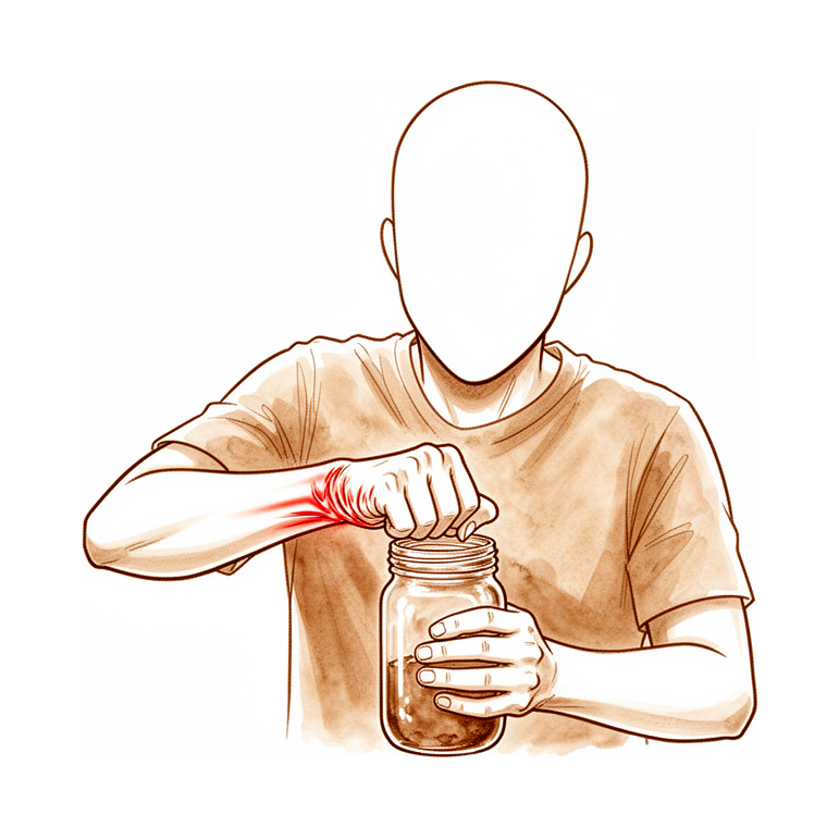
Patients with carpal injury commonly present with wrist pain as their primary complaint [40]. Pain, swelling, and ecchymosis around the region of the carpus may be present in the acute phase of carpal injury [40]. Carpal instability is characterized by wrist pain, loss of motion, and weakness [26]. Lack of treatment for carpal instability may lead to degenerative arthritis and disability [26]. The spectrum of carpal instability ranges from occult (predynamic) to dynamic to static [26]. Static instability is seen on standard radiographs [26]. Dynamic instability requires either stress radiographs or live fluoroscopy for detection [26]. Early accurate diagnosis of intrinsic carpal ligament injuries provides for best outcomes [15]. Delayed diagnosis of intrinsic carpal ligament injuries leads to arthritis within 10 years if not treated [15].
Physical Examination
The most constant and dependable sign of carpal injury is well-localized tenderness [40]. Tenderness in the anatomical snuffbox is suggestive of scaphoid injury [40]. Tenderness distal to Lister's tubercle is suggestive of scapholunate and lunate injury [40]. Tenderness on the dorsal margin, fingerbreadth distal to the ulnar head, is suggestive of triquetral, lunotriquetral ligament, and triquetrohamate ligament injury [40]. Swelling over the proximal carpal row is suggestive of a ligament avulsion with or without an associated fracture [40]. With carpal instability or dislocation, a gross deformity may be apparent, such as a marked prominence of the entire carpus dorsally suggestive of a perilunate dislocation [40].
Compressive stresses applied actively or passively may produce pain at the site of damage and cause a palpable and audible snap, click, shift, catch, or clunk [40]. Stress loading the wrist with compression and motion from radial to ulnar deviation may simulate midcarpal instability and produce a “catch-up clunk” as the proximal row of carpal bones snaps from flexion to extension [40]. Tendon displacements with audible snaps are easily produced by some persons but are seldom symptomatic [40]. For all specific physical tests, it is imperative to test the contralateral, uninjured wrist, which provides an internal control [165].
Scaphoid Shift Maneuver (Watson Test): The scaphoid shift maneuver (Watson test) is positive when a palpable clunk is felt as the wrist is moved from ulnar deviation/extension to radial deviation/flexion while the examiner’s thumb applies pressure to the volar aspect of the scaphoid tubercle [165]. In a positive scaphoid shift maneuver, pain is felt when the scaphoid subluxes over the dorsal lip of the distal radius because the scapholunate interosseous ligament is incompetent [165]. A modified Watson maneuver is positive when pain is reproduced but no clunk is felt [165]. The Watson shift test reproduces pain or a palpable clunk with the scaphoid shift test, where dorsally directed pressure over the volar scaphoid tubercle while the wrist is brought from ulnar to radial deviation subluxates or dislocates the scaphoid over the dorsal ridge of the distal radius [26]. Bilateral nonpainful clunks constitute a negative result for the Watson shift test [26].
Scapholunate Ballottement Test: The scapholunate ballottement test is positive when abnormal motion or pain is appreciated between the scaphoid and the lunate by grasping the two bones separately and shucking them in opposite directions [165].
Lunotriquetral Tests: The lunotriquetral shear test involves stabilizing the lunate and generating shear force across the articulation with volar pressure on the pisiform [165]. The lunotriquetral ballottement test is positive when the lunate is grasped between the thumb and index of one hand, and the triquetrum/pisiform are grasped between the thumb and index of the opposite hand and shucked in opposite directions [165].
Midcarpal Shift Test (Lichtman Test): The midcarpal shift test (Lichtman test) is performed by holding the forearm in pronation and applying a volar force with the thumb on the dorsum of the capitate while the hand is in 15° of radial deviation [165]. In a positive midcarpal shift test, the carpus assumes a VISI position with the distal row translated volarly on the proximal row, and a clunk is felt upon reduction with ulnar deviation [165]. The Lichtman test is defined as a palmar translation of the hand at the level of the distal capitate as the wrist is simultaneously loaded axially and moved from radial to ulnar deviation [25]. The Lichtman test is positive when a painful rapid 'catch-up' clunk occurs as the proximal carpal row jumps from flexion into extension, reproducing the patient's symptoms [25].
Dorsal wrist pain with loading and diminished grip strength are physical examination findings associated with DISI [26]. Radiocarpal instability presents with wrist swelling, deformity, and pain, with dorsal wrist swelling and tenderness most noticeable at the radiocarpal level and aggravated by wrist motion [52]. Deformity in radiocarpal instability may be due to an ulnar, dorsal, or palmar translation of the carpus [52]. With ulnar translation in radiocarpal instability, the wrist and hand are offset in an ulnar direction [52]. Patients with STT arthritis may present with carpal instability not related to radiographic scapholunate instability, characterized by a normal scapholunate angle with scaphoid extension [18]. Palmar midcarpal instability is diagnosed when the Lichtman test is found to be positive during both physical examination and during wrist cineradiography in the absence of any other wrist pathology [25].
Radiographic Findings
Static imaging techniques may accurately depict major wrist ligamentous injury [23]. Dynamic ultrasound and videofluoroscopy may demonstrate dynamic instability and kinematic dysfunction [23]. The four standard views commonly employed in the assessment of scaphoid fractures can be used to detect most injuries to the carpus [40]. These standard views include neutral posteroanterior (PA) and lateral radiographs, along with a 45-degree radial oblique and a 45-degree ulnar oblique views [40]. Additional extension and flexion views are advocated for detecting intercarpal ligament injury, along with a clenched-fist and stress views [40]. Some authors advocate contralateral wrist views because of the wide range of normal alignment [40].
Standard neutral PA and lateral radiographs are useful for determining the presence of clear fractures and assessing carpal alignment but are often poor for scaphoid fracture detection [40]. The VISI and DISI patterns of carpal malalignment are commonly detected using standard neutral lateral radiographs [40]. Additional views in maximal radial and ulnar deviation are used if the diagnosis of VISI or DISI is in doubt [40]. In the normal carpus with the wrist and hand in a neutral position, a line drawn through the axis of rotation parallel with the anatomic axis of the forearm will pass through the head and base of the third metacarpal, the capitate, the radial aspect of the lunate, and the center of the lunate fossa of the radius [40]. In the sagittal (lateral) plane with the wrist and hand in a neutral position, a line will pass through the longitudinal axis of the index finger metacarpal, capitate, lunate, and the radius, with the scaphoid lying on an axis at a 45-degree angle to this line [40]. Standard radiographs should demonstrate a constant space between the scaphoid, lunate, and triquetrum throughout the range of wrist motion [40].
A gap of 4 mm or greater between the scaphoid and lunate on the PA view suggests scapholunate ligament injury [56]. Ulnar translocation is defined as the carpus being displaced ulnarward, with more than 50% of the lunate lying ulnar to the lunate fossa [56]. Radiographs with slight radioscaphoid arthritis, such as osteophyte or “beaking” of the radial styloid, represent old injuries and are the earliest stage of scapholunate advanced collapse (SLAC) [56]. Disruption of one of the Gilula lines on PA radiographs suggests a carpal fracture or ligamentous injury [56]. The carpal height ratio is calculated by dividing the carpal height by the length of the third metacarpal, with a normal ratio of 0.54 ± 0.03 [56]. Collapse of the midcarpal joint in disease processes such as scapholunate dissociation, SLAC, and Kienböck disease produces a reduction in the carpal height ratio [56].
The normal scapholunate angle is 30° to 60°, with an average of 47° [26]. A scapholunate angle greater than 70 degrees is considered abnormal on radiographs [26]. A widened scapholunate interval greater than 3 mm is considered abnormal on radiographs [26]. The cortical ring sign showing flexion of the scaphoid is a radiographic finding associated with DISI [26]. The pencil test or bilateral clenched-fist (anteroposterior grip) comparison views can demonstrate dynamic DISI with a relatively widened SL interval on the affected side [26]. Clenched-fist PA views are used to evaluate scapholunate interosseous ligament disruption, as clenching pulls the capitate proximally, forcing the lunate and scaphoid apart and increasing the SL interval [165]. The normal SL interval is less than 4 to 5 mm [165]. Increased sensitivity for detecting SLIL disruption is achieved with ulnar deviation, supination, and AP views compared with PA views [165]. A PA view with 10° ulnar deviation is used to detect SLIL disruption [165]. The clenched pencil view involves the patient gripping a pencil or dowel with both fists, with index fingers tightly apposed and thumb metacarpals lying on the cassette for bilateral comparison [165]. An oblique view with 20° of pronation is used to visualize the dorsum of the triquetrum [165]. Dynamic fluoroscopic imaging enables dynamic testing of carpal stability and asynchronous carpal motion [165].
DISI is radiographically characterized by a scapholunate angle greater than 60° to 70° and a capitolunate/radiolunate angle greater than 15° to 30° in the dorsal direction [24]. Dorsal angulation of the capitate produces a zigzag radiolunocapitate alignment in DISI [24]. VISI is radiographically characterized by a scapholunate angle less than 30° and a radiolunate angle less than −15° [24]. DISI is defined by a lunate extending, dorsal displacement of the capitate, scapholunate angle >60 degrees, capitolunate angle >15 degrees, and radiolunate angle >10–15 degrees in the dorsal direction [42]. VISI is defined by a lunate flexing, volar displacement of the capitate, scapholunate angle <30 degrees, capitolunate angle >30 degrees, and radiolunate angle >10–15 degrees in the volar direction [42]. Dorsal tilt of the lunate greater than 15° on a true lateral radiograph is a radiographic abnormality associated with carpal instability [56]. Volar tilt of the lunate on the lateral radiograph is highly variable and should be compared with the contralateral uninjured side [56]. The “signet ring” sign, where the distal pole of the scaphoid appears as a ring on PA radiographs as it flexes, is associated with scapholunate instability [56]. A radiolunate angle greater than 15° dorsal indicates a DISI deformity on a true lateral radiograph [56]. Traditional radiographic indices measured on plain radiographs have poor diagnostic performance in the detection of carpal collapse in Kienböck's disease [19].
Advanced Imaging and Arthroscopy
MRI is the best but not perfect modality for detection of SL ligament injury [26]. Wrist arthroscopy is the gold standard to diagnose any wrist ligament injury, including the scapholunate ligament [26]. Arthroscopy is considered the reference standard for the diagnosis of intercarpal ligament injuries [56]. The roles of MRI and MRI with gadolinium arthrography are debated [56]. High-resolution MRI is an advanced imaging modality of choice, with sensitivity and specificity of ligament disruption increasing with intra-articular contrast injection and use of a 3-Tesla coil [165]. Indications for high-resolution MRI include pain localized over a particular ligament, positive provocative physical examination testing with negative radiograph, and unclear diagnosis with continued symptoms refractory to nonsurgical treatment [165]. Diagnostic arthroscopy is indicated for MRI findings of a low-grade or high-grade ligament tear without clear evidence of frank instability or a high index of clinical suspicion even without radiographic evidence [165]. Arthroscopy provides both anatomic and functional information of the interosseous and extrinsic wrist ligaments and allows simultaneous diagnosis and treatment [165]. Accurate diagnosis of ligament injuries in carpal instability is usually difficult without an arthroscopic evaluation [88].
The Geissler classification grades scapholunate ligament disruption based on arthroscopic findings [26]. Geissler Grade I is characterized by attenuation or hemorrhage of the interosseous ligament as seen from the radiocarpal space with no incongruity of carpal alignment in the midcarpal space [26]. Geissler Grade II is characterized by attenuation or hemorrhage of the interosseous ligament as seen from the radiocarpal space with a possible slight gap (less than width of probe) between carpal bones in the midcarpal space [26]. Geissler Grade III is characterized by incongruity or step-off of carpal alignment as seen from both radiocarpal and midcarpal spaces, where a probe may be passed through the gap between carpal bones [26]. A characteristic clinical pattern of predynamic SL ligament attenuation includes chronic dorsal wrist pain especially under loading in wrist extension, a positive Watson test or apprehension with pain during examination but without a reduction clunk, and normal static motion studies and grip view radiographs without SL diastasis [111]. This predynamic pattern may be either a Geissler grade 1 or 2 lesion [111]. In Geissler 1 and 2 lesions, there is no to minimal instability because of ligament attenuation without disruption [111]. Grade 3 and 4 lesions represent partial and complete tears with greater degrees of carpal instability [111].
Classification and Patterns
Carpal instability is classified based on chronicity, severity, etiology, location, direction, and pattern of instability [24]. Chronicity is classified as acute (less than 1 week), subacute (1 to 6 weeks), or chronic (more than 6 weeks) [24]. Severity/degree of instability is classified as predynamic, dynamic, or static [24]. Predynamic instability is characterized by pain with instability not evident on imaging but suggested by clinical examination [24]. Dynamic instability is characterized by carpal malalignment and pain under loading, where stress imaging with fluoroscopy is helpful and radiographs may appear normal unless certain provocative maneuvers are performed [24]. Static instability is characterized by a fixed alteration in carpal alignment demonstrable on standard static radiographs [24]. Etiology includes congenital, traumatic, inflammatory, arthritis, neoplastic, iatrogenic, or miscellaneous causes [24]. Location includes radiocarpal, intercarpal, midcarpal, carpometacarpal, specific bones, specific ligaments, or combination [24]. Direction includes dorsal intercalated segment instability (DISI), volar intercalated segment instability (
Investigations
Clinical Examination
Well-localized tenderness is the most constant and dependable sign of carpal injury [40]. Specific localization guides the differential diagnosis: tenderness in the anatomical snuffbox indicates scaphoid injury [40]; tenderness distal to Lister's tubercle indicates scapholunate and lunate injury [40]; and tenderness on the dorsal margin, fingerbreadth distal to the ulnar head, indicates triquetral, lunotriquetral ligament, and triquetrohamate ligament injury [40].
Dynamic testing provides diagnostic specificity for instability. The scaphoid shift test is diagnostic of scapholunate disruption, although up to 30% of normal wrists yield a positive result [40]. The midcarpal shift test is diagnostic of midcarpal instability (MCI) [40]. Lunotriquetral ballottement is diagnostic of lunotriquetral instability or arthritis [40], while the lunotriquetral shear test is diagnostic of lunotriquetral instability [40]. Stress loading the wrist with compression and motion from radial to ulnar deviation may simulate midcarpal instability and produce a “catch-up clunk” [40]. A full examination of the contralateral wrist can be helpful when assessing for instability [40].
Radiography
Plain radiography: The four standard views for assessing scaphoid fractures include neutral posteroanterior (PA), lateral, 45-degree radial oblique, and 45-degree ulnar oblique radiographs [40]. Standard neutral PA and lateral radiographs are useful for determining the presence of clear fractures and assessing carpal alignment [40]. However, these views are often poor for scaphoid fracture detection due to tubercle overhang on the PA view and overlap on the lateral view [40]. Plain radiography is approximately 50% sensitive for the detection of a scaphoid fracture [104] and less than 50% sensitive for the detection of other carpal bone fractures [104]. Up to 30% of patients with negative initial radiographs may have positive follow-up radiographs at 2 weeks [104].
Additional views are advocated for detecting intercarpal ligament injury, including extension and flexion views [40] and clenched-fist and stress views [40]. Ziter's view is an additional image that can aid in the diagnosis of scaphoid fractures [40]. VISI and DISI patterns of carpal malalignment are commonly detected using standard neutral lateral radiographs [40]. In the normal carpus, a line drawn through the axis of rotation parallel with the anatomic axis of the forearm passes through the head and base of the third metacarpal, the capitate, the radial aspect of the lunate, and the center of the lunate fossa of the radius [40]. Additionally, a line passes through the longitudinal axis of the index finger metacarpal, capitate, lunate, and radius, with the scaphoid lying on an axis at a 45-degree angle to this line [40].
Cineradiography: Cineradiography has a high diagnostic value for diagnosing scapholunate dissociations [196]. It is a qualitative rather than a quantitative tool [81]. More research is needed to determine the exact role of cineradiography in diagnosing carpal instability [81].
Advanced Imaging
MRI: MRI has the highest sensitivity in detecting an occult scaphoid fracture [53, 54] and is more sensitive than CT for making the diagnosis of scaphoid fracture [104]. A normal MRI study as early as 2 days after injury has a negative predictive value of 100% for scaphoid fracture [104]. MRI is the best imaging modality for assessing osteonecrosis of the proximal pole in scaphoid nonunion [203]. However, the diagnosis of osteonecrosis can be challenging because of the limited sensitivity of imaging modalities, including contrast-enhanced MRI [13]. MRI may be more helpful to exclude potential alternative diagnoses in the patient with ulnar wrist pain [186]. It is not clear whether diagnosis of subtle injuries only demonstrated on MRI improves outcomes [189]. Magnetic resonance imaging using advanced multiparametric sequences may facilitate accurate, noninvasive assessment of articular cartilage changes after distal radius fracture without the need for a contrast agent [195]. MRI can be used to determine the extent of ligamentous disruption in radiocarpal instability [52]. Notably, MRI is not recommended for the diagnosis of scapholunate ligament injury [207].
CT: CT may be required to better define associated bony injuries in radiocarpal instability [52]. Four-dimensional computed tomography aids assessment of chronic scapholunate instability [204] and allows the differentiation between patients without and those with definite or questionable scapholunate instability [204].
Other Considerations
Diagnostic Challenges: Carpal instability is a missed diagnosis in patients with clinically suspected scaphoid fracture [8]. Approximately 25% of perilunate dislocations are missed during initial emergency department evaluations [53, 54]. Despite the evolution of diagnostic and treatment options, the ideal treatment for scapholunate instability remains an unresolved problem with inconsistent results [7].
Fluoroscopic and Comparative Assessment: Measurements in the middle of the scapholunate joint in neutral and 30° of ulnar deviation under fluoroscopic imaging best capture all stages of ligamentous disruptions [199]. A comparison of the carpal height to that of the contralateral wrist allows the extent of collapse and scaphoid shortening to be estimated [13]. The influence of intraoperative instability of a scaphoid fracture that is seen to be nondisplaced on radiographs or CT on the risk of nonunion is currently unknown [205].
Treatment
General Principles and Outcomes
The ideal treatment for scapholunate instability remains an unresolved problem characterized by inconsistent results and ongoing concerns regarding complications [7]. Early accurate diagnosis of intrinsic carpal ligament injuries provides the best outcomes, whereas delayed diagnosis leads to arthritis within 10 years if not treated [15]. Nonsurgical management of perilunate fracture-dislocations results in progressive arthritis and poor long-term outcomes [47]. Surgical decision-making for carpal instability must encompass the extent of ligament injury, reparability, secondary stabilizers, reducibility, status of cartilage, and radiolunate alignment [170]. Arthritis is a contraindication for surgical treatment of carpal instability [170].
There is a disconnect between the understanding of carpal instability and the limitations of current reconstruction techniques [21]. The lack of long-term outcome measurements for bone-tissue-bone (BTB) surgeries makes it difficult to determine the appropriate use of these treatment modalities [140]. Referral of cases to specialized centers is highlighted as necessary to run randomized controlled trials due to the lack of objective evidence on long-term outcomes [11].
Non-Operative Management
Nonsurgical management options for carpal instability include NSAIDs, splinting, casting, and steroid injections [170]. Suspected predynamic scapholunate or lunotriquetral instability may be initially treated with a cast [170]. Nonoperative treatment or arthroscopic débridement is indicated for partial ligament injuries presenting as predynamic or dynamic instability [26]. Nonoperative approaches are recommended for appropriate candidates with predynamic or dynamic midcarpal instability [187]. Conservative management of palmar carpal subluxation has been reported to fail in cases of severe instability [32]. A wrist exercise program has been used for the management of palmar midcarpal instability [25].
Treatment of post-traumatic carpal instability in young children may be deferred in asymptomatic patients with normal or near-normal function [65]. Non-operative treatment, including a protective orthosis, can be used in young children with permanent structural changes that prevent reduction until skeletal maturity [65]. Patients with bilateral avascular necrosis of the trapezoid improved clinically and radiographically with nonoperative treatment, achieving complete pain freedom and full wrist movement at 5 years [202]. Contrary to existing literature, successful outcomes have been reported with nonsurgical management and delayed nonfusion surgery for traumatic volar carpal instability nondissociative (CINDT-VISI) [150].
Operative Management
Indications: Surgical intervention is indicated when nonoperative measures fail or when instability is dynamic or static. Acute scapholunate ligament rupture is rarely amenable to primary repair alone [26]. Perilunate dislocations almost always require surgical treatment [170]. Surgical fixation is recommended for all acute perilunate injuries, even after anatomic closed reduction, because of the extent of instability [155]. Intercarpal fusions may be used in Stage IV scapholunate dissociation with static irreducible deformities without arthritis [170].
Surgical Approach / Technique: For scapholunate instability, after delayed diagnosis of scapholunate ligament rupture, open reduction of the scapholunate interval and K-wire pinning with or without dorsal capsulodesis or tendon autograft reconstruction is performed [26]. Closed reduction and percutaneous pinning can be used for scapholunate ligament injuries if part of the ligament remains intact after débridement [170]. Temporary pins inserted dorsally into the scaphoid and lunate act as joysticks to correct scaphoid flexion and lunate extension during pinning [170]. Commonly, two 0.045-inch Kirschner wires are placed across the scapholunate interval, and one is placed across the scaphocapitate interval for further stabilization [170]. Arthroscopic ligament débridement with or without thermal shrinkage is used for low-grade tears (Geissler I/II) with no frank instability [170]. Open reduction and ligament repair and pinning is performed if the ligament is repairable and the scaphoid is reducible, using suture anchors or transosseous sutures [170].
Volar scapholunate reconstruction using the volar half of the radioscaphocapitate or long radiolunate ligament is used for isolated volar scapholunate tears or combined with dorsal repair [170]. Brunelli tenodesis involves harvesting the flexor carpi radialis tendon, pulling it through a tunnel in the scaphoid, and attaching it to the dorsal ulnar corner of the radius [170]. Tri-ligament tenodesis is a modification of the Brunelli technique where the flexor carpi radialis is fixed dorsally to the lunate and passed through a slit in the dorsal radiocarpal ligament [170]. The scapholunate ligament internal brace 360° tenodesis (SLITT) procedure uses a tendon graft and internal brace to reconstruct the volar and dorsal scapholunate interosseous ligament without Kirschner wires [170]. Dorsal capsulodesis can be performed in isolation if the ligament is not repairable and the scaphoid is reducible, or to augment ligament repair or reconstruction [170]. The Blatt capsulodesis involves attaching a flap of the dorsal wrist capsule to the dorsal pole of the scaphoid while leaving it proximally attached to the distal radius [170]. The Mayo capsulodesis involves detaching a proximal strip of the dorsal intercarpal ligament from the triquetrum, rotating it proximally, and attaching it to the dorsal lunate [170]. Reduction and association of the scaphoid and lunate (RASL) screw creates a pseudarthrosis using a partially threaded headless compression screw [170]. Reconstruction of the scapholunate ligament with bone-ligament-bone autograft from the foot or capitohamate interval is an available surgical option [170].
Arthroscopic grade I injury of the scapholunate or lunotriquetral ligament with no joint incongruence typically resolves with immobilization alone [160]. Acute tears of the scapholunate or lunotriquetral ligament (less than 4 to 6 weeks) that result in incongruence may be arthroscopically reduced and temporarily pinned [160]. Arthroscopic reduction association of the scapholunate joint (ARASL) uses a headless compression screw to resist scaphoid pronation and maintain alignment while fibrous union develops [134]. In a study of 18 consecutive patients undergoing ARASL, preoperative radiographic parameters were evaluated for association with surgical complications [134]. The modified Brunelli technique correction might correlate with improved carpal dynamics and improved clinical outcomes [39]. Arthroscopic scapholunate synthetic ligamentoplasty is described as an effective solution that is not highly invasive and avoids the consequences of capsulotomy [144]. Arthroscopic scapholunate ligamentoplasty is a demanding technique with more peroperative traps than open surgeries, resulting in elongated operative time and tourniquet ischemia time [144]. A clinical trial is currently underway to confirm the hypotheses regarding the benefits of arthroscopic scapholunate ligamentoplasty [144]. Arthroscopic-assisted volar scapholunate capsulodesis is a described technique for scapholunate instability [145]. Modified minimally invasive extensor carpi radialis longus tenodesis is a prospective observational study option for scapholunate dissociation [62]. Modified Viegas dorsal capsuloplasty has been used for chronic partial injury of the scapholunate ligament in young athletes [136]. Anatomical anterior and posterior reconstruction for scapholunate dissociation resulted in no patient requiring secondary surgery or treatment related to the carpal stabilization [6]. At 17.9-month average follow-up, radiographic and patient-reported outcome parameters improved after reconstruction of the critical dorsal and volar ligament stabilizers of the proximal carpal row with the ANAFAB technique [46]. The fact that arthritic changes were observed in only 1 of 8 cases suggests that carpal stability obtained by modified Brunelli tenodesis is probably sufficient to obtain good functional long-term results [5]. Although ongoing scapholunate instability resulted in early arthritic degeneration following dorsal intercarpal ligament capsulodesis, most patients had acceptable long-term function of the wrist [3]. There is convincing evidence supporting the role of arthroscopy in diagnosis and assessment of factors involved in the development of carpal instability, but weak evidence for the effectiveness of arthroscopic techniques in the actual treatment of this condition [12].
For lunotriquetral instability and VISI, options include arthroscopic débridement, closed reduction and percutaneous pinning, open reduction and ligament repair, open reduction and ligament reconstruction, and lunotriquetral arthrodesis [170]. Closed reduction and percutaneous pinning for lunotriquetral instability uses dorsal joysticks to correct the deformity [170]. Open reduction and ligament repair for lunotriquetral instability uses bone anchors or transosseous sutures [170]. Open reduction and ligament reconstruction for lunotriquetral instability uses a distally based strip of the extensor carpi ulnaris passed through the lunate and triquetrum and sutured to itself [170]. Lunotriquetral ligament reconstruction using the extensor carpi ulnaris may be superior to arthrodesis or primary repair of the lunotriquetral interosseous ligament [170]. Lunotriquetral arthrodesis is performed with bone graft and screws or staples [170]. Division of the dorsal or palmar part of the intrinsic lunotriquetral ligament can cause significant kinematic dysfunction, potentially resulting in synovitis, dynamic VISI deformity, and abnormal motion of the carpus [64]. In case of extrinsic ligament division, a highly dysfunctional static VISI deformity can arise [64].
For perilunate dislocations and fracture-dislocations, urgent reduction and immobilization are required to relieve pressure on the median nerve and soft tissues [170]. Closed reduction of perilunate dislocations can be performed using the Tavernier method, which involves traction, extending the wrist, and gradually flexing the wrist with volar pressure on the lunate [170]. Postreduction X-rays are important to evaluate reduction and possibly a CT scan to look for concomitant fractures after closed reduction of perilunate dislocations [170]. Closed reduction and percutaneous pinning for perilunate dislocations is dictated by the injury pattern, with fixation pins commonly placed across the lunotriquetral, scapholunate, and scaphocapitate intervals [170]. Headless compression screws may be used for fixation of concomitant scaphoid fractures in perilunate injuries [170]. Closed reduction and percutaneous pinning for perilunate dislocations is often combined with open carpal tunnel release as needed [170]. A carpal tunnel release may be added in the setting of median nerve symptoms for acute perilunate injuries [155]. Chronic perilunate injuries may be managed either in the same manner as acute injuries or with a proximal row carpectomy [155]. Arthroscopic reduction and percutaneous fixation of perilunate dislocations and fracture-dislocations was found to have satisfactory outcomes [155]. Interposed palmar capsular ligaments were found in all patients surgically treated after unsuccessful closed reduction of perilunate dislocations [155]. At a minimum 10-year follow-up, 18 patients with perilunate dislocation or fracture-dislocation had an average Mayo wrist score of 77 (range, 60 to 90), and 12 patients (67%) had radiographic evidence of arthritis [155]. Although intraoperative reduction was considered to be good in more patients with perilunate fracture-dislocation than perilunate dislocation, there were no significant differences in long-term clinical or radiographic parameters [155]. Much of the articular cartilage damage at the time of perilunate injury occurs because of shear and compression and cannot be fully mitigated by the reduction [155]. Most patients have a satisfactory long-term clinical outcome despite radiologic evidence of osteoarthritis progression following perilunate injury [155]. Operative management of volarly displaced transscaphoid, translunate, transtriquetrum fractures likely prevented more serious consequences despite radiographic evidence of cartilage loss between the lunate and capitate [201]. Primary wrist fusion is recommended in cases of pancarpal dissociation with significant displacement and unsalvageable devascularized fragments [78].
For scaphoid nonunion and SNAC wrist, an unstable scaphoid nonunion requires bone grafting to restore height and correct carpal malalignment, principally dorsal intercalated segment instability [13]. A systematic review found that both cancellous and corticocancellous bone grafting led to reliable union (95% or 92%, respectively) [13]. The 1,2 intercompartmental supraretinacular artery was commonly used as a vascularized pedicled bone graft but has begun to fall out of favor compared with other vascularized grafts due to a wide range of reported union rates [13]. The free medial femoral condyle vascularized graft has demonstrated superior performance in revision scenarios, with
Implant Selection: Treatment for SLAC wrist depends on the condition of articular surfaces and competency of the radioscaphocapitate ligament [26]. Radial styloidectomy with or without partial wrist denervation is indicated for stage 1 SLAC [26]. Proximal row carpectomy (PRC) is indicated for stage 2 SLAC when the proximal capitate is spared of arthrosis and requires a competent radioscapholunate ligament [26]. Scaphoid excision and four-corner or isolated capitolunate fusion is indicated for stage 2 or 3 SLAC if the proximal lunate is spared of arthrosis [26]. Scaphoid excision and four-corner or isolated capitolunate fusion is contraindicated in smokers [26]. Total wrist fusion is indicated for stage 3 SLAC with diffuse radiocarpal and midcarpal changes [26]. In the treatment of scapholunate advanced collapse and scaphoid nonunion advanced collapse wrists, proximal row carpectomy results in better outcomes and a lower complication rate compared to four-corner fusion [163]. The authors do not recommend proximal row carpectomy for the management of scapholunate dissociation in the absence of degenerative changes due to disappointing results compared to other treatments [59].
Other Considerations: Partial wrist denervation, involving excision of the terminal branch of the posterior interosseous nerve with or without the anterior interosseous nerve, is often done in conjunction with SLAC procedures or as an option on its own [26]. The outcome of scaphoid excision and four-corner arthrodesis is favorable at one year and does not deteriorate significantly between one and 10 years [74]. Scaphoid excision and four-corner fusion remains a viable option for patients with advanced wrist arthritis with functional results that are reliable, resilient, and remain stable over time [159]. Distal scaphoid resection is a durable procedure with good long-term results, with 94% of patients remaining satisfied and no further wrist collapse or radiocarpal arthritis developing [31]. Patients with established scaphoid non-union should be advised that osteoarthritis will most likely develop [82].
Complications
Natural History and Degenerative Progression
Neglected scaphoid nonunion is associated with osteonecrosis and progressive radiocarpal and midcarpal arthritis, a condition termed scaphoid nonunion advanced collapse [13]. Persistent carpal instability leads to degenerative changes resulting from increased shear forces and abnormal contact between individual carpal bones [42]. An isolated injury to the scapholunate interosseous ligament may herald a relentless progression to abnormal joint mechanics, cartilage wear, and degenerative change [29]. In the natural history of arthroscopically proven scapholunate rupture without initial SL widening or DISI, all patients developed SLAC changes and wrist pain at an average of seven years [35]. Although radiographic carpal collapse and ulnar translocation occurred in patients with Kienböck disease treated with scaphocapitate arthrodesis, these patients were not symptomatic [4].
Iatrogenic and Procedural Complications
Excision arthroplasty for scapho-trapezial-trapezoid (STT) arthritis can provoke severe malalignment and midcarpal instability, leading to an intercarpal arthrodesis with an outcome potentially worse than STT fusion [20]. Collapse after carpal bone tunneling is an underreported complication with potentially substantial consequences; patients should be warned of this possibility when discussing complications of ligament reconstruction for chronic scapholunate instability [72]. Scaphoid malunion following treatment for nonunion may cause poor clinical outcomes due to a dorsal intercalated segmental instability (DISI) deformity [181]. Prolonged malposition of the carpus in adaptive instability tends to stretch the dorsal capsuloligamentous structures with time, and the kinematic abnormalities may eventually become permanent [36]. Donor morbidity of the knee must be considered when using a free vascularized medial femoral trochlea osteocartilaginous flap for proximal one-fifth scaphoid nonunions [13]. The free medial femoral condyle vascularized graft technique for scaphoid nonunion requires the advanced skills of a microvascular flap surgeon [13]. Carpal fractures had a small negative effect on the Short Musculoskeletal Function Assessment Hand/Arm Index and EQ-5D scores 1 year after the injury [179].
Diagnostic and Management Challenges
There is a lack of objective evidence on long-term outcomes for the unstable wrist, highlighting the need for randomized controlled trials [11]. A diagnosis of osteonecrosis in scaphoid nonunion can be challenging because of the limited sensitivity of imaging modalities, including contrast-enhanced MRI [13]. Post-traumatic carpal instability in a young child may be difficult to diagnose early since the centers of ossification of the scaphoid and lunate do not usually appear before the age of four years [65]. Chronic unreduced fracture-dislocations should not be classified as a carpal instability problem but as a more complex injury that tends to progress to stiffness and degenerative disease [45]. The cumulative incidence of carpal instability in the second year after a fall onto an outstretched hand is higher than anticipated [76, 85].
Recovery
Light activity (weeks): The provided evidence does not specify a typical week range for the resumption of desk work, driving, or light activities of daily living.
Full activity (months): The provided evidence does not specify a month range for the return to manual work, sport, or full range of motion and strength.
Complete recovery / outcome plateau (months): The provided evidence does not specify a month range for the stabilization of pain, strength, and final functional outcomes.
Rehabilitation protocol: The provided evidence does not detail specific physical therapy phasing, immobilisation duration, weight-bearing or range-of-motion progression, or sling and brace removal timing.
Functional milestones: The provided evidence does not report validated patient-reported outcome trajectories or specific outcome-measure benchmarks such as Constant, ASES, or WOMAC scores.
Other Considerations: Long-term functional outcomes following carpal stabilization and reconstruction procedures are generally favorable, though specific radiographic findings may vary. Following dorsal intercarpal ligament capsulodesis, most patients achieved acceptable long-term wrist function despite ongoing scapholunate instability and early arthritic degeneration [3]. No patient required secondary surgery or treatment related to carpal stabilization following anatomical anterior and posterior reconstruction for scapholunate dissociation [6]. Both versions of scapholunate intercarpal ligamentoplasty yield satisfactory clinical and radiological results in the short to mid-term [190]. In contrast, the RASL procedure was abandoned for dynamic and static scapholunate instability due to a high radiographic failure rate observed in short-term radiographic findings from a limited series [212].
For Kienböck disease, the outcome of scaphoid excision and 4-corner arthrodesis is favorable at one year and does not deteriorate significantly between one and 10 years [74]. At an average follow-up of 10 years, proximal row carpectomy is a reliable and durable procedure for patients with Lichtman stage IIIA or IIIB Kienböck's disease [208]. Good clinical results observed in patients 10 years after radial shortening osteotomy for Kienböck disease are likely to remain stable at 20 years after surgery [213]. Static carpal malalignment does not preclude radius-shortening osteotomy given the high percentage of successful clinical outcomes [71]. Radiographic progression of Kienböck disease over 1 year or more seems slight on average regardless of treatment [210]. Although radiographic carpal collapse and ulnar translocation occurred, patients were not symptomatic following scaphocapitate arthrodesis for Kienböck disease [4].
In pediatric and adolescent populations, double bone forearm osteotomy in adolescence brought long-lasting functional improvement and provided long-term correction of distal radioulnar and radiocarpal subluxations for Madelung’s deformity [173]. Long-term follow-up at age 17 showed no pain, normal wrist motion, and no radiocarpal osteoarthritis in a child with Kienböck disease and cerebral palsy [175]. Prospective follow-up of over 200 patients has shown that both radiographic measures and patient-reported outcomes turned out great following vascularized bone flaps for carpal reconstruction [211].
Regarding post-injury instability, this study found a higher than anticipated cumulative incidence of carpal instability in the second year post injury [76]. This study found a higher than anticipated cumulative incidence of carpal instability in the second year after injury, which may reflect volunteer bias [85].
Key Evidence
- [L5] Carpal instability is a complex array of maladaptive and posttraumatic conditions that lead to the inability of the wrist to maintain anatomic relationships under normal loads. [2] (10.1016/j.hcl.2015.04.011)
- [L3] Although the consequent ongoing scapholunate instability resulted in early arthritic degeneration, most patients had acceptable long-term function of the wrist. [3] (10.1302/0301-620x.94b12.30007)
- [L4] Although radiographic carpal collapse and ulnar translocation occurred, patients were not symptomatic. [4] (10.1016/j.jhsa.2014.12.013)
- [L4] The fact that we observed arthritic changes in only 1 of 8 cases suggests that carpal stability obtained by this procedure is probably sufficient to obtain good functional long-term results. [5] (10.1016/j.jhsa.2013.02.022)
- [L4] No patient required secondary surgery or treatment related to the carpal stabilization. [6] (10.1177/1753193419886536)
- [L5] Despite the evolution of diagnostic and treatment options, the ideal treatment for scapholunate instability remains an unresolved problem with inconsistent results and ongoing concerns regarding complications. [7] (10.1177/17531934221148009)
- [L2] Bone scintigraphy was not useful for the detection or exclusion of carpal instability. [8] (10.1016/0020-1383(93)90182-6)
- [L5] Midcarpal instability is a collective term for conditions where wrist instability is predominantly between the proximal and distal carpal rows, with palmar midcarpal instability being the most common type. [9] (10.1177/1753193415617756)
- [L5] The aim of this themed issue is to provide answers regarding the investigation and management of the unstable wrist, highlighting the need for collaborative working and the referral of cases to specialized centers to run randomized controlled trials due to the lack of objective evidence on long-term outcomes. [11] (10.1177/1753193415617100)
- [L4] There is convincing evidence supporting the role of arthroscopy in diagnosis and assessment of factors involved in the development of carpal instability, but weak evidence for the effectiveness of arthroscopic techniques in the actual treatment of this condition. [12] (10.1177/1753193415616276)
- [L5] Early accurate diagnosis of intrinsic carpal ligament injuries provides for best outcomes, while delayed diagnosis leads to arthritis within 10 years if not treated. [15] (10.1016/j.hcl.2015.01.003)
- [L4] Prompt recognition and surgical treatment with anatomic reduction of carpal malalignment improve the likelihood of optimal, long-term clinical success and patient satisfaction. [16] (10.1016/j.jhsa.2012.07.034)
- [L4] Patients with STT arthritis may present with carpal instability not related to radiographic scapholunate instability, characterized by a normal scapholunate angle with scaphoid extension. [18] (10.1016/j.jhsa.2006.10.021)
- [L3] Traditional radiographic indices measured on plain radiographs have poor diagnostic performance in the detection of carpal collapse in Kienböck's disease. [19] (10.1177/17531934231153966)
- [L4] The procedure can provoke severe malalignment and midcarpal instability, leading to an intercarpal arthrodesis with an outcome potentially worse than STT fusion. [20] (10.1177/1753193408098903)
- [L5] Despite these advances, there remains a disconnect between the understanding of carpal instability and the limitations of current reconstruction techniques. [21] (10.1016/j.jhsa.2016.07.105)
- [L4] Static imaging techniques may accurately depict major wrist ligamentous injury, while dynamic ultrasound and videofluoroscopy may demonstrate dynamic instability and kinematic dysfunction. [23] (10.1177/1753193415610515)
- [L4] [25] (10.1055/s-0037-1612594)
- [L4] Carpal fractures other than the scaphoid are exceedingly rare and comprise approximately 1.1% of all fractures; management depends on the specific fracture pattern, displacement, and associated ligamentous injuries, with outcomes generally good for isolated injuries but complicated by nonunion and instability in cases with concomitant injuries. [28] (10.1016/j.jhsa.2013.10.030)
- [L5] [29] (10.1016/j.jhsa.2008.04.027)
- [L4] Distal scaphoid resection is a durable procedure with good long-term results. 94% of patients remained satisfied, and no further wrist collapse or radiocarpal arthritis developed. [31] (10.1016/s0363-5023(11)60002-6)
- [L4] These two cases show the results of the failure of conservative management in two extremes of palmar carpal subluxation. [32] (10.2106/00004623-198365070-00016)
- [L5] This correction might correlate with improved carpal dynamics and improved clinical outcomes. [39] (10.1016/j.jhsa.2010.06.029)
- [L4] Correct diagnosis and proper surgical indications are necessary to reduce the number and enhance the efficacy of operations required to treat carpal boss. [41] (10.1016/j.jhsa.2007.11.029)
- [L4] At 17.9-month average follow-up, radiographic and patient-reported outcome parameters improved after reconstruction of the critical dorsal and volar ligament stabilizers of the proximal carpal row with the ANAFAB technique. [46] (10.1016/j.jhsa.2023.12.012)
- [L4] The authors do not recommend PRC for the management of scapholunate dissociation in the absence of degenerative changes due to disappointing results compared to other treatments. [59] (10.1177/1753193410382719)
- [L5] [61] (10.5435/00124635-201209000-00004)
- [L4] [62] (10.1186/s12891-017-1414-7)
- [L4] [64] (10.1177/1753193415595167)
- [Case_report] [65] (10.2106/00004623-198062010-00021)
- [L5] Midcarpal instability is a term representing multiple distinct entities involving abnormal force transmission across the midcarpal joint. [69] (10.5435/jaaos-d-22-00777)
- [L4] Static carpal malalignment does not preclude radius-shortening osteotomy given the high percentage of successful clinical outcomes. [71] (10.1016/j.jhsa.2010.08.017)
- [L4] Although the incidence of carpal bone tunnel collapse is unknown, the consequences could be substantial; therefore, patients should be warned of this possibility when discussing complications of ligament reconstruction for chronic scapholunate instability requiring the creation of a bone tunnel. [72] (10.1016/j.jhsa.2023.07.008)
- [L4] The technique addresses critical stabilizers to prevent carpal instability and collapse. [73] (10.1016/j.jhsa.2024.10.019)
- [L4] The outcome of scaphoid excision and 4-corner arthrodesis is favorable at one year and does not deteriorate significantly between one and 10 years. [74] (10.1016/j.jhsa.2010.01.025)
- [L4] This study found a higher than anticipated cumulative incidence of carpal instability in the second year post injury. [76] (10.1016/j.jht.2017.11.014)
- [L4] Excisional arthroplasty is highly recommended for idiopathic, symptomatic, isolated STT osteoarthritis without midcarpal instability, as it provides reliable results, is less technically demanding, requires less prolonged immobilization, and has fewer complications than localized arthrodesis. [77] (10.1016/j.jhsa.2010.12.016)
- [Case_report] They recommend primary wrist fusion in cases of such significant displacement with unsalvageable devascularized fragments. [78] (10.1016/j.otsr.2016.12.023)
- [L3] Cineradiography is a qualitative rather than a quantitative tool, and more research is needed to determine its exact role in diagnosing carpal instability. [81] (10.1177/1753193417694820)
- [L4] Patients with established scaphoid non-union should be advised that osteoarthritis will most likely develop. [82] (10.2106/00004623-198567030-00013)
- [L5] The choice of procedure depends on whether the joint is isolated or associated with carpal malalignment and other joint osteoarthritis. [84] (10.1177/17531934241295345)
- [L2] This study found a higher than anticipated cumulative incidence of carpal instability in the second year after injury, which may reflect volunteer bias. [85] (10.1016/j.jht.2017.08.006)
- [L5] However, during simple unresisted wrist motions, the force did not exceed 20 N. [87] (10.1016/j.jhsa.2015.04.007)
- [L4] [88] (10.1007/s11552-008-9155-6)
- [L4] [111] (10.1016/j.hcl.2011.06.005)
- [L4] The SLIL generates proprioceptive stimuli at every wrist position. [125] (10.1016/j.jhsa.2010.10.002)
- [L5] [129] (10.1016/j.otsr.2013.06.015)
- [L4] [134] (10.1177/1753193415577335)
- [L4] [136] (10.1177/1753193420939490)
- [L4] [140] (10.1016/j.jhsa.2006.11.011)
- [L5] [144] (10.1016/j.eats.2021.05.022)
- [L4] [145] (10.1016/j.jhsa.2022.05.018)
- [L4] Contrary to existing literature, the authors report successful outcomes with nonsurgical management and delayed nonfusion surgery for traumatic volar carpal instability nondissociative (CINDT-VISI). [150] (10.1016/j.jhsg.2023.07.001)
- [Letter] The authors propose that the term midcarpal instability be subclassified into traumatic and nontraumatic categories, as the pathology often involves dissociative ligament disruptions rather than being truly nondissociative. [151] (10.1016/j.jhsa.2022.05.005)
- [L4] Scaphoid excision and four-corner fusion remains a viable option for patients with advanced wrist arthritis with functional results that are reliable, resilient, and remain stable over time. [159] (10.1016/j.jhsa.2014.06.117)
- [L1] In the treatment of scapholunate advanced collapse and scaphoid nonunion advanced collapse wrists, PRC results in better outcomes and a lower complication rate compared to 4CF. [163] (10.1016/j.jhsa.2024.01.011)
- [L4] Double bone forearm osteotomy in adolescence brought long-lasting functional improvement and provided long-term correction of distal radioulnar and radiocarpal subluxations. [173] (10.1054/jhsb.1999.0304)
- [Case_report] Long-term follow-up at age 17 showed no pain, normal wrist motion, and no radiocarpal osteoarthritis. [175] (10.2106/00004623-199610000-00016)
- [L5] [178] (10.1177/1753193410374690)
- [L4] Carpal fractures had a small negative effect on the Short Musculoskeletal Function Assessment Hand/Arm Index and EQ-5D scores 1 year after the injury. [179] (10.1177/17531934231202012)
- [L5] Midcarpal instability is a recently discovered carpal derangement that represents a transverse break in the ring, most significantly at the triquetrohamate joint. [180] (10.1016/s0749-0712(21)00640-5)
- [L4] [181] (10.1177/1753193416680133)
- [Paper] [183] (10.1016/j.hcl.2006.07.005)
- [L3] MRI may be more helpful to exclude potential alternative diagnoses in the patient with ulnar wrist pain. [186] (10.1016/j.jhsa.2013.05.040)
- [L4] The authors recommend these nonoperative approaches for appropriate candidates, particularly those with predynamic or dynamic instability. [187] (10.1177/1753193415613050)
- [L2] A scaphoid fracture was by far the most common injury, but it is not clear whether diagnosis of subtle injuries only demonstrated on MRI improves outcomes. [189] (10.1016/j.jhsa.2012.09.034)
- [L3] Both versions of the scapholunate intercarpal ligamentoplasty yield satisfactory clinical and radiological results in the short to mid-term. [190] (10.1177/1753193420940498)
- [L4] Magnetic resonance imaging using advanced multiparametric sequences may facilitate accurate, noninvasive assessment of articular cartilage changes after distal radius fracture without the need for a contrast agent. [195] (10.1016/j.jhsa.2020.02.009)
- [L3] Cineradiography has a high diagnostic value for diagnosing scapholunate dissociations. [196] (10.1177/1753193413489056)
- [L5] Measurements in the middle of the scapholunate joint in neutral and 30° of ulnar deviation under fluoroscopic imaging best capture all stages of ligamentous disruptions. [199] (10.1177/1558944717729219)
- [L5] [200] (10.1016/j.jhsa.2013.06.019)
- [Case_report] Operative management likely prevented more serious consequences despite radiographic evidence of cartilage loss between the lunate and capitate. [201] (10.1016/j.jhsa.2014.02.034)
- [L4] The patient with bilateral avascular necrosis of the trapezoid improved clinically and radiographically with nonoperative treatment, achieving complete pain freedom and full wrist movement at 5 years. [202] (10.1016/j.jhsa.2011.07.022)
- [L5] MRI is the best imaging modality for assessing osteonecrosis of the proximal pole in scaphoid nonunion. [203] (10.1016/j.jhsa.2013.03.055)
- [L2] Four-dimensional computed tomography aids assessment of chronic scapholunate instability, which allows the differentiation between patients without and those with definite or questionable scapholunate instability. [204] (10.1177/1753193419893890)
- [L3] The influence, with regard to the risk of nonunion, of intraoperative instability of a scaphoid fracture that is seen to be nondisplaced on radiographs or CT is currently unknown. [205] (10.2106/jbjs.k.00993)
- [L3] MRI is not recommended for the diagnosis of scapholunate ligament injury. [207] (10.1054/jhsb.2000.0450)
- [L4] At an average follow-up of 10 years, proximal row carpectomy is a reliable and durable procedure for patients with Lichtman stage IIIA or IIIB Kienböck's disease. [208] (10.1016/j.jhsa.2008.02.031)
- [L4] Radiographic progression of Kienböck over 1 year or more seems slight on average regardless of treatment. [210] (10.1016/j.jhsa.2016.02.016)
- [Paper] Prospective follow-up of over 200 patients has shown that both radiographic measures and patient-reported outcomes turned out great. [211] (10.1016/j.jhsa.2026.05.018)
- [L4] Based on short-term radiographic findings in this limited series, the authors have abandoned the RASL procedure for the treatment of dynamic and static scapholunate instability due to a high radiographic failure rate. [212] (10.1016/j.jhsa.2014.12.032)
- [L4] Good clinical results observed in patients 10 years after radial shortening osteotomy are likely to remain stable at 20 years after surgery. [213] (10.1016/j.jhsa.2025.04.018)
See Also
- Distal Radius Fracture
- Scaphoid Fracture
- Scapholunate Ligament Injury
- Kienböck's Disease
- Wrist Arthroscopy
- Carpal Tunnel Release
- Proximal Row Carpectomy
- Total Wrist Fusion
References
[1] Green S Operative Hand Surgery. CARPAL INSTABILITY > Classification of Carpal Instability.
[2] Carpal Ligament Injuries, Pathomechanics, and Classification. Hand Clinics. 2015. DOI: 10.1016/j.hcl.2015.04.011
[3] Long-term results of dorsal intercarpal ligament capsulodesis for the treatment of chronic scapholunate instability. The Journal of Bone and Joint Surgery. British volume. 2012. DOI: 10.1302/0301-620x.94b12.30007
[4] Scaphocapitate Arthrodesis for Kienböck Disease. The Journal of Hand Surgery. 2015. DOI: 10.1016/j.jhsa.2014.12.013
[5] Treatment of Static Scapholunate Instability With Modified Brunelli Tenodesis: Results Over 10 Years. The Journal of Hand Surgery. 2013. DOI: 10.1016/j.jhsa.2013.02.022
[6] Anatomical anterior and posterior reconstruction for scapholunate dissociation: preliminary outcome in ten patients. Journal of Hand Surgery (European Volume). 2019. DOI: 10.1177/1753193419886536
[7] Scapholunate instability: why are the surgical outcomes still so far from ideal?. Journal of Hand Surgery (European Volume). 2023. DOI: 10.1177/17531934221148009
[8] Carpal instability, the missed diagnosis in patients with clinically suspected scaphoid fracture. Injury. 1993. DOI: 10.1016/0020-1383(93)90182-6
[9] Midcarpal instability. Journal of Hand Surgery (European Volume). 2015. DOI: 10.1177/1753193415617756
[11] Special issue: the unstable wrist. Journal of Hand Surgery (European Volume). 2015. DOI: 10.1177/1753193415617100
[12] The role of arthroscopy in carpal instability. Journal of Hand Surgery (European Volume). 2015. DOI: 10.1177/1753193415616276
[13] Orthopaedic Knowledge Update Trauma. Hand/Carpal Fractures and Dislocations > Scaphoid Fractures > Scaphoid Nonunion.
[15] Management of Complications of Ligament Injuries of the Wrist. Hand Clinics. 2015. DOI: 10.1016/j.hcl.2015.01.003
[16] Perilunate Dislocations and Fracture Dislocations. The Journal of Hand Surgery. 2012. DOI: 10.1016/j.jhsa.2012.07.034
[18] The Clinical Implications of Scaphotrapezium-Trapezoidal Arthritis With Associated Carpal Instability. The Journal of Hand Surgery. 2007. DOI: 10.1016/j.jhsa.2006.10.021
[19] Diagnostic performance of traditional radiographic indices in detection of carpal collapse in Kienböck’s disease. Journal of Hand Surgery (European Volume). 2023. DOI: 10.1177/17531934231153966
[20] Midcarpal instability after excision arthroplasty for scapho-trapezial-trapezoid (STT) arthritis. Journal of Hand Surgery (European Volume). 2009. DOI: 10.1177/1753193408098903
[21] Carpal Kinematics and Kinetics. The Journal of Hand Surgery. 2016. DOI: 10.1016/j.jhsa.2016.07.105
[23] Imaging in carpal instability. Journal of Hand Surgery (European Volume). 2015. DOI: 10.1177/1753193415610515
[24] Aaos Comprehensive Orthopaedic Review 3. Carpal Instability* > III. Classifications.
[25] Long-Term Functional Results of a Wrist Exercise Program for Patients with Palmar Midcarpal Instability. Journal of Wrist Surgery. 2017. DOI: 10.1055/s-0037-1612594
[26] Miller S Review Of Orthopaedics. SECTION 16 PATELLAR TRACKING IN TOTAL KNEE ARTHROPLASTY > 2. Scaphoid fractures > 4. Carpal instability.
[28] Carpal Fractures. The Journal of Hand Surgery. 2014. DOI: 10.1016/j.jhsa.2013.10.030
[29] Scapholunate Instability: Current Concepts in Diagnosis and Management. The Journal of Hand Surgery. 2008. DOI: 10.1016/j.jhsa.2008.04.027
[31] Distal Scaphoid Resection for Arthritis Secondary to Scaphoid Nonunion: A Twenty-year Experience. The Journal of Hand Surgery. 2011. DOI: 10.1016/s0363-5023(11)60002-6
[32] Post-traumatic palmar carpal subluxation. Report of two cases.. The Journal of Bone & Joint Surgery. 1983. DOI: 10.2106/00004623-198365070-00016
[35] Green S Operative Hand Surgery. CARPAL INSTABILITY DISSOCIATIVE.
[36] Green S Operative Hand Surgery. AUTHORS' PREFERRED METHOD OF TREATMENT: SL DISSOCIATION > CARPAL INSTABILITY ADAPTIVE (CIA).
[39] Radiographic Evaluation of the Modified Brunelli Technique Versus the Blatt Capsulodesis for Scapholunate Dissociation in a Cadaver Model. The Journal of Hand Surgery. 2010. DOI: 10.1016/j.jhsa.2010.06.029
[40] Rockwood And Green S Fractures In Adults. 42: Fractures of the Distal Radius and Ulna > Signs and Symptoms of Carpal Fractures and Dislocations.
[41] The Carpal Boss: Review of Diagnosis and Treatment. The Journal of Hand Surgery. 2008. DOI: 10.1016/j.jhsa.2007.11.029
[42] Rockwood And Green S Fractures In Adults. 42: Fractures of the Distal Radius and Ulna > Carpal Ligament Injuries.
[45] Carpal Instability. 2021.
[46] One-Year Outcomes of the Anatomical Front and Back Reconstruction for Scapholunate Dissociation. The Journal of Hand Surgery. 2024. DOI: 10.1016/j.jhsa.2023.12.012
[47] Chapter 24 Carpal Instability. 2019.
[48] Aaos Comprehensive Orthopaedic Review 3. Carpal Instability* > II. Anatomy and Biomechanics (See Chapter 92).
[49] Green S Operative Hand Surgery. WRIST BIOMECHANICS > Carpal Kinematics.
[51] Rockwood And Green S Fractures In Adults. 42: Fractures of the Distal Radius and Ulna > Pathoanatomy and Applied Anatomy Relating to Carpal Fractures and Dislocations.
[52] Rockwood And Green S Fractures In Adults. 42: Fractures of the Distal Radius and Ulna > Assessment of Radiocarpal Instability.
[53] Miller S Review Of Orthopaedics. CARPAL FRACTURES AND INSTABILITY.
[54] Miller S Review Of Orthopaedics. SECTION 16 PATELLAR TRACKING IN TOTAL KNEE ARTHROPLASTY > CARPAL FRACTURES AND INSTABILITY.
[56] Aaos Comprehensive Orthopaedic Review 3. Wrist Fractures and Dislocations, Carpal Dissociation, and Distal Radius Fractures > II. Carpal Ligament Injury and Perilunate Dislocation.
[57] Green S Operative Hand Surgery. CARPAL INSTABILITY > Definition.
[59] Proximal row carpectomy for scapholunate dissociation. Journal of Hand Surgery (European Volume). 2010. DOI: 10.1177/1753193410382719
[61] Carpal Instability Nondissociative. Journal of the American Academy of Orthopaedic Surgeons. 2012. DOI: 10.5435/00124635-201209000-00004
[62] Modified minimally invasive extensor carpi radialis longus tenodesis for scapholunate dissociation: a prospective observational study. BMC Musculoskeletal Disorders. 2017. DOI: 10.1186/s12891-017-1414-7
[64] Management of lunotriquetral instability: a review of the literature. Journal of Hand Surgery (European Volume). 2015. DOI: 10.1177/1753193415595167
[65] Post-traumatic carpal instability in a young child. A case report.. The Journal of Bone & Joint Surgery. 1980. DOI: 10.2106/00004623-198062010-00021
[69] Midcarpal Instability. Journal of the American Academy of Orthopaedic Surgeons. 2023. DOI: 10.5435/jaaos-d-22-00777
[71] Joint Leveling for Advanced Kienböck's Disease. The Journal of Hand Surgery. 2010. DOI: 10.1016/j.jhsa.2010.08.017
[72] Collapse After Carpal Bone Tunneling: An Underreported Complication?. The Journal of Hand Surgery. 2025. DOI: 10.1016/j.jhsa.2023.07.008
[73] Minimally Invasive Radiolunate Imbrication Neutralization (MIRLIN) Procedure. The Journal of Hand Surgery. 2025. DOI: 10.1016/j.jhsa.2024.10.019
[74] The Outcome of Scaphoid Excision and Four-Corner Arthrodesis for Advanced Carpal Collapse at a Minimum of Ten Years. The Journal of Hand Surgery. 2010. DOI: 10.1016/j.jhsa.2010.01.025
[76] Cumulative Incidence of Carpal Instability in the Second Year after a Fall onto Outstretched Hand. Journal of Hand Therapy. 2018. DOI: 10.1016/j.jht.2017.11.014
[77] Excisional Arthroplasty for Scaphotrapeziotrapezoidal Osteoarthritis. The Journal of Hand Surgery. 2011. DOI: 10.1016/j.jhsa.2010.12.016
[78] Pancarpal dissociation with palmar Capitate fracture-dislocation: Case report. Orthopaedics & Traumatology: Surgery & Research. 2017. DOI: 10.1016/j.otsr.2016.12.023
[81] Wrist cineradiography: a protocol for diagnosing carpal instability. Journal of Hand Surgery (European Volume). 2017. DOI: 10.1177/1753193417694820
[82] The natural history of scaphoid non-union. A review of fifty-five cases.. The Journal of Bone & Joint Surgery. 1985. DOI: 10.2106/00004623-198567030-00013
[84] Surgical options and outcomes for treatment of osteoarthritis of the scaphotrapeziotrapezoidal joint. Journal of Hand Surgery (European Volume). 2025. DOI: 10.1177/17531934241295345
[85] Cumulative incidence of carpal instability 12-24 months after fall onto outstretched hand. Journal of Hand Therapy. 2018. DOI: 10.1016/j.jht.2017.08.006
[87] Force in the Scapholunate Interosseous Ligament During Active Wrist Motion. The Journal of Hand Surgery. 2015. DOI: 10.1016/j.jhsa.2015.04.007
[88] The Floating Lunate: Arthroscopic Treatment of Simultaneous Complete Tears of the Scapholunate and Lunotriquetral Ligaments. HAND. 2008. DOI: 10.1007/s11552-008-9155-6
[93] Green S Operative Hand Surgery. Intrinsic Ligaments > Intercarpal Ligaments.
[94] Rockwood And Green S Fractures In Adults. 42: Fractures of the Distal Radius and Ulna > Neurovascular Anatomy of Carpal Fractures and Dislocations.
[104] Campbell S Operative Orthopaedics 4 Volume Set. OVERCORRECTION OSTEOTOMY AND LIGAMENTOUS REPAIR OR RECONSTRUCTION > SCAPHOID AND CARPAL FRACTURES.
[111] The Use of Thermal Shrinkage for Scapholunate Instability. Hand Clinics. 2011. DOI: 10.1016/j.hcl.2011.06.005
[125] The Scapholunate Interosseous Ligament Afferent Proprioceptive Pathway: A Human In Vivo Experimental Study. The Journal of Hand Surgery. 2011. DOI: 10.1016/j.jhsa.2010.10.002
[129] Post-traumatic carpal instability. Orthopaedics & Traumatology: Surgery & Research. 2014. DOI: 10.1016/j.otsr.2013.06.015
[134] Outcome of arthroscopic reduction association of the scapholunate joint. Journal of Hand Surgery (European Volume). 2015. DOI: 10.1177/1753193415577335
[136] Modified Viegas dorsal capsuloplasty for chronic partial injury of the scapholunate ligament in young athletes: outcomes at 24 months. Journal of Hand Surgery (European Volume). 2020. DOI: 10.1177/1753193420939490
[137] Green S Operative Hand Surgery. AUTHORS' PREFERRED METHOD OF TREATMENT: SL DISSOCIATION > Carpal Malalignment Secondary to Deformity of the Lunate.
[140] Bone–Tissue–Bone Repairs for Scapholunate Dissociation. The Journal of Hand Surgery. 2007. DOI: 10.1016/j.jhsa.2006.11.011
[144] Arthroscopic Scapholunate Synthetic Ligamentoplasty. Arthroscopy Techniques. 2021. DOI: 10.1016/j.eats.2021.05.022
[145] Arthroscopic-Assisted Volar Scapholunate Capsulodesis: A New Technique. The Journal of Hand Surgery. 2022. DOI: 10.1016/j.jhsa.2022.05.018
[150] Traumatic Volar Carpal Instability Nondissociative: A Case Series. Journal of Hand Surgery Global Online. 2023. DOI: 10.1016/j.jhsg.2023.07.001
[151] Letter Regarding “Traumatic Nondissociative Carpal Instability: A Case Series”. The Journal of Hand Surgery. 2022. DOI: 10.1016/j.jhsa.2022.05.005
[155] Orthopaedic Knowledge Update Trauma. Hand/Carpal Fractures and Dislocations > Perilunate Dislocations and Fracture-Dislocations.
[159] A Novel Approach to Treatment of Dynamic Scaphoid Instability: A 20-Year Prospective Outcome Study. The Journal of Hand Surgery. 2014. DOI: 10.1016/j.jhsa.2014.06.117
[160] Green S Operative Hand Surgery. AUTHOR'S PREFERRED METHOD OF TREATMENT: ARTHROSCOPIC PARTIAL WRIST FUSION > Carpal Instability.
[163] Four-Corner Fusion Versus Proximal Row Carpectomy for Scapholunate Advanced Collapse and Scaphoid Nonunion Advanced Collapse Wrist: A Systematic Review and Meta-Analysis. The Journal of Hand Surgery. 2024. DOI: 10.1016/j.jhsa.2024.01.011
[165] Aaos Comprehensive Orthopaedic Review 3. Carpal Instability* > IV. Evaluation.
[170] Aaos Comprehensive Orthopaedic Review 3. Carpal Instability* > V. Treatment.
[173] Long-Term Follow-Up of Surgical Correction of Madelung’s Deformity with Conservation of the Distal Radioulnar Joint in Teenagers. Journal of Hand Surgery. 2000. DOI: 10.1054/jhsb.1999.0304
[175] Kienböck Disease in a Child Who Has Cerebral Palsy. A Case Report. The Journal of Bone & Joint Surgery*. 1996. DOI: 10.2106/00004623-199610000-00016
[178] The classification and treatment of Kienböck’s disease: the state of the art and a look at the future. Journal of Hand Surgery (European Volume). 2010. DOI: 10.1177/1753193410374690
[179] Carpal fractures: epidemiology, classification and treatment of 6542 fractures from the Swedish Fracture Registry. Journal of Hand Surgery (European Volume). 2023. DOI: 10.1177/17531934231202012
[180] Midcarpal Instability. Hand Clinics. 1987. DOI: 10.1016/s0749-0712(21)00640-5
[181] Dorsal intercalated segmental instability associated with malunion of a reconstructed scaphoid. Journal of Hand Surgery (European Volume). 2016. DOI: 10.1177/1753193416680133
[183] Idiopathic Avascular Necrosis of the Scaphoid: Preiser's Disease. Hand Clinics. 2006. DOI: 10.1016/j.hcl.2006.07.005
[186] Utility of Magnetic Resonance Imaging for Detection of Longitudinal Split Tear of the Ulnotriquetral Ligament. The Journal of Hand Surgery. 2013. DOI: 10.1016/j.jhsa.2013.05.040
[187] Conservative management of midcarpal instability. Journal of Hand Surgery (European Volume). 2015. DOI: 10.1177/1753193415613050
[189] The Benefit of Magnetic Resonance Imaging for Patients With Posttraumatic Radial Wrist Tenderness. The Journal of Hand Surgery. 2013. DOI: 10.1016/j.jhsa.2012.09.034
[190] Intercarpal ligamentoplasty for scapholunate dissociation: comparison of two techniques. Journal of Hand Surgery (European Volume). 2020. DOI: 10.1177/1753193420940498
[195] Assessment of Articular Cartilage Disorders After Distal Radius Fracture Using Biochemical and Morphological Nonenhanced Magnetic Resonance Imaging. The Journal of Hand Surgery. 2020. DOI: 10.1016/j.jhsa.2020.02.009
[196] The diagnostic accuracy of wrist cineradiography in diagnosing scapholunate dissociation. Journal of Hand Surgery (European Volume). 2013. DOI: 10.1177/1753193413489056
[199] The Optimal Location to Measure Scapholunate Diastasis on Screening Radiographs. HAND. 2017. DOI: 10.1177/1558944717729219
[200] Preiser Disease. The Journal of Hand Surgery. 2013. DOI: 10.1016/j.jhsa.2013.06.019
[201] Volarly Displaced Transscaphoid, Translunate, Transtriquetrum Fracture of the Carpus: Case Report. The Journal of Hand Surgery. 2014. DOI: 10.1016/j.jhsa.2014.02.034
[202] Bilateral Avascular Necrosis of the Trapezoid. The Journal of Hand Surgery. 2011. DOI: 10.1016/j.jhsa.2011.07.022
[203] The Role of Magnetic Resonance Imaging in Scaphoid Fractures. The Journal of Hand Surgery. 2013. DOI: 10.1016/j.jhsa.2013.03.055
[204] Assessment of scapholunate instability with dynamic computed tomography. Journal of Hand Surgery (European Volume). 2019. DOI: 10.1177/1753193419893890
[205] Diagnostic Performance of Radiographs and Computed Tomography for Displacement and Instability of Acute Scaphoid Waist Fractures. Journal of Bone and Joint Surgery. 2012. DOI: 10.2106/jbjs.k.00993
[207] Mri Versus Arthroscopy in the Diagnosis of Scapholunate Ligament Injury. Journal of Hand Surgery. 2001. DOI: 10.1054/jhsb.2000.0450
[208] Proximal Row Carpectomy for Advanced Kienböck's Disease: Average 10-Year Follow-Up. The Journal of Hand Surgery. 2008. DOI: 10.1016/j.jhsa.2008.02.031
[210] Radiographic Progression of Kienböck Disease: Radial Shortening Versus No Surgery. The Journal of Hand Surgery. 2016. DOI: 10.1016/j.jhsa.2016.02.016
[211] Innovator’s Corner: Vascularized Bone Flaps for Carpal Reconstruction—Challenging Dogma. The Journal of Hand Surgery. 2026. DOI: 10.1016/j.jhsa.2026.05.018
[212] Reduction and Association of the Scaphoid and Lunate Procedure: Short-Term Outcomes. The Journal of Hand Surgery. 2015. DOI: 10.1016/j.jhsa.2014.12.032
[213] Long-Term Outcomes of Radial Shortening Osteotomy for Kienböck Disease: Minimum 20-Year Follow-Up Study. The Journal of Hand Surgery. 2026. DOI: 10.1016/j.jhsa.2025.04.018