Clinicians › Elbow
Ligament Injuries
MUCL injuries: diagnostic approach, non-operative vs UCLR/repair indications, and considerations for revision cases.
Overview¶
Cruciate ligament injuries are common with a rising incidence [1], yet optimal graft locations and surgical techniques for ACL reconstruction remain areas of ongoing research [1]. Isolated PCL injuries can often be managed nonsurgically [1]. There is no scientifically proven superior method for ACL or PCL reconstruction due to a lack of well-designed randomized clinical trials [61], meaning the field continues to rely on unproven assumptions and varying opinions [61]. Long-term clinical outcome studies involving a larger number of patients will be required to corroborate the approach to augmentation for partial ACL ruptures [18].
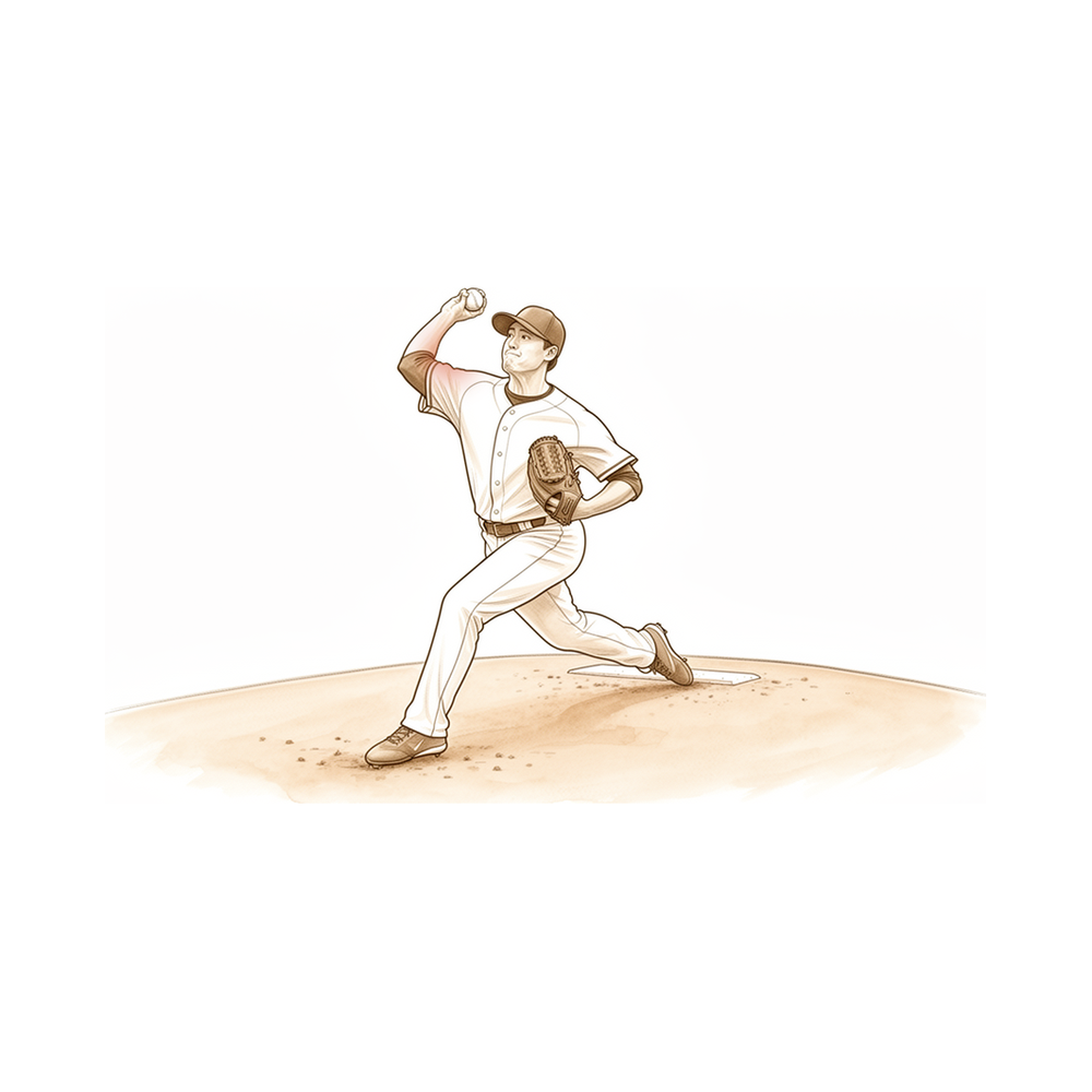
Most isolated medial collateral ligament injuries are treated nonsurgically [10], while concomitant damage to the anterior or posterior cruciate ligaments is a common indication for surgical management of high-grade medial collateral ligament injuries [10]. For ulnar collateral ligament injuries, nonoperative treatment can offer favorable outcomes for younger patients with acute or partial injuries [38]. However, the current evidence for ulnar collateral ligament injury treatment is not convincing [16], and expert consensus should be viewed with caution due to methodological biases [16]. Future high-quality randomized studies are required for ulnar collateral ligament injury treatment [16], and future prospective and comparative studies are necessary to better define the optimal operative treatment for elbow ulnar collateral ligament injuries in throwing athletes [163]. Injury prevention programs and improved nonoperative treatment protocols hold promise in decreasing the need for surgical repair for medial elbow injuries in throwing athletes [63].
Biomechanically, there are no treatments of repair or reconstruction using native tissues that provide equivalent strength to the preinjured ligament for thumb metacarpophalangeal joint ulnar collateral ligament injuries [5]. Chronic thumb ulnar collateral ligament injuries are traditionally managed with tendon graft ligament reconstruction or tendon transfers [27], though evidence for their management is limited to case reports and retrospective series [27]. Repair of a chronic thumb ulnar collateral ligament injury with available local tissue appears to be a reasonable alternative to ligament reconstruction [11], resulting in durable long-term outcomes despite the majority of patients progressing to osteoarthritis [11]. Early return to play without immobilization should not be encouraged until the ligament is adequately healed after surgery for thumb metacarpophalangeal joint ulnar collateral ligament injuries [12]. Failure of ligament healing for ulnar collateral ligament injuries of the metacarpophalangeal joint was probably a result of the ligament lying within the joint [4], although functional results were excellent after surgical repair for these injuries [4].
Anatomy & Pathophysiology¶
Knee Ligaments¶
The differential healing capacity of human ACL and MCL stem cells may be attributed to their distinct properties [6]. Specifically, tumor necrosis factor-alpha-induced expression of lysyl oxidases and matrix metalloproteinases is higher in injured MCL fibroblasts and ACL fibroblasts, respectively [6]. Anatomically, the sartorial branch of the saphenous nerve is in relation to medial knee ligament repair or reconstruction [6]. The medial collateral ligament maintains a specific topographical and biomechanical relationship with the medial meniscus [6].
ACL injuries are among the most frequent knee ligament injuries in sport and usually require reconstruction [20]. In English Premiership Rugby (2011–2018), ACL injuries have been in the top five most severe injuries for six of the last seven seasons [20]. In 2016–2017, ACL injuries in English Premiership Rugby accounted for 224 days of absence per 1000 hours [20]. These injuries lead to muscle weakness, altered movement, joint effusion, and reduced functional performance [20]. Furthermore, ACL injuries are associated with continuing clinical sequelae such as chondral lesions, meniscal tears, and increased risk of early-onset post-traumatic osteoarthritis [20].
Mechanisms of injury in rugby union are predominantly contact-based. Video analysis showed that 57% of ACL injuries occurred through contact [20]. Specifically, 43% occur when a player is being tackled, 29% when tackling, and 14% in general collisions [20]. Non-contact mechanisms account for the remainder, with 14% occurring through twisting and turning [20]. In rugby union, 43% of ACL injuries occur through non-contact mechanisms, mainly sidestepping maneuvers [20].
Risk factors for ACL injury are categorized as intrinsic and extrinsic. Intrinsic risk factors include age, anthropometry, sex, previous injury, anatomical variation, neuromuscular and cognitive factors, and genetics [20]. Extrinsic risk factors include environmental conditions, shoes/surfaces, training errors, and mechanical errors [20]. Trauma to the knee is a fundamental requirement for ACL injury [20].
The mechanism responsible for combined tibiofemoral injuries and knee ligament injury is not clear [22]. In cases of trauma at a site where both bone and ligament are present, failure of the weakest component, i.e., the ligament, is plausible [22]. A common mechanism for damage to cruciate ligaments and capsule in traffic accidents is when knees are trapped beneath the dashboard and the trunk is carried up and over the top, hyperextending the knee [22]. If dashboard hyperextension is coupled with rotation or varus/valgus stress, damage of the collateral ligaments can also result [22]. Posterior displacement of the upper portion of the tibia on the femur may cause rupture of the posterior cruciate ligament [22]. Ipsilateral femoral and tibial shaft fracture and knee ligament injury is part of a continuum of combined injuries resulting from complex high-energy forces [22].
Elbow Bony Anatomy¶
The elbow is a trocho-ginglymoid joint consisting of medial and lateral articulations which afford bony stability [72]. On the medial side, the trochlea articulates with the ulna within the greater sigmoid notch to create the ulnohumeral, hinged or trochoid, portion of the elbow joint [72]. There is highly congruent anatomy through almost 180° of articular contact on the medial side, with the exception of the bare area of the greater sigmoid notch of the ulna which is devoid of cartilage [72]. The coronoid has a medial and lateral facet which buttresses the trochlea anteriorly [72]. The sublime tubercle, located just distal and medial to the coronoid, provides the attachment site of the anterior bundle of the medial ulnar collateral ligament [72]. The medial epicondyle forms the attachment site for the origins of the flexor pronator mass and is larger and more posteriorly oriented than the lateral epicondyle [72].
Laterally, the capitellum and radial head form the radiocapitellar joint [72]. The radius is held in close approximation to the ulna at the proximal radioulnar joint by the annular ligament [72]. The area of the ulna which articulates with the margin of the radial head at the proximal radioulnar joint is also known as the lesser sigmoid notch [72]. The radial head is a concave elliptical structure, covered with articular cartilage along the radiocapitellar joint and approximately 270° of the articular margin [72]. The less prominent lateral epicondyle is the origin of the lateral extensor musculature [72]. Just distal to the lateral epicondyle at the geometric center of the radiocapitellar articulation is the origin of the lateral ulnar collateral ligamentous complex [72].
The distal humeral articulation is angled 30° from the longitudinal axis [72]. The anterior humeral line should pass through the center of the axis of rotation [72]. The axis of rotation is 5° to 7° angulated in the coronal plane to the epicondylar axis with the medial side more distal than the lateral side [72]. The angulation of the distal humerus accounts for the change in a valgus carrying angle to a more varus position as the elbow is flexed [72]. In the same individual, there is a high correlation between the sizes of the radius and capitellum on the left and right sides [72]. The olecranon allows for a broad attachment site of the triceps posteriorly [72]. The ulna medially bends approximately 8° at 8 cm from the tip of the olecranon [72]. The articulation to the tip of the coronoid is approximately 30° from the long axis of the ulna in the sagittal plane [72].
The normal elbow has a range of motion from 0° to 140° from extension to flexion and 75° and 85° in pronation and supination respectively [30]. A functional arc in each plane for the elbow is 100° for flexion and extension and forearm rotation [30]. The ulnohumeral joint allows the flexion extension of the joint, and the radiocapitellar joint allows forearm rotation [81]. The trochlea has a 300-degree arc of cartilage [81]. The medial column diverges from the humeral shaft at a 45-degree angle, and the lateral column diverges at a 20-degree angle [81].
The articular surface of the distal humerus is in 5° to 6° of valgus with respect to the humeral shaft [82]. The articular surface of the distal humerus is in 30° of flexion relative to the shaft [82]. The medial epicondyle is the origin of the flexor-pronator muscle group and the medial collateral ligament [82]. The lateral epicondyle is the origin of the extensor-supinator muscle group and the lateral collateral ligament complex [82]. The ulnar nerve lies in a bony groove covered and restrained by the arcuate ligament posterior to the medial epicondyle [82].
The distal humeral shaft is triangular shaped in cross section with its apex directed anteriorly [84]. The medial column diverges approximately 45 degrees from the humeral shaft in the coronal plane and terminates as the medial epicondyle [84]. The lateral column diverges at approximately 20 degrees from the shaft in the coronal plane [84]. As the lateral column extends distally, it curves anteriorly creating a 35 to 40 degrees angle with the shaft in the sagittal plane [84]. In the coronal plane, the trochlea is more distal than the capitellum resulting in a valgus alignment of 4 to 8 degrees [84]. Including the ulna, the elbow has a valgus angle in extension of 10 to 17 degrees, termed the carrying angle [84]. Axially, the distal humerus articular surface is internally rotated 3 to 8 degrees [84]. The trochlea is covered by articular cartilage anteriorly, inferiorly, and posteriorly, creating an arc of almost 270 degrees [84]. The trochlea is shaped like a spool with a central sulcus which articulates with the central ridge of the greater sigmoid notch of the proximal ulna [84]. The olecranon fossa and coronoid fossa are separated by a thin bony septum [84]. The tolerances of the olecranon and coronoid fossae to accommodate their respective bony processes are narrow [84]. The articular surface of the capitellum starts at the most distal aspect of the lateral column and encompasses an arc of approximately 180 degrees in the sagittal plane [84].
The normal range of elbow flexion/extension is 0 to 150 degrees [79]. Normal forearm pronosupination is 80 to 85 degrees in each direction [79]. Functional range of motion for the elbow is 30 to 130 degrees flexion/extension and 50 degrees pronosupination [79]. The normal valgus carrying angle of the elbow is 5 to 10 degrees for men and 10 to 15 degrees for women [79]. In full extension, 60% of axial load is transmitted through the radiocapitellar joint [79].
Elbow Ligaments¶
Elbow stability is determined by primary and secondary stabilizers [30]. The three primary stabilizers of the elbow are the ulnohumeral articulation, the medial ulnar collateral ligament (MUCL), and the lateral ulnar collateral ligament (LUCL) complex [30]. Secondary stabilizers of the elbow are the radiocapitellar articulation, the common flexor tendon, the common extensor tendon, and the joint capsule [30].
The medial or ulnar collateral ligament is the primary valgus stabilizer [74]. The anterior bundle of the medial collateral ligament is most important for stability [74]. The posterior bundle of the medial collateral ligament has the greatest change and length, becoming taut at flexion beyond 120 degrees [74]. The lateral ulnar collateral ligament is the posterolateral stabilizer [74]. Osborne’s ligament stabilizes the ulnar nerve in the cubital tunnel [74]. The ligament of Struthers is a variant anatomy arising from the supracondylar process to attach to the medial epicondyle and is a potential site of median nerve compression [74].
The medial ulnar collateral ligament complex consists of the anterior bundle, posterior bundle, and transverse ligament [71]. The anterior bundle is the strongest component of the medial ulnar collateral ligament complex and the primary restraint to valgus stress [71]. The anterior bundle of the medial ulnar collateral ligament is subdivided into anterior and posterior bands which provide reciprocal function, with the anterior band tight in extension and the posterior band tight in flexion [71]. The lateral ulnar collateral ligament origin center is 10.7 mm from the lateral epicondyle [71]. The lateral ulnar collateral ligament insertion is 3.3 mm from the apex of the supinator crest [71].
The medial ulnar collateral ligament originates on the posterior medial epicondyle and inserts on the sublime tubercle of the medial coronoid process [79]. The anterior bundle of the medial collateral ligament is the primary restraint to valgus stress within functional elbow range of motion [79]. The secondary restraint to valgus stress within functional elbow range of motion is the radial head [79]. The posterior bundle of the medial collateral ligament is the primary restraint to valgus stress with the elbow in maximal flexion [79]. Stability in full extension is provided by the medial collateral ligament, joint capsule, and ulnohumeral articulation [79].
The lateral collateral ligament complex consists of the radial collateral ligament, the lateral ulnar collateral ligament, and the annular ligament [84]. The annular ligament attaches to the anterior and posterior margins of the lesser sigmoid notch [84]. The radial collateral ligament originates from an isometric point on the lateral epicondyle and fans out to attach to the annular ligament [84]. The lateral ulnar collateral ligament arises from the isometric point on the lateral epicondyle and attaches to the crista supinatoris of the proximal ulna [84]. The lateral collateral ligament complex functions as an important restraint to varus and posterolateral rotatory instability [84].
The most important portion of the medial or ulnar collateral ligament is the anterior portion, which attaches to a small process on the medial surface of the coronoid [81]. The capsule allows maximum distension at approximately 70 to 80 degrees of flexion [74]. The anterior capsule attaches at a point approximately 6 mm distal to the tip of the coronoid [74]. Tensile forces are present at the medial elbow, while compressive forces are present at the lateral elbow [74].
Elbow Pathophysiology & Biomechanics¶
The kinematics of the elbow deviate increasingly from those of the native joint with a 2 mm to a 4 mm lengthening of the radius [73]. Musculoskeletal ultrasonography provides a dynamic, functional assessment of elbow structures, allowing visualization of pathology under stress and motion [85]. An understanding of the relevant anatomy and the factors associated with elbow stability allows the application of a systematic algorithm for treatment to ensure sufficient elbow stability for early motion [88].
Biomechanical and clinical outcomes show that the External Joint Stabilizer – Elbow (EJS-E) via the posterior approach can restore mobility and stability in all patients [92]. From a biomechanical perspective, the enhancement of elbow stability with a monopolar radial head prosthesis is superior to that with a bipolar design [94]. Dynamic analyses using a 3-dimensional elbow model showed that none of the configurations for double-strand lateral ulnar collateral ligament reconstruction were isometric [95].
Gripping does not change ulnohumeral joint space width or medial elbow tissue stiffness in the joint testing configuration and external loading conditions applied in the study [99]. Medial elbow joint space increases under a valgus load and then decreases when a maximal grip contraction is performed [102]. Forearm position was not associated with the elbow varus moment, but the supination moment was associated with the elbow varus moment in collegiate baseball pitchers [103]. Valgus torque at the elbow during baseball pitching is associated with 6 biomechanical variables of sequential body motion [104]. No kinematic or kinetic differences were noted between throwing balls and strikes in professional baseball pitchers [105].
Ulnar collateral ligament reconstruction using a suspension button fixation technique reliably restored elbow kinematics to the intact state [106]. Increased medial elbow torque was associated with greater ball velocity regardless of the history of medial elbow injuries in youth baseball pitchers [107]. An improvement in isometric contraction in flexion of the elbow was observed after tenotomy of the long head of biceps tendon, but this did not reach the flexion power of the contralateral healthy arm [108]. Elbow valgus torque is poorly suited as a standalone metric for predicting injury risk due to narrow data ranges, modeling noise, and crude assumptions [109]. The available current evidence possesses a high degree of fragility regarding the optimal elbow flexion angle for graft fixation in ulnar collateral ligament reconstruction [110].
The suture-augmented lateral ulnar collateral ligament and radial collateral ligament reconstruction method provides a reproducible, anatomically based construct that restores posterolateral elbow stability [111]. No significant relationships between adaptations in shoulder strength or range of motion were related to chronic structural adaptations of the elbow in professional baseball pitchers [113]. The anatomic model of terrible triad of the elbow was successfully created and validated by exerting axial compression on an elbow in 15° flexion and maximal pronation at speeds of 100 and 10 mm/min [116]. The posterior (Boyd) approach for terrible triad injuries allows better visualization of the lateral structures for repair and confers excellent stability to the elbow joint [120]. Following ulnar collateral ligament repairs and reconstructions, elbow range of motion is reliably preserved or improved with a predictable trajectory of rapid improvement within the first 2 to four months [121]. When controlling for an individual pitcher, peak kinetics at the shoulder and elbow can be strongly predicted by ball velocity [124]. Posterolateral rotatory instability (PLRI) of the elbow remains to be fully understood [125]. Both elbow varus torque and swing velocity were greatest when swinging to the outside location in baseball hitting [126]. Complex elbow instability remains a challenging clinical entity requiring a balance between stability
Classification¶
UCL MRI-Based Classifications: Multiple MRI-based grading systems exist for ulnar collateral ligament (UCL) injuries, designed to guide operative versus nonoperative management and predict valgus laxity in throwing athletes [26, 187]. A 6-stage classification addressing tear grade and location demonstrates substantial to near-perfect agreement among fellowship-trained observers [64]. Ford et al. classify UCL injuries into four levels of severity: Grade I (intact ligament with or without edema), Grade IIA (partial tear with incomplete fiber disruption), Grade IIB (chronic healed injury with thickened ligament but no fiber disruption), and Grade III (complete tear with fiber disruption) [44]. Joyner et al. propose a four-level system: Type I (edema in the UCL only), Type II (partial tear with no extravasation of fluid on MRA), Type III (complete full-thickness tear with extravasation of fluid on MRA), and Type IV (tear in more than one location) [44]. In adolescent baseball players, preoperative MRI classifies injuries into four grades: Grade 0 (intact ligament continuity), Grade 1 (low-grade partial tear of 50% or less of the ligament substance), Grade 2 (high-grade partial tear of more than 50% of the ligament substance), and Grade 3 (complete tear) [201]. A systematic review of UCL tears treated with platelet-rich plasma defines Type I (low-grade partial tears with edema), Type II (high-grade partial tears with no extravasation of fluid), Type III (full-thickness tears in one location), and Type IV (full-thickness tears in multiple locations) [211].
UCL Tear Criteria and Location: Criteria for a partial UCL tear on MRI include a thickened ligament with abnormal signal intensity, diffuse or focal thinning, partial detachment from the origin or insertion site, partial-thickness defect of the articular surface as determined by extension of fluid, and undersurface partial detachment characterized by the distal extension of fluid along the medial margin of the coronoid producing a “T sign” [201]. The site of UCL injury is further classified into origin, midsubstance, or insertion site injury [201].
Other Considerations: A systematic review proposed a contemporary injury classification system for pectoralis major tears that includes injury timing, injury location, and standardized terminology addressing tear extent [35]. A comprehensive classification of complex fracture-dislocations of the elbow was created that appeared to be reproducible and may represent a useful tool for the management of such difficult injuries [170]. An intraoperative arthroscopic classification tool for posterolateral elbow instability (PLRI) can be used as a standardized grading system for further research and communication between orthopedic surgeons [172]. A classification system for partial distal biceps tendon tears based on 3-Tesla magnetic resonance imaging tear morphology may have implications for operative and non-operative management [49]. A proposed MRI classification has emerged as one of the most reliable methods to define stages of chronic lateral epicondylitis [190].
Clinical Presentation¶
General Assessment and Diagnostic Challenges¶
Clinicians in the Emergency Department lack proficiency in performing the assessment methods used for diagnosing acute ACL injury [32]. High variability exists among surgeons regarding the evaluation, treatment, and rehabilitation of medial ulnar collateral ligament injuries [115]. The current evidence for ulnar collateral ligament injury treatment is not convincing, and expert consensus should be viewed with caution due to methodological biases [16]. Publicly available information on YouTube regarding ulnar collateral ligament injuries is of low quality, with differential diagnoses, accurate surgical indications, and thorough discussions of adverse outcomes being the most lacking [41].
Knee Ligaments (ACL/PCL)¶
Cruciate ligament injuries are common with a rising incidence [1]. ACL injuries are among the most frequent knee ligament injuries in sport and usually require reconstruction [20]. These injuries frequently lead to muscle weakness, altered movement, joint effusion, and reduced functional performance [20]. Continuing clinical sequelae associated with ACL injury include chondral lesions, meniscal tears, and an increased risk of early-onset post-traumatic osteoarthritis [20]. In rugby, the primary mechanisms of ACL injury are a player being tackled (43%), tackling (29%), or general collisions (14%) [20]. Fourteen percent of ACL injuries in rugby occurred through non-contact mechanisms such as twisting and turning [20]. Video analysis of ACL injury mechanisms in rugby showed that 57% occurred through contact and 43% through non-contact mechanisms, mainly sidestepping maneuvers [20]. Recent studies have shed new light on the natural history of injury to the posterior cruciate ligament, as well as its complex anatomy and functional mechanical behavior [14]. New information on PCL injury is likely to alter the way orthopaedic surgeons have traditionally treated injuries to this structure [14].
Elbow Ligaments (UCL/MCL)¶
Patients with acute UCL injuries should be assessed with history, clinical examination, and radiographs [9]. Ultrasound is a valuable adjunct to clinical examination in a specialist clinic for differentiating displaced from undisplaced thumb UCL tears, provided it is performed soon after presentation [39]. Valgus stress ultrasound can be used as a diagnostic tool for complete ulnar collateral ligament ruptures but may not be sufficient alone for partial injuries [40]. MRI has typically been the standard in identifying UCL injuries, particularly full-thickness tears [44]. Magnetic resonance arthrogram (MRA) may provide additional imaging details for evaluating the severity of UCL injuries, particularly partial tears [44]. Proper coil selection, pulse sequence parameters, and patient positioning enhance the ability of MR imaging to demonstrate subtle injuries to elbow ligaments and regional osseous and soft tissue structures [51].
Two primary grading systems for UCL injuries are utilized: * Ford et al. MRI grading scale: Grade I (intact ligament with or without edema), Grade IIA (partial tear with incomplete fiber disruption), Grade IIB (chronic healed injury with thickened ligament but no fiber disruption), and Grade III (complete tear with fiber disruption) [44]. * Joyner et al. 4-level system: Type I (edema in the UCL only), Type II (partial tear with no extravasation of fluid on MRA), Type III (complete full-thickness tear with extravasation of fluid on MRA), and Type IV (tear in more than 1 location) [44].
A correlation of pain must be present to make the diagnosis of UCL injury based on newer grading schemes [44]. In a study of 49 patients, ultrasound identified Stener lesions in 11 patients but only correctly in 4, resulting in a sensitivity of 36% [122]. In the same study, ultrasound demonstrated a low specificity (61%) and sensitivity (65%) for diagnosing displaced UCL ruptures [122]. MRI and ultrasound of suspected UCL injury did not demonstrate good enough sensitivity to recommend their implementation and use in clinical situations for obviating the need for surgical exploration [122]. The most common indication for acute treatment of an isolated medial ulnar collateral ligament injury was the presence of a complete distal tear, agreed upon by 85% of respondents [115]. No other tear pattern for medial ulnar collateral ligament injury resulted in agreement greater than 50% among surgeons regarding the need for acute surgical treatment [115]. Diagnosis of posterolateral rotatory instability is confirmed by the posterolateral rotatory instability test [117]. Future research may elucidate the diagnostic value of a pop sign for ulnar collateral ligament injury [48].
Hand and Finger Ligaments¶
Thumb ulnar collateral ligament (UCL) tears are stratified into three grades: * Grade I: Minor tearing without instability [112]. * Grade II: Partial tear with increased joint laxity but a definitive end point [112]. * Grade III: Complete tear and gross instability [112].
Grade I and II thumb UCL injuries are best treated nonoperatively with immobilization [112]. Grade III thumb UCL injuries, particularly those with a Stener lesion, are treated with surgical repair or reconstruction depending on the chronicity of injury [112]. A displaced fleck sign on radiographs is consistent with Grade III thumb UCL tears with Stener lesions [112]. Most authors agree that both acute and chronic grade 3 radial collateral ligament tears of the thumb should be surgically treated [33].
Finger metacarpophalangeal joint collateral ligament injuries occur less frequently than their counterparts in the thumb [118]. There are little data to guide diagnosis and/or treatment of finger metacarpophalangeal joint collateral ligament injuries [118]. Normal parameters for collateral ligament laxity of the fingers have not been firmly established [118]. It is not known whether the contralateral, uninjured digit can be used as a comparison in the assessment of finger collateral ligament injuries [118]. The cam shape of the metacarpal head results in a tightening of the collateral ligaments with joint flexion [118]. A diagnostic technique based on specific questions and three clinical tests is useful for diagnosing triquetrolunate ligament injuries before arthroscopy [34]. Diagnosing the cause of ulnar collateral ligament locking in the middle finger may be complicated by the lack of evidence in imaging studies [42].
Combined and Complex Injuries¶
The mechanism responsible for a combined fracture of the femoral and tibial shaft and knee ligament injury is not clear [22]. The most common mechanism of damage to the cruciate ligaments and capsule in traffic accidents is when the knees are trapped beneath the dashboard, hyperextending the knee [22]. If hyperextension is coupled with rotation or varus/valgus stress, damage of the collateral ligaments can also result [22]. Posterior displacement of the tibia on the femur may cause rupture of the posterior cruciate ligament [22]. There is no clear correlation between the site or type of the fracture and which of the ligaments of the knee are injured [22]. Posterolateral rotatory instability (PLER) is the main mechanism of injury in terrible triad injuries and should be managed individually based on injury mechanisms presenting different instability patterns [21]. Physicians must maintain heightened vigilance for associated physeal injuries in pediatric transolecranon fracture dislocations that may not be obvious on initial radiographs [52]. Elbow fracture–dislocation with triceps avulsion is a rare injury combination that should be recognized [46].
Investigations¶
Clinical Examination and History: Assessment of acute ulnar collateral ligament (UCL) injuries requires a combination of history, clinical examination, and radiographs [9]. The physical examination of the elbow is directed by the history and the specific location of the patient's pain in the anterior, posterior, medial, or lateral aspect [30]. Elbow stability is determined by primary and secondary stabilizers; injury to these structures causes elbow instability [30]. Secondary stabilizers include the radiocapitellar articulation, the common flexor tendon, the common extensor tendon, and the joint capsule [30]. In professional baseball pitchers, distal UCL tears described on MRI show significantly higher rates of failure with nonoperative management compared to proximal tears [194]. There was no relationship between UCL tear type and age, competition level, and plain radiographic abnormalities in adolescent baseball pitchers [208]. A traffic light model based on initial radiographic displacement can predict the need for collateral ligament repair in transolecranon fractures: >10 mm requires repair, <5 mm does not, and 5-10 mm requires careful screening for instability [209].
Plain radiography: Plain radiographs remain the hallmark and the best screening test for elbow evaluation [30]. Standard AP and lateral radiographs typically show osteophyte formation at the coronoid process, coronoid fossa, radial fossa, radial head, olecranon tip, and olecranon fossa in elbow osteoarthritis [96]. Radiographs typically underestimate the number of loose bodies present in elbow osteoarthritis [96]. The coronoid opening angle is a novel radiographic technique that can be of value alongside 3-dimensional imaging in evaluating elbow injuries and used as an adjunct in clinical decision making [204]. Physicians must maintain heightened vigilance for associated physeal injuries that may not be obvious on initial radiographs in pediatric transolecranon fracture dislocations [52].
CT: The nature of an anteromedial facet coronoid process injury can be most reliably documented using computed tomography (CT) with three-dimensional reconstructions [28]. With correct identification, classification, and understanding using CT scans followed by appropriate surgical treatment that addresses all components of the injury, good to excellent mid-term results can be achieved for Monteggia-like lesions [45]. CT is helpful when assessing for malunion architecture and the location and pattern of osteophytes and/or loose bodies in the context of elbow stiffness [89]. Three-dimensional CT is used to check for heterotopic ossification in the evaluation of elbow stiffness [89]. CT with two-dimensional reconstruction and three-dimensional surface rendering best visualizes the pathology of valgus extension overload syndrome [91]. The value of MRI and CT in identifying ligament injuries in elbow fractures is noted, along with the challenge of determining surgical necessity [206].
MRI: A reliable 6-stage MRI-based classification addressing UCL tear grade and location may confer decision making between operative and nonoperative management [26]. A newly proposed 6-stage MRI-based classification utilizing grade and location of the injury had substantial to near perfect agreement among and within fellowship-trained observers [64]. MRI grading of UCL injuries can help predict return to play and the need for surgery in professional baseball players [68]. Proper coil selection, pulse sequence parameters, and patient positioning enhance the ability of MR imaging to demonstrate subtle injuries to the ligaments and the regional osseous and soft tissue structures [51]. Conventional MRI technique demonstrates difficulties in the evaluation of ligaments after simple elbow dislocation, as shown by weak inter- and intraobserver agreement [55]. The FEVER (Flexed Elbow Valgus External Rotation) MRI may be both sensitive and specific for detecting UCL injuries and guiding surgical decision making while obviating the need for joint contrast fluid [203]. MRI may be most helpful in evaluating associated injuries including partial or complete tears of the medial collateral ligament (MCL) in valgus extension overload syndrome [91]. MRI can be used to evaluate ligaments and tendons in the context of elbow stiffness, but it is rarely indicated [89]. Ultrasound and MRI are helpful in evaluating acute traumatic brachialis rupture and monitoring its resolution [123]. Preoperative MRI and intraoperative assessments agreed in 80% of cases for myotendinous junction tears of the pectoralis major, a value that was significantly higher for complete over partial tears [179]. The clinical use of MRI in the management of patients with enthesopathy of the extensor carpi radialis longus origin merits further study [183]. Although there is variation in the use of MRI for lateral epicondylitis and its use is associated with downstream effects, the routine use of MRI for the diagnosis of lateral epicondylitis is low [198]. Preoperative MRI could be used to exclude subtle instability in patients with lateral epicondylitis, especially when patients have a history of multiple corticosteroid injections (≥3) or severe pain [199]. Radiographic evaluations are essential when diagnosing an osteochondritis dissecans (OCD) lesion of the elbow; however, important aspects of the OCD lesions may be better seen with MRI [93].
Ultrasound: Ultrasound is a valuable adjunct to clinical examination in a specialist clinic, particularly for differentiating displaced from undisplaced tears of the thumb UCL, provided it is performed soon after presentation [39]. Valgus stress ultrasound can be used as a diagnostic tool for complete ruptures of the medial UCL but may not be sufficient alone for partial injuries [40]. Thickened UCL may exhibit a decrease in function; therefore, physicians should not evaluate the joint status solely on the basis of the structural properties on conventional ultrasound [213].
Other Considerations: The current evidence for ulnar collateral ligament injury treatment is not convincing, and while expert consensus provides guidance, it should be viewed with caution due to methodological biases [16].
Treatment¶
General Principles and Diagnostic Considerations¶
MRI grading of UCL injuries assists in predicting return to play and the necessity for surgical intervention [68]. In professional pitchers, distal UCL tears demonstrate significantly higher odds of failure with nonoperative management compared with proximal tears [164]. Several prognostic factors associated with the failure of nonoperative treatment have been identified among patients with UCL injuries [157]. Furthermore, the classification of partial distal biceps tendon tears may have implications for both operative and non-operative management decisions [49].
Non-Operative¶
Nonoperative treatment allows 42% of athletes to return to their previous level of competition at an average of 24.5 weeks after diagnosis [66]. Incomplete UCL injuries are more likely to lead to nonoperative treatment and a higher return to the same or higher level of play than complete tears [173]. Non-operative management of isolated LCL injuries is associated with a 100% return to pre-injury level of sport, reasonable recovery times, and no significant residual varus instability [133]. UCL injuries in high school baseball players can be successfully treated nonoperatively in most cases [152]. Non-operative treatment of UCL injuries will likely be more successful in younger players, lower grade tears, and humeral-sided injuries [156]. Nonoperative treatment of partial ligament injuries with cast or splint immobilization will result in a stable and painless thumb [177]. Conservative intervention has demonstrated effectiveness in eliminating recurrent annular ligament displacement events in pediatric patients [150]. Injured UCL ligaments were naturally stable after reduction and did not need surgical repair [29]. Non-operative management of PLRI is ineffective; while bracing, dynamic stabiliser strengthening, and activity modification can be attempted, surgical treatment is generally required to stabilise the joint [184]. As with many chronic tendinopathies, nonsurgical therapy is the mainstay of treatment for medial epicondylitis [188]. Injury prevention programs and improved nonoperative treatment protocols hold promise in decreasing the need for surgical repair [63]. There is no consensus about the treatment of partial ACL ruptures; anterior-medial bundle ruptures may do well with non-operative treatment, while posterior-lateral bundle ruptures often need surgery due to pivot shift development [161]. Wrestlers showed high return to sport rates following both nonsurgical and surgical UCL treatment; however, primary non-operative management failed in 44% of patients, particularly those with complete tears, necessitating subsequent surgery [171]. Isolated UCL injuries are relatively uncommon in nonquarterback NFL players, with the vast majority being grade I sprains treated nonoperatively [65]. First-line management in essentially all patients with incomplete UCL tears consists of non-operative management with early physical therapy with or without biologic augmentation [60]. Conservative measures may also be considered in younger patients with full thickness tears in order to avoid operative intervention during years of skeletal immaturity and should also be considered in high school/college athletes who may plan to refrain from further competitive play in the future [60].
Operative¶
Indications: Surgery is indicated only for Grade 3 injuries where there is no definitive end point when tested for collateral instability due to extensive injury of the surrounding soft tissues [24]. Surgical repair is indicated in active persons with complete tears and for incomplete tears with concomitant loss of strength, with good to excellent results reported for distal triceps rupture [62]. Professional athletes and those with complete tears were indicated for surgery by consensus, whereas opinion was more divided on how to treat partial tears or nonprofessionals [158]. Treatment of UCL injuries depends on the type of tear, with nonoperative management for low-grade partial tears and UCL reconstruction as the gold standard for complete tears [149]. The current gold standard for management of full thickness, attritional, midsubstance UCL tears in high-demand athletes, or elite throwers remains operative reconstruction [60]. UCL reconstruction remains the benchmark for tears not amendable to nonsurgical treatment [153]. Failing nonsurgical management, ulnar collateral ligament reconstruction is a viable option to return the throwing athlete to competition [168]. While UCL reconstruction remains a mainstay for high-demand athletes with full-thickness tears, nonreconstructive options such as rehabilitation, biologic strategies, and repair may be beneficial for specific clinical scenarios including lower-demand athletes, adolescents, and aging athletes [175]. Treatment should be unique to the patient and sport specific in nature, with nonoperative management requiring a multimodal approach for well-selected patients [159].
Surgical Approach / Technique: ACL repair has demonstrated significant clinical improvement in an appropriately selected patient cohort [8]. Surgical repair of fourteen fingers with complete rupture yielded satisfactory results with restored joint stability and pain relief [25]. Completely ruptured collateral ligaments frequently result in prolonged disability when treated non-operatively [25]. The functional result was excellent after surgical repair of a ruptured Ulnar Collateral Ligament of the Metacarpophalangeal Joint of the Index Finger [4]. Failure of ligament healing was probably a result of the ligament lying within the joint [4]. Correct understanding and reasonable treatment plans, including bony restoration and ligament repair, can achieve good function for trans-olecranon fracture posterior dislocation [2]. Repair of a chronic UCL injury with available local tissue appears to be a reasonable alternative to ligament reconstruction, resulting in durable long-term outcomes despite the majority of patients progressing to osteoarthritis [11]. No significant difference in clinical outcome or range of motion was observed after direct repair of traumatic tears of the lateral ulnar collateral ligament between acute and delayed treatment cohorts [23]. UCL repair is an effective procedure in helping female athletes return to sport with low complication rates [58]. Repair was most appropriate for partial and full-thickness distal tears but relatively contraindicated for complete midsubstance UCL tears [160]. UCL repair may be a good alternative in contact athletes [176]. Simultaneous surgical treatment including open débridement and ligament reconstruction provides satisfactory pain relief and functional improvement in patients with lateral epicondylitis and LCL insufficiency [139]. The Ligamentotaxor is a safe and effective device for managing intra-articular PIPJ injuries, offering practical advantages and comparable efficacy to other devices [119]. Encouraging results for treating chronic longitudinal radioulnar dissociation injuries have been achieved by bone-ligament-bone reconstruction using a patellar tendon graft [15]. Treatment of late instability is focused on lateral ligament reconstruction from the humerus to the ulna using tendon grafts with reasonably good outcomes [154]. Ulnar collateral ligament reconstruction is currently the most commonly performed surgical treatment option for UCL insufficiency, with a commonly quoted success rate of 85% defined as the ability to return to a preinjury level of play for at least one year [151]. At 2 years after ACL reconstruction with tibialis anterior allografts, this subject group displayed satisfactory functional outcomes [54].
Other Considerations: There is no scientifically proven superior method for ACL or PCL reconstruction due to a lack of well-designed randomized clinical trials, and the field continues to rely on unproven assumptions and varying opinions [61]. Biomechanically, there are no treatments of repair or reconstruction using native tissues that provide equivalent strength to the preinjured ligament for thumb MCP joint UCL injuries [5]. Early return to play without immobilization should not be encouraged until the ligament is adequately healed after surgery for thumb MCP joint UCL injuries [12]. Nevertheless, long-term clinical outcome studies involving a larger number of patients will be required to corroborate this approach to augmentation for partial ACL ruptures [18].
Adjunctive Therapies and Rehabilitation¶
Conflicting data on the cellular and molecular effects of PRP for ligament injuries were observed secondary to the finding that included studies were heterogeneous, limiting interpretation across studies and the ability to draw meaningful conclusions [50]. Cell-based therapies and regenerative medicine offer safe and potentially efficacious treatment for sports-related musculoskeletal injuries, but more clinical evidence is necessary to define the indications and parameters for their use [155]. There was unanimous agreement regarding the lack of evidence for orthobiologics and specific areas for nonoperative management, as well as indications for operative management and return to sport criteria [143]. The current evidence for ulnar collateral ligament injury treatment is not convincing, and while expert consensus provides guidance, it should be viewed with caution due to methodological biases; future high-quality randomized studies are required [16]. In addition to the ACL, there is emerging, but limited evidence, for suture augmentation for injuries to the posterior cruciate ligament [47]. Most specific interventions for postoperative management of MUCL reconstruction have not been thoroughly examined, and randomized trials are lacking [191]. Bernas et al examined strain on reconstructed MUCLs in 8 cadavers and found that full extension does not strain the MUCL beyond known detrimental thresholds whereas flexion beyond 50° may place deleterious strain on the reconstruction [191]. Isometric flexion and extension exercises did not increase MUCL strain but became a concern at 90° of flexion [191]. Differences exist in protocol composition and in the timing of the introduction of specific interventions between protocols used in orthopedic residency programs and published protocols in the peer-reviewed literature [191]. Most agreement occurred regarding early protection and symptomatic interventions (bracing or splinting, modalities, and so on) whereas the variations were more apparent for therapeutic exercise interventions proximal and distal to the elbow [191].
Complications¶
Degenerative Joint Disease: Chronic thumb ulnar collateral ligament injuries are associated with progression to osteoarthritis in the majority of patients [11].
Functional Disability: Completely ruptured collateral ligaments of the proximal interphalangeal joint frequently result in prolonged disability when treated non-operatively [25].
Surgical Outcomes and Revision Rates: The long-term outcome with surgical management of complex elbow injuries is unknown [67]. In young patients undergoing primary medial ulnar collateral ligament repair with no additional ligamentous, fracture, and dislocation-related diagnoses, the incidence of early postoperative complications was 7.3% [169]. The 2-year revision rate for medial ulnar collateral ligament surgery in this specific cohort was 1.0% [169]. Ulnar collateral ligament reconstruction of the elbow demonstrates low complication and revision rates at medium-term follow-up [56]. Female athletes have low complication rates following ulnar collateral ligament repair or reconstruction [58]. UCL repair is associated with low complication rates and high rates of return to sport [186]. Midterm results for box-loop ligament reconstruction of the elbow for medial and lateral instability were generally excellent with few complications [185].
Other Considerations: Short-term follow-up of 20 subpectoral biceps tenodesis procedures has not shown any failure of fixation or residual biceps discomfort [69]. Biomechanically, there are no treatments of repair or reconstruction using native tissues that provide equivalent strength to the preinjured ligament in thumb metacarpophalangeal joint ulnar collateral ligament injuries [5].
Recovery¶
Knee Ligaments¶
Other Considerations: At 2 years after ACL reconstruction with tibialis anterior allografts, the subject group displayed satisfactory functional outcomes [54]. Long-term clinical outcome studies involving a larger number of patients are required to corroborate the approach to augmentation for partial ACL ruptures [18].
Elbow Ligaments¶
Other Considerations: Ulnar collateral ligament reconstruction provides excellent patient-reported and clinical outcomes at medium-term follow-up with low complication and revision rates [56]. Surgical reconstruction of the medial ulnar collateral ligament returns athletes to their previous level of play more than 80% of the time [225]. In nonthrowing athletes, patients undergoing UCL repair and UCL reconstruction show favorable outcomes at minimum 2-year follow-up [192]. Augmented UCL repair demonstrated excellent return to previous level of play and Kerlan Jobe Orthopaedic Clinic scores with modest complications and time to return [228]. Short-term results for repair and internal brace augmentation of the medial ulnar collateral ligament are on par with traditional UCL reconstruction techniques but with shorter return to play times [195]. Elbow ligament reconstruction by the technique of O'Driscoll et al effectively restores stability and limits progression to osteoarthritis in the long term [193]. Long-term outcome with surgical management of complex elbow injuries is unknown [67]. The postoperative course for elbow UCL reconstruction in javelin throwers is associated with an extended period of time until return to previous level of competition when compared with baseball players [226]. Correlations of risk for revision surgery after primary ulnar collateral ligament reconstruction may be younger age and less MLB experience at the time of the primary reconstruction [227].
Hand and Finger Ligaments¶
Other Considerations: Repair of a chronic thumb UCL injury with available local tissue appears to be a reasonable alternative to ligament reconstruction, resulting in durable long-term outcomes despite the majority of patients progressing to osteoarthritis [11]. Injured UCL ligaments in the thumb metacarpophalangeal joint were naturally stable after reduction and did not need surgical repair [29]. Surgical repair of fourteen fingers with complete rupture of the proximal interphalangeal joint collateral ligament yielded satisfactory results with restored joint stability and pain relief [25].
Shoulder and Biceps¶
Other Considerations: Distal biceps short head tears have good outcomes with acute and delayed reconstruction [13]. Clinical and functional outcome at more than 1 year after distal biceps tendon repair was excellent in both bioabsorbable and nonabsorbable screw groups [181]. Most morbidity from repair of the distal biceps tendon can be attributed primarily to a delay in the timing of the repair and secondarily to an extensive anterior exposure [224]. Early diagnosis and surgical repair allow for a successful return to competitive sports for avulsion injury of the serratus anterior muscle [43].
Key Evidence¶
- [L4] Correct understanding and reasonable treatment plans, including bony restoration and ligament repair, can achieve good function. [2] (10.1186/s13018-023-03563-5)
- [L4] Thus, surgical strategies including collateral ligament repair stabilize the joint and result in favorable clinical outcomes. [3] (10.1016/j.jse.2014.07.010)
- [L4] Failure of ligament healing was probably a result of the ligament lying within the joint, but the functional result was excellent after surgical repair. [4] (10.1054/jhsb.1999.0334)
- [L5] Biomechanically, there are no treatments of repair or reconstruction using native tissues that provide equivalent strength to the preinjured ligament. [5] (10.5435/jaaos-d-22-00112)
- [L4] ACL repair has demonstrated significant clinical improvement in an appropriately selected patient cohort. [8] (10.1177/2325967126s00016)
- [L1] Patients with acute UCL injuries should be assessed with history, clinical examination, and radiographs. [9] (10.1177/17531934241274612)
- [L5] Most isolated medial collateral ligament injuries are treated nonsurgically, while concomitant damage to the anterior or posterior cruciate ligaments is a common indication for surgical management of high-grade injuries. [10] (10.5435/00124635-200903000-00004)
- [L4] Repair of a chronic UCL injury with available local tissue appears to be a reasonable alternative to ligament reconstruction, resulting in durable long-term outcomes despite the majority of patients progressing to osteoarthritis. [11] (10.1177/1558944716628482)
- [L5] Early return to play without immobilization should not be encouraged until the ligament is adequately healed after surgery. [12] (10.1177/1558944717725380)
- [L4] They present acutely, have a poor natural history akin to complete tears, and have good outcomes with acute and delayed reconstruction. [13] (10.1016/j.jse.2020.04.038)
- [L4] Recent studies have shed new light on the natural history of injury to the posterior cruciate ligament, as well as on its complex anatomy and functional mechanical behavior, and this new information is likely to alter the way that orthopaedic surgeons have traditionally treated injuries to this structure. [14] (10.2106/00004623-199309000-00014)
- [L5] Encouraging results for treating chronic injuries have been achieved by bone-ligament-bone reconstruction using a patellar tendon graft. [15] (10.1016/j.hcl.2007.01.005)
- [L5] The current evidence for ulnar collateral ligament injury treatment is not convincing, and while expert consensus provides guidance, it should be viewed with caution due to methodological biases; future high-quality randomized studies are required. [16] (10.1016/j.arthro.2023.02.003)
- [L4] Nevertheless, long-term clinical outcome studies involving a larger number of patients will be required to corroborate this approach to augmentation for partial ACL ruptures. [18] (10.1007/s00167-010-1068-6)
- [L4] [20] (10.3390/sports7060138)
- [L4] PLER is the main mechanism of injury and should be managed individually based on injury mechanisms presenting different instability patterns. [21] (10.1016/j.jse.2020.12.015)
- [L4] [22] (10.1007/bf00393877)
- [L3] No significant difference in clinical outcome or range of motion was observed after direct repair of traumatic tears of the lateral ulnar collateral ligament between acute and delayed treatment cohorts. [23] (10.1016/j.jhsa.2014.02.011)
- [Case_report] Surgery is indicated only for Grade 3 injuries where there is no definitive end point when tested for collateral instability due to extensive injury of the surrounding soft tissues. [24] (10.1177/17531934211054769)
- [L4] Completely ruptured collateral ligaments frequently result in prolonged disability when treated non-operatively, whereas surgical repair of fourteen fingers with complete rupture yielded satisfactory results with restored joint stability and pain relief. [25] (10.2106/00004623-196749020-00009)
- [L4] A reliable 6-stage MRI-based classification addressing UCL tear grade and location may confer decision making between operative and nonoperative management. [26] (10.1016/j.jse.2018.11.063)
- [L4] Chronic UCL injuries are traditionally managed with tendon graft ligament reconstruction or tendon transfers, though evidence is limited to case reports and retrospective series. [27] (10.1016/j.jhsa.2011.06.004)
- [L4] The nature of the injury can be most reliably documented using computed tomography with three-dimensional reconstructions. [28] (10.1111/j.1758-5740.2009.00044.x)
- [L4] Injured UCL ligaments were naturally stable after reduction and did not need surgical repair. [29] (10.1177/1753193418790502)
- [L1] Clinicians in the Emergency Department are not proficient in performing the assessment methods that are used for diagnosis in acute ACL injury. [32] (10.1186/s12891-022-05595-0)
- [L5] Most authors agree that both acute and chronic grade 3 RCL tears should be surgically treated. [33] (10.1016/j.jhsa.2008.01.037)
- [L3] The authors describe a diagnostic technique based on specific questions and three clinical tests that is useful for diagnosing triquetrolunate ligament injuries before arthroscopy. [34] (10.1054/jhsb.1999.0269)
- [L4] A contemporary injury classification system is proposed that includes injury timing, injury location, and standardized terminology addressing tear extent to more accurately reflect the musculotendinous morphology of PM injuries and better inform surgical management, rehabilitation, and research. [35] (10.1016/j.jse.2011.04.035)
- [L5] Nonoperative treatment can offer favorable outcomes for younger patients with acute or partial ulnar collateral ligament injuries. [38] (10.1016/j.otsm.2020.150733)
- [L2] Ultrasound is a valuable adjunct to clinical examination in a specialist clinic, particularly for differentiating displaced from undisplaced tears, provided it is performed soon after presentation. [39] (10.1054/jhsb.1999.0283)
- [L3] It can be used as a diagnostic tool for complete ruptures but may not be sufficient alone for partial injuries. [40] (10.1016/j.jse.2019.12.005)
- [L4] The quality of ulnar collateral ligament-related information on YouTube is low, with differential diagnoses, accurate surgical indications, and thorough discussions of adverse outcomes being the most lacking information. [41] (10.1016/j.asmr.2023.100769)
- [L4] Although diagnosing the cause of UCL locking may be complicated by the lack of evidence in imaging studies, open surgical treatment has traditionally been the most often used with a high success rate. [42] (10.1016/j.jhsg.2022.08.003)
- [Case_report] Early diagnosis and surgical repair allow for a successful return to competitive sports. [43] (10.1016/j.jse.2006.12.014)
- [L4] [44] (10.1177/2325967117731296)
- [L3] With correct identification, classification, and understanding using CT scans followed by appropriate surgical treatment that addresses all components of the injury, good to excellent mid-term results can be achieved. [45] (10.1302/0301-620x.100b2.bjj-2017-0398.r2)
- [L4] This rare injury combination should be recognized, and early motion protocols may be feasible. [46] (10.1016/j.jhsa.2010.12.027)
- [L5] [47] (10.1016/j.arthro.2023.01.012)
- [L4] Future research may elucidate the diagnostic value of a pop sign for UCL injury. [48] (10.1016/j.jse.2019.01.017)
- [L3] Classification of tears may have implications for operative and non-operative management. [49] (10.5397/cise.2023.00458)
- [L1] Conflicting data on the cellular and molecular effects of PRP for ligament injuries were observed secondary to the finding that included studies were heterogeneous, limiting interpretation across studies and the ability to draw meaningful conclusions. [50] (10.1177/23259671211066504)
- [L5] Proper coil selection, pulse sequence parameters, and patient positioning enhance the ability of MR imaging to demonstrate subtle injuries to the ligaments and the regional osseous and soft tissue structures, including those not easily visualized at surgery. [51] (10.1016/j.mric.2004.02.006)
- [L4] Physicians must maintain heightened vigilance for associated physeal injuries that may not be obvious on initial radiographs. [52] (10.1016/j.jhsa.2012.02.037)
- [L4] At 2 years after ACL reconstruction with tibialis anterior allografts, this subject group displayed satisfactory functional outcomes. [54] (10.1007/s00167-003-0371-x)
- [L4] This study shows difficulties in the evaluation of ligaments by conventional MRI technique as demonstrated by a weak inter- and intraobserver agreement. [55] (10.1186/s12891-017-1451-2)
- [L4] UCLR provides excellent patient-reported and clinical outcomes to patients at medium-term follow-up with low complication and revision rates. [56] (10.1136/jisakos-2021-000614)
- [L4] The authors recommend early operative repair with a standard protocol for these injuries. [57] (10.2106/jbjs.d.02933)
- [L4] UCL repair is an effective procedure in helping female athletes return to sport with low complication rates. [58] (10.1016/j.xrrt.2025.02.007)
- [L4] We recommend early operative repair with a standard protocol for these injuries. [59] (10.2106/00004623-200406000-00002)
- [L4] [60] (10.1007/s12178-020-09637-9)
- [L5] There is no scientifically proven superior method for ACL or PCL reconstruction due to a lack of well-designed randomized clinical trials, and the field continues to rely on unproven assumptions and varying opinions. [61] (10.1007/s00167-001-0251-1)
- [L5] Surgical repair is indicated in active persons with complete tears and for incomplete tears with concomitant loss of strength, with good to excellent results reported. [62] (10.5435/00124635-201001000-00005)
- [L5] Injury prevention programs and improved nonoperative treatment protocols hold promise in decreasing the need for surgical repair. [63] (10.1016/j.jse.2023.09.012)
- [L2] The newly proposed 6-stage MRI-based classification utilizing grade and location of the injury had substantial to near perfect agreement among and within fellowship-trained observers. [64] (10.1177/0363546518786970)
- [L4] Isolated UCL injuries are relatively uncommon in nonquarterback NFL players, with the vast majority being grade I sprains treated nonoperatively. [65] (10.1016/j.jseint.2025.04.010)
- [L4] Nonoperative treatment allowed 42% of athletes to return to their previous level of competition at an average of 24.5 weeks after diagnosis. [66] (10.1177/03635465010290010601)
- [L5] Long-term outcome with surgical management of complex elbow injuries is unknown. [67] (10.5435/00124635-200605000-00003)
- [L4] MRI grading of UCL injuries can help predict return to play and the need for surgery. [68] (10.1177/0363546515621756)
- [L5] Short-term follow-up of 20 procedures has not shown any failure of fixation or residual biceps discomfort. [69] (10.1007/s00167-014-3348-z)
- [L5] The kinematics of the elbow deviated increasingly from those of the native joint with a 2 mm to a 4 mm lengthening of the radius. [73] (10.1302/0301-620x.106b10.bjj-2024-0405.r1)
- [L5] Musculoskeletal ultrasonography provides a dynamic, functional assessment of elbow structures, allowing visualization of pathology under stress and motion. [85] (10.5435/jaaos-d-20-00935)
- [L5] Despite the complexities of this injury, an understanding of the relevant anatomy and the factors associated with elbow stability allows the application of a systematic algorithm for treatment that can help ensure sufficient elbow stability to allow early motion, thereby leading to improved outcomes in most patients. [88] (10.5435/00124635-200903000-00003)
- [L4] Biomechanical and clinical outcomes show that EJS-E via the posterior approach can restore mobility and stability in all patients, thus serving as a valuable alternative option for the treatment of persistent instability of the elbow. [92] (10.1186/s12891-022-06103-0)
- [L5] From a biomechanical perspective, the enhancement of elbow stability with a monopolar radial head prosthesis is superior to that with a bipolar design. [94] (10.1016/j.jse.2010.10.033)
- [L5] Dynamic analyses using a 3-dimensional elbow model showed that none of the configurations for double-strand LUCL reconstruction were isometric. [95] (10.1016/j.jse.2018.11.070)
- [L4] Gripping does not change ulnohumeral joint space width or medial elbow tissue stiffness in the joint testing configuration and external loading conditions applied in this study. [99] (10.1186/s12891-025-08343-2)
- [L4] Medial elbow joint space increases under a valgus load and then decreases when a maximal grip contraction is performed. [102] (10.1177/0363546518755149)
- [L4] The results demonstrated that forearm position was not associated with the elbow varus moment, but the supination moment was associated with the elbow varus moment. [103] (10.1177/0363546517733471)
- [L4] Valgus torque at the elbow during baseball pitching is associated with 6 biomechanical variables of sequential body motion. [104] (10.1177/0363546509336721)
- [L3] No kinematic or kinetic differences were noted between throwing balls and strikes. [105] (10.1177/0363546517730052)
- [L5] Ulnar collateral ligament reconstruction using a suspension button fixation technique reliably restored elbow kinematics to the intact state. [106] (10.1177/0363546509350109)
- [L2] Increased medial elbow torque was associated with greater ball velocity regardless of the history of medial elbow injuries. [107] (10.1016/j.arthro.2022.07.016)
- [L3] An improvement in isometric contraction in flexion of the elbow was observed, but this did not reach the flexion power of the contralateral healthy arm. [108] (10.1007/s00167-018-5007-2)
- [L5] Elbow valgus torque is poorly suited as a standalone metric for predicting injury risk due to narrow data ranges, modeling noise, and crude assumptions; future efforts should focus on integrated, longitudinal metrics rather than single-session proxies. [109] (10.1002/arj.70098)
- [L4] However, the available current evidence possesses a high degree of fragility, and further studies are needed with objective measurements to determine the optimal elbow flexion angle for graft fixation. [110] (10.1016/j.jse.2018.07.029)
- [L5] The described method provides a reproducible, anatomically based construct that restores posterolateral elbow stability and addresses the complex spectrum of lateral-sided injuries observed in PLRI. [111] (10.1016/j.eats.2025.103797)
- [L4] [112] (10.1016/j.jhsa.2024.12.003)
- [L3] However, no significant relationships between adaptations in shoulder strength or ROM were related to chronic structural adaptations of the elbow. [113] (10.1177/03635465251317509)
- [L5] [115] (10.1016/j.xrrt.2024.01.011)
- [L5] The study successfully created and validated an anatomic model of terrible triad of the elbow by exerting axial compression on an elbow in 15° flexion and maximal pronation at speeds of 100 and 10 mm/min. [116] (10.1186/s13018-024-05069-0)
- [L5] Diagnosis is confirmed by the posterolateral rotatory instability test, and management typically involves ligament repair or isometric reconstruction using a tendon graft. [117] (10.5435/00124635-200411000-00005)
- [L2] [118] (10.1016/j.jhsa.2014.02.033)
- [L4] The Ligamentotaxor is a safe and effective device for managing intra-articular PIPJ injuries, offering practical advantages and comparable efficacy to other devices. [119] (10.1177/1753193415578305)
- [L4] The authors suggest that the approach allows better visualization of the lateral structures for repair and confers excellent stability to the elbow joint. [120] (10.1016/j.jseint.2021.11.011)
- [L4] Following UCL repairs and reconstructions, elbow ROM is reliably preserved or improved with a predictable trajectory of rapid improvement within the first 2 to four months. [121] (10.1016/j.jse.2025.10.002)
- [L4] [122] (10.1177/1753193420932496)
- [Letter] Ultrasound and MRI are helpful in evaluating this injury and monitoring its resolution. [123] (10.1016/j.jse.2013.01.016)
- [L4] However, when controlling for an individual pitcher, peak kinetics at the shoulder and elbow can be strongly predicted by ball velocity. [124] (10.1016/j.jse.2021.04.017)
- [L4] PLRI of the elbow remains to be fully understood. [125] (10.1016/j.arthro.2014.02.029)
- [L5] Additionally, both elbow varus torque and swing velocity were greatest when swinging to the outside location. [126] (10.1016/j.jse.2025.02.001)
- [L4] Non-operative management of isolated LCL injuries is associated with high return to pre-injury level of sport (100%), reasonable recovery times, and no significant residual varus instability. [133] (10.1177/2325967126s00022)
- [L4] Simultaneous surgical treatment including open débridement and ligament reconstruction provides satisfactory pain relief and functional improvement in patients with LE and LCL insufficiency. [139] (10.1016/j.jse.2018.06.011)
- [L5] There was unanimous agreement regarding the lack of evidence for orthobiologics and specific areas for nonoperative management, as well as indications for operative management and return to sport criteria. [143] (10.1016/j.arthro.2022.12.033)
- [L5] Treatment of UCL injuries depends on the type of tear, with nonoperative management for low-grade partial tears and UCL reconstruction as the gold standard for complete tears. [149] (10.1016/j.arthro.2020.02.022)
- [Case_report] The conservative intervention demonstrated effectiveness in eliminating recurrent annular ligament displacement events in this pediatric patient. [150] (10.1016/j.jht.2025.02.005)
- [L4] Ulnar collateral ligament reconstruction is currently the most commonly performed surgical treatment option for UCL insufficiency, with a commonly quoted success rate of 85% defined as the ability to return to a preinjury level of play for at least one year. [151] (10.1136/bjsm.2005.025072)
- [L3] UCL injuries in high school baseball players can be successfully treated nonoperatively in most cases. [152] (10.1016/j.jse.2020.09.022)
- [L5] While nonsurgical management can result in successful return to sport in carefully selected patients, UCL reconstruction remains the benchmark for tears not amendable to nonsurgical treatment. [153] (10.5435/jaaos-d-24-00392)
- [L5] Treatment of late instability is focused on lateral ligament reconstruction from the humerus to the ulna using tendon grafts with reasonably good outcomes. [154] (10.1016/j.jhsa.2012.10.030)
- [L4] Cell-based therapies and regenerative medicine offer safe and potentially efficacious treatment for sports-related musculoskeletal injuries, but more clinical evidence is necessary to define the indications and parameters for their use. [155] (10.1177/2325967113519935)
- [L3] Non-operative treatment of UCL injuries will likely be more successful in younger players, lower grade tears, and humeral-sided injuries. [156] (10.1177/2325967119s00311)
- [L3] Among patients with UCL injuries, several prognostic factors were identified that are associated with failure of nonoperative treatment. [157] (10.1177/2325967126s00494)
- [L4] Professional athletes and those with complete tears were indicated for surgery by consensus, whereas opinion was more divided on how to treat partial tears or nonprofessionals. [158] (10.1016/j.jse.2017.08.005)
- [L5] Treatment should be unique to the patient and sport specific in nature, with nonoperative management requiring a multimodal approach for well-selected patients. [159] (10.2106/jbjs.rvw.m.00057)
- [L4] Repair was most appropriate for partial and full-thickness distal tears but relatively contraindicated for complete midsubstance UCL tears. [160] (10.1016/j.jse.2023.01.001)
- [L5] There is no consensus about the treatment of partial ACL ruptures; anterior-medial bundle ruptures may do well with non-operative treatment, while posterior-lateral bundle ruptures often need surgery due to pivot shift development. [161] (10.1007/s00167-007-0384-y)
- [L5] Future prospective and comparative studies are necessary to better define the optimal operative treatment for these injuries. [163] (10.1016/j.jhsa.2021.11.026)
- [L3] In professional pitchers, distal UCL tears showed significantly higher odds of failure with nonoperative management compared with proximal tears. [164] (10.1177/0363546517699832)
- [L5] Failing nonsurgical management, ulnar collateral ligament reconstruction is a viable option to return the throwing athlete to competition. [168] (10.5435/jaaos-22-05-315)
- [L4] There was a low incidence of early postoperative complications (7.3%) and 2-year revision MUCL surgery (1.0%) in young patients who underwent primary MUCL repair with no additional ligamentous, fracture, and dislocation-related diagnoses. [169] (10.1016/j.asmr.2023.100828)
- [L3] The authors created a comprehensive classification of complex fracture-dislocations of the elbow that appeared to be reproducible and may represent a useful tool for the management of such difficult injuries. [170] (10.1016/j.jse.2011.06.003)
- [L4] Wrestlers showed high return to sport (RTS) rates following both nonsurgical and surgical UCL treatment; however, primary non-operative management failed in 44% of patients, particularly those with complete tears, necessitating subsequent surgery. [171] (10.1177/2325967126s00505)
- [L4] This new classification is a tool for an arthroscopic assessment of PLRI and can be used as a standardized grading system for further research and communication between orthopedic surgeons. [172] (10.1016/j.jseint.2023.02.016)
- [L4] Incomplete UCL injuries are more likely to lead to nonoperative treatment and a higher return to the same or higher level of play than complete tears. [173] (10.1177/2325967114s00021)
- [L4] Nonsurgical management of the TT injury can result in acceptable functional outcomes when a patient meets the criteria set for nonsurgical treatment. [174] (10.1016/j.jse.2017.05.012)
- [L5] While UCL reconstruction remains a mainstay for high-demand athletes with full-thickness tears, nonreconstructive options such as rehabilitation, biologic strategies, and repair may be beneficial for specific clinical scenarios including lower-demand athletes, adolescents, and aging athletes. [175] (10.1177/0363546517692548)
- [L5] While UCL reconstruction has typically been recommended as the accepted treatment for UCL tears that require operative treatment, UCL repair may be a good alternative in contact athletes. [176] (10.1007/s12178-022-09785-0)
- [L5] Nonoperative treatment of partial ligament injuries with cast or splint immobilization will result in a stable and painless thumb. [177] (10.5435/00124635-201105000-00006)
- [L4] Preoperative MRI and intraoperative assessments agreed in 80% of cases, a value that was significantly higher for complete over partial tears. [179] (10.1016/j.jseint.2023.06.019)
- [L3] Clinical and functional outcome at more than 1 year after distal biceps tendon repair was excellent in both groups. [181] (10.1016/j.jse.2015.12.007)
- [L3] The clinical use of MRI in the management of patients with enthesopathy of the ECRB origin merits further study. [183] (10.1016/j.jhsa.2009.02.023)
- [L5] [184] (10.1302/2058-5241.160033)
- [L4] The midterm results were generally excellent with few complications. [185] (10.1016/j.jse.2014.12.008)
- [L4] It also found that UCL repair is safe and effective, with patients having low complication rates and high rates of return to sport. [186] (10.1016/j.xrrt.2025.05.020)
- [L4] We propose a new classification for UCL injuries based on MRI findings that helps predict valgus laxity, improve communication, and guide treatment for UCL pathology in throwing athletes. [187] (10.1016/j.jse.2016.05.006)
- [L5] [188] (10.5435/JAAOS-D-14-00145)
- [L4] The proposed MRI classification has emerged as one of the most reliable methods to define stages of chronic lateral epicondylitis. [190] (10.1186/s12891-022-05758-z)
- [L5] [191] (10.1016/j.jse.2023.07.034)
- [L4] In nonthrowing athletes, patients undergoing UCL repair and UCL reconstruction show favorable outcomes at minimum 2-year follow-up. [192] (10.1177/03635465221120654)
- [L4] Elbow ligament reconstruction by the technique of O'Driscoll et al effectively restores stability and limits progression to osteoarthritis in the long term. [193] (10.1016/j.jseint.2022.12.009)
- [L3] In professional pitchers, distal ulnar collateral ligament tears described on MRI show significantly higher rates of failure with nonoperative management compared to proximal tears. [194] (10.1177/2325967116s00169)
- [L5] Short-term results are on par with the results for traditional UCL reconstruction techniques but with shorter return to play times. [195] (10.1016/j.csm.2020.04.001)
- [L3] Although there is variation in the use of MRI for lateral epicondylitis and its use is associated with downstream effects, the routine use of MRI for the diagnosis of lateral epicondylitis is low. [198] (10.1016/j.jhsa.2023.03.025)
- [L4] Preoperative MRI could be used to exclude subtle instability, and surgeons should consider checking for subtle instability, especially when patients have a history of multiple corticosteroid injections (≥3) or severe pain. [199] (10.1186/s12891-018-2069-8)
- [L4] [201] (10.1016/j.jse.2016.09.007)
- [L2] Therefore, the FEVER MRI may be both sensitive and specific for detecting UCL injuries and guiding surgical decision making while obviating the need for joint contrast fluid. [203] (10.1177/2325967125s00096)
- [L4] It can be of value alongside 3-dimensional imaging in evaluating elbow injuries and used as an adjunct in clinical decision making. [204] (10.1016/j.jse.2021.12.039)
- [L5] This CORR Insights commentary discusses a retrospective study by Rhyou and colleagues on injury mechanisms and ligament status in elbow fractures, noting the value of MRI and CT in identifying ligament injuries and the challenge of determining surgical necessity. [206] (10.1007/s11999-017-5384-8)
- [L3] There was no relationship between UCL tear type and age, competition level, and plain radiographic abnormalities. [208] (10.1177/03635465221094326)
- [L3] A traffic light model based on initial radiographic displacement can predict the need for collateral ligament repair: >10 mm requires repair, <5 mm does not, and 5-10 mm requires careful screening for instability. [209] (10.1016/j.jse.2023.06.006)
- [L1] [211] (10.1016/j.arthro.2024.03.017)
- [L4] Thickened UCL may exhibit a decrease in function; therefore, physicians should not evaluate the joint status solely on the basis of the structural properties on conventional ultrasound. [213] (10.1016/j.jse.2025.01.027)
- [L4] Most morbidity from repair of the distal biceps tendon can be attributed primarily to a delay in the timing of the repair and secondarily to an extensive anterior exposure. [224] (10.2106/00004623-200011000-00010)
- [L5] Surgical reconstruction of the medial ulnar collateral ligament gets athletes back to their previous level of play more than 80% of the time and has more favorable outcomes compared with primary repair. [225] (10.1016/j.hcl.2015.07.002)
- [L4] However, the postoperative course is associated with an extended period of time until return to previous level of competition when compared with baseball players. [226] (10.1177/0363546511422350)
- [L2] However, correlations of risk may be younger age and less MLB experience at the time of the primary reconstruction. [227] (10.1016/j.jse.2016.11.008)
- [L4] Clinically, augmented UCL repair demonstrated excellent return to previous level of play and Kerlan Jobe Orthopaedic Clinic scores with modest complications and time to return. [228] (10.1016/j.arthro.2023.09.030)
See Also¶
References¶
[1] Chapter 16 Cruciate Ligament Injuries. 2019.
[2] Trans-olecranon fracture posterior dislocation: a novel type of elbow injury. Journal of Orthopaedic Surgery and Research. 2023. DOI: 10.1186/s13018-023-03563-5
[3] How should anteromedial coronoid facet fracture be managed? A surgical strategy based on O'Driscoll classification and ligament injury. Journal of Shoulder and Elbow Surgery. 2015. DOI: 10.1016/j.jse.2014.07.010
[4] Rupture of the Ulnar Collateral Ligament of the Metacarpophalangeal Joint of the Index Finger. Journal of Hand Surgery. 2000. DOI: 10.1054/jhsb.1999.0334
[5] Thumb Metacarpophalangeal Joint Ulnar Collateral Ligament Injuries: Management and Biomechanical Evaluation. Journal of the American Academy of Orthopaedic Surgeons. 2023. DOI: 10.5435/jaaos-d-22-00112
[6] Chapter 17 Collateral Ligament Injuries. 2019.
[8] ACL Repair is a valid alternative to ACL Reconstruction in appropriately chosen patients. Orthopaedic Journal of Sports Medicine. 2025. DOI: 10.1177/2325967126s00016
[9] Guideline on managing thumb ulnar collateral ligament injuries: the British Society of Surgery for the Hand Evidence for Surgical Treatment (BEST) findings and recommendations. Journal of Hand Surgery (European Volume). 2024. DOI: 10.1177/17531934241274612
[10] Treatment of Medial Collateral Ligament Injuries. Journal of the American Academy of Orthopaedic Surgeons. 2009. DOI: 10.5435/00124635-200903000-00004
[11] Long-Term Outcomes of Primary Repair of Chronic Thumb Ulnar Collateral Ligament Injuries. HAND. 2016. DOI: 10.1177/1558944716628482
[12] A Biomechanical Evaluation of a 2-Suture Anchor Repair Technique for Thumb Metacarpophalangeal Joint Ulnar Collateral Ligament Injuries. HAND. 2017. DOI: 10.1177/1558944717725380
[13] Distal biceps short head tears: repair, reconstruction, and systematic review. Journal of Shoulder and Elbow Surgery. 2020. DOI: 10.1016/j.jse.2020.04.038
[14] Injuries of the posterior cruciate ligament.. The Journal of Bone & Joint Surgery. 1993. DOI: 10.2106/00004623-199309000-00014
[15] Longitudinal Radioulnar Dissociation: Identification and Treatment of Acute and Chronic Injuries. Hand Clinics. 2007. DOI: 10.1016/j.hcl.2007.01.005
[16] Editorial Commentary: Diagnosis, Treatment, Rehabilitation and Return to Sport After Ulnar Collateral Ligament Injury: Agreement Does not Equal Consensus. Arthroscopy. 2023. DOI: 10.1016/j.arthro.2023.02.003
[18] Augmentation technique for partial ACL ruptures using semitendinosus tendon in the over‐the‐top position. Knee Surgery, Sports Traumatology, Arthroscopy. 2010. DOI: 10.1007/s00167-010-1068-6
[20] Tendon and Ligament Injuries in Elite Rugby: The Potential Genetic Influence. Sports. 2019. DOI: 10.3390/sports7060138
[21] Patterns of injury mechanism observed in terrible triad. Journal of Shoulder and Elbow Surgery. 2021. DOI: 10.1016/j.jse.2020.12.015
[22] Knee ligament injuries combined with ipsilateral tibial and femoral diaphyseal fractures: The “floating knee”. Archives of Orthopaedic and Trauma Surgery. 1991. DOI: 10.1007/bf00393877
[23] Direct Repair for Managing Acute and Chronic Lateral Ulnar Collateral Ligament Disruptions. The Journal of Hand Surgery. 2014. DOI: 10.1016/j.jhsa.2014.02.011
[24] A distal interphalangeal joint collateral ligament repair using a soft mini-anchor: a case report. Journal of Hand Surgery (European Volume). 2021. DOI: 10.1177/17531934211054769
[25] Rupture of a Collateral Ligament of the Proximal Interphalangeal Joint of the Fingers. The Journal of Bone & Joint Surgery. 1967. DOI: 10.2106/00004623-196749020-00009
[26] Prognostic utility of an magnetic resonance imaging-based classification for operative versus nonoperative management of ulnar collateral ligament tears: one-year follow-up. Journal of Shoulder and Elbow Surgery. 2019. DOI: 10.1016/j.jse.2018.11.063
[27] Chronic Thumb Metacarpophalangeal Joint Ulnar Collateral Ligament Insufficiency. The Journal of Hand Surgery. 2012. DOI: 10.1016/j.jhsa.2011.06.004
[28] Non-operatively Treated Fractures of the Anteromedial Facet of the Coronoid Process: A Report of Six Cases. Shoulder & Elbow. 2010. DOI: 10.1111/j.1758-5740.2009.00044.x
[29] Arthroscopic findings of injured ulnar and radial collateral ligaments in the thumb metacarpophalangeal joint. Journal of Hand Surgery (European Volume). 2018. DOI: 10.1177/1753193418790502
[30] Orthopaedic Knowledge Update 13 Ebook Without Multimedia. Anatomy, Biomechanics, Physical Examination, and Imaging of the Elbow > Summary and Conclusions.
[32] Evaluating the diagnostic pathway for acute ACL injuries in trauma centres: a systematic review. BMC Musculoskeletal Disorders. 2022. DOI: 10.1186/s12891-022-05595-0
[33] Radial Collateral Ligament Injuries of the Thumb. The Journal of Hand Surgery. 2008. DOI: 10.1016/j.jhsa.2008.01.037
[34] Clinical Diagnosis of Triquetrolunate Ligament Injuries. Journal of Hand Surgery. 1999. DOI: 10.1054/jhsb.1999.0269
[35] A systematic review and comprehensive classification of pectoralis major tears. Journal of Shoulder and Elbow Surgery. 2012. DOI: 10.1016/j.jse.2011.04.035
[38] Nonoperative Treatment of Ulnar Collateral Ligament Injuries. Operative Techniques in Sports Medicine. 2020. DOI: 10.1016/j.otsm.2020.150733
[39] The Use of Ultrasound in the Diagnosis of Injuries of the Ulnar Collateral Ligament of the Thumb. Journal of Hand Surgery. 2000. DOI: 10.1054/jhsb.1999.0283
[40] Valgus stress ultrasound for medial ulnar collateral ligament injuries in athletes: is ultrasound alone enough for diagnosis?. Journal of Shoulder and Elbow Surgery. 2020. DOI: 10.1016/j.jse.2019.12.005
[41] The Quality of YouTube Content on Ulnar Collateral Ligament Injuries Is Low: A Systematic Review of Video Content. Arthroscopy, Sports Medicine, and Rehabilitation. 2023. DOI: 10.1016/j.asmr.2023.100769
[42] Middle Finger Metacarpophalangeal Joint Locked in Flexion Caused by Entrapped Ulnar Collateral Ligament. Journal of Hand Surgery Global Online. 2022. DOI: 10.1016/j.jhsg.2022.08.003
[43] Avulsion injury of the serratus anterior muscle in a high-school underhand pitcher: A case report. Journal of Shoulder and Elbow Surgery. 2007. DOI: 10.1016/j.jse.2006.12.014
[44] Trends in Sports-Related Elbow Ulnar Collateral Ligament Injuries. Orthopaedic Journal of Sports Medicine. 2017. DOI: 10.1177/2325967117731296
[45] The challenge of Monteggia-like lesions of the elbow. The Bone & Joint Journal. 2018. DOI: 10.1302/0301-620x.100b2.bjj-2017-0398.r2
[46] Elbow Fracture–Dislocation With Triceps Avulsion: Report of 2 Cases. The Journal of Hand Surgery. 2011. DOI: 10.1016/j.jhsa.2010.12.027
[47] Suture Augmentation in Orthopaedic Surgery Offers Improved Time‐Zero Biomechanics and Promising Short‐Term Clinical Outcomes. Arthroscopy. 2023. DOI: 10.1016/j.arthro.2023.01.012
[48] The pathoanatomy of the anterior bundle of the medial ulnar collateral ligament. Journal of Shoulder and Elbow Surgery. 2019. DOI: 10.1016/j.jse.2019.01.017
[49] Classification system for partial distal biceps tendon tears: a descriptive 3-Tesla magnetic resonance imaging study of tear morphology. Clinics in Shoulder and Elbow. 2023. DOI: 10.5397/cise.2023.00458
[50] The Efficacy of Platelet-Rich Plasma for Ligament Injuries: A Systematic Review of Basic Science Literature With Protocol Quality Assessment. Orthopaedic Journal of Sports Medicine. 2022. DOI: 10.1177/23259671211066504
[51] MR imaging of ligament injuries to the elbow. Magnetic Resonance Imaging Clinics of North America. 2004. DOI: 10.1016/j.mric.2004.02.006
[52] Pediatric Variants of the Transolecranon Fracture Dislocation: Recognition and Tension Band Fixation: Report of 3 Cases. The Journal of Hand Surgery. 2012. DOI: 10.1016/j.jhsa.2012.02.037
[54] Two‐year outcomes following ACL reconstruction with allograft tibialis anterior tendons: a retrospective study. Knee Surgery, Sports Traumatology, Arthroscopy. 2003. DOI: 10.1007/s00167-003-0371-x
[55] Interobserver and intraobserver agreement of ligamentous injuries on conventional MRI after simple elbow dislocation. BMC Musculoskeletal Disorders. 2017. DOI: 10.1186/s12891-017-1451-2
[56] Ulnar collateral ligament reconstruction of the elbow at minimum 48-month mean follow-up demonstrates excellent clinical outcomes with low complication and revision rates: systematic review. Journal of ISAKOS. 2021. DOI: 10.1136/jisakos-2021-000614
[57] Standard Surgical Protocol to Treat Elbow Dislocations with Radial Head and Coronoid Fractures. Journal of Bone and Joint Surgery. 2005. DOI: 10.2106/jbjs.d.02933
[58] Female athletes have high return to sport and low complication rates following ulnar collateral ligament (UCL) repair or reconstruction: a systematic review. JSES Reviews, Reports, and Techniques. 2025. DOI: 10.1016/j.xrrt.2025.02.007
[59] Standard Surgical Protocol to Treat Elbow Dislocations with Radial Head and Coronoid Fractures. The Journal of Bone and Joint Surgery-American Volume. 2004. DOI: 10.2106/00004623-200406000-00002
[60] The Role of Biologic Agents in the Non-operative Management of Elbow Ulnar Collateral Ligament Injuries. Current Reviews in Musculoskeletal Medicine. 2020. DOI: 10.1007/s12178-020-09637-9
[61] Treatment of ACL and PCL. Knee Surgery, Sports Traumatology, Arthroscopy. 2001. DOI: 10.1007/s00167-001-0251-1
[62] Distal Triceps Rupture. American Academy of Orthopaedic Surgeon. 2010. DOI: 10.5435/00124635-201001000-00005
[63] Medial elbow injuries in the throwing athlete. Journal of Shoulder and Elbow Surgery. 2024. DOI: 10.1016/j.jse.2023.09.012
[64] Interobserver and Intraobserver Reliability of an MRI-Based Classification System for Injuries to the Ulnar Collateral Ligament. The American Journal of Sports Medicine. 2018. DOI: 10.1177/0363546518786970
[65] Incidence, management, and outcomes following elbow ulnar collateral ligament injuries in nonquarterback professional football players. JSES International. 2025. DOI: 10.1016/j.jseint.2025.04.010
[66] Nonoperative Treatment of Ulnar Collateral Ligament Injuries in Throwing Athletes. The American Journal of Sports Medicine. 2001. DOI: 10.1177/03635465010290010601
[67] Complex Elbow Instability. Journal of the American Academy of Orthopaedic Surgeons. 2006. DOI: 10.5435/00124635-200605000-00003
[68] Return-to-Play Outcomes in Professional Baseball Players After Medial Ulnar Collateral Ligament Injuries. The American Journal of Sports Medicine. 2016. DOI: 10.1177/0363546515621756
[69] Subpectoral biceps tenodesis: a new technique using an all‐suture anchor fixation. Knee Surgery, Sports Traumatology, Arthroscopy. 2014. DOI: 10.1007/s00167-014-3348-z
[71] Orthopaedic Knowledge Update 13 Ebook Without Multimedia. Anatomy, Biomechanics, Physical Examination, and Imaging of the Elbow > Annotated References.
[72] Orthopaedic Knowledge Update 13 Ebook Without Multimedia. Anatomy, Biomechanics, Physical Examination, and Imaging of the Elbow > Anatomy > Bony Anatomy.
[73] Elbow kinematics with increased lengthening of a radial head arthroplasty evaluated with dynamic radiostereometric analysis. The Bone & Joint Journal. 2024. DOI: 10.1302/0301-620x.106b10.bjj-2024-0405.r1
[74] Miller S Review Of Orthopaedics. SECTION 16 PATELLAR TRACKING IN TOTAL KNEE ARTHROPLASTY > TABLE 2.3 Shoulder Spaces.
[79] Miller S Review Of Orthopaedics. ELBOW.
[81] A Lange Medical Book Current Diagnosis Treatment In Orthopedics Fifth Edition. 2Musculoskeletal Trauma Surgery > INJURIES AROUND THE ELBOW.
[82] Orthopaedic Knowledge Update Trauma. Distal Humerus Fracture > Anatomy.
[84] Rockwood And Green S Fractures In Adults. 29: Principles of Nonunion and Bone Defect Treatment > Pathoanatomy and Applied Anatomy Related to Distal Humerus Fractures.
[85] Ultrasound Examination Techniques for Elbow Injuries in Overhead Athletes. Journal of the American Academy of Orthopaedic Surgeons. 2020. DOI: 10.5435/jaaos-d-20-00935
[88] Terrible Triad Injury of the Elbow: Current Concepts. Journal of the American Academy of Orthopaedic Surgeons. 2009. DOI: 10.5435/00124635-200903000-00003
[89] Aaos Comprehensive Orthopaedic Review 3. Elbow Stiffness* > IV. Evaluation.
[91] Aaos Comprehensive Orthopaedic Review 3. Elbow Injuries in the Athlete* > III. Valgus Extension Overload Syndrome and Posterior Impingement.
[92] Using External Joint Stabilizer – Elbow (EJS-E) for treating elbow instability—biomechanical assessment and clinical outcomes. BMC Musculoskeletal Disorders. 2022. DOI: 10.1186/s12891-022-06103-0
[93] Orthopaedic Knowledge Update. Osteochondritis Dissecans of the Knee and Elbow* > Summary.
[94] Radiocapitellar stability: the effect of soft tissue integrity on bipolar versus monopolar radial head prostheses. Journal of Shoulder and Elbow Surgery. 2011. DOI: 10.1016/j.jse.2010.10.033
[95] Three-dimensional computed tomography modeling for kinematic analysis of double-strand lateral ulnar collateral ligament reconstruction. Journal of Shoulder and Elbow Surgery. 2019. DOI: 10.1016/j.jse.2018.11.070
[96] Aaos Comprehensive Orthopaedic Review 3. Arthritis and Arthroplasty of the Elbow > I. Osteoarthritis.
[99] An ultrasound and shear wave elastography study: effect of grip on medial elbow joint morphology during valgus stress. BMC Musculoskeletal Disorders. 2025. DOI: 10.1186/s12891-025-08343-2
[102] Medial Elbow Joint Space Increases With Valgus Stress and Decreases When Cued to Perform A Maximal Grip Contraction. The American Journal of Sports Medicine. 2018. DOI: 10.1177/0363546518755149
[103] A Biomechanical Analysis of the Association Between Forearm Mechanics and the Elbow Varus Moment in Collegiate Baseball Pitchers. The American Journal of Sports Medicine. 2017. DOI: 10.1177/0363546517733471
[104] Correlation of Throwing Mechanics With Elbow Valgus Load in Adult Baseball Pitchers. The American Journal of Sports Medicine. 2009. DOI: 10.1177/0363546509336721
[105] Biomechanical Comparisons Among Fastball, Slider, Curveball, and Changeup Pitch Types and Between Balls and Strikes in Professional Baseball Pitchers. The American Journal of Sports Medicine. 2017. DOI: 10.1177/0363546517730052
[106] Revision Ulnar Collateral Ligament Reconstruction Using a Suspension Button Fixation Technique. The American Journal of Sports Medicine. 2009. DOI: 10.1177/0363546509350109
[107] Increased Medial Elbow Torque Is Associated With Ball Velocity Rather Than a History of Medial Elbow Injuries in Youth Baseball Pitchers. Arthroscopy. 2022. DOI: 10.1016/j.arthro.2022.07.016
[108] Increased fatigue of the biceps after tenotomy of the long head of biceps tendon. Knee Surgery, Sports Traumatology, Arthroscopy. 2018. DOI: 10.1007/s00167-018-5007-2
[109] Editorial Commentary : The Limitations of Elbow Valgus Torque as an Injury Predictor. Arthroscopy. 2026. DOI: 10.1002/arj.70098
[110] Elbow flexion angle during graft fixation for ulnar collateral ligament reconstruction: a systematic review of outcomes and complications. Journal of Shoulder and Elbow Surgery. 2018. DOI: 10.1016/j.jse.2018.07.029
[111] Suture‐Augmented Lateral Ulnar Collateral Ligament and Radial Collateral Ligament Reconstruction for Subacute and Chronic Posterolateral Rotatory Instability. Arthroscopy Techniques. 2025. DOI: 10.1016/j.eats.2025.103797
[112] The Displaced Fleck Sign: Description of a Radiographic Finding Consistent with Grade III Thumb Ulnar Collateral Ligament Tears with Stener Lesions. The Journal of Hand Surgery. 2025. DOI: 10.1016/j.jhsa.2024.12.003
[113] Chronic Structural Adaptations of the Shoulder and Elbow Are Correlated in Professional Baseball Pitchers. The American Journal of Sports Medicine. 2025. DOI: 10.1177/03635465251317509
[115] High variability among surgeons in evaluation, treatment, and rehabilitation of medial ulnar collateral ligament injuries. JSES Reviews, Reports, and Techniques. 2024. DOI: 10.1016/j.xrrt.2024.01.011
[116] Creation of a replicable anatomic model of terrible triad of the elbow. Journal of Orthopaedic Surgery and Research. 2024. DOI: 10.1186/s13018-024-05069-0
[117] Posterolateral Rotatory Instability of the Elbow. Journal of the American Academy of Orthopaedic Surgeons. 2004. DOI: 10.5435/00124635-200411000-00005
[118] Collateral Ligament Laxity of the Finger Metacarpophalangeal Joints: An In Vivo Study. The Journal of Hand Surgery. 2014. DOI: 10.1016/j.jhsa.2014.02.033
[119] Treatment of fracture subluxations of the proximal interphalangeal joint using a ligamentotaxis device: a multidisciplinary approach. Journal of Hand Surgery (European Volume). 2015. DOI: 10.1177/1753193415578305
[120] Posterior (Boyd) approach to terrible triad injuries. JSES International. 2022. DOI: 10.1016/j.jseint.2021.11.011
[121] Elbow range of motion is stable or improves following ulnar collateral ligament repairs and reconstructions. Journal of Shoulder and Elbow Surgery. 2026. DOI: 10.1016/j.jse.2025.10.002
[122] The value of magnetic resonance imaging and ultrasound in diagnosing displaced rupture of the thumb ulnar collateral ligament. Journal of Hand Surgery (European Volume). 2020. DOI: 10.1177/1753193420932496
[123] Regarding “Acute traumatic brachialis rupture in a young rugby player: a case report” and “Acute brachialis muscle rupture caused by closed elbow dislocation in a professional American football player”. Journal of Shoulder and Elbow Surgery. 2013. DOI: 10.1016/j.jse.2013.01.016
[124] Intra- versus inter-pitcher comparisons: Associations of ball velocity with throwing-arm kinetics in professional baseball pitchers. Journal of Shoulder and Elbow Surgery. 2021. DOI: 10.1016/j.jse.2021.04.017
[125] Surgical Treatment of Posterolateral Rotatory Instability of the Elbow. Arthroscopy. 2014. DOI: 10.1016/j.arthro.2014.02.029
[126] Medial elbow torque during baseball hitting: considerations for return to play. Journal of Shoulder and Elbow Surgery. 2025. DOI: 10.1016/j.jse.2025.02.001
[133] Isolated Clinically Diagnosed Grade I-II Lateral Collateral Ligament Injuries in Elite Athletes Do Not Require Surgery. Orthopaedic Journal of Sports Medicine. 2025. DOI: 10.1177/2325967126s00022
[139] Surgical management of lateral epicondylitis combined with ligament insufficiency. Journal of Shoulder and Elbow Surgery. 2018. DOI: 10.1016/j.jse.2018.06.011
[143] Elbow Ulnar Collateral Ligament Tears: A Modified Consensus Statement. Arthroscopy. 2023. DOI: 10.1016/j.arthro.2022.12.033
[149] Elbow Ulnar Collateral Ligament Injuries: Indications, Management, and Outcomes. Arthroscopy. 2020. DOI: 10.1016/j.arthro.2020.02.022
[150] A case report of recurrent annular ligament displacement in a pediatric patient. Journal of Hand Therapy. 2025. DOI: 10.1016/j.jht.2025.02.005
[151] Evolution of the treatment options of ulnar collateral ligament injuries of the elbow. British Journal of Sports Medicine. 2006. DOI: 10.1136/bjsm.2005.025072
[152] Return-to-play outcomes in high school baseball players after ulnar collateral ligament injuries: dynamic contributions of flexor digitorum superficialis function. Journal of Shoulder and Elbow Surgery. 2021. DOI: 10.1016/j.jse.2020.09.022
[153] Ulnar Collateral Ligament Injuries in Overhead Athletes: Diagnosis, Management, and Clinical Outcomes. Journal of the American Academy of Orthopaedic Surgeons. 2024. DOI: 10.5435/jaaos-d-24-00392
[154] Elbow Lateral Collateral Ligament Injuries. The Journal of Hand Surgery. 2013. DOI: 10.1016/j.jhsa.2012.10.030
[155] Emerging Applications of Stem Cell and Regenerative Medicine to Sports Injuries. Orthopaedic Journal of Sports Medicine. 2014. DOI: 10.1177/2325967113519935
[156] Outcomes of Non-Operatively Treated Elbow Ulnar Collateral Ligament Injuries in Professional Baseball Players by Magnetic Resonance Imaging Tear Grade and Location. Orthopaedic Journal of Sports Medicine. 2019. DOI: 10.1177/2325967119s00311
[157] Poster 197. Predicting Cross-Over to Surgical Management in Patients with Nonoperatively Treated Ulnar Collateral Ligament Injuries: A Machine Learning Analysis. Orthopaedic Journal of Sports Medicine. 2026. DOI: 10.1177/2325967126s00494
[158] Management of ulnar collateral ligament injury in throwing athletes: a survey of the American Shoulder and Elbow Surgeons. Journal of Shoulder and Elbow Surgery. 2017. DOI: 10.1016/j.jse.2017.08.005
[159] Treatment of Medial Collateral Ligament Injuries of the Elbow with Use of the “Tommy John” Operation: Indications and Results. JBJS Reviews. 2014. DOI: 10.2106/jbjs.rvw.m.00057
[160] What are the indications for medial ulnar collateral ligament surgery in baseball players? An MRI case-based study. Journal of Shoulder and Elbow Surgery. 2023. DOI: 10.1016/j.jse.2023.01.001
[161] Partial ACL injuries. Knee Surgery, Sports Traumatology, Arthroscopy. 2007. DOI: 10.1007/s00167-007-0384-y
[163] Elbow Ulnar Collateral Ligament Injuries in Throwing Athletes: Diagnosis and Management. The Journal of Hand Surgery. 2022. DOI: 10.1016/j.jhsa.2021.11.026
[164] Magnetic Resonance Imaging Predictors of Failure in the Nonoperative Management of Ulnar Collateral Ligament Injuries in Professional Baseball Pitchers. The American Journal of Sports Medicine. 2017. DOI: 10.1177/0363546517699832
[168] Ulnar Collateral Ligament Injuries in the Throwing Athlete. Journal of the American Academy of Orthopaedic Surgeons. 2014. DOI: 10.5435/jaaos-22-05-315
[169] Low Rates of Postoperative Complications and Revision Surgery After Primary Medial Elbow Ulnar Collateral Ligament Repair. Arthroscopy, Sports Medicine, and Rehabilitation. 2024. DOI: 10.1016/j.asmr.2023.100828
[170] Complex fracture-dislocations of the proximal ulna and radius in adults: a comprehensive classification. Journal of Shoulder and Elbow Surgery. 2011. DOI: 10.1016/j.jse.2011.06.003
[171] Poster 208. Evaluating Outcomes and Return to Sport of Ulnar Collateral Ligament Injuries in Wrestlers. Orthopaedic Journal of Sports Medicine. 2026. DOI: 10.1177/2325967126s00505
[172] Intraoperative arthroscopic classification tool for posterolateral elbow instability. JSES International. 2023. DOI: 10.1016/j.jseint.2023.02.016
[173] Return to Play after Nonsurgical Treatment of Elbow Ulnar Collateral Ligament Injuries in Professional Baseball Players. Orthopaedic Journal of Sports Medicine. 2014. DOI: 10.1177/2325967114s00021
[174] Evaluation of functional outcome after nonsurgical management of terrible triad injuries of the elbow. Journal of Shoulder and Elbow Surgery. 2017. DOI: 10.1016/j.jse.2017.05.012
[175] Avoiding Tommy John Surgery: What Are the Alternatives?. The American Journal of Sports Medicine. 2017. DOI: 10.1177/0363546517692548
[176] Medial Ulnar Collateral Ligament Injuries in Contact Athletes. Current Reviews in Musculoskeletal Medicine. 2022. DOI: 10.1007/s12178-022-09785-0
[177] Collateral Ligament Injuries of the Thumb Metacarpophalangeal Joint. Journal of the American Academy of Orthopaedic Surgeons. 2011. DOI: 10.5435/00124635-201105000-00006
[179] Myotendinous junction tears of the pectoralis major are occurring more frequently and discrepancies exist between intraoperative and radiographic assessments. JSES International. 2023. DOI: 10.1016/j.jseint.2023.06.019
[181] Distal biceps tendon repair: comparison of clinical and radiological outcome between bioabsorbable and nonabsorbable screws. Journal of Shoulder and Elbow Surgery. 2016. DOI: 10.1016/j.jse.2015.12.007
[183] Magnetic Resonance Imaging Signal Abnormalities in Enthesopathy of the Extensor Carpi Radialis Longus Origin. The Journal of Hand Surgery. 2009. DOI: 10.1016/j.jhsa.2009.02.023
[184] Lateral collateral ligament injuries of the elbow – chronic posterolateral rotatory instability (PLRI). EFORT Open Reviews. 2016. DOI: 10.1302/2058-5241.160033
[185] Box-loop ligament reconstruction of the elbow for medial and lateral instability. Journal of Shoulder and Elbow Surgery. 2015. DOI: 10.1016/j.jse.2014.12.008
[186] Elbow ulnar collateral ligament repair: a systematic review of trends and outcomes. JSES Reviews, Reports, and Techniques. 2025. DOI: 10.1016/j.xrrt.2025.05.020
[187] Magnetic resonance imaging–based classification for ulnar collateral ligament injuries of the elbow. Journal of Shoulder and Elbow Surgery. 2016. DOI: 10.1016/j.jse.2016.05.006
[188] Medial Epicondylitis. Journal of the American Academy of Orthopaedic Surgeons. 2015. DOI: 10.5435/JAAOS-D-14-00145
[190] Validation of a novel magnetic resonance imaging classification and recommended treatment for lateral elbow tendinopathy. BMC Musculoskeletal Disorders. 2022. DOI: 10.1186/s12891-022-05758-z
[191] Rehabilitation of the painful elbow. Journal of Shoulder and Elbow Surgery. 2024. DOI: 10.1016/j.jse.2023.07.034
[192] Clinical Outcomes of Ulnar Collateral Ligament Surgery in Nonthrowing Athletes. The American Journal of Sports Medicine. 2022. DOI: 10.1177/03635465221120654
[193] Lateral elbow ligament reconstruction for posterolateral rotatory instability: 10 years follow-up in 32 patients. JSES International. 2023. DOI: 10.1016/j.jseint.2022.12.009
[194] MRI Predictors of Failure in Non-operative Management of Ulnar Collateral Ligament Injuries in Professional Baseball Pitchers. Orthopaedic Journal of Sports Medicine. 2016. DOI: 10.1177/2325967116s00169
[195] Repair and InternalBrace Augmentation of the Medial Ulnar Collateral Ligament. Clinics in Sports Medicine. 2020. DOI: 10.1016/j.csm.2020.04.001
[198] The Use and Downstream Associations of Magnetic Resonance Imaging for Lateral Epicondylitis. The Journal of Hand Surgery. 2024. DOI: 10.1016/j.jhsa.2023.03.025
[199] Subtle elbow instability associated with lateral epicondylitis. BMC Musculoskeletal Disorders. 2018. DOI: 10.1186/s12891-018-2069-8
[201] Valgus extension overload syndrome in adolescent baseball players: clinical characteristics and surgical outcomes. Journal of Shoulder and Elbow Surgery. 2016. DOI: 10.1016/j.jse.2016.09.007
[203] Paper 39: Can the Flexed Elbow Valgus External Rotation (FEVER) View MRI Eliminate the Need for an MR Arthrogram or Dynamic Stress Ultrasound in Athletes with Ulnar Collateral Ligament Injuries of the Elbow? A Prospective Study of Professional and Varsity Athletes. Orthopaedic Journal of Sports Medicine. 2025. DOI: 10.1177/2325967125s00096
[204] The coronoid opening angle: a novel radiographic technique to assess bone loss in coronoid trauma. Journal of Shoulder and Elbow Surgery. 2022. DOI: 10.1016/j.jse.2021.12.039
[206] CORR Insights®: What Injury Mechanism and Patterns of Ligament Status Are Associated With Isolated Coronoid, Isolated Radial Head, and Combined Fractures?. Clinical Orthopaedics & Related Research. 2017. DOI: 10.1007/s11999-017-5384-8
[208] Adolescent Baseball Pitchers With Ulnar Collateral Ligament Tears Exhibit a High Proportion of Partial Tears. The American Journal of Sports Medicine. 2022. DOI: 10.1177/03635465221094326
[209] Predicting the need for collateral ligament repair in transolecranon fractures of the elbow: a traffic light model. Journal of Shoulder and Elbow Surgery. 2023. DOI: 10.1016/j.jse.2023.06.006
[211] Return to Sport After Ulnar Collateral Ligament Tears Treated with Platelet‐Rich Plasma Injections is Influenced by Length of Rehabilitation and Leukocyte Content of Injections: A Systematic Review. Arthroscopy. 2024. DOI: 10.1016/j.arthro.2024.03.017
[213] Shear wave elastography ultrasound assessment of the ulnar collateral ligament in the elbow of college baseball players. Journal of Shoulder and Elbow Surgery. 2025. DOI: 10.1016/j.jse.2025.01.027
[224] Complications of Repair of the Distal Biceps Tendon with the Modified Two-Incision Technique†. The Journal of Bone and Joint Surgery-American Volume*. 2000. DOI: 10.2106/00004623-200011000-00010
[225] Elbow Trauma in the Athlete. Hand Clinics. 2015. DOI: 10.1016/j.hcl.2015.07.002
[226] Elbow Ulnar Collateral Ligament Reconstruction in Javelin Throwers at a Minimum 2-Year Follow-up. The American Journal of Sports Medicine. 2011. DOI: 10.1177/0363546511422350
[227] Major League pitching workload after primary ulnar collateral ligament reconstruction and risk for revision surgery. Journal of Shoulder and Elbow Surgery. 2017. DOI: 10.1016/j.jse.2016.11.008
[228] Elbow Ulnar Collateral Ligament Repair With Suture Augmentation Is Biomechanically Equivalent to Reconstruction and Clinically Demonstrates Excellent Outcomes: A Systematic Review. Arthroscopy. 2023. DOI: 10.1016/j.arthro.2023.09.030