Clinicians › Elbow
Olecranon Bursectomy (Bursa Excision)
Open olecranon bursectomy: indications, technique, and the wound-healing/recurrence profile (corpus-synthesised).
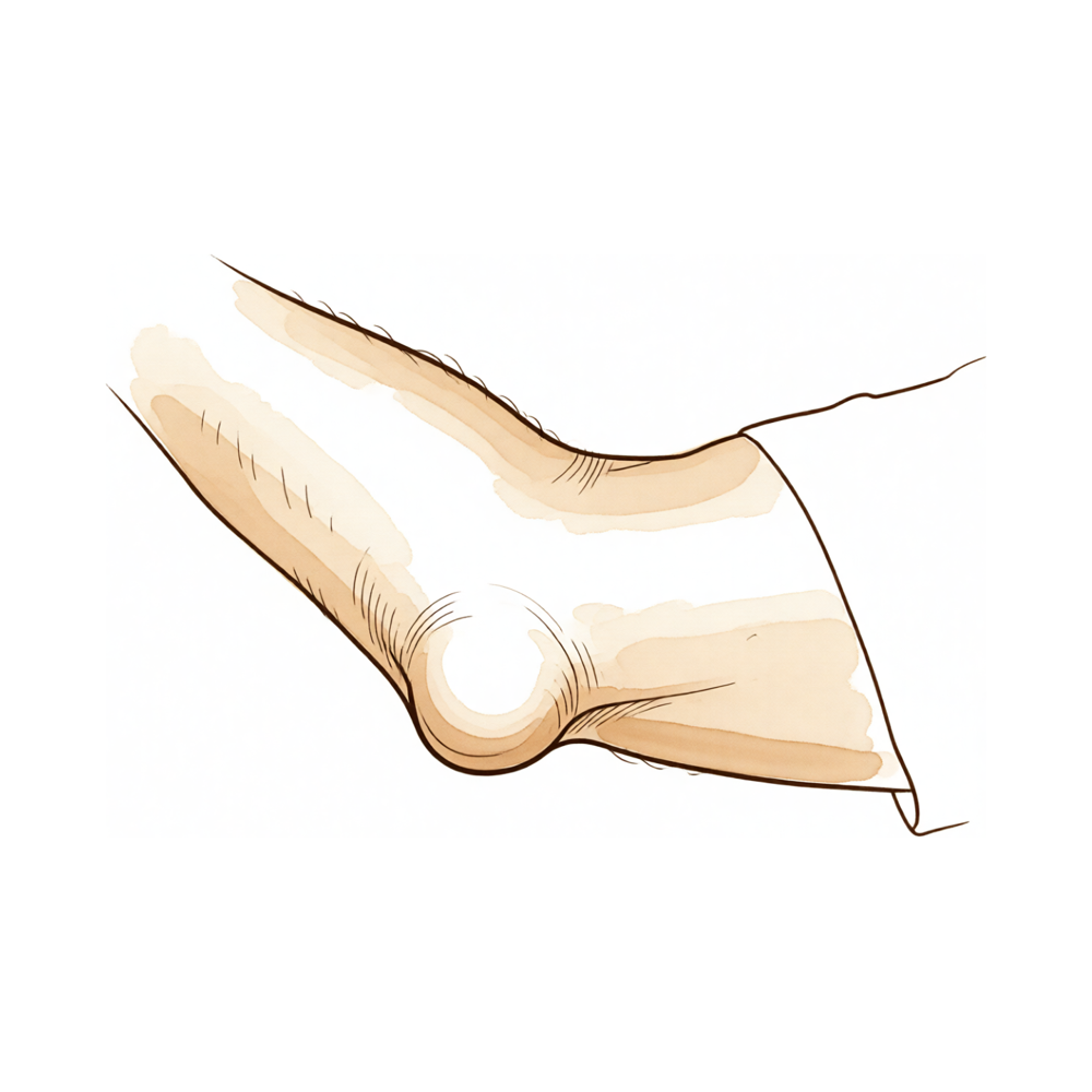
For patients: a plain-language version of this topic is available. See the patient guide.
Overview
Olecranon bursectomy involves the surgical resection of the olecranon bursa, a procedure reserved for patients with chronic or recalcitrant olecranon bursitis who have failed conservative management [2, 5]. While nonsurgical management is significantly more effective and safer than surgical intervention based primarily on level IV evidence [10], open excisional procedures allow for the complete removal of pathological bursal tissue in unresponsive patients [5]. Arthroscopy is increasingly considered a suitable new modality for these cases, with endoscopic bursectomies serving as a useful treatment option [3, 5]. For refractory olecranon bursitis, particularly if aseptic, patients tend to gain significant physical health benefits from open bursectomy [14]. Excision has been curative for all lesions of the olecranon bursa, whereas multiple medications have been tried for cutaneous and systemic infections without clear-cut success [13].
The operation appears to give satisfactory results for chronic olecranon bursitis [4]. Since changing to the olecranon extrabursal endoscopic bursectomy technique, authors have reported no recurrences of bursitis [7]. Patients who underwent endoscopic olecranon bursectomy for recalcitrant olecranon bursitis experienced no recurrences or wound-healing complications necessitating return to the operating room [8]. Endoscopic debridement combined with compression suture for aseptic olecranon bursitis offers advantages including simple operation, minimal invasiveness, minimal postoperative pain, rapid recovery, a low recurrence rate, and satisfactory overall efficacy [18]. Arthroscopic debridement, olecranon spur excision, and loose body removal allow return to throwing sports and reliable subjective and objective results in carefully selected patients [6].
Despite these benefits, the revision rate after bursectomy for olecranon bursitis was 11.5% [1]. Patients with olecranon cords were less satisfied after surgical excision compared to those without cords [16]. Bursal suture repair is a viable alternative to bursectomy in selected patients with chronic traumatic olecranon bursitis, combining functional and cosmetic benefits [9]. Hydrothermal ablation at temperatures between 50C and 52C is a safe treatment option for recurrent or chronic olecranon bursitis with fewer complications than open bursectomy and a comparable efficacy [11]. Intrabursal doxycycline sclerotherapy may be an effective alternative to surgical bursectomy for patients with recurrent olecranon bursitis refractory to conservative management [12]. Nontuberculous mycobacterial olecranon bursitis should be considered in any patient with a swollen bursa and protracted course, regardless of immune status [15]. The medial single-window approach utilizing a triceps-on technique provides safe and adequate exposure of the elbow joint while reducing potential complications associated with dissection through the olecranon bursa [21].
Anatomy & Pathophysiology
Bursal Anatomy and Physiology
The olecranon bursa is the most commonly affected of the 150 human bursae by an inflammatory process [25]. Its lining consists of a poorly vascularized synovial membrane with a low coefficient of friction, which allows the bony olecranon to glide under the skin during flexion and extension of the elbow [25]. The superficial position and limited vascularity of the olecranon bursa render it particularly vulnerable to injury and inflammation [25]. This limited vascularity is the proposed reason for infection via a transcutaneous route rather than haematogenous spread, even when no obvious wound is present [25]. During bursitis, the condition is characterized by an abnormal increase in the volume of fluid within the bursal cavity [25].
Microbiology
Staphylococcus aureus predominates as the causative bacteria in olecranon bursitis [25]. Beta-haemolytic strep is also a common causative bacteria in olecranon bursitis [25].
Clinical Presentation and Diagnosis
Olecranon bursitis presents with an abnormal increase in the volume of fluid within the bursal cavity [25]. Endoscopic bursectomies permit excellent visualization of different anatomical structures along the entire bursal extent [19]. Pathologic tissue in olecranon bursitis can be biopsied and excised under endoscopic vision [19].
Pathophysiology of Recurrence and Revision
Open excisional procedures allow for the complete removal of pathological bursal tissue in unresponsive patients [5].
Classification
Anatomy and Pathophysiology
Olecranon bursitis is defined by an abnormal increase in the volume of fluid within the bursal cavity [25]. The bursal lining consists of a poorly vascularized synovial membrane with a low coefficient of friction, a structure that allows the bony olecranon to glide under the skin during flexion and extension of the elbow [25]. The superficial position and limited vascularity of the olecranon bursa render it particularly vulnerable to injury and inflammation [25]. This limited vascularity is the proposed reason for infection via a transcutaneous route rather than haematogenous spread, even when no obvious wound is present [25]. Among the 150 human bursae, the olecranon is the most commonly affected by an inflammatory process [25].
Differential Diagnosis
Pyoderma gangrenosum: This condition must be considered in the differential diagnosis whenever a patient presents with ulcerative cutaneous lesions that resemble an infectious process such as olecranon bursitis [41].
Other Considerations
Microbiology: Staphylococcus aureus predominates as the causative bacteria in olecranon bursitis, with beta-haemolytic strep also being common [25].
Evidence Base: There is no randomized control data available for olecranon bursitis treatment [25]. Multiple small number studies often provide conflicting findings regarding olecranon bursitis treatment [25]. Consequently, there is currently no consensus on the treatment of olecranon bursitis [25].
Investigations
Plain radiography: Anteroposterior and lateral radiographs of the elbow are required for olecranon fractures [30]. A true lateral radiograph is necessary to accurately identify the plane of the fracture and the number of fracture fragments [27]. Plain radiographs are usually sufficient for isolated fractures of the olecranon [27]. If there is concern for a radial head fracture, a radiocapitellar view of the elbow can be obtained [30]. In the setting of an elbow dislocation, post-reduction radiographs should be obtained [30].
CT: Computed tomography (CT) may help delineate comminution or impaction better in more complex cases, but this is not routinely required [27]. CT can be used for preoperative planning for comminuted fractures of the olecranon if there is an associated radial head or coronoid fracture, but this is not routinely utilized [30].
MRI: Early imaging, such as ultrasound or MRI, may have decreased the likelihood of a second surgery by alerting the surgeon to the underlying process in cases of infected olecranon bursitis [81].
Treatment
Non-Operative
Nonsurgical management of olecranon bursitis is significantly more effective and safer than surgical management [10]. Recent literature demonstrates adverse effects of intrabursal injections and surgery compared with noninvasive management for the initial treatment of nonseptic olecranon bursitis [23].
Operative
Indications: The available evidence did not support the central European concept of immediate bursectomy in cases of septic bursitis [24]. Excision has been curative for all lesions of the olecranon bursa in cases of protothecal infection, whereas multiple medications have been tried for cutaneous and systemic infections without clear-cut success [13].
Surgical Approach / Technique: Open excisional procedures allow for the complete removal of pathological bursal tissue [5]. Arthroscopy is increasingly being considered as a suitable new modality of management for unresponsive patients [5].
Complications
Recurrence: Endoscopic olecranon bursectomy has demonstrated no recurrences of bursitis in reported series [7]. Similarly, patients undergoing this procedure experienced no recurrences necessitating a return to the operating room [8].
Wound complications: In the population undergoing endoscopic olecranon bursectomy, no wound-healing complications were observed that required reoperation [8].
Other Considerations: More recent literature indicates that intrabursal injections and surgery carry adverse effects compared with noninvasive management for the initial treatment of nonseptic olecranon bursitis [23].
Recovery
Other Considerations: In patients who remain unresponsive to initial management, open excisional procedures allow for the complete removal of pathological bursal tissue, although arthroscopy is increasingly considered a suitable new modality for management [5]. Arthroscopic bursectomy results in significant improvements in shoulder function and pain; however, outcomes are significantly worse in patients with a longer duration of preoperative complaints and those with peroperative identification of degenerative glenoid cartilage [22]. While older studies showed resolution with injections and surgery, more recent literature demonstrates adverse effects of intrabursal injections and surgery compared with noninvasive management for the initial treatment of nonseptic olecranon bursitis [23].
Key Evidence
- [L3] The revision rate after bursectomy for olecranon bursitis was 11.5%. [1] (10.1016/j.jse.2020.09.033)
- [L5] Endoscopic bursectomies are a useful treatment option for patients with recalcitrant olecranon bursitis. [3] (10.1016/j.eats.2023.09.004)
- [L4] The operation appears to give satisfactory results for chronic olecranon bursitis. [4] (10.1016/s0020-1383(77)80050-8)
- [L4] In unresponsive patients, although open excisional procedures allow to completely remove the pathological bursal tissue, arthroscopy is increasingly being considered as a suitable new modality of management. [5] (10.1016/j.surge.2012.02.002)
- [L4] Arthroscopic debridement, olecranon spur excision, and loose body removal allow return to throwing sports and reliable subjective and objective results in carefully selected patients. [6] (10.1016/j.arthro.2011.06.012)
- [L4] Since changing to this technique, the authors have had no recurrences of bursitis. [7] (10.1097/bth.0b013e31829c0535)
- [L4] In this population, patients who underwent endoscopic olecranon bursectomy experienced no recurrences or wound-healing complications necessitating return to the operating room. [8] (10.1016/j.asmr.2023.100832)
- [L5] Bursal suture repair is a viable alternative to bursectomy in selected patients with chronic traumatic olecranon bursitis, combining functional and cosmetic benefits. [9] (10.1016/j.xrrt.2025.100597)
- [L4] Based primarily on level IV evidence, nonsurgical management of olecranon bursitis is significantly more effective and safer than surgical management. [10] (10.1007/s00402-014-2088-3)
- [L4] Hydrothermal ablation at temperatures between 50C and 52C is a safe treatment option for recurrent or chronic olecranon bursitis with fewer complications than open bursectomy and a comparable efficacy. [11] (10.1016/j.jse.2024.03.021)
- [L4] This may be an effective alternative to surgical bursectomy for patients with recurrent olecranon bursitis refractory to conservative management. [12] (10.1016/j.jhsg.2024.03.006)
- [Case_report] Excision has been curative for all lesions of the olecranon bursa, whereas multiple medications have been tried for cutaneous and systemic infections without clear-cut success. [13] (10.2106/00004623-198062050-00024)
- [L4] Patients with refractory olecranon bursitis, particularly if aseptic, tend to gain significant physical health benefits from open bursectomy. [14] (10.7759/cureus.43696)
- [L4] Nontuberculous mycobacterial olecranon bursitis should be considered in any patient with a swollen bursa and protracted course, regardless of immune status. [15] (10.1016/j.jse.2008.07.009)
- [L4] Patients with olecranon cords were less satisfied after surgical excision compared to those without cords. [16] (10.1016/j.jse.2015.04.016)
- [L4] Endoscopic debridement combined with compression suture for the treatment of aseptic olecranon bursitis has several advantages: simple operation, minimal invasiveness, minimal postoperative pain, rapid recovery, a low recurrence rate, and satisfactory overall efficacy. [18] (10.1186/s13018-024-05090-3)
- [L5] The technique permits excellent visualization of different anatomical structures along the entire bursal extent, and pathologic tissue can be biopsied and excised under endoscopic vision. [19] (10.1016/j.eats.2022.02.017)
- [L4] The medial single-window approach utilizing a triceps-on technique provides safe and adequate exposure of the elbow joint while reducing potential complications associated with dissection through the olecranon bursa. [21] (10.1016/j.xrrt.2025.08.016)
- [L4] Arthroscopic bursectomy resulted in significant improvements in shoulder function and pain, but outcomes were significantly worse in patients with a longer duration of preoperative complaints and those with peroperative identification of degenerative glenoid cartilage. [22] (10.1016/j.jseint.2020.10.021)
- [L5] Older studies showed resolution with injections and surgery, but more recent literature demonstrates adverse effects of intrabursal injections and surgery compared with noninvasive management for initial treatment of nonseptic olecranon bursitis. [23] (10.1016/j.jhsa.2021.02.006)
- [L1] The available evidence did not support the central European concept of immediate bursectomy in cases of septic bursitis. [24] (10.1007/s00402-013-1882-7)
- [L4] [25] (10.1177/1758573214532787)
- [Case_report] PG must be considered in the differential diagnosis whenever a patient presents with ulcerative cutaneous lesions that resemble an infectious process such as olecranon bursitis. [41] (10.1016/j.jse.2014.06.032)
- [L5] Early imaging, such as ultrasound or MRI, may have decreased the likelihood of a second surgery by alerting the surgeon to the underlying process. [81] (10.1055/s-0040-1701149)
See Also
References
[1] Factors associated with revision surgery for olecranon bursitis after bursectomy. Journal of Shoulder and Elbow Surgery. 2021. DOI: 10.1016/j.jse.2020.09.033
[2] Complications following resection of the olecranon bursa. Acta Orthopaedica Belgica. 2006.
[3] Endoscopic Olecranon Bursectomy in the Treatment of Recalcitrant Olecranon Bursitis: Patient Selection and Operative Technique. Arthroscopy Techniques. 2023. DOI: 10.1016/j.eats.2023.09.004
[4] A useful procedure in the treatment of chronic olecranon bursitis. Injury. 1977. DOI: 10.1016/s0020-1383(77)80050-8
[5] Diagnosis and management of olecranon bursitis. The Surgeon. 2012. DOI: 10.1016/j.surge.2012.02.002
[6] Posteromedial Elbow Impingement: Magnetic Resonance Imaging Findings in Overhead Throwing Athletes and Results of Arthroscopic Treatment. Arthroscopy. 2011. DOI: 10.1016/j.arthro.2011.06.012
[7] Olecranon Extrabursal Endoscopic Bursectomy. Techniques in Hand & Upper Extremity Surgery. 2013. DOI: 10.1097/bth.0b013e31829c0535
[8] No Wound Healing Complications or Recurrences Were Seen and a High Level of Satisfaction Was Reported in Patients Who Underwent Endoscopic Olecranon Bursectomy for Recalcitrant Olecranon Bursitis. Arthroscopy, Sports Medicine, and Rehabilitation. 2024. DOI: 10.1016/j.asmr.2023.100832
[9] Olecranon bursal repair for chronic traumatic bursitis: a surgical technique. JSES Reviews, Reports, and Techniques. 2026. DOI: 10.1016/j.xrrt.2025.100597
[10] Treatment of olecranon bursitis: a systematic review. Archives of Orthopaedic and Trauma Surgery. 2014. DOI: 10.1007/s00402-014-2088-3
[11] Hydrothermal ablation in recurrent or chronic olecranon bursitis: a prospective study. Journal of Shoulder and Elbow Surgery. 2024. DOI: 10.1016/j.jse.2024.03.021
[12] Intrabursal Doxycycline Sclerotherapy for Recurrent Olecranon Bursitis of the Elbow: A Case Control Study. Journal of Hand Surgery Global Online. 2024. DOI: 10.1016/j.jhsg.2024.03.006
[13] Protothecal olecranon bursitis. A case report and review of the literature.. The Journal of Bone & Joint Surgery. 1980. DOI: 10.2106/00004623-198062050-00024
[14] Clinical Outcomes Following Open Olecranon Bursa Excision for Septic and Aseptic Olecranon Bursitis: An Observational Study. Cureus. 2023. DOI: 10.7759/cureus.43696
[15] Nontuberculous mycobacterial olecranon bursitis: Case reports and literature review. Journal of Shoulder and Elbow Surgery. 2009. DOI: 10.1016/j.jse.2008.07.009
[16] The existence of cords in olecranon bursae. Journal of Shoulder and Elbow Surgery. 2015. DOI: 10.1016/j.jse.2015.04.016
[18] Clinical efficacy of endoscopic debridement combined with compression suture in the treatment of recalcitrant aseptic olecranon bursitis. Journal of Orthopaedic Surgery and Research. 2024. DOI: 10.1186/s13018-024-05090-3
[19] “All‐Endoscopic” Management of Refractory Elbow Bicipitoradial Bursitis and Partial Distal Biceps Tendon Tears. Arthroscopy Techniques. 2022. DOI: 10.1016/j.eats.2022.02.017
[21] Medial single-window approach to the elbow: a triceps-on technique that does not violate the olecranon bursa. JSES Reviews, Reports, and Techniques. 2026. DOI: 10.1016/j.xrrt.2025.08.016
[22] Arthroscopic bursectomy less effective in the degenerative shoulder with chronic subacromial pain. JSES International. 2021. DOI: 10.1016/j.jseint.2020.10.021
[23] Clinical Management of Olecranon Bursitis: A Review. The Journal of Hand Surgery. 2021. DOI: 10.1016/j.jhsa.2021.02.006
[24] Prepatellar and olecranon bursitis: literature review and development of a treatment algorithm. Archives of Orthopaedic and Trauma Surgery. 2013. DOI: 10.1007/s00402-013-1882-7
[25] Olecranon bursitis: a systematic overview. Shoulder & Elbow. 2014. DOI: 10.1177/1758573214532787
[27] Aaos Comprehensive Orthopaedic Review 3. Fractures of the Elbow > II. Olecranon Fractures.
[30] Rockwood And Green S Fractures In Adults. 40: Fractures of the Proximal Forearm: Olecranon, Proximal Radius, and Radial Head > Assessment of Olecranon Fractures.
[41] Case report: misdiagnosed olecranon bursitis: pyoderma gangrenosum. Journal of Shoulder and Elbow Surgery. 2014. DOI: 10.1016/j.jse.2014.06.032
[81] Epidermoid Cyst in an Infected Olecranon Bursa. Journal of Hand and Microsurgery. 2020. DOI: 10.1055/s-0040-1701149