Clinicians › Shoulder
Sternoclavicular Joint Disorders
Sternoclavicular joint instability, dislocation and arthritis (corpus-synthesised).
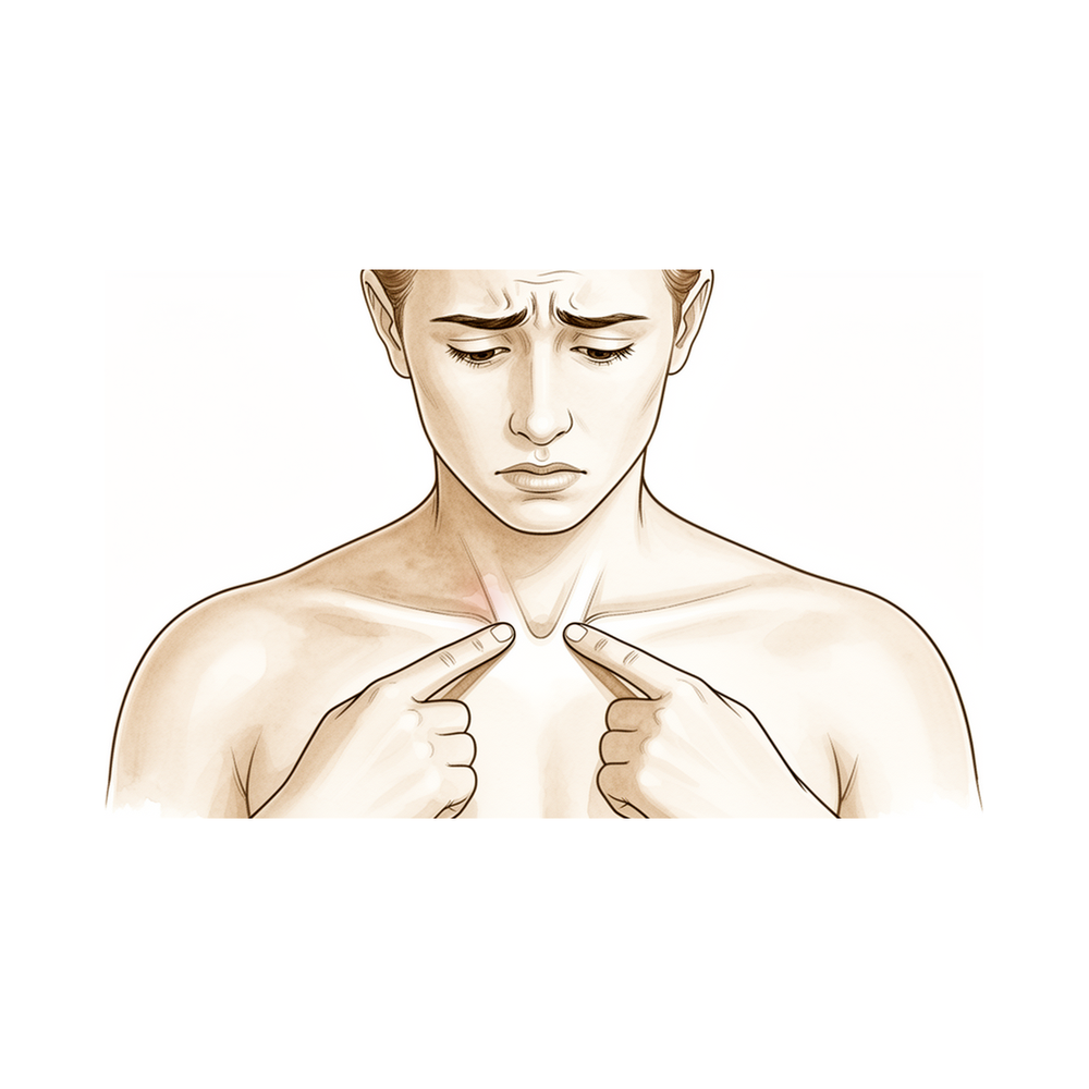
For patients: a plain-language version of this topic is available. See the patient guide.
Overview
Sternoclavicular joint disorders encompass a spectrum of traumatic and atraumatic pathologies, where greater than 10% of patients exhibit substantial anatomical asymmetry that may be misinterpreted as pathological [4]. The majority of these injuries are managed non-operatively [8], with most atraumatic conditions treated nonsurgically and joint resection reserved for patients with persistent symptoms [3]. Nonoperative treatment of atraumatic dislocation improves symptoms and function over time, as no alternative treatment is currently recommended due to poor surgical outcomes [6]. While most anterior dislocations are managed non-surgically, a small subgroup develops persistent symptomatic anterior instability requiring surgical stabilisation [10]. Conservative options include sternoclavicular injections for degenerative disorders [5], where two-thirds of patients report clinically significant improvement regardless of CT abnormalities [11]. The best treatment for chronic instability is prevention through early recognition and treatment of acute instability [1].
Posterior sternoclavicular dislocations present a distinct clinical urgency, carrying a 25% to 30% rate of serious, life-threatening complications involving vital structures [7]. These injuries necessitate immediate evaluation and treatment with a thoracic surgeon present [8], and primary open reduction is preferred in acute cases [116]. The most effective way to avoid serious complications is accurate and prompt diagnosis with appropriate treatment [7]. Surgical procedures for the sternoclavicular joint offer benefits as well as risks and should be performed by experienced surgeons with a good understanding of the neurovascular anatomy and with a thoracic surgeon on call [27].
Digital tomograms provide a safe, accurate, and economically beneficial investigation for sternoclavicular joint pathology [15]. Operative intervention is indicated for specific refractory cases, including excision arthroplasty for arthritis resistant to nonoperative measures [44] and open debridement for symptomatic osteoarthritis unresponsive to conservative management [41]. Arthroscopic treatment may be appropriate in selected cases with adequate joint cavity to improve functional recovery and reduce the risk of secondary instability post-excision arthroplasty [13], potentially lowering the threshold for operative management of a limited number of disorders [9]. Short-term pain relief immediately after injection serves as a strong predictor of future success for operative treatment [5].
Anatomy & Pathophysiology
Bony Anatomy & Development
The sternoclavicular (SC) joint is the only true diarthrodial articulation between the upper appendicular and axial skeletons [62]. It functions as a double gliding joint with an articular disc [73]. The clavicle serves as the primary stabilizer between the axial skeleton via the SC joint and the appendicular skeleton via the acromioclavicular joint [68]. As the first bone to ossify, occurring in the fifth intra-uterine week [26], the clavicle is the only long bone to ossify by intramembranous ossification [62]. The medial (sternal) epiphysis is the last ossification center to fuse, a process occurring between the ages of 20 and 25 years [26, 62]. The primary blood supply to the clavicle is periosteal, with no nutrient artery present [62].
Ligaments & Soft Tissue Stabilizers
The posterior SC joint capsule and ligaments are the primary stabilizers to anterior and posterior translation of the medial clavicle [62]. Specifically, the posterior sternoclavicular ligament is the strongest and primary restraint to anteroposterior instability [73]. The subclavius muscle has a tendinous origin from the first rib immediately lateral to the costoclavicular ligament and a long direct insertion onto the inferior surface of the clavicle [26]. This muscle is enveloped by the clavipectoral fascia, which is contiguous with the investing cervical fascia above the clavicle and continuous with the costoclavicular ligament medially below the clavicle [26]. The form and attachments of the subclavius muscle suggest it acts to reduce the rate and range of upward displacement of the clavicle [26].
The SC joint is bounded by closely applied muscular layers anteriorly and posteriorly, with sternohyoid and sternothyroid muscles located behind [26]. The indirect aponeurotic insertion of the superficial part of the clavicular insertion of the sternocleidomastoid is contiguous with the more direct insertion of the clavicular and sternal parts of pectoralis major below, covering the SC joint in a musculo-aponeurotic layer [26]. In most cases, the tendinous extension of the sternomastoid part of the sternocleidomastoid covers the medial aspect of the SC joint [26].
Regional Anatomy & Vital Structures
The SC joint lies immediately above and medial to the thoracic outlet, which is bounded by the first thoracic vertebra, first rib, and upper border of the manubrium [26]. SC joint pathology may interfere with the trachea and esophagus posteromedially [26]. Conversely, pathology may interfere with the subclavian vessels and supraclavicular brachial plexus posterolaterally [26]. SC joint injuries can result in damage to adjacent structures such as the trachea, esophagus, lungs, and great vessels [51]. Posterior dislocations can be associated with mediastinal compromise in up to 25% of patients [21]. The risk of major complications is associated with open surgical intervention because of the proximity of major vital structures to the SC joint [51].
Biomechanics & Kinematics
The SC joint rotates 30 degrees with shoulder motion [73]. Substantial force to the shoulder girdle or the medial clavicle is required to disrupt the strong ligamentous complex surrounding the SC joint [21]. Consequently, SC joint injuries are uncommon because of the high energy required to disrupt the SC ligaments [51]. Despite technology innovations, a precise, easy to use and low-cost non-invasive method able to draw and analyze the kinematics of the shoulder complex has not been developed yet [18].
Pathophysiology & Injury Mechanisms
Anterior dislocation usually results from an indirect force to the shoulder, with forces transmitted through the clavicle to the SC joint [51]. Specifically, an indirect force to the anterolateral shoulder, which causes the shoulder to roll backward, results in an anterior SC dislocation [21]. Posterior SC joint dislocation can result from both indirect and direct forces [51]. A direct force to the anteromedial clavicle results in a posterior SC dislocation [21]. An indirect force to the posterolateral shoulder, which causes the shoulder to roll forward, also results in a posterior SC dislocation [21]. In these cases, direct anteromedial force usually results in the clavicle being pushed posteriorly into the mediastinum [51].
In patients with an open physis, injuries often result in physeal disruption rather than ligamentous injury [21]. Trauma to the SC joint can result in an intra-articular disk injury without ligamentous instability, causing localized pain, swelling, and early arthritis [21]. Osteoarthritis of the sternoclavicular joint can cause tertiary subacromial impingement due to decreased clavicle motion [129].
Classification
Epidemiology and Mechanism
Traumatic dislocations of the sternoclavicular joint comprise 1% of all joint dislocations and 3% of all upper extremity dislocations [21]. SC joint injuries constitute 3% of all injuries involving the shoulder girdle [51]. The most common causes are motor vehicle accidents, athletic accidents, and falls from a height [21]. Athletic injuries and motor vehicle accidents constitute more than 80% of injuries to the SC joint [51]. Anterior dislocations are more common than posterior dislocations [21], with anterior dislocation being the most common form of SC joint dislocation [51]. SC joint dislocation usually requires a large force because of the strong support provided by the surrounding ligaments [51]. Posterior SC joint dislocation can result from both indirect and direct forces [51]. Direct anteromedial force usually results in the clavicle being pushed posteriorly into the mediastinum [51]. The medial clavicle epiphysis does not fuse with the shaft until age 23 to 25 years [51]. The medial physis of the clavicle is the last to fuse, between the ages of 23 and 25 years [26].
Clinical Presentation and Diagnosis
Patients typically report pain and swelling after trauma to the shoulder girdle or the chest [21]. Patients with a posterior dislocation show higher levels of pain and may report shortness of breath, difficulty swallowing, or a sensation of choking [21]. Posterior sternoclavicular dislocations are often initially overlooked because of their rarity [19]. In a retrospective review of 14 patients, 10 (71.4%) had a missed diagnosis with an average of 13 months until diagnosis [33]. In the same retrospective review of 14 patients, 4 (28.6%) presented with dysphagia or dyspnea [33]. Ultrasound scans of the sternoclavicular joint are more accurately interpreted and have a higher diagnostic yield compared to plain films [29]. CT scan is the imaging modality of choice due to its availability and quickness in the trauma setting [135]. MRI is a useful adjunct to distinguish between SC joint dislocation and physeal injuries in patients under the age of 25 [135].
Complications and Risk Stratification
Posterior sternoclavicular dislocations carry a 25% to 30% rate of serious, life-threatening complications involving vital structures [7]. Vascular injury occurred only in severely polytraumatized patients with an Injury Severity Score (ISS) ≥ 15 [104]. SCJ pathology may interfere with the trachea and oesophagus posteromedially and the subclavian vessels and supraclavicular brachial plexus posterolaterally [26]. SC joint injuries sometimes result in damage to adjacent structures such as the trachea, esophagus, lungs, and great vessels [51].
Atraumatic and Degenerative Conditions
Osteomyelitis of the sternoclavicular joint is a rare disease that has become more prevalent in recent years and can be associated with increasing use of long-term indwelling catheters [20]. Injuries to the SC joint are uncommon, and recognition and classification are critical to proper management to minimize long-term sequelae [108].
Clinical Presentation
Traumatic Dislocations
Traumatic sternoclavicular joint dislocations typically result from motor vehicle accidents, athletic accidents, or falls from a height [21]. Disruption of the strong ligamentous complex surrounding the joint requires substantial force to the shoulder girdle or the medial clavicle [21]. Anterior dislocations are more common than posterior dislocations [21]. The mechanism of injury helps distinguish the direction of subluxation or dislocation [21]. A direct force to the anteromedial clavicle results in a posterior sternoclavicular joint dislocation [21]. An indirect force to the posterolateral shoulder, which causes the shoulder to roll forward, results in a posterior sternoclavicular joint dislocation [21]. Conversely, an indirect force to the anterolateral shoulder, which causes the shoulder to roll backward, results in an anterior sternoclavicular joint dislocation [21].
Patients with a posterior sternoclavicular dislocation show higher levels of pain and may report shortness of breath, difficulty swallowing, or a sensation of choking [21]. Posterior sternoclavicular dislocations can be associated with mediastinal compromise in up to 25% of patients [21]. These injuries are often initially overlooked due to their rarity [19]. Dislocation of the sternoclavicular joint should always be considered in a trauma patient with pain, swelling, or deformity over the medial clavicle [24]. Posterior sternoclavicular disruption and ipsilateral clavicle fracture carry significant risk of intrathoracic injury [95]. Trauma to the sternoclavicular joint can also result in an intra-articular disk injury without ligamentous instability, causing localized pain, swelling, and early arthritis [21].
Atraumatic and Degenerative Conditions
Transient sternoclavicular joint arthropathy may often be a self-limited disease that can be effectively treated solely by the administration of nonsteroidal anti-inflammatory drugs [25]. Atraumatic posterior subluxation of the sternoclavicular joint is likely a self-limiting pathology [36]. Sternoclavicular hyperostosis is a disease of unknown etiology characterised by periosteal reaction and endosteal hyperossification [30]. Patients who have a sternoclavicular joint pain with dermatosis should be considered as a candidate for SAPHO Syndrome [86]. Sternoclavicular joint infections and osteomyelitis of the clavicle are extremely rare infections, especially in the pediatric population [42]. Early signs of sternoclavicular joint infections and clavicular osteomyelitis are nonspecific and can be mistaken for common upper respiratory infections [42].
Diagnostic Evaluation
In mild sprains with intact ligaments, tenderness and swelling of the sternoclavicular joint are present [21]. The location of pain originating from the sternoclavicular joint can be diverse and patients are often not able to identify the exact location [90]. The pain pattern of sternoclavicular joint arthropathy can overlap those of the acromioclavicular joint, the subacromial space and cervical nerves [90]. Mechanical pain in the area of the sternoclavicular region is itself a reliable sign for sternoclavicular arthropathy [90].
The most sensitive clinical test for identifying sternoclavicular arthropathy is examination for local sternoclavicular joint tenderness with 93% sensitivity [90]. Pain during active protraction has a sensitivity of 86% for identifying sternoclavicular arthropathy [90]. CT imaging shows a sensitivity of 84% for identifying sternoclavicular arthropathy [90]. Sternoclavicular joint injections are a useful conservative treatment option for degenerative sternoclavicular joint disorders [5]. The short-term pain relief immediately after sternoclavicular injection is a strong predictor of future success for operative treatment [5]. Definitive diagnosis of sternoclavicular joint infections is with sternoclavicular joint aspiration or bone culture [42].
Investigations
Plain radiography: The serendipity view is a special radiographic view indicated for the sternoclavicular joint, obtained with the patient supine and a 40° cephalic tilt centered on the sternum [83]. In patients with symptomatic sternoclavicular arthritis, the mean anterior-posterior and superior-inferior distances are significantly larger compared to normal joints [110]. This prominence is caused by both osseous enlargement and subluxation [110].
CT: CT scans should be interpreted with the knowledge that the prevalence of osteoarthritis of the sternoclavicular joint is a factor when assessing symptomatic pathology [117]. Posterior sternoclavicular dislocations can be dangerous due to the proximity of vital structures such as the esophagus, trachea, and neurovascular bundle [55]. Consequently, prompt and accurate diagnosis is critical for posterior sternoclavicular dislocations [55].
Ultrasound: Ultrasound-guided injections of the sternoclavicular joint are a safe and accurate alternative diagnostic method that saves the patient from harmful radiation and additional appointments [111].
Other Considerations: The short-term pain relief immediately after a sternoclavicular injection is a strong predictor of future success for operative treatment [5].
Treatment
Non-Operative
Most atraumatic conditions of the sternoclavicular joint, including most anterior dislocations, are managed nonsurgically [3] [10]. Nonoperative treatment of atraumatic sternoclavicular dislocation results in improved symptoms and function over time [6]. Most acromioclavicular and sternoclavicular joint injuries are ligamentous sprains treated with a period of sling immobilisation and pain control followed by physical therapy [88]. Atraumatic posterior subluxation is likely a self-limiting pathology that should be treated conservatively [36]. Sternoclavicular osteomyelitis can be successfully treated by medical management alone with antibiotics and hyperbaric oxygen, avoiding hazardous surgical intervention [101].
Immobilization duration varies by injury type. Type I sternoclavicular joint injuries require only a short period of immobilization in a sling, approximately 1 week or less, with return to activity as symptoms improve [96]. Type II injuries are often treated with immobilization using either a sling or figure-of-eight brace for 4 to 6 weeks [96]. Type III sternoclavicular joint injuries should be treated with closed reduction [96]. Closed reduction of anterior sternoclavicular dislocation is recommended if the patient presents within 7 to 10 days of injury [37].
Operative
Indications: Joint resection is reserved for patients with persistent symptoms of atraumatic sternoclavicular joint conditions [3]. A small subgroup of patients with anterior sternoclavicular joint dislocations develop persistent symptomatic anterior instability that may require surgical stabilisation [10]. Patients who fail to have acceptable outcomes with nonoperative treatment for anterior sternoclavicular joint instability should be considered for surgical treatment [96]. Excision arthroplasty for patients with sternoclavicular arthritis resistant to nonoperative measures is only considered rarely [44].
Surgical Approach / Technique: Suture anchors are recommended for the treatment of symptomatic sternoclavicular joint instability [2]. Sternoclavicular joint reconstruction using a sternocleidomastoid tendon graft is safe and offers reliable pain relief and functional improvement for patients with chronic debilitating anterior instability of the sternoclavicular joint [34]. The unicortical sternoclavicular joint reconstruction using synthetic graft is relatively safe and allows for early postoperative functional rehabilitation [12]. The novel sternoclavicular hook plate is effective for the treatment of posterior sternoclavicular dislocation [91]. Resection arthroplasty for septic arthritis of the sternoclavicular joint is safe and effective, resulting in a low rate of complications, eradication of infection, and good functional recovery of the shoulder [48].
Other Considerations: The results for both open and arthroscopic excision arthroplasty for sternoclavicular arthritis appear to be consistently good [44]. Surgical stabilization of atraumatic posterior subluxation of the sternoclavicular joint is difficult and may lead to complications like graft erosion [36]. The approach for current presentation and optimal surgical management of sternoclavicular joint infections has minimal impact on upper extremity function [38]. Placement of antibiotic-impregnated calcium sulfate beads into the surgical site cleared the infection in all cases where they were used for pediatric sternoclavicular joint infections [42].
Complications
Posterior Dislocation and Mediastinal Injury
Patients with posterior sternoclavicular dislocation may present with shortness of breath, difficulty swallowing, or a sensation of choking [21]. Chronic posterior dislocation poses significant risks to adjacent mediastinal structures. It can result in obstruction of the innominate vein [113], compression of the subclavian artery [113], and compression brachial plexopathy [113].
Anterior Dislocation and Instability
Most acute anterior dislocations remain unstable following reduction [37]. In patients older than 23 to 25 years, persistent prominence of the anterior clavicle is expected after anterior dislocation [37]. This persistent prominence does not seem to interfere with usual activities and, in some cases, has not interfered with heavy manual labor [37].
Surgical and Procedural Complications
Surgical stabilization of atraumatic posterior subluxation is difficult and may lead to complications such as graft erosion [36]. In a series of 14 patients undergoing sternoclavicular joint reconstruction, 6 complications occurred in 5 patients [33]. Arthroscopic treatment of sternoclavicular arthropathy carries a risk of secondary instability post-excision arthroplasty [13]. Migration of pins used in operative treatment is a recognized complication [125].
Infection and Osteomyelitis
Osteomyelitis of the sternoclavicular joint is a rare disease that has become more prevalent in recent years [20]. It can be associated with increasing use of long-term indwelling catheters [20].
Associated Injuries
Sternoclavicular joint subluxation can occur as part of an unusual constellation of shoulder girdle trauma, including clavicle fracture and long thoracic nerve injury [31].
Recovery
Non-Operative Management: In a 1990 report of 10 patients with traumatic anterior sternoclavicular dislocations treated nonoperatively, good results were observed in 7 patients, fair results in 2, and poor results in 1 at a mean follow-up interval of 5 years [37]. For patients older than 23 to 25 years, persistent prominence of the anterior clavicle is expected following nonoperative management of anterior dislocation [37]. This persistent prominence does not appear to interfere with usual activities and, in some cases, has not even interfered with heavy manual labor [37].
Operative Management: A small subgroup of patients with anterior sternoclavicular dislocation develop persistent symptomatic anterior instability that may require surgical stabilisation [10]. The unicortical sternoclavicular joint reconstruction using synthetic graft is a relatively safe technique that allows for early postoperative functional rehabilitation [12]. Stabilization of the sternoclavicular joint with the figure-of-eight technique using hamstrings seems to be a feasible alternative for young and active patients with remaining instability following conservative treatment [17]. Resection of the medial clavicle results in good functional outcome when the costoclavicular ligament is preserved or reconstructed [103]. Anatomic reconstruction after excessive medial clavicle resection provides better restoration of shoulder mechanics and function compared to nonanatomic soft tissue only reconstruction techniques [23]. Temporary sternoclavicular plating was successful in achieving an excellent long-term outcome for an unusual double clavicle fracture with medial nonunion and lateral acute fracture [114].
Prognosis and Complications: At 1-year follow-up, a patient with posterior sternoclavicular epiphyseal fracture-dislocation was doing well, without symptoms, and had full use of his shoulder [52]. A patient with positional anterior sternoclavicular joint dislocation achieved a perfect Western Ontario Shoulder Instability Index score of zero points at two years with no recurrence of sternoclavicular joint dislocation [50].
Key Evidence
- [L5] The text emphasizes that the best treatment for most cases of chronic sternoclavicular instability is prevention through early recognition and treatment of acute instability. [1] (10.1016/s0030-5898(05)70140-1)
- [L4] The technique is recommended for symptomatic sternoclavicular joint instability. [2] (10.1016/j.jse.2005.07.005)
- [L5] Most atraumatic conditions of the sternoclavicular joint can be managed nonsurgically, with joint resection reserved for patients with persistent symptoms. [3] (10.5435/00124635-200503000-00007)
- [L4] Greater than 10% of patients show substantial asymmetry in the sternoclavicular joints, which may be misinterpreted as pathological. [4] (10.1007/s00256-009-0689-7)
- [L4] Sternoclavicular injections are a useful conservative treatment option for degenerative sternoclavicular joint disorders, and the short-term pain relief immediately after injection is a strong predictor of future success for operative treatment. [5] (10.1177/1758573220916904)
- [L4] Nonoperative treatment of atraumatic sternoclavicular dislocation results in improved symptoms and function over time, with no alternative treatment currently recommended due to poor surgical outcomes. [6] (10.1016/j.jse.2019.04.060)
- [Textbook] Posterior sternoclavicular dislocations carry a 25% to 30% rate of serious, life-threatening complications involving vital structures, necessitating operative reduction and stabilization; the most effective way to avoid these complications is accurate and prompt diagnosis with appropriate treatment. [7] (10.1016/s0278-5919(03)00013-9)
- [L4] Most anterior sternoclavicular joint dislocations can be managed non-surgically, although a small subgroup of patients develop persistent symptomatic anterior instability that may require surgical stabilisation. [10] (10.5312/wjo.v7.i4.244)
- [L4] Two thirds of patients having sternoclavicular joint injections of corticosteroids and local anesthetics report clinically significant improvement regardless of the abnormalities detected on their CT images. [11] (10.2214/ajr.10.4501)
- [L4] The authors report a technique for managing sternoclavicular joint injuries that is relatively safe and allows for early postoperative functional rehabilitation. [12] (10.1177/1758573218790964)
- [L4] Arthroscopic treatment of sternoclavicular arthropathy may be appropriate in selected cases with adequate joint cavity to improve functional recovery and reduce the risk of secondary instability post-excision arthroplasty. [13] (10.1007/s00167-008-0692-x)
- [L5] It is shared as a salvage option to address continued sternoclavicular joint insufficiency. [14] (10.1016/j.xrrt.2026.100867)
- [L4] Digital tomograms are a safe, accurate and economically beneficial investigation for sternoclavicular joint pathology. [15] (10.1007/s00590-019-02433-3)
- [L4] Stabilization of the sternoclavicular joint with the figure-of-eight technique seems to be a feasible alternative for young and active patients with remaining instability following conservative treatment. [17] (10.1016/j.jse.2012.02.009)
- [L5] Despite technology innovations, a precise, easy to use and low-cost non-invasive method able to draw and analyze the kinematics of the shoulder complex has not been developed yet. [18] (10.1177/17585732221090226)
- [L4] Posterior sternoclavicular dislocations, because of their rarity, are often initially overlooked. [19] (10.1007/s00590-008-0363-0)
- [L4] Osteomyelitis of the sternoclavicular joint is a rare disease that has become more prevalent in recent years and can be associated with increasing use of long-term indwelling catheters. [20] (10.1155/2014/747315)
- [L5] This anatomic reconstruction provides better restoration of shoulder mechanics and function compared to nonanatomic soft tissue only reconstruction techniques. [23] (10.1016/j.eats.2021.08.020)
- [L4] Dislocation of the sternoclavicular joint should always be considered in a trauma patient with pain, swelling, or deformity over the medial clavicle. [24] (10.1007/bf00197665)
- [L4] Transient sternoclavicular joint arthropathy may often be a self-limited disease that can be effectively treated solely by the administration of nonsteroidal anti-inflammatory drugs. [25] (10.1016/j.jse.2013.08.013)
- [L4] [26] (10.1302/0301-620x.95b6.31064)
- [L5] Sternoclavicular joint surgical procedures offer benefits as well as risks and should be performed by experienced surgeons with a good understanding of the neurovascular anatomy and with a thoracic surgeon on call. [27] (10.1016/j.arthro.2020.02.023)
- [L4] This novel sternoclavicular hook plate shows excellent efficacy for the treatment of unstable sternoclavicular joint dislocations. [28] (10.1177/2309499016684488)
- [L5] Ultrasound scans of the sternoclavicular joint however are more accurately interpreted, and have a higher diagnostic yield compared to plain films. [29] (10.1016/s0020-1383(02)00350-9)
- [L5] Sternoclavicular hyperostosis is a disease of unknown etiology characterised by periosteal reaction and endosteal hyperossification. [30] (10.1016/s0020-1383(02)00259-0)
- [L4] The case represents an unusual constellation of shoulder girdle trauma where an initially isolated clavicle fracture was accompanied by sternoclavicular joint subluxation and long thoracic nerve injury. [31] (10.1177/03635465000280062301)
- [L4] [33] (10.1097/bot.0000000000000483)
- [L4] Sternoclavicular joint reconstruction using a sternocleidomastoid tendon graft is safe and offers reliable pain relief and functional improvement for patients with chronic debilitating anterior instability of the sternoclavicular joint. [34] (10.2106/jbjs.m.00681)
- [L4] The authors conclude that atraumatic posterior subluxation of the sternoclavicular joint is likely a self-limiting pathology that should have been treated conservatively, as surgical stabilization is difficult and may lead to complications like graft erosion. [36] (10.1007/s004020050424)
- [L4] [37] (10.5435/00124635-199609000-00005)
- [L4] This approach has minimal impact on upper extremity function. [38] (10.1016/s0003-4975(01)03390-2)
- [L4] Open sternoclavicular debridement has proved to be a simple, safe and highly effective new surgical treatment for patients with symptomatic sternoclavicular osteoarthritis unresponsive to non-operative management. [41] (10.1177/1758573220972093)
- [L5] [42] (10.5435/jaaosglobal-d-21-00302)
- [L4] Excision arthroplasty for patients with sternoclavicular arthritis resistant to nonoperative measures is only considered rarely, but the results for both open and arthroscopic excision arthroplasty appear to be consistently good. [44] (10.1053/j.otsm.2014.02.019)
- [L4] The procedure was found to be safe and effective, resulting in a low rate of complications, eradication of infection, and good functional recovery of the shoulder. [48] (10.1016/j.jse.2011.05.020)
- [Case_report] The patient achieved a perfect Western Ontario Shoulder Instability Index score of zero points at two years with no recurrence of sternoclavicular joint dislocation. [50] (10.1016/j.jseint.2020.04.007)
- [L5] At 1-year follow-up the patient was doing well, without symptoms, and had full use of his shoulder. [52] (10.1007/s00256-005-0076-y)
- [L4] [55] (10.1177/2325967126s00191)
- [L5] The patients who have a sternoclavicular joint pain with dermatosis should be considered as a candidate for SAPHO Syndrome. [86] (10.1016/s0020-1383(13)70161-x)
- [L4] [88] (10.1136/bjsm.2009.059295)
- [L3] [90] (10.1186/1471-2474-15-421)
- [L4] The novel sternoclavicular hook plate is effective for the treatment of posterior SCD. [91] (10.1186/s13018-023-04436-7)
- [L5] Posterior sternoclavicular disruption and ipsilateral clavicle fracture carry significant risk of intrathoracic injury and often require surgical intervention. [95] (10.1097/bot.0b013e3181aa7d3d)
- [L4] [96] (10.5435/jaaos-d-19-00611)
- [L4] Sternoclavicular osteomyelitis can be successfully treated by medical management alone with antibiotics and hyperbaric oxygen, avoiding hazardous surgical intervention. [101] (10.1111/j.1758-5740.2010.00061.x)
- [L4] Resection of the medial clavicle results in good functional outcome when the costoclavicular ligament is preserved or reconstructed. [103] (10.1007/s00402-009-0911-z)
- [L3] Among all patients with sternoclavicular dislocation, vascular injury was rare, occurring only in severely polytraumatized patients with an Injury Severity Score (ISS) ≥ 15. [104] (10.1097/bot.0000000000001685)
- [L1] Injuries to the SC joint are uncommon, and recognition and classification are critical to proper management to minimize long-term sequelae. [108] (10.1177/0363546513498990)
- [L4] It found that the mean anterior-posterior and superior-inferior distances were significantly larger in patients with symptomatic sternoclavicular arthritis compared to normal joints, supporting the hypothesis that prominence is caused by both osseous enlargement and subluxation. [110] (10.2106/jbjs.m.00623)
- [L4] The use of ultrasound-guided injections of the sternoclavicular joint is a safe and accurate alternative diagnostic method, which saves the patient from harmful radiation and additional appointments. [111] (10.1016/j.xrrt.2021.08.010)
- [L5] [113] (10.1067/mse.2002.119394)
- [L4] Temporary sternoclavicular plating was successful in achieving an excellent long-term outcome for an unusual double clavicle fracture with medial nonunion and lateral acute fracture. [114] (10.1155/2014/206125)
- [L4] Primary open reduction should probably be preferred in acute cases of sternoclavicular dislocation. [116] (10.3109/17453678608994382)
- [L3] This should be taken into consideration when using a CT scan to assess a patient with symptomatic SCJ pathology. [117] (10.1016/j.jse.2016.04.029)
- [L4] [125] (10.1016/j.jse.2005.04.005)
- [L4] Osteoarthritis of the sternoclavicular joint can cause tertiary subacromial impingement due to decreased clavicle motion. [129] (10.1111/j.1758-5740.2010.00060.x)
See Also
References
[1] CONSERVATIVE MANAGEMENT OF STERNOCLAVICULAR INJURIES. Orthopedic Clinics of North America. 2000. DOI: 10.1016/s0030-5898(05)70140-1
[2] Suture anchors for treatment of sternoclavicular joint instability. Journal of Shoulder and Elbow Surgery. 2006. DOI: 10.1016/j.jse.2005.07.005
[3] Atraumatic Disorders of the Sternoclavicular Joint. Journal of the American Academy of Orthopaedic Surgeons. 2005. DOI: 10.5435/00124635-200503000-00007
[4] Variations in normal sternoclavicular joints; a retrospective study to quantify SCJ asymmetry. Skeletal Radiology. 2009. DOI: 10.1007/s00256-009-0689-7
[5] Sternoclavicular joint injections and their predictive value for the outcome of surgery. Shoulder & Elbow. 2020. DOI: 10.1177/1758573220916904
[6] Evolution of nonoperative treatment of atraumatic sternoclavicular dislocation. Journal of Shoulder and Elbow Surgery. 2019. DOI: 10.1016/j.jse.2019.04.060
[7] 22. Complications of Sternoclavicular Joint Injuries. Clinics in Sports Medicine. 2003. DOI: 10.1016/s0278-5919(03)00013-9
[8] 20. Non-operative Management of Sternoclavicular Joint Injuries: Indications, Techniques, and Outcomes. 2011.
[9] Arthroscopy_of_the_Sternoclavicular_Joint_(SS-23)_S0749806312003696. 2003.
[10] Sternoclavicular joint dislocation and its management: A review of the literature. World Journal of Orthopedics. 2016. DOI: 10.5312/wjo.v7.i4.244
[11] CT-Guided Sternoclavicular Joint Injections: Description of the Procedure, Reliability of Imaging Diagnosis, and Short-Term Patient Responses. American Journal of Roentgenology. 2010. DOI: 10.2214/ajr.10.4501
[12] The unicortical sternoclavicular joint reconstruction using synthetic graft. Shoulder & Elbow. 2018. DOI: 10.1177/1758573218790964
[13] Arthroscopic excision of sternoclavicular joint. Knee Surgery, Sports Traumatology, Arthroscopy. 2008. DOI: 10.1007/s00167-008-0692-x
[14] Reconstruction of Sternoclavicular Joint with Excision of Medial Clavicle and Internal Fixation to the First Rib. JSES Reviews, Reports, and Techniques. 2026. DOI: 10.1016/j.xrrt.2026.100867
[15] Digital tomography is an effective investigation for sternoclavicular joint pathology. European Journal of Orthopaedic Surgery & Traumatology. 2019. DOI: 10.1007/s00590-019-02433-3
[17] Reconstruction of the sternoclavicular joint in active patients with the figure-of-eight technique using hamstrings. Journal of Shoulder and Elbow Surgery. 2013. DOI: 10.1016/j.jse.2012.02.009
[18] Evaluation of the range of motion of scapulothoracic, acromioclavicular and sternoclavicular joints: State of the art. Shoulder & Elbow. 2022. DOI: 10.1177/17585732221090226
[19] Posterior sternoclavicular dislocations. European Journal of Orthopaedic Surgery & Traumatology. 2008. DOI: 10.1007/s00590-008-0363-0
[20] Treatment of Sternoclavicular Joint Osteomyelitis with Debridement and Delayed Resection with Muscle Flap Coverage Improves Outcomes. Surgery Research and Practice. 2014. DOI: 10.1155/2014/747315
[21] Chapter 77 Disorders of the Sternoclavicular Joint. 2019.
[23] Reconstruction of the Sternoclavicular Joint After Excessive Medial Clavicle Resection. Arthroscopy Techniques. 2021. DOI: 10.1016/j.eats.2021.08.020
[24] Dislocations of the sternoclavicular joint. Skeletal Radiology. 1976. DOI: 10.1007/bf00197665
[25] Transient sternoclavicular joint arthropathy, a self-limited disease. Journal of Shoulder and Elbow Surgery. 2014. DOI: 10.1016/j.jse.2013.08.013
[26] Instability of the sternoclavicular joint. The Bone & Joint Journal. 2013. DOI: 10.1302/0301-620x.95b6.31064
[27] Editorial Commentary: Sternoclavicular Joint Osteoarthritis Surgical Treatment Offers Benefits as Well as Risks. Arthroscopy. 2020. DOI: 10.1016/j.arthro.2020.02.023
[28] Efficacy analysis of a novel sternoclavicular hook plate for treatment of unstable sternoclavicular joint dislocation or fracture. Journal of Orthopaedic Surgery. 2017. DOI: 10.1177/2309499016684488
[29] Posterior sternoclavicular joint dislocation: the value of intra-operative ultrasound. Injury. 2003. DOI: 10.1016/s0020-1383(02)00350-9
[30] Sternoclavicular hyperostosis with pathological fracture of the clavicle—a case report. Injury. 2003. DOI: 10.1016/s0020-1383(02)00259-0
[31] Ipsilateral Clavicle Fracture, Sternoclavicular Joint Subluxation, and Long Thoracic Nerve Injury: An Unusual Constellation of Injuries Sustained during Wrestling. The American Journal of Sports Medicine. 2000. DOI: 10.1177/03635465000280062301
[33] Sternoclavicular Reconstruction in the Young Active Patient. Journal of Orthopaedic Trauma. 2016. DOI: 10.1097/bot.0000000000000483
[34] Clinical Outcome After Reconstruction for Sternoclavicular Joint Instability Using a Sternocleidomastoid Tendon Graft. Journal of Bone and Joint Surgery. 2014. DOI: 10.2106/jbjs.m.00681
[36] Atraumatic spontaneous posterior subluxation of the sternoclavicular joint. Archives of Orthopaedic and Trauma Surgery. 2005. DOI: 10.1007/s004020050424
[37] Acute and Chronic Traumatic Injuries of the Sternoclavicular Joint. Journal of the American Academy of Orthopaedic Surgeons. 1996. DOI: 10.5435/00124635-199609000-00005
[38] Current presentation and optimal surgical management of sternoclavicular joint infections. The Annals of Thoracic Surgery. 2002. DOI: 10.1016/s0003-4975(01)03390-2
[41] Open sternoclavicular osteophyte debridement in the surgical management of sternoclavicular osteoarthritis: clinical outcome of a new procedure. Shoulder & Elbow. 2020. DOI: 10.1177/1758573220972093
[42] Delayed Diagnosis of Pediatric Sternoclavicular Joint Infections and Clavicular Osteomyelitis During the COVID-19 Pandemic: A Report of 3 Cases. JAAOS: Global Research and Reviews. 2022. DOI: 10.5435/jaaosglobal-d-21-00302
[44] Sternoclavicular Joint Arthritis: Arthroscopic and Open Resection. Operative Techniques in Sports Medicine. 2014. DOI: 10.1053/j.otsm.2014.02.019
[48] Resection arthroplasty for septic arthritis of the sternoclavicular joint. Journal of Shoulder and Elbow Surgery. 2012. DOI: 10.1016/j.jse.2011.05.020
[50] Positional anterior sternoclavicular joint dislocation in the acceleration phase of throwing: a case report. JSES International. 2020. DOI: 10.1016/j.jseint.2020.04.007
[51] Chapter 2 Disorders of the Acromioclavicular Joint, Sternoclavicular Joint, and Clavicle. 2019.
[52] Posterior sternoclavicular epiphyseal fracture-dislocation with delayed diagnosis. Skeletal Radiology. 2006. DOI: 10.1007/s00256-005-0076-y
[55] Get in Line! Diagnostic Accuracy and Outcomes of Pediatric Posterior Sternoclavicular Joint Dislocations. Orthopaedic Journal of Sports Medicine. 2026. DOI: 10.1177/2325967126s00191
[62] Aaos Comprehensive Orthopaedic Review 3. Anatomy of the Shoulder, Arm, and Elbow > I. Shoulder.
[68] Aaos Comprehensive Orthopaedic Review 3. Fractures of the Clavicle, Scapula, and Glenoid > I. Clavicular Fractures.
[73] Miller S Review Of Orthopaedics. Genetics of musculoskeletal conditions and abnormalities are summarized in Table 1.27 > UPPER EXTREMITY > SHOULDER.
[83] Aaos Comprehensive Orthopaedic Review 3. Imaging of the Shoulder and Elbow > I. Shoulder.
[86] PS12 SAPHO syndrome should be considered in patients who have a sternoclavicular joint pain with dermatosis: a case report. Injury. 2013. DOI: 10.1016/s0020-1383(13)70161-x
[88] Biomechanics and treatment of acromioclavicular and sternoclavicular joint injuries. British Journal of Sports Medicine. 2010. DOI: 10.1136/bjsm.2009.059295
[90] Diagnostic value of active protraction and retraction for sternoclavicular joint pain. BMC Musculoskeletal Disorders. 2014. DOI: 10.1186/1471-2474-15-421
[91] Novel sternoclavicular hook plate for the treatment of posterior sternoclavicular dislocation: a retrospective study. Journal of Orthopaedic Surgery and Research. 2023. DOI: 10.1186/s13018-023-04436-7
[95] Posterior Sternoclavicular Disruption With Ipsilateral Clavicle Fracture in a Nine-Year-Old Hockey Player. Journal of Orthopaedic Trauma. 2010. DOI: 10.1097/bot.0b013e3181aa7d3d
[96] Sternoclavicular Joint Instability and Reconstruction. Journal of the American Academy of Orthopaedic Surgeons. 2022. DOI: 10.5435/jaaos-d-19-00611
[101] Idiopathic Sternoclavicular Osteomyelitis Masquerading as a Pyomyositis Medically Managed with Antibiotics and Hyperbaric Oxygen. Shoulder & Elbow. 2010. DOI: 10.1111/j.1758-5740.2010.00061.x
[103] Long-term outcome after joint reconstruction or medial resection arthroplasty for anterior SCJ instability. Archives of Orthopaedic and Trauma Surgery. 2009. DOI: 10.1007/s00402-009-0911-z
[104] Posterior Sternoclavicular Dislocation: Do We Need “Cardiothoracic Backup”? Insights From a National Sample. Journal of Orthopaedic Trauma. 2020. DOI: 10.1097/bot.0000000000001685
[108] Instability and Degenerative Arthritis of the Sternoclavicular Joint. The American Journal of Sports Medicine. 2013. DOI: 10.1177/0363546513498990
[110] Relationship of the Medial Clavicular Head to the Manubrium in Normal and Symptomatic Degenerated Sternoclavicular Joints. Journal of Bone and Joint Surgery. 2014. DOI: 10.2106/jbjs.m.00623
[111] Ultrasound-guided sternoclavicular joint injection: technique and case series. JSES Reviews, Reports, and Techniques. 2021. DOI: 10.1016/j.xrrt.2021.08.010
[113] Ligamentous restraints to anterior and posterior translation of the sternoclavicular joint. Journal of Shoulder and Elbow Surgery. 2002. DOI: 10.1067/mse.2002.119394
[114] Temporary Sternoclavicular Plating for an Unusual Double Clavicle Fracture (Medial Nonunion, Lateral Acute) Complicated by an Intraoperative Pneumothorax. Case Reports in Orthopedics. 2014. DOI: 10.1155/2014/206125
[116] Sternoclavicular dislocation: A plea for open treatment. Acta Orthopaedica Scandinavica. 1986. DOI: 10.3109/17453678608994382
[117] The prevalence of osteoarthritis of the sternoclavicular joint on computed tomography. Journal of Shoulder and Elbow Surgery. 2017. DOI: 10.1016/j.jse.2016.04.029
[125] Operative treatment of the painful sternoclavicular joint: A new technique using interpositional arthroplasty. Journal of Shoulder and Elbow Surgery. 2006. DOI: 10.1016/j.jse.2005.04.005
[129] Sternoclavicular Joint Arthritis: Unrecognized Cause of Shoulder Impingement. Shoulder & Elbow. 2010. DOI: 10.1111/j.1758-5740.2010.00060.x
[135] 19. Epidemiology, Clinical Evaluation, Imaging, and Classification of Sternoclavicular Joint Injuries. 2011.