Clinicians › Elbow
Distal Triceps Repair
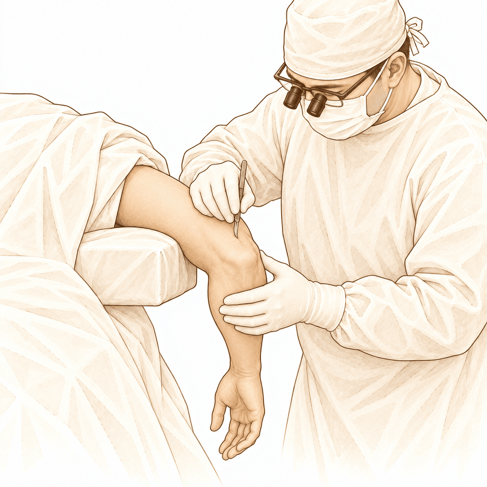
For patients: a plain-language version of this topic is available. See the patient guide.
Overview
Distal triceps tendon rupture is a rare but potentially challenging injury that traditionally occurs in an active population, most frequently in men aged 40 to 59 years [11, 14]. While acute partial ruptures may achieve good outcomes with nonoperative management, surgical repair is strongly recommended for complete ruptures [23]. Operative intervention is required in most cases, particularly for active patients, laborers, and athletes wishing to return to competition [31], as partial rupture is not well tolerated in patients with high functional demands [56]. Early surgical repair, performed within three weeks after injury, is the treatment of choice [6].
Surgical repair offers a predictable return of function with a small risk of loss of elbow motion [2, 12]. Distal triceps repair reliably restores elbow function and provides good patient outcomes with a low complication rate [1]. Primary repair of both acute and chronic ruptures yields satisfactory results in the majority of patients with a low rerupture rate, regardless of the repair construct used [10, 13]. Triceps tendon ruptures may result in uniformly good to excellent results if recognized and treated surgically [16].
Despite a heightened risk of perioperative complications, favorable functional outcomes and no cases of re-rupture were identified at short- to mid-term follow-up [7, 9]. However, distal triceps repair for traumatic injuries is associated with 14% complication and 13.1% reoperation rates [8], and surgery generally carries a relatively high complication and reoperation rate regardless of technique [17]. Patients experience improvements in postoperative outcomes, though there is a moderate reported risk of rerupture or complication [4]. Approximately 93% of patients returned to work by 2.2 ± 3.2 months postoperatively [3], and 89.7% returned to sport by 5.9 ± 4.4 months [5]. The overall return-to-sport rate was high (89.3%), with no consensus on return-to-sport criteria [18]. Current knowledge regarding triceps tendon repairs is derived from low level evidence [19].
Anatomy & Pathophysiology
Bony Anatomy
The elbow functions as a trocho-ginglymoid joint comprising medial and lateral articulations that provide inherent bony stability [61]. The ulnohumeral joint forms where the trochlea articulates with the ulna within the greater sigmoid notch, while the radiocapitellar joint involves the articulation of the capitellum with the radial head [61]. The proximal ulna articulates with the trochlea and features the olecranon process posteriorly and the coronoid process anteriorly [69]. The olecranon serves as a broad posterior attachment site for the triceps [61].
The distal humerus consists of medial and lateral columns, with the articular surface angled 30 degrees anterior to the humeral shaft axis [67]. In the coronal plane, the medial column diverges from the humeral shaft at a 45-degree angle, whereas the lateral column diverges at a 20-degree angle [69]. The trochlea is covered by articular cartilage forming an arc of almost 270 degrees [72]. The olecranon fossa accepts the olecranon during extension and is separated from the coronoid fossa by a thin bony septum [72].
Soft Tissue Anatomy
The triceps acts as the primary elbow extensor, inserting on the olecranon process [62]. This insertion is broad and tendinous, located 1.1 cm from the tip of the olecranon with a width of 2.6 cm [60, 69]. The distal triceps tendon insertional anatomy comprises three distinct areas: the posterior capsular insertion, the deep muscular portion, and the superficial tendinous portion [60]. The deep muscular head corresponds to the medial head, while the tendinous portion corresponds to the long and lateral heads [60]. Magnetic resonance imaging describes a bipartite insertion between these superficial and deep components into the olecranon [25]. A defined interval known as the "triceps decussation" exists between the lateral triceps expansion and medial triceps tendon just proximal to the olecranon [25].
The brachialis, the strongest elbow flexor, attaches to the coronoid 11 mm distal to the tip [62]. The medial ulnar collateral ligament originates on the posterior medial epicondyle and inserts on the sublime tubercle of the medial coronoid process [67]. The lateral collateral ligament complex consists of the radial collateral ligament, the lateral ulnar collateral ligament, and the annular ligament [72].
Pathophysiology & Mechanism of Injury
The usual mechanism for distal triceps rupture is a forceful sudden flexion of an extended elbow [25]. Lacerations and open injuries, with or without elbow fracture-dislocation, can also cause distal triceps rupture [25]. Distal triceps tendon lesions may result from acute traumatic events or pathological chronic tendinitis in patients with cumulative submaximal loading [25]. Iatrogenic conditions involving prior surgery that violated the extensor mechanism are another recognized cause [25].
In fall on an outstretched hand injuries, the lateral and long heads of the triceps brachii play primary roles in eccentric contraction, resulting in only superficial tendon tears [29]. In contrast, direct injury groups exhibit more full-thickness tendon tears than indirect mechanism injuries [29]. Most traumatic distal triceps tendon ruptures developing by indirect mechanisms are partial tears [29]. Repair of the triceps insertion is recommended to prevent weakness of elbow extension [40]. Differences in triceps strength are greatest at increasing positions of elbow extension, where triceps strength is more functionally important [28].
Classification
No Formal System: No formal classification system for distal triceps rupture has been established [55]. Distal triceps injuries are described based on results of imaging studies, including ultrasonography and MRI, as well as intraoperative findings [55].
Anatomic Factors: The degree of the tear is an anatomic factor important to the description of distal triceps injuries, categorized as complete, partial, or intact [55]. The location of the tear is an anatomic factor important to the description of distal triceps injuries, categorized as muscle belly, musculotendinous junction, tendinous insertion, or avulsion off bone [55]. The integrity of the lateral expansion is an anatomic factor important to the description of distal triceps injuries, categorized as intact versus torn [55].
Other Considerations: Nonanatomic factors that must be considered when managing triceps ruptures include the functional and medical status of the patient [55]. Nonanatomic factors that must be considered when managing triceps ruptures include the chronicity and atrophy of the muscle-tendon unit [55]. The main causes of triceps tendon lesions are acute traumatic events or pathological chronic tendinitis in patients with cumulative submaximal loading [25]. Iatrogenic conditions with a history of surgery that violated the extensor mechanism can cause triceps tendon lesions [25]. In fall on an outstretched hand injuries, all patients in the studied group had superficial distal triceps tendon tears [29]. In direct injury groups, three of seven elbows had full-thickness tendon tears [29]. The lateral and long heads of the triceps brachii have primary roles in eccentric contraction in fall on an outstretched hand injuries, resulting in only superficial tendon tears [29].
Clinical Presentation
Epidemiology and Demographics
Triceps tendon rupture is a rare injury, estimated to account for less than 1% of all upper limb tendon injuries [86]. This distal triceps tendon rupture is a rare but potentially challenging injury, traditionally occurring in an active population [14].
Mechanism and Etiology
The usual mechanism of injury is a forceful sudden flexion of an extended elbow [25]. Triceps tendon ruptures may result from acute traumatic events or pathological chronic tendinitis in patients with cumulative submaximal loading [25]. These ruptures can occur through healthy tissue [89].
Diagnosis and Clinical Challenges
These injuries can be difficult to diagnose and are frequently missed in the emergency department [86]. Missing the diagnosis potentially results in considerable loss of function [86].
Injury Classification and Severity
Partial ruptures of the distal triceps tendon typically occur in the superficial, lateral portion of the tendon [47]. Partial ruptures of the distal triceps tendon demonstrate a not statistically significant lower ultimate load to failure than intact tendons [47].
Investigations
Plain radiography: Plain radiographs remain the hallmark and best screening test for elbow evaluation [41]. Standard imaging views include AP, lateral, and oblique projections [41]. In the context of triceps tendon pathology, most ruptures are accompanied by an avulsion fracture of the olecranon [136]. Surgeons must suspect triceps tendon rupture when radiographs demonstrate a small fleck of bone avulsed from the olecranon [136].
CT: CT is helpful when assessing for malunion architecture and the location and pattern of osteophytes and/or loose bodies [41]. Three-dimensional CT is used to check for heterotopic ossification [41]. CT is not necessary when the stiffness is entirely soft-tissue related [41].
MRI: MRI can be used to evaluate ligaments and tendons, but it is rarely indicated for elbow stiffness [41].
Other Considerations: Electromyography/nerve conduction velocity studies should be performed if any question about neurologic dysfunction exists [41]. An assessment for ulnar nerve subluxation should be performed during physical examination [41]. Subluxation of the ulnar nerve is a relative contraindication for an arthroscopic procedure secondary to possible iatrogenic nerve injury [41].
Treatment
Non-Operative
Conservative management is appropriate for acute partial triceps tendon ruptures, which may achieve good outcomes without surgery [23]. Complete triceps tendon ruptures require surgical repair to restore active elbow extension, whereas partial tears may be treated conservatively [34]. In cases of bilateral partial triceps tendon rupture, conservative management has resulted in excellent functional outcomes [32]. Surgical treatment is also beneficial for severe triceps tears even after failed conservative treatment [46].
Operative
Indications: Most complete triceps tendon injuries should be managed with surgical repair [2]. Operative management is required for complete distal triceps tendon ruptures, particularly in active patients, laborers, and athletes wishing to return to competition [31].
Surgical Approach / Technique: Anatomic repair of triceps tendon ruptures demonstrates the most anatomic restoration and shows statistically significantly less repair-site motion when cyclically loaded [27]. The suture bridge repair technique provides an anatomic repair of the distal triceps tendon to its footprint, with potential advantages of increased load to failure, better footprint coverage, higher load resistance, and allowance for early motion [51]. Whether the stronger biomechanical properties and anatomic footprint coverage of the suture bridge technique translate to improved healing and functional outcome must be further assessed in future clinical studies [52]. Anatomical reconstruction using a 'double row' footprint reconstruction technique for traumatic rupture of the triceps yields favourable results [37]. In a patient who had undergone operative repair of the contralateral triceps, operative repair using bone tunnels led to a good outcome and should be considered over conservative management [26]. A V-shaped double-row distal triceps tendon repair with unicortical button fixation resulted in full elbow range of motion and extension muscle strength (5/5) compared to the uninjured arm 12 weeks after surgery [44].
Implant Selection: Primary repair of triceps ruptures with transosseous fixation has a significantly higher rerupture rate, higher reoperation rate, and longer release from medical care than does repair with anchor fixation [43]. The authors endorse restoring the anatomic footprint of the distal triceps using suture anchors and high-strength sutures to potentially improve fixation strength and allow for more rapid rehabilitation compared to traditional techniques [21]. The biomechanical strength of an all-suture construct is not different from that of suture anchors for repair of distal triceps avulsions [134]. Augmented triceps repair is superior to direct triceps repair for a distal triceps avulsion produced in a cadaver model [49].
Outcomes and Complications: Surgical repair offers a predictable return of function with a small risk of loss of elbow motion [2]. Patients undergoing distal triceps tendon rupture repair experience improvements in postoperative outcomes; however, there is a moderate reported risk of rerupture or complication [4]. Despite the heightened risk of perioperative complications after primary repair of distal triceps tendon injuries, favorable functional outcomes and no cases of re-rupture were identified at short- to mid-term follow-up [7]. Similarly, despite the heightened risk of perioperative complications, favorable functional outcomes and no cases of reruptures were found at short-term to midterm follow-up [9].
Return to Work and Sport: Surgical repair of a distal triceps injury results in reliable return to work and sport, with the majority of patients returning to the same physical intensity of work and the same intensity of sporting, respectively [15].
Complications
Overall Complication and Reoperation Rates: Distal triceps tendon repair is associated with a 14% complication rate and a 13.1% reoperation rate [8]. Patients undergoing repair for distal triceps tendon rupture experience a moderate reported risk of rerupture or complication [4]. Primary repair of distal triceps tendon injuries carries a heightened risk of perioperative complications [7, 9]. Distal triceps tendon repair surgery has a relatively high complication and reoperation rate regardless of repair technique [17]. In a cohort of American military personnel, the complication rate following triceps repair was 8% [83], and the retear rate was 3% [83].
Rerupture and Fixation-Related Complications: Primary repair of triceps ruptures with transosseous fixation has a significantly higher rerupture rate than repair with anchor fixation [43]. Primary repair of triceps ruptures with transosseous fixation has a significantly higher reoperation rate than repair with anchor fixation [43]. Iatrogenic fracture has been described with suture anchor placement during distal triceps repair [57]. Suture anchors require bone of a certain density and quality and must be placed at a specific location and trajectory to avoid ulnohumeral joint penetration [57]. Suboptimal suture anchor constructs are prone to screw pullout or under-restoration of the footprint, leading to a biomechanically weaker construct [57].
Wound Complications: Potential postoperative complications of direct repair of acute distal triceps ruptures include surgical site infections, wound dehiscence, and irritation from nonabsorbable suture knots [38]. Knot irritation is sometimes unavoidable in patients with deficient soft tissues following distal triceps repair [38].
Other Considerations: The use of autogenous semi-tendinous and gracilis tendons permits a good functional outcome in cases of triceps insufficiency after total elbow arthroplasty [143].
Recovery
Light activity (weeks): The evidence provided does not specify a distinct week range for light activities such as desk work, driving, or light activities of daily living.
Full activity (months): Surgical repair of a distal triceps injury results in reliable return to work and sport, with the majority of patients returning to the same physical intensity of work and the same intensity of sporting [15]. The overall return-to-sport rate after distal triceps repair was 89.3%, with no consensus on return-to-sport criteria [18].
Complete recovery / outcome plateau (months): Distal triceps tendon repair reliably restores elbow function and provides good patient outcomes with a low complication rate [1]. Surgical repair of complete triceps tendon injuries offers a predictable return of function with a small risk of loss of elbow motion [2]. Patients undergoing distal triceps tendon rupture repair experience improvements in postoperative outcomes, with a moderate reported risk of rerupture or complication [4].
Rehabilitation protocol: Early surgical repair within three weeks after injury is the treatment of choice for distal triceps tendon ruptures [6]. Restoring the anatomic footprint of the distal triceps using suture anchors and high-strength sutures may improve fixation strength and allow for more rapid rehabilitation compared to traditional techniques [21].
Functional milestones: Primary repair of distal triceps tendon injuries is associated with favorable functional outcomes and no cases of re-rupture at short- to mid-term follow-up [7]. Primary repair of distal triceps tendon injuries is associated with favorable functional outcomes and no cases of reruptures at short-term to midterm follow-up [9].
Other Considerations: Partial triceps tendon ruptures can heal without functional deficit [30].
Key Evidence
- [L4] Distal triceps tendon repair reliably restores elbow function, provides good patient outcomes and has a low complication rate. [1] (10.1016/j.jse.2016.12.062)
- [L5] Most complete triceps tendon injuries should be managed with surgical repair, which offers a predictable return of function with a small risk of loss of elbow motion. [2] (10.1016/j.hcl.2015.06.010)
- [L4] Approximately 93% of patients who underwent distal triceps repair returned to work by 2.2 ± 3.2 months postoperatively. [3] (10.1016/j.jse.2020.07.036)
- [L4] Patients undergoing distal triceps tendon rupture repair experience improvements in postoperative outcomes; however, there is a moderate reported risk of rerupture or complication. [4] (10.1016/j.jse.2021.06.019)
- [L4] Distal triceps repair enables 89.7% of patients to return to sport by 5.9 ± 4.4 months following surgery. [5] (10.1016/j.jhsa.2021.11.021)
- [L4] Early surgical repair, within three weeks after the injury, is the treatment of choice for distal triceps tendon ruptures. [6] (10.2106/00004623-200310000-00015)
- [L4] Despite heightened risk of perioperative complications after primary repair of distal triceps tendon injuries, the current series identifies favorable functional outcomes and no cases of re-rupture at short- to mid-term follow-up. [7] (10.1177/2325967118s00163)
- [L3] Distal triceps repair for traumatic injuries is associated with 14% complication and 13.1% reoperation rates. [8] (10.1016/j.xrrt.2022.05.004)
- [L4] Despite the heightened risk of perioperative complications after primary repair of distal triceps tendon injuries, the current series found favorable functional outcomes and no cases of reruptures at short-term to midterm follow-up. [9] (10.1177/2325967119839998)
- [L3] Primary repair of distal triceps tendon ruptures yields good, durable patient outcomes with minimal rerupture regardless of repair construct. [10] (10.1016/j.jse.2017.08.006)
- [L3] Distal triceps repairs in this large cohort study occur most frequently in men aged 40 to 59 years. [11] (10.1177/15589447221095114)
- [Case_report] Surgical repair of distal triceps tendon ruptures can be accomplished with predictable results. [12] (10.1016/j.jse.2006.06.002)
- [L4] Primary repair of acute and chronic distal triceps tendon ruptures in a general population yields satisfactory results in the majority of patients with a low rerupture rate. [13] (10.1302/0301-620x.100b5.bjj-2017-1057.r2)
- [L4] A distal triceps tendon rupture is a rare but potentially challenging injury, traditionally occurring in an active population, and often requires surgical repair. [14] (10.1177/03635465241283970)
- [L3] Surgical repair of a distal triceps injury results in reliable return to work and sport, with the majority of patients returning to the same physical intensity of work and the same intensity of sporting, respectively. [15] (10.1177/2325967120s00370)
- [L4] Triceps tendon ruptures may result in uniformly good to excellent results if recognized and treated surgically. [16] (10.1177/036354659302100327)
- [L4] Regardless of repair technique, distal triceps tendon repair surgery has a relatively high complication and reoperation rate. [17] (10.1016/j.xrrt.2024.06.008)
- [L4] The overall return-to-sport rate after distal triceps repair was high (89.3%), with no consensus on return-to-sport criteria. [18] (10.1177/23259671241275956)
- [L1] What is known about triceps tendon repairs comes from low level evidence. [19] (10.1016/j.jht.2025.02.006)
- [Paper] The authors endorse restoring the anatomic footprint of the distal triceps using suture anchors and high-strength sutures to potentially improve fixation strength and allow for more rapid rehabilitation compared to traditional techniques. [21] (10.1016/j.eats.2018.04.013)
- [L4] Endoscopic repair of partial distal triceps tendon tears leads to good clinical and radiological results after 12 months, with patients benefiting especially from an improvement of extension strength. [22] (10.1016/j.arthro.2013.03.049)
- [L4] Acute partial triceps tendon ruptures may have good outcomes with nonoperative management, whereas surgical repair is strongly recommended for complete ruptures. [23] (10.2106/jbjs.rvw.19.00172)
- [L5] In the Mayo experience of 900 elbow replacements, 25 reoperations were performed for triceps insufficiency after the surgical technique described above. [24] (10.1097/00132589-200203000-00007)
- [L5] [25] (10.1007/s12306-015-0359-y)
- [L5] It also demonstrates that operative repair using bone tunnels in a patient who has had an operative repair of the contralateral triceps led to a good outcome and should be considered over conservative management. [26] (10.1093/milmed/usx075)
- [L5] Anatomic repair of triceps tendon ruptures demonstrated the most anatomic restoration of distal triceps ruptures and showed statistically significantly less repair-site motion when cyclically loaded. [27] (10.1177/0363546509358319)
- [L5] These differences are greatest at increasing positions of elbow extension where triceps strength is more functionally important. [28] (10.1053/jhsu.2003.50036)
- [L3] [29] (10.1097/corr.0000000000001550)
- [L4] Partial triceps tendon ruptures can heal without functional deficit. [30] (10.1177/0095399703258707)
- [L5] Operative management of complete distal biceps and triceps tendon ruptures is required in most cases, especially for active patients, laborers, and athletes wishing to return to competition. [31] (10.1016/j.hcl.2016.08.019)
- [L4] Conservative management was entirely appropriate for this patient with bilateral partial triceps tendon rupture, resulting in excellent functional outcome. [32] (10.1177/0363546503258903)
- [L5] Complete triceps tendon ruptures must be repaired to provide active extension at the elbow, while partial tears may be treated conservatively. [34] (10.1016/j.jhsa.2015.05.016)
- [L4] Anatomical reconstruction using a 'double row' footprint reconstruction technique for traumatic rupture of triceps yields favourable results in our patient group. [37] (10.1111/j.1758-5740.2012.00218.x)
- [L5] [38] (10.1097/bth.0000000000000082)
- [L5] To prevent weakness of elbow extension, we recommend repair of the triceps insertion. [40] (10.1016/s0020-1383(98)00208-3)
- [L3] Primary repair of triceps ruptures with transosseous fixation has a significantly higher rerupture rate, higher reoperation rate, and longer release from medical care than does repair with anchor fixation. [43] (10.1177/0363546518757426)
- [L5] The patient reached full elbow range of motion and extension muscle strength (5/5) compared to the uninjured arm 12 weeks after surgery. [44] (10.1186/s40001-017-0250-4)
- [L4] Surgical treatment seems to be beneficial in severe triceps tears even after failed conservative treatment. [46] (10.1007/s00402-011-1319-0)
- [L5] Partial ruptures of the distal triceps tendon demonstrate a not statistically significant lower ultimate load to failure than intact tendons and typically occur in the superficial, lateral portion of the tendon. [47] (10.1186/s12891-023-06720-3)
- [L5] Augmented triceps repair is superior to direct triceps repair for a distal triceps avulsion produced in a cadaver model. [49] (10.1016/j.jse.2010.08.017)
- [L4] The suture bridge repair technique provides an anatomic repair of the distal triceps tendon to its footprint with potential advantages of increased load to failure, better footprint coverage, higher load resistance, and allows for early motion. [51] (10.1055/s-0038-1636729)
- [L4] Whether the stronger biomechanical properties and anatomic footprint coverage of this technique translates to improved healing and functional outcome must be further assessed in future clinical studies. [52] (10.1097/bte.0b013e3182274292)
- [L5] [55] (10.5435/00124635-201001000-00005)
- [L4] Partial rupture of brachii triceps tendon is not well tolerated in high functional demand patients. [56] (10.1016/j.otsr.2011.09.022)
- [L4] [57] (10.1097/bth.0000000000000196)
- [L4] [83] (10.1177/1558944717745499)
- [L4] [86] (10.1177/1758573217706358)
- [L4] Triceps tendon ruptures can occur through healthy tissue, and the time of delay from injury to repair seems to have little effect on the functional outcome. [89] (10.1177/036354658401200415)
- [L5] These findings suggest that the biomechanical strength of an all-suture construct is not different from that of suture anchors for repair of distal triceps avulsions. [134] (10.1016/j.jse.2018.05.025)
- [L5] Most triceps tendon ruptures are accompanied by avulsion fracture of the olecranon, and it is important to suspect this injury when radiographs show a small fleck of bone avulsed from the olecranon. [136] (10.1007/s00590-012-1127-4)
- [L5] The use of autogenous semi-tendinous and gracilis tendons permits a good functional outcome when direct reattachment of the triceps brachial is impossible due to tendon retraction or loss of substance, or in cases of triceps insufficiency after total elbow arthroplasty. [143] (10.1016/j.main.2007.05.006)
See Also
References
[1] Outcomes following distal triceps tendon repair. Journal of Shoulder and Elbow Surgery. 2017. DOI: 10.1016/j.jse.2016.12.062
[2] Distal Triceps Tendon Injuries. Hand Clinics. 2015. DOI: 10.1016/j.hcl.2015.06.010
[3] Return to work following distal triceps repair. Journal of Shoulder and Elbow Surgery. 2021. DOI: 10.1016/j.jse.2020.07.036
[4] The surgical management of distal triceps tendon ruptures: a systematic review. Journal of Shoulder and Elbow Surgery. 2022. DOI: 10.1016/j.jse.2021.06.019
[5] Return to Sport Following Distal Triceps Repair. The Journal of Hand Surgery. 2023. DOI: 10.1016/j.jhsa.2021.11.021
[6] SURGICAL TREATMENT OF DISTAL TRICEPS RUPTURES. The Journal of Bone and Joint Surgery-American Volume. 2003. DOI: 10.2106/00004623-200310000-00015
[7] Surgical Repair of Distal Triceps Tendon Injuries: Short-Term Clinical Outcomes and Re-Rupture Rate. Orthopaedic Journal of Sports Medicine. 2018. DOI: 10.1177/2325967118s00163
[8] Complications after traumatic distal triceps tears: an analysis of 107 cases. JSES Reviews, Reports, and Techniques. 2022. DOI: 10.1016/j.xrrt.2022.05.004
[9] Surgical Repair of Distal Triceps Tendon Injuries: Short-term to Midterm Clinical Outcomes and Risk Factors for Perioperative Complications. Orthopaedic Journal of Sports Medicine. 2019. DOI: 10.1177/2325967119839998
[10] Functional outcomes of distal triceps tendon repair comparing transosseous bone tunnels with suture anchor constructs. Journal of Shoulder and Elbow Surgery. 2017. DOI: 10.1016/j.jse.2017.08.006
[11] Surgical Outcomes, Trends, and Risk Factors of Distal Triceps Repairs. HAND. 2022. DOI: 10.1177/15589447221095114
[12] Distal triceps tendon rupture and repair in an otherwise healthy pediatric patient: A case report and review of the literature. Journal of Shoulder and Elbow Surgery. 2007. DOI: 10.1016/j.jse.2006.06.002
[13] Results of primary repair of distal triceps tendon ruptures in a general population. The Bone & Joint Journal. 2018. DOI: 10.1302/0301-620x.100b5.bjj-2017-1057.r2
[14] Return to Work, Sport, and Sport- Related Activity After Distal Triceps Tendon Repair: A Systematic Review. The American Journal of Sports Medicine. 2025. DOI: 10.1177/03635465241283970
[15] Return to Work and Sport Following Distal Triceps Repair. Orthopaedic Journal of Sports Medicine. 2020. DOI: 10.1177/2325967120s00370
[16] Rupture of the triceps tendon associated with steroid injections. The American Journal of Sports Medicine. 1993. DOI: 10.1177/036354659302100327
[17] A comparison of distal triceps tendon repair outcomes by surgical technique. JSES Reviews, Reports, and Techniques. 2024. DOI: 10.1016/j.xrrt.2024.06.008
[18] Postoperative Rehabilitation and Return-to-Sport Criteria After Distal Triceps Rupture Repair: A Scoping Review. Orthopaedic Journal of Sports Medicine. 2024. DOI: 10.1177/23259671241275956
[19] Rehabilitation following distal triceps repair: A scoping review. Journal of Hand Therapy. 2025. DOI: 10.1016/j.jht.2025.02.006
[21] Distal Triceps Speed Bridge Repair. Arthroscopy Techniques. 2018. DOI: 10.1016/j.eats.2018.04.013
[22] The Endoscopic Repair of Partial Lesions of the Distal Triceps Tendon: First Prospective Results of 14 Cases (SS‐42). Arthroscopy. 2013. DOI: 10.1016/j.arthro.2013.03.049
[23] Triceps Tendon Ruptures. JBJS Reviews. 2020. DOI: 10.2106/jbjs.rvw.19.00172
[24] Bryan–Morrey Triceps-Reflecting Approach. Techniques in Shoulder & Elbow Surgery. 2002. DOI: 10.1097/00132589-200203000-00007
[25] Triceps tendon rupture: the knowledge acquired from the anatomy to the surgical repair. MUSCULOSKELETAL SURGERY. 2015. DOI: 10.1007/s12306-015-0359-y
[26] A Case of Non-simultaneous Bilateral Partial Triceps Tendon Repair. Military Medicine. 2017. DOI: 10.1093/milmed/usx075
[27] The Distal Triceps Tendon Footprint and a Biomechanical Analysis of 3 Repair Techniques. The American Journal of Sports Medicine. 2010. DOI: 10.1177/0363546509358319
[28] Partial olecranon excision: The relationship between triceps insertion site and extension strength of the elbow. The Journal of Hand Surgery. 2003. DOI: 10.1053/jhsu.2003.50036
[29] Differences in Rupture Patterns and Associated Lesions Related to Traumatic Distal Triceps Tendon Rupture Between Outstretched Hand and Direct Injuries. Clinical Orthopaedics & Related Research. 2020. DOI: 10.1097/corr.0000000000001550
[30] Triceps Tendon Ruptures in Professional Football Players. The American Journal of Sports Medicine. 2004. DOI: 10.1177/0095399703258707
[31] Biceps and Triceps Ruptures in Athletes. Hand Clinics. 2017. DOI: 10.1016/j.hcl.2016.08.019
[32] Bilateral Partial Rupture of Triceps Tendon. The American Journal of Sports Medicine. 2004. DOI: 10.1177/0363546503258903
[34] Triceps Tendon Repair. The Journal of Hand Surgery. 2015. DOI: 10.1016/j.jhsa.2015.05.016
[37] Triceps Rupture: A Case Series, Anatomical Study of the Triceps Footprint and Description of Surgical Technique. Shoulder & Elbow. 2013. DOI: 10.1111/j.1758-5740.2012.00218.x
[38] A Treatment Algorithm for the Management of Distal Triceps Ruptures. Techniques in Hand & Upper Extremity Surgery. 2015. DOI: 10.1097/bth.0000000000000082
[40] Rupture of the triceps muscle at its attachments. Injury. 1999. DOI: 10.1016/s0020-1383(98)00208-3
[41] Orthopaedic Knowledge Update 13 Ebook Without Multimedia. Anatomy, Biomechanics, Physical Examination, and Imaging of the Elbow > Summary and Conclusions.
[43] Operative Management of Acute Triceps Tendon Ruptures: Review of 184 Cases. The American Journal of Sports Medicine. 2018. DOI: 10.1177/0363546518757426
[44] V-shaped double-row distal triceps tendon repair: a novel technique using unicortical button fixation. European Journal of Medical Research. 2017. DOI: 10.1186/s40001-017-0250-4
[46] Triceps tears in athletes: different injury patterns and surgical treatment. Archives of Orthopaedic and Trauma Surgery. 2011. DOI: 10.1007/s00402-011-1319-0
[47] Partial ruptures of the distal triceps tendons show only slightly lower ultimate load to failure: a biomechanical study. BMC Musculoskeletal Disorders. 2023. DOI: 10.1186/s12891-023-06720-3
[49] Triceps tendons: a biomechanical comparison of intact and repaired strength. Journal of Shoulder and Elbow Surgery. 2011. DOI: 10.1016/j.jse.2010.08.017
[51] Triceps Tendon Anatomic Repair Utilizing the “Suture Bridge” Technique. Journal of Hand and Microsurgery. 2018. DOI: 10.1055/s-0038-1636729
[52] Distal Triceps Tendon Repair Using an Anatomic Footprint Repair. Techniques in Shoulder & Elbow Surgery. 2011. DOI: 10.1097/bte.0b013e3182274292
[55] Distal Triceps Rupture. American Academy of Orthopaedic Surgeon. 2010. DOI: 10.5435/00124635-201001000-00005
[56] Distal partial ruptures of triceps brachii tendon in an athlete. Orthopaedics & Traumatology: Surgery & Research. 2012. DOI: 10.1016/j.otsr.2011.09.022
[57] Triceps Repair and Restoration of Triceps Footprint With Anchorless Suture Fixation. Techniques in Hand & Upper Extremity Surgery. 2018. DOI: 10.1097/bth.0000000000000196
[60] Orthopaedic Knowledge Update 13 Ebook Without Multimedia. Anatomy, Biomechanics, Physical Examination, and Imaging of the Elbow > Annotated References.
[61] Orthopaedic Knowledge Update 13 Ebook Without Multimedia. Anatomy, Biomechanics, Physical Examination, and Imaging of the Elbow > Anatomy > Bony Anatomy.
[62] Miller S Review Of Orthopaedics. SECTION 16 PATELLAR TRACKING IN TOTAL KNEE ARTHROPLASTY > TABLE 2.3 Shoulder Spaces.
[67] Miller S Review Of Orthopaedics. ELBOW.
[69] A Lange Medical Book Current Diagnosis Treatment In Orthopedics Fifth Edition. 2Musculoskeletal Trauma Surgery > INJURIES AROUND THE ELBOW.
[72] Rockwood And Green S Fractures In Adults. 29: Principles of Nonunion and Bone Defect Treatment > Pathoanatomy and Applied Anatomy Related to Distal Humerus Fractures.
[83] Outcomes of Triceps Rupture in the US Military: Minimum 2-Year Follow-up. HAND. 2017. DOI: 10.1177/1558944717745499
[86] Triceps tendon rupture: repair and rehabilitation. Shoulder & Elbow. 2017. DOI: 10.1177/1758573217706358
[89] Triceps tendon avulsion in a professional body builder. The American Journal of Sports Medicine. 2004. DOI: 10.1177/036354658401200415
[134] Distal triceps transosseous cruciate versus suture anchor repair using equal constructs: a biomechanical comparison. Journal of Shoulder and Elbow Surgery. 2018. DOI: 10.1016/j.jse.2018.05.025
[136] Triceps tendon avulsion: a case report and discussion about the olecranon ossification nucleus. European Journal of Orthopaedic Surgery & Traumatology. 2012. DOI: 10.1007/s00590-012-1127-4
[143] Réparation d'une rupture complète du triceps brachial par greffe tendineuse au DIDT. À propos d'un cas et revue de la littérature. Chirurgie de la Main. 2007. DOI: 10.1016/j.main.2007.05.006