Clinicians › Shoulder
Rehabilitation After Labral / SLAP Repair
Phased rehabilitation protocols after arthroscopic labral / SLAP repair. Protection phase, loading restrictions, return-to-overhead timelines. Modifications for combined nerve decompression.
Overview
Arthroscopic repair of superior labral anterior-posterior (SLAP) lesions yields predictably good functional results and an acceptable rate of return to sport or work [4]. Long-term outcomes after isolated labral repair are good and independent of age [1], with a good or excellent functional outcome anticipated in 87% of cases following type II SLAP repair [11]. Patients undergoing superior labral repair following acute trauma experience the most reliable functional recovery, characterized by increased forward flexion, lower revision risk, and higher return to play compared with other cohorts [2]. In the general population, arthroscopic type 2 SLAP repair induces good short- and long-term clinical outcomes, return to overhead activities, and subjective satisfaction regardless of age [15]. For patients younger than 50 years with a coexistent rotator cuff tear, predictable short-term surgical results and return to activity can be expected after repair of type II SLAP lesions [6]. Arthroscopic treatment of both rotator cuff and labral lesions yields good clinical outcomes, restoration of motion, and a high degree of patient satisfaction [7].
Surgical indications require caution, particularly in overhead throwing athletes where return to play after SLAP repair is poor [35]. SLAP repair should be considered only after nonsurgical management has failed [36], although nonoperative treatment in athletes can be successful if the rehabilitation program is completed before attempting a return to play [13]. In a young active population, primary arthroscopic biceps tenodesis is a viable surgical alternative to labral repair for type II SLAP lesions [9]. High-demand patients with biceps tendonitis and a SLAP lesion with labral instability who undergo combined tenodesis and labral repair have significantly worse outcomes than those who undergo either isolated labral repair or isolated biceps tenodesis [57]. Neither labral repair nor biceps tenodesis provided significant clinical benefit over sham surgery for patients with SLAP II lesions in the studied population [5].
Return to play criteria show minimal variability between North American and European shoulder surgeons despite the absence of evidence-based guidelines [3]. Athletes who undergo an objective return to play criteria based testing protocol have lower rates of recurrent instability following arthroscopic shoulder stabilization surgery than those cleared by time from surgery [38]. In active duty males, arthroscopic repair of isolated Type II SLAP had comparable results to combined Type II SLAP repairs, with the combined treatment group having significantly better results in two of three parameters measured [12]. Following arthroscopic repair, patients with Type V SLAP tears had a similar overall rate of return to play compared directly to a control group of patients who underwent arthroscopic Bankart repair alone [17]. Both arthroscopic-assisted subpectoral biceps tenodesis combined with anterior labral repair and arthroscopic SLAP repair led to statistically and clinically significant increases in outcome scores, marked improvements in pain, and high rates of return to unrestricted active duty in military patients with type V SLAP lesions [21]. Arthroscopic posterior labral repair has demonstrated favorable outcomes with high rates of return to play, although clinical evidence is limited [10]. Surgical positioning for arthroscopic posterior shoulder labral repair did not affect postoperative clinical and patient-reported outcomes [16]. Arthroscopic capsulolabral repair for posterior shoulder instability improved long-term shoulder pain and function and facilitated return to sport in the majority of patients at a mean follow-up of 15.4 years, although a notable proportion of patients met various criteria for failure [197].
Anatomy & Pathophysiology
Glenoid and Labrum
The glenoid is a convex structure of shallow depth shaped like an inverted pear [72]. Its subchondral bone is relatively flat, with the articular concavity augmented by cartilage and a circumferential labrum [76]. The glenoid averages 5° of retroversion in relation to the axis of the scapular body [76]. The glenoid labrum provides concavity and up to 50% of marginal glenoid socket depth [76]. Composed of dense fibrocartilaginous tissue, the labrum increases the depth of the socket by 50% around the humeral head [86]. The glenoid articular surface and the labrum combine to create a socket that is approximately 9 mm deep in the superoinferior direction and 5 mm deep in the anteroposterior direction [86]. Adding the glenoid labrum increases the glenoid surface to 75% of the humeral head vertically and 57% horizontally [86]. Biomechanical testing of cadaver shoulder specimens showed that the labrum affects the distribution of contact stresses when a compressive load is applied to the shoulder at 90 degrees of abduction [86].
Labral anatomic variants include a sublabral foramen (anterosuperior) and the Buford complex (absence of anterosuperior labrum and cordlike middle glenohumeral ligament) [87]. Repairing an anterosuperior labral variant may cause loss of external rotation [87].
Proximal Humerus
The proximal humerus anatomy comprises four main parts: the humeral head, greater tuberosity (GT), lesser tuberosity (LT), and humeral shaft [72]. The articular head is spherical and has a diameter of 37 to 57 mm [72]. The most superior portion of the articular surface of the humeral head averages 8 mm above the GT [72]. Humeral version averages 29.8 degrees, with a range of 10 to 55 degrees [72]. The humeral head is inclined approximately 130 degrees with respect to the humeral shaft [72]. The bicipital groove lies between the GT and LT and serves as a pathway for the long head of the biceps as it traverses from its intraarticular origin into the proximal arm [72]. The distal aspect of the bicipital groove is internally rotated with respect to the proximal portion [72]. The anatomic neck of the proximal humerus is located at the junction of the articular surface and the tuberosities [72]. The surgical neck represents an indistinct region (metadiaphyseal junction) below the tuberosities but above the humeral shaft [72]. The GT serves as the attachment site for the supraspinatus, infraspinatus, and teres minor tendons of the rotator cuff [72]. The LT serves as the attachment site for the subscapularis tendon [72].
The humeral head averages 19° of retroversion and 41° of inclination (neck-shaft angle) [76]. The humeral head is retroverted an average of 30 degrees [74]. The neck-shaft angle measures an average of 135 degrees [74]. The articular surface of the humeral head is essentially spherical, with an arc of approximately 160 degrees covered by articular cartilage [85]. The radius of curvature of the humeral head is approximately 25 mm and is slightly larger in men than in women [85]. The average neck-shaft angle is 45 degrees (±5 degrees), with a range of 30 to 50 degrees [85]. The superior margin of the humeral head articular surface normally is superior to the top of the greater tuberosity by 8 to 10 mm [85]. The distance from the lateral base of the coracoid process to the lateral margin of the greater tuberosity is called the lateral humeral offset [85]. A significant decrease in lateral humeral offset reduces the lever arms for the deltoid and supraspinatus muscles, which weakens abduction and impairs function [85]. A significant increase in lateral humeral offset causes excessive tension on the soft tissues ("overstuffing" of the joint), which results in loss of motion and likely accelerates polyethylene wear [85]. Humeral articular malposition of more than 4 mm led to increased subacromial contact [85]. An offset of 8 mm in any direction significantly decreased passive range of motion [85]. Proximal humeral retroversion is highly variable, ranging from 0 to 55 degrees, depending on the method used for measurement [85].
Scapula and Shoulder Girdle
The scapula is attached to the axial skeleton by the clavicle, specifically by the acromioclavicular (AC) and sternoclavicular (SC) joints [75]. The scapula is separated from the chest wall by thin gliding fibro-fatty tissue, allowing its smooth excursion over the chest wall [75]. The glenoid is connected with the flat body of the scapula by the scapular neck [75]. The hook-shaped coracoid process curves forwards from the superior surface of the scapular neck [75]. The scapular spine ends in a flattened bony process, the acromion, which curves forwards [75]. The highest concentration of bony mass in the scapula is found in the glenoid, the scapular neck (including the base of the coracoid process), and the lateral border of the scapular body [75]. Two bony pillars transmit compressive forces from the glenoid fossa: the lateral pillar and the spinal pillar [75]. The lateral pillar connects the inferior border of the glenoid with the inferior angle [75]. The spinal pillar arises from the central part of the glenoid and continues medially to become part of the base of the scapular spine [75]. The weakest bone in the scapula is located primarily in the central part of the biomechanical body, i.e., in the infraspinous fossa [75]. The weakest area of the circumference of the biomechanical body of the scapula is the spinomedial angle [75].
Normal shoulder motion is approximately two-thirds glenohumeral and one third scapulothoracic [76]. The scapula has only one true diarthrodial articulation, the acromioclavicular (AC) joint [76]. The acromion has three ossification centers: the metacromion (base), the mesoacromion (middle), and the preacromion (tip) [76]. Failure of fusion of the acromial ossification centers results in os acromiale [76]. The coracobrachialis muscle and the short head of the biceps tendon originate from the coracoid process [76]. The pectoralis minor muscle inserts onto the medial coracoid process [76]. The superior shoulder suspensory complex (SSSC) provides a stable connection between the scapula and the axial skeleton [76]. The SSSC is composed of the glenoid, the coracoid process, the coracoclavicular ligaments, the distal clavicle, the AC joint, and the acromion [76]. The superior strut of the SSSC comprises the middle clavicle [76]. The inferior strut of the SSSC comprises the lateral scapular border/spine of the scapula [76].
The scapula spans second through seventh ribs and serves as an attachment for 17 muscles [87]. The scapula is anteverted on the chest wall approximately 30 degrees relative to the body [87]. The coracoacromial ligament contributes to anterosuperior stability in rotator cuff deficiency and should be preserved with irreparable cuff tears to prevent anterosuperior escape [87]. The acromial branch of the thoracoacromial artery runs on the medial aspect of the coracoacromial ligament [87]. The coracoacromial ligament is the arthroscopic landmark for a complete release of the rotator interval for adhesive capsulitis [87].
The clavicle is the first bone to ossify (fifth week of gestation) and is the only long bone to ossify by intramembranous ossification [76]. The medial (sternal) epiphysis of the clavicle is the last ossification center to fuse, at age 20 to 25 years [76]. The primary blood supply to the clavicle is periosteal; no nutrient artery is present [76]. The clavicle serves as the primary stabilizer between the axial skeleton (via the sternoclavicular joint) and the appendicular skeleton (via the acromioclavicular joint) [82]. The coracoclavicular ligaments are the primary stabilizers to superior (vertical) translation of the distal clavicle [82]. The SSSC is a bone–soft-tissue ring that provides a stable connection of the glenoid and scapula to the clavicle [82]. The SSSC is composed of four bony landmarks—distal clavicle, acromion, coracoid process, and glenoid neck—and the supporting ligamentous complexes of the AC joint and the CC ligaments [82].
The scapula in humans is suspended by muscles alone and has shifted caudally from the cervical position in lower animals [83]. Broadening of the infraspinatus fossa has resulted in a change in the vector of muscle pull from the axillary border of the scapula to the glenoid fossa [83]. This adaptation allows the infraspinatus and teres minor muscles to be more effective in their roles as depressors and external rotators of the humeral head [83]. The acromion has enlarged over time, reflecting the increasing role of the deltoid muscle in shoulder function [83]. The broader attachment of the deltoid on the acromion and its more distal insertion on the humerus have increased its mechanical advantage in shoulder motion [83]. The coracoid process has undergone an increase in size over time [83]. With the shoulder in 90 degrees of abduction, the coracoid extension over the glenohumeral joint can mechanically limit anterior translation of the humerus relative to the glenoid [83].
Joints and Ligaments
The sternoclavicular (SC) joint is the only true diarthrodial articulation between the upper appendicular and axial skeletons [76]. The posterior SC joint capsule and ligaments are the primary stabilizers to anterior and posterior translation of the medial clavicle [76]. The AC joint is a small diarthrodial joint with an interposed fibrocartilaginous disk [76]. The superior and posterior AC ligaments are the primary stabilizers to anterior and posterior (horizontal) translation of the clavicle [76]. The coracoclavicular ligaments (conoid: medial; trapezoid: lateral) are the primary stabilizers to superior (vertical) translation of the distal clavicle [76].
The rotator cuff stabilizes the glenohumeral joint via joint compression [76]. Static stabilizers of the glenohumeral joint include articular congruity, the glenoid labrum, concavity-compression, negative intra-articular pressure, and the glenohumeral capsule and ligaments [76]. The rotator interval is defined medially by the base of the coracoid, superiorly by the supraspinatus tendon, and inferiorly by the subscapularis tendon [76]. The rotator interval contains the coracohumeral (CH) ligament, the superior glenohumeral ligament (SGHL), and the intra-articular portion of the long head of the biceps tendon [76]. Laxity of the rotator interval results in inferior laxity (the sulcus sign) [76]. Contracture of the rotator interval is seen with adhesive capsulitis [76].
The CH ligament restricts external rotation in adduction and is a static restraint to inferior and posterior translation in adduction and external rotation [76]. The SGHL is a primary static restraint against anterior translation with the arm at the side [76]. With the CH ligament, the SGHL forms a pulley that provides restraint against medial subluxation of the long head of the biceps tendon [76]. The middle glenohumeral ligament (MGHL) is a primary static restraint against anterior translation with the arm in external rotation and 45° of abduction [76]. The anterior band of the inferior glenohumeral ligament (AB-IGHL) is a primary static restraint against anterior-inferior dislocation of the glenohumeral joint in 90° of abduction and external rotation (position of apprehension) [76]. The posterior band of the IGHL (PB-IGHL) is a primary static restraint against posterior-inferior translation in internal rotation and adduction [76].
The superior transverse scapular ligament arises from the medial base of the coracoid overlying the suprascapular notch [76]. The suprascapular artery runs superior to the superior transverse scapular ligament, and the nerve runs deep to the ligament [76]. Entrapment of the suprascapular nerve at the superior transverse scapular ligament causes denervation of both the supraspinatus and the infraspinatus [76]. The spinoglenoid ligament overlies the suprascapular nerve at the spinoglenoid notch [76]. Entrapment, traction, or compression of the suprascapular nerve at the spinoglenoid notch causes denervation of the infraspinatus [76].
The glenohumeral joint is a ball and socket joint with the greatest range of motion in the body [87]. Motion at the glenohumeral joint is at the expense of stability, which is provided by static and dynamic restraints [87]. Static restraints of the glenohumeral joint include articular anatomy, glenoid labrum, glenohumeral ligaments, capsule, and negative intraarticular pressure [87]. Dynamic stabilizers of the glenohumeral joint include the rotator cuff and biceps tendon [87]. Scapulothoracic mechanics contribute to stability of the glenohumeral joint [87]. The fibrocartilaginous glenoid labrum deepens the socket 50% and provides a bumper to translation [87]. The coracohumeral ligament restrains inferior translation and external rotation of the adducted arm [87]. The superior glenohumeral ligament restrains external rotation and inferior translation of the adducted or slightly abducted arm [87]. The middle glenohumeral ligament is absent in up to 30% of shoulders [87]. The middle glenohumeral ligament restrains anterior translation with the arm abducted to 45 degrees [87]. The inferior glenohumeral ligament, anterior band, restrains anterior and inferior translation with the arm externally rotated and abducted to 90 degrees (position of apprehension) [87]. The inferior glenohumeral ligament, posterior band, restrains posterior and inferior translation with the arm internally rotated and abducted to 90 degrees [87].
The sternoclavicular joint is a double gliding joint with an articular disc [87]. The sternoclavicular joint is the only true joint connecting the upper extremity with the axial skeleton [87]. The posterior sternoclavicular ligament is the strongest and primary restraint to anteroposterior instability [87]. The sternoclavicular joint rotates 30 degrees with shoulder motion [87]. The AC joint is a plane/gliding joint with a fibrocartilaginous disc [87]. AC ligaments prevent anteroposterior displacement [87]. The posterior and superior AC ligaments are considered the strongest [87]. Coracoclavicular ligaments prevent superior displacement of the distal clavicle [87]. The trapezoid ligament is anterolateral and approximately 25 mm from the AC joint [87]. The conoid ligament is posteromedial, stronger, and approximately 45 mm from the AC joint [87].
The shoulder joint is composed of four articulations: the sternoclavicular, acromioclavicular, glenohumeral, and scapulothoracic [86]. The bony anatomy contributes little to stability and has been compared with a golf ball on a tee [86]. The ligamentous constraints are the primary stabilizers at extremes of motion [86]. The superior glenohumeral ligament is the primary restraint to inferior humeral subluxation in 0 degrees of abduction and is the primary stabilizer to anterior and posterior stress in the same position [86]. Tightening of the rotator interval decreases posterior and inferior translation [86].
Classification
Snyder: The Snyder classification system categorizes SLAP tears based on the degree of labral tearing and bicipital tendon involvement [28]. For experienced shoulder surgeons, this system is a reliable method for identifying SLAP lesions [157]. The classification defines four distinct types: * Type I: Characterized by degenerative fraying of the superior labrum with the biceps anchor intact [146, 160]. * Type II: Involves fraying of the superior labrum and the detachment of the biceps anchor from the superior glenoid [28]. This type is characterized by a detached superior labrum with detachment of the biceps anchor [160]. Type II lesions are the most common type overall [28] and are the most common type seen in overhead athletes [146]. * Type III: Characterized by a bucket-handle tear of the superior labrum without involvement of the long head of biceps [160]. * Type IV: Characterized by a bucket-handle tear of the superior labrum with involvement of the long head of biceps [160].
Types III and IV lesions are found in overhead throwing athletes and those with instability [146].
Other Considerations: A mechanistic classification for superior labral injuries has been proposed to guide operative management [34].
Clinical Presentation
Patients with SLAP lesions typically present with shoulder pain, particularly during overhead activity [119]. In overhead athletes, this pain is likely multifactorial, related to pitch counts, throwing mechanics, internal rotation deficits, and rotator cuff "internal impingement" [27]. It remains unclear whether preoperative pain is generated from the labrum, the biceps, the anterior ligaments, or from all of these structures [27]. Patients with type VIII SLAP lesions report pain with both overload activity and axial load at 90° of elevation [46]. In a series of 21 shoulders with posterior labral detachment following a traumatic event, pain was the major symptom in 15 patients [152].
Special Tests: The O’Brien test was positive in 26 of 26 patients in the isolated SLAP group and 9 of 10 patients in the combined group in a study of 50 patients [119]. All patients with type VIII SLAP lesions in a military cohort had pain provoked by the Kim test [46].
Severity and Functional Impact: All patients with type VIII SLAP lesions in a military cohort had shoulder pain severe enough to interfere with activities of daily living and military job duties [46].
Associated Pathology and Prognostic Factors: A significant tear of the infraspinatus in combination with glenohumeral internal rotation deficit and SLAP tears in the throwing athlete results in a guarded prognosis for return to play at the same level [149]. Humeral head abrasion is common in patients with a pathologic biceps-labral complex, especially those with failed SLAP repair [151].
Postoperative Symptoms: Symptoms after SLAP repair can be caused by postoperative stiffness, unrecognized pathology of the long head of the biceps tendon, implant problems such as prominence or failure, chondral injury, associated pathology such as rotator cuff tears, AC joint impingement, arthritis, or a true unhealed or recurrent SLAP tear [18]. The cause of persistent pain or recurrent symptoms after SLAP repair is likely multifactorial [18].
Diagnostic and Treatment History: All patients in a study of 50 with arthroscopically documented type II SLAP lesions had a preoperative MRI consistent with the lesion [119]. All patients in a study of 50 with arthroscopically documented type II SLAP lesions had a failed course of conservative treatment lasting at least 3 months [119].
Investigations
Plain radiography: Standardized plain films are almost always sufficient for shoulder evaluation, and the temptation to "overimage" should be resisted [42]. At least two views are required: an anteroposterior (AP) view in the plane of the scapula and an axillary projection with the arm in abduction [94]. The AP view demonstrates the superoinferior position of the humeral head relative to the glenoid, presence of osteophytes, joint space narrowing, and degree of medial displacement of the humerus [42]. The axillary view, taken with the arm in the functional position of elevation, is referred to as the "truth view" because it demonstrates glenohumeral relationships in that position [42]. This standardized axillary view enables measurement of posterior subluxation or "functional decentering" that is not evident in images taken with the arm at the side [42]. In patients presenting with shoulder instability and dislocations, standard radiographs provide an initial overview of bony anatomy and assessment for bony Bankart and Hill–Sachs lesions [107]. However, in a systematic review of posterior shoulder dislocations, 73% of patients had a missed initial diagnosis due to the lack of an axillary view, Y view, or CT imaging [107]. When axillary or Y-view radiographs were obtained subsequently in patients with suspected posterior dislocation, the diagnosis was confirmed in 100% of patients [107].
CT: CT scans may offer increased precision in the measurement of glenoid version, but this precision does not improve the quality of the surgery or the clinical outcome [42]. CT is helpful for planning fracture surgery and shoulder joint replacement [94]. CT with three-dimensional reconstructions is the advanced imaging study of choice for determining the extent of glenoid bone loss in the setting of shoulder instability [104]. When MRI or MR arthrography is contraindicated, CT arthrography is indicated [103]. A contrast-filled gap between the labrum and glenoid on neutral CT arthrography after SLAP repair is frequently observed even in patients with satisfactory clinical outcomes [50].
MRI: MRI is the modality of choice for evaluating the rotator cuff, biceps, and subacromial/subdeltoid bursa [103]. It is useful to identify osteonecrosis of the humeral head, bone tumours, labral tears, and rotator cuff tears [94]. Radiographs should be the first imaging study obtained, and magnetic resonance imaging provides the best overall evaluation of the rotator cuff [210]. T1-weighted MRI can reveal Hill–Sachs lesions and is often used with magnetic resonance (MR) arthrograms to provide a more detailed picture of the joint surfaces [103]. T2-weighted MRI provides better visualization of full thickness rotator cuff tears [103]. The accuracy of MRI for identifying labral tears and rotator cuff tears is enhanced by combining the scan with arthrography [94]. Traditional MRI accuracy in identifying labral and rotator cuff tears ranges from 70% to 100% [100]. MR arthrography is considered the benchmark for evaluation of labral tears and is rarely indicated for evaluation of rotator cuff pathology [103]. MR arthrography increases the sensitivity and specificity for detecting injuries to the capsulolabral–ligamentous complex compared to traditional MRI [100]. A meta-analysis found greater diagnostic test accuracy for MRA over MRI in the detection of glenoid labral lesions, with MRA sensitivity of 88% and specificity of 93% versus MRI sensitivity of 76% and specificity of 87% [100]. Abduction and external rotation (ABER) positioning is utilized to increase the sensitivity and specificity for detecting anteroinferior labroligamentous injury [100]. A systematic approach to interpreting preoperative MRI scans by orthopaedic surgeons resulted in improved accuracy in diagnosing subscapularis tendon tears compared with previous studies [208]. The position of the musculotendinous junction (MTJ) with respect to the glenoid face is a reliable, identifiable marker on MRI scans that can be predictive of healing after rotator cuff repair [212]. No features on postoperative MR arthrogram predicted either functional outcome or recurrent instability in a pilot study evaluating Bankart repair [191].
Ultrasonography: Ultrasonography is a simple and accurate test for identifying rotator cuff tears and calcific tendinitis [94]. It is a low-cost alternative to MRI and arthrography for evaluating both skeletal and soft-tissue structures of the shoulder [103]. Ultrasonography can provide immediate, real-time visualization of the rotator cuff, biceps tendon, and calcific deposits [103]. It can be used to measure the subacromial space and detect atrophy of rotator cuff muscles [103]. Ultrasonography can be useful in guiding injections or barbotage [94]. The most commonly performed joint examination using ultrasonography is the shoulder examination, with accuracy depending on the skill of the scanner operator and awareness of pitfalls [90]. Ultrasonography is highly operator dependent and is not as useful for evaluating labral tears or rotator cuff tears that are very small or larger than 3 cm [103]. When patients with clinical suspicion of rotator cuff tear present with radiographic signs of greater tuberosity spurs and narrow acromiohumeral intervals, a prompt MRI examination and a referral to a shoulder specialist are recommended [53].
Other Considerations: Glenoid Bone Loss was identified as a significant prognostic factor for patients not returning to sport following arthroscopic labral repair [51]. No correlations between functional outcomes and radiographic shoulder findings at mid-term were identified in patients undergoing superior capsular reconstruction using xenograft [44]. Arthroscopic evaluation and repair of posterior labral lesions resulted in 93% of patients returning to sport and 82% returning without any limitations [23]. Arthroscopic SLAP repairs show favorable clinical and radiological outcomes, though return to play may still be problematic in elite baseball players [62]. Arthroscopic repair of isolated type II SLAP lesions with suture anchors leads to a satisfactory functional outcome and return to pre-injury sports levels, with delayed but significant pain relief observed 6 months after surgery [19].
Treatment
Non-Operative
Nonoperative management is an appropriate and effective initial treatment for superior labral injuries, particularly in young active patients with isolated SLAP tears or middle-aged patients with symptomatic lesions [34, 52, 177]. Conservative treatment can resolve pain and restore function in patients with SLAP tears or biceps lesions [14]. In athletes, nonoperative treatment can be successful, especially when patients complete their rehabilitation program before attempting a return to play [13]. Athletes completing nonoperative treatment return to play at 6 months on average, compared to 12 months for surgical counterparts [188].
Operative
Indications: Arthroscopic proximal biceps reattachment (SLAP repair) is indicated for type II SLAP lesions in active younger patients with consistent history, physical examination, and imaging findings, and no identifiable concomitant pathologic abnormality [20]. Treatment decisions are driven primarily by the presence of pain, overhead activity level, and prior non-operative management [180]. SLAP repair should be considered only after nonsurgical management has failed [36].
Surgical Approach / Technique: SLAP repair remains the most commonly performed index procedure; however, biceps tenodesis (BT) appears equally efficacious and may represent an attractive alternative [59]. Biceps tenodesis is indicated for proximal biceps pathologic abnormality, including SLAP tears and intrinsic biceps disorders, and may allow better ability to return to physical activity, improved cosmesis, and closer approximation of normal anatomy despite longer rehabilitation times and increased technical difficulty when compared with biceps tenotomy [20]. Numerous arthroscopic fixation methods for biceps tenodesis have been described; however, the use of an interference screw or suture anchor construct is supported by most clinical evidence [20]. Arthroscopic biceps tenodesis provides consistently favorable outcomes in terms of function and pain relief, without any long-term difference in clinical outcomes or complications when compared with open subpectoral tenodesis [20]. The detailed approach described allows the arthroscopist access to the entire glenohumeral joint and transforms repair of complex intra-articular pathology into simple exercises in arthroscopic surgical technique [163].
Adjuncts: Concomitant shoulder pathology should be treated at the time of SLAP repair [8]. In patients with rotator cuff and labral lesions, arthroscopic treatment of both lesions yields good clinical outcomes, restoration of motion, and a high degree of patient satisfaction [7]. Predictable short-term surgical results and return to activity can be expected after repair of type II SLAP lesions in patients younger than 50 years who have coexistent rotator cuff tear [6]. An effective arthroscopic technique exists for managing combined anterior shoulder instability and type IV SLAP lesions by repairing all pathoanatomy present, including the superior labrum and biceps tendon split [32].
Pain Management: Patients prescribed NSAIDs as part of a postoperative pain management regimen following primary arthroscopic labral repair for glenohumeral instability had similar patient-reported outcomes, revision rates, and rates of return to preinjury activities compared to those who were not prescribed NSAIDs [205].
Other Considerations: SLAP repair and biceps tenodesis both present viable treatment options but come with specific advantages and disadvantages, with the decision ultimately made individually with the patient [192]. Neither labral repair nor biceps tenodesis had any significant clinical benefit over sham surgery for patients with SLAP II lesions in the population studied [5]. Implant related complications should be considered when patients present with recurrent pain after arthroscopic SLAP repair using suture anchors, in particular during overhead activity [70]. It is anatomically possible that suprascapular nerve could sustain iatrogenic injury during labral anchor placement during SLAP repair [206].
Outcomes and Return to Play: Patients undergoing superior labral repair following acute trauma experienced the most reliable functional recovery, with increased forward flexion, lower revision risk, and higher RTP compared with the other cohorts [2]. In 87% of cases, a good or excellent functional outcome can be anticipated after arthroscopic repair of type II SLAP lesions with the described techniques [11]. Arthroscopic SLAP repair of type II lesions with bioabsorbable suture anchors provides a significant improvement in functional capacity and pain relief [215]. The majority of patients returned to their pre-injury activity level and the subjective patient-administered evaluations appeared to improve after arthroscopic repair of type-2 SLAP lesions using one double-looped Corkscrew anchor [217]. Return to play after SLAP repair in overhead throwing athletes is poor, with surgeons needing to be cautious with indications [35]. Arthroscopic SLAP repairs show favorable clinical and radiological outcomes; however, the study findings raise a concern that return to play may still be problematic in elite baseball players [62]. Although clinical evidence is limited, arthroscopic posterior labral repair has demonstrated favorable outcomes with high rates of return to play [10]. Following arthroscopic repair, patients with Type V SLAP tears had a similar overall rate of return to play when compared directly to a control group of patients who underwent arthroscopic Bankart repair alone [17]. Combined posterior labral and SLAP repair led to statistically and clinically significant increases in outcome scores and high rates of return to active-duty military service that did not differ significantly from the results after isolated posterior labral repair [24]. In a population of active duty males, arthroscopic repair of isolated Type II SLAP had comparable results with a cohort of Type II SLAP repairs treated in combination with other shoulder conditions, with the combined treatment group having significantly better results in two of three parameters measured [12].
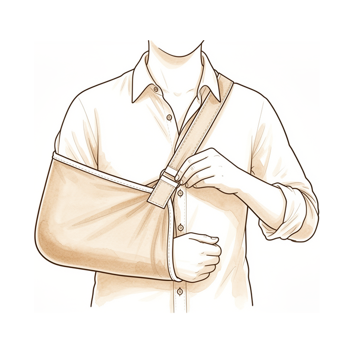
Rehabilitation and Return-to-Sport Protocols: Despite the absence of evidence-based guidelines, there exists minimal variability in recommendations between North American and European shoulder surgeons regarding return to play criteria [3]. Criteria for determining successful return to sport and return to preinjury level after superior labral pathophysiology treatment were not reported by most studies [37]. There is no clear consensus regarding optimal post-operative rehabilitation following arthroscopic shoulder stabilisation [196]. The postoperative protocol for revision arthroscopic type II SLAP repairs did not differ from the standard protocol for primary arthroscopic type II SLAP repairs, which included initial placement in a sling for 7 to 10 days [64].
Complications
Failed Repair and Revision Outcomes
Outcomes following surgical management of failed SLAP repair are inferior to those of primary repair [18]. The etiology of unsatisfactory outcomes remains debated, with the source of preoperative pain—whether labral, biceps, anterior ligamentous, or multifactorial—remaining unclear [27]. In a cohort of 348 patients, associated shoulder pathology such as Bankart lesions or rotator cuff pathology did not affect the SLAP reoperation rate [26]. However, patients undergoing SLAP repair face a higher risk of revision surgery and subsequent rotator cuff strain diagnosis [22]. A 10.1% incidence of subsequent surgery was identified after isolated SLAP repair, often related to an additional diagnosis [31]. In 12 patients (19.7%) undergoing concomitant rotator cuff and SLAP repair, radiologic evidence of an unhealed SLAP lesion was identified at a minimum of 1 year postoperatively; however, results of this combined procedure are not affected by the presence of an unhealed SLAP lesion [139].
Implant and Technical Complications
Poly-L/D-lactic acid anchors are associated with reoperation and failure of SLAP repairs [26]. Posterosuperior suture granuloma impingement is a complication that should be considered when patients present with recurrent pain after arthroscopic SLAP repair using suture anchors, particularly during overhead activity [70].
Rehabilitation and Return to Activity
No current studies have described a postoperative protocol for rehabilitation of either a revision SLAP repair or biceps tenodesis for failed SLAP repair [18]. For failed SLAP repair, a typical rehabilitation program includes 4 to 6 weeks in a sling, with active range of motion beginning at approximately 6 weeks postoperatively [18]. Strengthening exercises are typically begun at approximately 8 to 10 weeks postoperatively [18]. For overhead athletes undergoing failed SLAP repair, a throwing program is instituted 3 to 4 months postoperatively, with average return to play at 4 to 6 months [18].
Other Considerations
The SLAP literature is characterized by substantial variability in outcomes reporting, with time to return to play noted in few studies [227]. In 87% of cases, a good or excellent functional outcome can be anticipated after arthroscopic repair of type II SLAP lesions [11]. Recent outcomes studies have shown predictably good functional results and an acceptable rate of return to sport and/or work with arthroscopic treatment of SLAP tears [4]. Repairing rotator cuff tears and SLAP tears together results in significant clinical benefits compared to repairing just rotator cuff tears and analogous results against SLAP-only repair [235]. There are no advantages in repairing a type II SLAP lesion when associated with a rotator cuff tear in patients over 50 years of age [45].
Biceps tenodesis is indicated for the management of proximal biceps pathologic abnormality, including SLAP tears, and may allow better ability to return to physical activity, improved cosmesis, and closer approximation of normal anatomy despite longer rehabilitation times and increased technical difficulty when compared with biceps tenotomy [20]. Primary subpectoral open biceps tenodesis for SLAP tears or pathology of the long head of the biceps tendon provides significant improvement in shoulder outcomes with a reliable return to activity level with low risk for complications [252]. Biceps tenodesis can be successfully performed as an index operation rather than SLAP repair, as well as in a younger patient population [256]. SLAP repair remains the most commonly performed index procedure; however, biceps tenodesis appears equally efficacious and may represent an attractive alternative [59].
Outcomes after SLAP repair are largely favorable, although less successful outcomes have been demonstrated in certain populations, such as overhead athletes, patients older than 35 years of age, workers' compensation patients, and patients with concomitant shoulder pathologic abnormality [20]. The authors suggest that too many SLAP repairs are being performed in elderly patients despite evidence questioning its use in this population [230]. In overhead athletes, the chronic attritional loads placed on the superior labrum in the late cocking stage of throwing may be too high for even a repaired labrum to withstand, but the pain associated is likely multifactorial and related to a number of other issues such as pitch counts, throwing mechanics, internal rotation deficits and rotator cuff "internal impingement" [27]. Stabilizing a symptomatic detached biceps is believed to be of primary importance, but overconstraining or shortening the tendon is likely detrimental [27]. The ideal surgical technique for SLAP tears, using suture anchors or otherwise, is not well understood, nor is the ideal postoperative rehabilitation program or the biologic healing potential of these repairs [27].
A period of 3–6 months of non-operative treatment is often advocated for SLAP lesions [28]. In a prospective case series of 46 patients, three months of non-operative treatment improved patient ASES scores compared with prior to treatment [28]. In studies advocating 3–6 months of non-operative treatment for SLAP lesions, several patients (15% and 51%, respectively) still required surgery after this period [28].
Recovery
Light activity (weeks): The postoperative protocol for revision arthroscopic type II SLAP repairs includes initial placement in a sling for 7 to 10 days [64]. A typical rehabilitation program for failed SLAP repair extends this immobilization, requiring 4 to 6 weeks in a sling before active range of motion begins at approximately 6 weeks postoperatively [18].
Full activity (months): Strengthening exercises are typically initiated at approximately 8 to 10 weeks after failed SLAP repair [18]. For overhead athletes, a throwing program is instituted 3 to 4 months postoperatively, with an average return to play at 4 to 6 months [18]. Undergoing repair of the throwing or trail batting shoulder can delay return to sport by several months, though pitching workload returns to baseline by the second season postoperatively [174].
Rehabilitation protocol: The postoperative protocol for revision arthroscopic type II SLAP repairs did not differ from the standard protocol for primary repairs, which included initial placement in a sling for 7 to 10 days [64]. For failed SLAP repairs, the protocol mandates 4 to 6 weeks in a sling, with active range of motion beginning at approximately 6 weeks postoperatively [18]. Strengthening exercises are typically begun at approximately 8 to 10 weeks [18]. In overhead athletes, a throwing program is instituted 3 to 4 months postoperatively, leading to an average return to play at 4 to 6 months [18].
Functional milestones: Arthroscopic repair of isolated type II SLAP lesions with suture anchors leads to a satisfactory functional outcome and return to pre-injury sports levels [19]. Delayed but significant pain relief is observed 6 months after arthroscopic repair of isolated type II SLAP lesions [19]. Return to preinjury level of competition for elite overhead athletes after type II SLAP lesion repairs was 57%, despite high American Shoulder and Elbow Surgeons scores [69]. Studies reviewed reported moderate return to play and return to previous level of performance rates following SLAP repairs in competitive overhead athletes [218]. Arthroscopic posterior labral repair has demonstrated favorable outcomes with high rates of return to play [10]. Arthroscopic evaluation and repair of posterior labral lesions resulted in 26 of 28 patients (93%) returning to sport and 23 of 28 (82%) returning without any limitations [23]. Most patients successfully returned to sports after posterior labral repair with capsular plication, with nearly 80% returning at their preinjury level [67].
Other Considerations: Nearly a quarter of athletes were unable to return to play after arthroscopic superior labral repair [60]. Criteria-based return-to-sport testing in young athletes after posterior labral repair did not reduce recurrence or improve return to play compared to time-based clearance [221]. Two-thirds of athletes who underwent criteria-based return-to-sport testing after posterior labral repair failed at least one section, indicating some functional deficit [221]. Participants with microtraumatic posterior shoulder instability demonstrated significant improvements in patient-reported outcome measures and high rates of return to sport following a 24-week conservative rehabilitation program [195]. Predictable short-term surgical results and return to activity can be expected after repair of type II SLAP lesions in patients younger than 50 years who have coexistent rotator cuff tears [6]. Concomitant shoulder pathology should be treated at the time of SLAP repair [8].
Key Evidence
- [L4] Long-term outcomes after isolated labral repair for SLAP lesions are good and independent of age. [1] (10.1016/j.arthro.2012.02.025)
- [L4] Patients undergoing superior labral repair following acute trauma experienced the most reliable functional recovery, with increased forward flexion, lower revision risk, and higher RTP compared with the other cohorts. [2] (10.1177/2325967126s00526)
- [L4] Despite the absence of evidence-based guidelines, there exists minimal variability in recommendations between North American and European shoulder surgeons regarding return to play criteria. [3] (10.1016/j.jse.2021.01.026)
- [L5] Recent outcomes studies have shown predictably good functional results and an acceptable rate of return to sport and/or work with arthroscopic treatment of SLAP tears. [4] (10.5435/00124635-200910000-00005)
- [L1] Neither labral repair nor biceps tenodesis had any significant clinical benefit over sham surgery for patients with SLAP II lesions in the population studied. [5] (10.1136/bjsports-2016-097098)
- [L3] Predictable short-term surgical results and return to activity can be expected after repair of type II SLAP lesions in patients younger than 50 years who have coexistent rotator cuff tear. [6] (10.1177/0363546509347364)
- [L3] In patients with rotator cuff and labral lesions, arthroscopic treatment of both lesions yields good clinical outcomes, restoration of motion, and a high degree of patient satisfaction. [7] (10.1177/0363546507300062)
- [L4] The results suggest that concomitant shoulder pathology should be treated at the time of SLAP repair. [8] (10.1016/j.jse.2006.05.015)
- [L3] In a young active population, primary arthroscopic biceps tenodesis is a viable surgical alternative to labral repair for type II SLAP lesions. [9] (10.1007/s00167-020-05971-0)
- [L4] Although clinical evidence is limited, arthroscopic posterior labral repair has demonstrated favorable outcomes with high rates of return to play. [10] (10.5435/jaaos-d-24-00287)
- [L4] In 87% of cases, a good or excellent functional outcome can be anticipated after arthroscopic repair of type II SLAP lesions with the described techniques. [11] (10.1016/j.arthro.2011.09.005)
- [L3] In a population of active duty males, arthroscopic repair of isolated Type II SLAP had comparable results with a cohort of Type II SLAP repairs treated in combination with other shoulder conditions, with the combined treatment group having significantly better results in two of three parameters measured. [12] (10.1007/s00167-007-0334-8)
- [L4] Overall, nonoperative treatment of SLAP tears in athletes can be successful, especially in the subset of patients who are able to complete their rehabilitation program before attempting a return to play. [13] (10.1016/j.jse.2021.12.022)
- [L4] Arthroscopic type 2 SLAP repair induced good short- and long-term clinical outcomes, return to overhead activities, and subjective satisfaction in the general population, regardless of age. [15] (10.1007/s00167-021-06608-6)
- [L3] Surgical positioning for arthroscopic posterior shoulder labral repair did not affect postoperative clinical and patient-reported outcomes. [16] (10.1177/03635465221095243)
- [L3] Following arthroscopic repair, patients with Type V SLAP tears had a similar overall rate of return to play when compared directly to a control group of patients who underwent arthroscopic Bankart repair alone. [17] (10.1007/s00167-020-06388-5)
- [L5] [18] (10.5435/jaaos-22-09-554)
- [L4] Arthroscopic repair of isolated type II SLAP lesions with suture anchors leads to a satisfactory functional outcome and return to pre-injury sports levels, with delayed, but significant pain relief observed 6 months after surgery. [19] (10.1186/s12891-017-1620-3)
- [Paper] [20] (10.1016/j.csm.2015.08.001)
- [L3] Both arthroscopic-assisted subpectoral biceps tenodesis combined with anterior labral repair and arthroscopic SLAP repair led to statistically and clinically significant increases in outcome scores, marked improvements in pain, and high rates of return to unrestricted active duty in military patients with type V SLAP lesions. [21] (10.1177/03635465231169238)
- [L3] Patients who underwent SLAP repair were associated with a higher risk of revision surgery and subsequent rotator cuff strain diagnosis. [22] (10.1016/j.jse.2023.12.015)
- [L4] Arthroscopic evaluation and repair of these posterior labral lesions resulted in 26 of 28 patients (93%) returning to sport and 23 of 28 (82%) returning without any limitations. [23] (10.1016/j.arthro.2010.01.006)
- [L3] Combined posterior labral and SLAP repair led to statistically and clinically significant increases in outcome scores and high rates of return to active-duty military service that did not differ significantly from the results after isolated posterior labral repair. [24] (10.1177/03635465231181702)
- [L3] [26] (10.1016/j.arthro.2011.06.021)
- [L5] [27] (10.1016/j.jse.2010.11.013)
- [L3] [28] (10.1177/17585732211015825)
- [L3] We identified a 10.1% incidence of subsequent surgery after isolated SLAP repair, often related to an additional diagnosis, suggesting that clinicians should consider other potential causes of shoulder pain when considering surgery for patients with SLAP lesions. [31] (10.1016/j.arthro.2016.01.053)
- [L4] The authors present an effective arthroscopic technique for managing combined anterior shoulder instability and type IV SLAP lesions by repairing all pathoanatomy present, including the superior labrum and biceps tendon split. [32] (10.1016/j.arthro.2009.04.075)
- [L5] Nonoperative management is often an appropriate and effective initial treatment for superior labral injuries, unless there are obvious pathologic changes altering the mechanics of the glenohumeral joint or other associated injuries. [34] (10.1016/j.arthro.2025.03.059)
- [L4] Return to play after SLAP repair in overhead throwing athletes is poor, with surgeons needing to be cautious with indications. [35] (10.1016/j.csm.2016.05.003)
- [L4] SLAP repair should continue to be considered as an option for SLAP tear treatment only after nonsurgical management has failed. [36] (10.1177/0363546517728256)
- [L1] Criteria for determining successful return to sport and return to preinjury level after superior labral pathophysiology treatment were not reported by most studies. [37] (10.1016/j.arthro.2024.09.053)
- [L3] Athletes who undergo an objective return to play criteria based testing protocol have lower rates of recurrent instability following arthroscopic shoulder stabilization surgery than those cleared by time from surgery. [38] (10.1177/2325967120s00381)
- [L5] No correlations between functional outcomes and radiographic shoulder findings at mid-term were identified. [44] (10.1016/j.arthro.2025.07.020)
- [L1] There are no advantages in repairing a type II SLAP lesion when associated with a rotator cuff tear in patients over 50 years of age. [45] (10.1177/0363546507308194)
- [L4] [46] (10.1016/j.arthro.2022.03.021)
- [L4] A contrast-filled gap between the labrum and glenoid on neutral CT arthrography after SLAP repair is frequently observed even in patients with satisfactory clinical outcomes. [50] (10.1007/s00167-014-3350-5)
- [L3] Glenoid Bone Loss was identified as a significant prognostic factor for patients not returning to sport following Arthroscopic Labral Repair. [51] (10.1016/j.jse.2023.02.060)
- [L3] An initial trial of nonoperative management may be considered in young active patients with isolated SLAP tear. [52] (10.1016/j.jse.2015.09.008)
- [L4] When patients with clinical suspicion of rotator cuff tear present with combinations of these radiographic signs, a prompt MRI examination and a referral to a shoulder specialist are recommended. [53] (10.1016/j.jseint.2020.09.015)
- [L3] High-demand patients with biceps tendonitis in the setting of a SLAP lesion with labral instability who undergo combined tenodesis and labral repair have significantly worse outcomes than patients who undergo either isolated labral repair for type II SLAP tears or isolated biceps tenodesis for a SLAP tear and biceps tendonitis. [57] (10.1007/s00167-015-3774-6)
- [L4] SLAP repair remains the most commonly performed index procedure; however, BT appears equally efficacious and may represent an attractive alternative. [59] (10.1016/j.arthro.2018.12.015)
- [L4] Overall, nearly a quarter of athletes were unable to return to play after arthroscopic superior labral repair. [60] (10.1177/03635465241246122)
- [L4] Arthroscopic SLAP repairs show favorable clinical and radiological outcomes; however, the study findings raise a concern that return to play may still be problematic in elite baseball players. [62] (10.1177/0363546513485361)
- [L4] [64] (10.1177/0363546511398648)
- [L3] Good to excellent results and high return to prior level of activity can be expected for the majority of properly indicated patients who undergo isolated type II superior labral anterior posterior repairs, regardless of age. [66] (10.1177/0363546508328417)
- [L5] Most patients successfully returned to sports, with nearly 80% returning at their preinjury level. [67] (10.1016/j.arthro.2025.07.040)
- [L3] Return to preinjury level of competition for elite overhead athletes after type II SLAP lesion repairs was 57%, despite high American Shoulder and Elbow Surgeons scores. [69] (10.1177/0363546510379971)
- [Case_report] Implant related complications should be considered when patients present with recurrent pain after arthroscopic SLAP repair using suture anchors, in particular during overhead activity. [70] (10.1007/s00167-008-0524-z)
- [L4] [119] (10.1177/0363546506296735)
- [L4] [139] (10.1016/j.jse.2010.04.003)
- [L5] [146] (10.1016/j.csm.2004.01.005)
- [L4] A significant tear of the infraspinatus in combination with glenohumeral internal rotation deficit and SLAP tears in the throwing athlete results in a guarded prognosis in return to play at the same level. [149] (10.1177/0363546512459481)
- [L4] Humeral head abrasion is common in patients with a pathologic biceps-labral complex, especially those with failed SLAP repair. [151] (10.1016/j.jse.2010.05.013)
- [L4] In this series of 21 shoulders, pain was the major symptom in 15 patients with posterior labral detachment after a traumatic event. [152] (10.1111/j.1758-5740.2010.00069.x)
- [L3] For experienced shoulder surgeons, the Snyder classification is a reliable system for identifying SLAP lesions. [157] (10.1177/0363546510392332)
- [L4] [160] (10.1016/j.arthro.2018.01.031)
- [L5] The detailed approach described allows the arthroscopist access to the entire glenohumeral joint and transforms repair of complex intra-articular pathology into simple exercises in arthroscopic surgical technique. [163] (10.1016/j.arthro.2009.09.019)
- [L4] Undergoing repair of the throwing or trail batting shoulder can delay return to sport by several months, though pitching workload returns to baseline by the second season postoperatively. [174] (10.1016/j.jse.2025.01.024)
- [L4] Non-operative treatment with an appropriate regimen provided satisfactory clinical outcomes in middle-aged patients with symptomatic SLAP lesions and should be considered before recommending operative treatment. [177] (10.1007/s00167-016-4226-7)
- [L5] Treatment decisions for SLAP lesions are driven primarily by the presence of pain, overhead activity level, and prior non-operative management. [180] (10.1016/j.jisako.2026.101087)
- [L4] Conservative management is preferred as athletes completing nonoperative treatment return to play at 6 months on average compared to 12 months for surgical counterparts. [188] (10.1016/j.xrrt.2021.08.007)
- [L4] In this pilot study, MR arthrogram was used to evaluate the labrum in detail 6 months postoperatively, but no features on postoperative MR arthrogram predicted either functional outcome or recurrent instability. [191] (10.1177/1758573214550839)
- [L5] SLAP repair and biceps tenodesis both present viable treatment options but come with specific advantages and disadvantages, with the decision ultimately made individually with the patient. [192] (10.1016/j.arthro.2019.02.026)
- [L4] Participants with microtraumatic posterior shoulder instability demonstrated significant improvements in patient-reported outcome measures and high rates of return to sport following a 24-week conservative rehabilitation program. [195] (10.1016/j.jseint.2024.09.016)
- [L4] There is no clear consensus regarding optimal post-operative rehabilitation following arthroscopic shoulder stabilisation. [196] (10.1177/17585732231154889)
- [L4] Arthroscopic capsulolabral repair for posterior shoulder instability was a durable treatment option that improved long-term shoulder pain and function and facilitated return to sport in the majority of patients at a mean follow-up of 15.4 years, although a notable proportion of patients met various criteria for failure. [197] (10.1177/03635465231162271)
- [L3] Patients who were prescribed NSAIDs as part of a postoperative pain management regimen following primary arthroscopic labral repair for glenohumeral instability had similar patient-reported outcomes, revision rates, and rates of return to preinjury activities compared to those who were not prescribed NSAIDs. [205] (10.1016/j.jse.2025.02.048)
- [L5] It is anatomically possible that suprascapular nerve could sustain iatrogenic injury during labral anchor placement during SLAP repair. [206] (10.1007/s00167-016-4086-1)
- [L3] Preoperative MRI scans of the shoulder interpreted by orthopaedic surgeons with the described systematic approach resulted in improved accuracy in diagnosing subscapularis tendon tears compared with previous studies. [208] (10.1016/j.arthro.2012.04.142)
- [Paper] Radiographs should be the first imaging study obtained, and magnetic resonance imaging provides the best overall evaluation of the cuff. [210] (10.1016/j.csm.2012.07.010)
- [L4] The position of the MTJ with respect to the glenoid face is a reliable, identifiable marker on MRI scans that can be predictive of healing. [212] (10.1016/j.arthro.2016.12.010)
- [L4] Arthroscopic SLAP repair of type II lesions with bioabsorbable suture anchors provides a significant improvement in functional capacity and pain relief. [215] (10.1016/j.jse.2010.03.004)
- [L4] The majority of patients returned to their pre-injury activity level and the subjective patient-administered evaluations appeared to improve after arthroscopic repair of type-2 SLAP lesions using one double-looped Corkscrew anchor. [217] (10.1007/s00167-003-0462-8)
- [L4] Studies reviewed reported moderate return to play and return to previous level of performance rates following SLAP repairs in competitive overhead athletes. [218] (10.1016/j.arthro.2022.03.026)
- [L3] While RTS testing in young athletes after posterior labral repair did not reduce recurrence or improve return to play compared to time-based clearance, two-thirds of athletes who underwent testing failed at least 1 section, indicating some functional deficit. [221] (10.1016/j.jseint.2023.01.002)
- [L4] The SLAP literature is characterized by substantial variability in outcomes reporting, with time to return to play noted in few studies. [227] (10.1016/j.jse.2016.04.020)
- [L3] The authors suggest that too many SLAP repairs are being performed in elderly patients despite evidence questioning its use in this population. [230] (10.1016/j.jse.2012.02.001)
- [L3] Repairing RCT and SLAP tears together results in significant clinical benefits compared to repairing just RCT and analogous results against SLAP-only repair. [235] (10.1016/j.jseint.2020.07.014)
- [L4] Primary subpectoral open biceps tenodesis for SLAP tears or pathology of the LHBT provides significant improvement in shoulder outcomes with a reliable return to activity level with low risk for complications. [252] (10.1016/j.arthro.2019.06.035)
- [L1] It can be successfully performed as an index operation rather than SLAP repair, as well as in a younger patient population. [256] (10.1177/0363546520921177)
See Also
- Rotator Cuff
- Biceps Tenodesis
- Shoulder Instability
- Os Acromiale
- Rotator cuff repair
- Calcific Tendinitis
References
[1] Long‐Term Results After SLAP Repair: A 5‐Year Follow‐up Study of 107 Patients With Comparison of Patients Aged Over and Under 40 Years. Arthroscopy. 2012. DOI: 10.1016/j.arthro.2012.02.025
[2] Poster 231. Outcomes following Superior Labral Repairs Based on Mechanism of Injury. Orthopaedic Journal of Sports Medicine. 2026. DOI: 10.1177/2325967126s00526
[3] Return to play criteria among shoulder surgeons following shoulder stabilization. Journal of Shoulder and Elbow Surgery. 2021. DOI: 10.1016/j.jse.2021.01.026
[4] Superior Labral Tears of the Shoulder: Pathogenesis, Evaluation, and Treatment. Journal of the American Academy of Orthopaedic Surgeons. 2009. DOI: 10.5435/00124635-200910000-00005
[5] Sham surgery versus labral repair or biceps tenodesis for type II SLAP lesions of the shoulder: a three-armed randomised clinical trial. British Journal of Sports Medicine. 2017. DOI: 10.1136/bjsports-2016-097098
[6] The Effect of Rotator Cuff Tears on Surgical Outcomes after Type II Superior Labrum Anterior Posterior Tears in Patients Younger than 50 Years. The American Journal of Sports Medicine. 2009. DOI: 10.1177/0363546509347364
[7] Outcomes of Combined Arthroscopic Rotator Cuff and Labral Repair. The American Journal of Sports Medicine. 2007. DOI: 10.1177/0363546507300062
[8] Arthroscopic superior labrum anterior-posterior repair in military patients. Journal of Shoulder and Elbow Surgery. 2007. DOI: 10.1016/j.jse.2006.05.015
[9] Arthroscopic treatment of type II superior labral anterior to posterior (SLAP) lesions in a younger population: minimum 2‐year outcomes are similar between SLAP repair and biceps tenodesis. Knee Surgery, Sports Traumatology, Arthroscopy. 2020. DOI: 10.1007/s00167-020-05971-0
[10] Batter's Shoulder: Diagnosis, Management, and Outcomes. Journal of the American Academy of Orthopaedic Surgeons. 2024. DOI: 10.5435/jaaos-d-24-00287
[11] Long‐Term Outcome After Arthroscopic Repair of Type II SLAP Lesions: Results According to Age and Workers' Compensation Status. Arthroscopy. 2012. DOI: 10.1016/j.arthro.2011.09.005
[12] Isolated and combined Type II SLAP repairs in a military population. Knee Surgery, Sports Traumatology, Arthroscopy. 2007. DOI: 10.1007/s00167-007-0334-8
[13] Return to play following nonsurgical management of superior labrum anterior-posterior tears: a systematic review. Journal of Shoulder and Elbow Surgery. 2022. DOI: 10.1016/j.jse.2021.12.022
[14] Chapter 78 Superior Labrum Anterior to Posterior Tears and Lesions of the Proximal Biceps Tendon. 2019.
[15] Posterior repair of isolated type 2 superior labrum anterior–posterior lesion prevents external rotation deficiency: long‐term outcome study. Knee Surgery, Sports Traumatology, Arthroscopy. 2021. DOI: 10.1007/s00167-021-06608-6
[16] Beach-Chair Versus Lateral Decubitus Positioning for Arthroscopic Posterior Shoulder Labral Repair: A Retrospective Comparison of Clinical and Patient-Reported Outcomes. The American Journal of Sports Medicine. 2022. DOI: 10.1177/03635465221095243
[17] Type V superior labral anterior–posterior tears results in lower rates of return to play. Knee Surgery, Sports Traumatology, Arthroscopy. 2021. DOI: 10.1007/s00167-020-06388-5
[18] Etiology, Diagnosis, and Management of Failed SLAP Repair. Journal of the American Academy of Orthopaedic Surgeons. 2014. DOI: 10.5435/jaaos-22-09-554
[19] Progression of function and pain relief as indicators for returning to sports after arthroscopic isolated type II SLAP repair—a prospective study. BMC Musculoskeletal Disorders. 2017. DOI: 10.1186/s12891-017-1620-3
[20] Arthroscopic Surgical Techniques for the Management of Proximal Biceps Injuries. Clinics in Sports Medicine. 2016. DOI: 10.1016/j.csm.2015.08.001
[21] Midterm Outcomes Following Combined Biceps Tenodesis and Anterior Labral Repair in Active Duty Military Patients Younger Than 35 Years. The American Journal of Sports Medicine. 2023. DOI: 10.1177/03635465231169238
[22] Superior labrum anterior to posterior (SLAP) repair is associated with increased rate of subsequent rotator cuff diagnoses and revision surgery: a propensity-matched comparison. Journal of Shoulder and Elbow Surgery. 2024. DOI: 10.1016/j.jse.2023.12.015
[23] Arthroscopic Posterior Labral Repair in Athletes: Outcome Analysis at 2‐Year Follow‐up. Arthroscopy. 2010. DOI: 10.1016/j.arthro.2010.01.006
[24] Outcomes Following Combined Posterior Labral and SLAP Repair in Military Patients Younger Than 35 Years. The American Journal of Sports Medicine. 2023. DOI: 10.1177/03635465231181702
[26] Poly‐L/D‐Lactic Acid Anchors Are Associated With Reoperation and Failure of SLAP Repairs. Arthroscopy. 2011. DOI: 10.1016/j.arthro.2011.06.021
[27] Superior labral tears: repair versus biceps tenodesis. Journal of Shoulder and Elbow Surgery. 2011. DOI: 10.1016/j.jse.2010.11.013
[28] Is timing of superior labrum anterior to posterior (SLAP) repair important? A cohort study evaluating the effect of the duration of symptoms prior to surgery on the outcomes of patients who underwent type II SLAP repair. Shoulder & Elbow. 2021. DOI: 10.1177/17585732211015825
[31] Subsequent Shoulder Surgery After Isolated Arthroscopic SLAP Repair. Arthroscopy. 2016. DOI: 10.1016/j.arthro.2016.01.053
[32] Combined Arthroscopic Repair of a Type IV SLAP Tear and Bankart Lesion. Arthroscopy. 2009. DOI: 10.1016/j.arthro.2009.04.075
[34] A Mechanistic Classification for Superior Labral Injuries Guides Operative Management. Arthroscopy. 2025. DOI: 10.1016/j.arthro.2025.03.059
[35] Return to Play After Shoulder Surgery in Throwers. Clinics in Sports Medicine. 2016. DOI: 10.1016/j.csm.2016.05.003
[36] Return to Play and Outcomes in Baseball Players After Superior Labral Anterior-Posterior Repairs. The American Journal of Sports Medicine. 2017. DOI: 10.1177/0363546517728256
[37] Ill‐defined Return‐to‐Sport Criteria and Inconsistent Unsuccessful Return Rates Caused by Various Reasons Not Necessarily Related to Treatment After Superior Labral Treatments: A Systematic Review. Arthroscopy. 2024. DOI: 10.1016/j.arthro.2024.09.053
[38] Recurrence Rates Following Arthroscopic Shoulder Stabilization are Improved Following a Criteria Based Return to Sport Testing Protocol. Orthopaedic Journal of Sports Medicine. 2020. DOI: 10.1177/2325967120s00381
[42] Rockwood And Matsen S The Shoulder. Arthroscopic Management of Prearthritic and Arthritic Conditions of the Shoulder and the Postarthroplasty Shoulder > Radiographic Evaluation.
[44] Shoulder Superior Capsular Reconstruction Using Xenograft Shows No Deterioration in Functional Improvement at 5-Year Follow-Up. Arthroscopy: The Journal of Arthroscopic & Related Surgery. 2025. DOI: 10.1016/j.arthro.2025.07.020
[45] No Advantages in Repairing a Type II Superior Labrum Anterior and Posterior (SLAP) Lesion When Associated with Rotator Cuff Repair in Patients over Age 50. The American Journal of Sports Medicine. 2007. DOI: 10.1177/0363546507308194
[46] Biceps Tenodesis Combined With Arthroscopic Posterior Labral Repair for Type VIII SLAP Lesions in Active‐Duty Military Patients Yields Excellent Return to Military Duty. Arthroscopy. 2022. DOI: 10.1016/j.arthro.2022.03.021
[50] External rotation and active supination CT arthrography for the postoperative evaluation of type II superior labral anterior to posterior lesions. Knee Surgery, Sports Traumatology, Arthroscopy. 2014. DOI: 10.1007/s00167-014-3350-5
[51] Prognostic Factors For Patients Not To Return To Sport Following Arthroscopic Labral Repair: A Retrospective Multicentre Study. Journal of Shoulder and Elbow Surgery. 2023. DOI: 10.1016/j.jse.2023.02.060
[52] Predictive factors associated with failure of nonoperative treatment of superior labrum anterior-posterior tears. Journal of Shoulder and Elbow Surgery. 2016. DOI: 10.1016/j.jse.2015.09.008
[53] Radiographic greater tuberosity spurs and narrow acromiohumeral intervals are associated with advanced retraction of the supraspinatus tendon in patients with symptomatic rotator cuff tears. JSES International. 2021. DOI: 10.1016/j.jseint.2020.09.015
[57] Combined SLAP repair and biceps tenodesis for superior labral anterior–posterior tears. Knee Surgery, Sports Traumatology, Arthroscopy. 2015. DOI: 10.1007/s00167-015-3774-6
[59] Labral Repair Versus Biceps Tenodesis for Primary Surgical Management of Type II Superior Labrum Anterior to Posterior Tears: A Systematic Review. Arthroscopy. 2019. DOI: 10.1016/j.arthro.2018.12.015
[60] Return to Play After Arthroscopic Superior Labral Repair: A Systematic Review. The American Journal of Sports Medicine. 2025. DOI: 10.1177/03635465241246122
[62] Clinical and Radiological Outcomes of Type 2 Superior Labral Anterior Posterior Repairs in Elite Overhead Athletes. The American Journal of Sports Medicine. 2013. DOI: 10.1177/0363546513485361
[64] Outcomes of Revision Arthroscopic Type II Superior Labral Anterior Posterior Repairs. The American Journal of Sports Medicine. 2011. DOI: 10.1177/0363546511398648
[66] Isolated Type II Superior Labral Anterior Posterior Lesions. The American Journal of Sports Medicine. 2009. DOI: 10.1177/0363546508328417
[67] Posterior Labral Repair With Capsular Plication Shows Enduring Mid-Term Outcomes and Return to Sports With Low Recurrence Rates. Arthroscopy: The Journal of Arthroscopic & Related Surgery. 2025. DOI: 10.1016/j.arthro.2025.07.040
[69] Outcome of Type II Superior Labral Anterior Posterior Repairs in Elite Overhead Athletes. The American Journal of Sports Medicine. 2010. DOI: 10.1177/0363546510379971
[70] Posterosuperior suture granuloma impingement after arthroscopic SLAP repair using suture anchors: a case report. Knee Surgery, Sports Traumatology, Arthroscopy. 2008. DOI: 10.1007/s00167-008-0524-z
[72] Rockwood And Matsen S The Shoulder. Shoulder and Elbow Specialty Clinic Workers’ Survey > ANATOMY.
[74] A Lange Medical Book Current Diagnosis Treatment In Orthopedics Fifth Edition. 2Musculoskeletal Trauma Surgery > SHOULDER AND ARM INJURIES.
[75] Rockwood And Green S Fractures In Adults. 29: Principles of Nonunion and Bone Defect Treatment > Applied Anatomy Related to Scapular Fractures.
[76] Aaos Comprehensive Orthopaedic Review 3. Anatomy of the Shoulder, Arm, and Elbow > I. Shoulder.
[82] Aaos Comprehensive Orthopaedic Review 3. Fractures of the Clavicle, Scapula, and Glenoid > I. Clavicular Fractures.
[83] Rockwood And Matsen S The Shoulder. Developmental Anatomy of the Shoulder and Anatomy of the Glenohumeral Joint > Development of Individual Regions.
[85] Campbell S Operative Orthopaedics 4 Volume Set. RECONSTRUCTIVE PROCEDURES OF THE SHOULDER AND ELBOW IN ADULTS > ANATOMY AND BIOMECHANICS.
[86] Campbell S Operative Orthopaedics 4 Volume Set. ANTERIOR CRUCIATE LIGAMENT RECONSTRUCTION WITH BONE-PATELLAR TENDON-BONE GRAFT > SHOULDER INJURIES > ANATOMY AND BIOMECHANICS.
[87] Miller S Review Of Orthopaedics. Genetics of musculoskeletal conditions and abnormalities are summarized in Table 1.27 > UPPER EXTREMITY > SHOULDER.
[90] Orthopaedic Knowledge Update Sports Medicine 6. Diagnostic Ultrasonography and Ultrasonography-Guided Procedures > Annotated References.
[94] Apley And Solomon S Concise System Of Orthopaedics And Trauma. INVESTIGATION.
[100] Rockwood And Green S Fractures In Adults. 29: Principles of Nonunion and Bone Defect Treatment > Magnetic Resonance Imaging and Arthrography.
[103] Orthopaedic Knowledge Update 13 Ebook Without Multimedia. Shoulder Anatomy and Biomechanics, Clinical Evaluation, Imaging > Clinical Evaluation > Imaging.
[104] Aaos Comprehensive Orthopaedic Review 3. Imaging of the Shoulder and Elbow > I. Shoulder.
[107] Rockwood And Green S Fractures In Adults. 29: Principles of Nonunion and Bone Defect Treatment > Radiography.
[119] Arthroscopic Repair of Type II Superior Labral Anterior Posterior Lesions with and without Acromioplasty. The American Journal of Sports Medicine. 2007. DOI: 10.1177/0363546506296735
[139] Results of concomitant rotator cuff and SLAP repair are not affected by unhealed SLAP lesion. Journal of Shoulder and Elbow Surgery. 2011. DOI: 10.1016/j.jse.2010.04.003
[146] Return to play for rotator cuff injuries and superior labrum anterior posterior (SLAP) lesions. Clinics in Sports Medicine. 2004. DOI: 10.1016/j.csm.2004.01.005
[149] Return to High-Level Throwing After Combination Infraspinatus Repair, SLAP Repair, and Release of Glenohumeral Internal Rotation Deficit. The American Journal of Sports Medicine. 2012. DOI: 10.1177/0363546512459481
[151] Humeral head abrasion: An association with failed superior labrum anterior posterior repairs. Journal of Shoulder and Elbow Surgery. 2011. DOI: 10.1016/j.jse.2010.05.013
[152] Arthroscopic Treatment of Traumatic Posterior Shoulder Instability in Athletes. Shoulder & Elbow. 2010. DOI: 10.1111/j.1758-5740.2010.00069.x
[157] Reproducibility and Reliability of the Snyder Classification of Superior Labral Anterior Posterior Lesions Among Shoulder Surgeons. The American Journal of Sports Medicine. 2011. DOI: 10.1177/0363546510392332
[160] Arthroscopic Management of SLAP Lesions With Concomitant Spinoglenoid Notch Ganglion Cysts: A Systematic Review Comparing Repair Alone to Repair With Decompression. Arthroscopy. 2018. DOI: 10.1016/j.arthro.2018.01.031
[163] Four‐Quadrant Approach to Capsulolabral Repair: An Arthroscopic Road Map to the Glenoid. Arthroscopy. 2010. DOI: 10.1016/j.arthro.2009.09.019
[174] Return-to-sport and performance outcomes after isolated posterior labral repair in professional baseball players. Journal of Shoulder and Elbow Surgery. 2025. DOI: 10.1016/j.jse.2025.01.024
[177] Clinical outcomes of non-operative treatment for patients presenting SLAP lesions in diagnostic provocative tests and MR arthrography. Knee Surgery, Sports Traumatology, Arthroscopy. 2016. DOI: 10.1007/s00167-016-4226-7
[180] Factors influencing surgeons’ decision-making in the treatment of SLAP lesions: A case-vignette study. Journal of ISAKOS. 2026. DOI: 10.1016/j.jisako.2026.101087
[188] Management of common upper extremity injuries in throwing athletes: a critical review of current outcomes. JSES Reviews, Reports, and Techniques. 2021. DOI: 10.1016/j.xrrt.2021.08.007
[191] Functional outcome and the structural integrity of arthroscopic Bankart repair: a prospective trial. Shoulder & Elbow. 2014. DOI: 10.1177/1758573214550839
[192] Editorial Commentary: Which to Fix—the Biceps or the Labrum? The Shoulder SLAP Tear Is Still Controversial. Arthroscopy. 2019. DOI: 10.1016/j.arthro.2019.02.026
[195] The efficacy of conservative management of micro-traumatic posterior shoulder instability. JSES International. 2025. DOI: 10.1016/j.jseint.2024.09.016
[196] Rehabilitation following shoulder arthroscopic stabilisation surgery: A survey of UK practice. Shoulder & Elbow. 2023. DOI: 10.1177/17585732231154889
[197] Minimum 10-Year Clinical Outcomes After Arthroscopic Capsulolabral Repair for Isolated Posterior Shoulder Instability. The American Journal of Sports Medicine. 2023. DOI: 10.1177/03635465231162271
[205] No difference in clinical outcomes and return to sport and work with use of postoperative nonsteroidal anti-inflammatory medications following primary arthroscopic glenoid labral repair. Journal of Shoulder and Elbow Surgery. 2025. DOI: 10.1016/j.jse.2025.02.048
[206] Drilling through lateral transmuscular portal lowers the risk of suprascapular nerve injury during arthroscopic SLAP repair. Knee Surgery, Sports Traumatology, Arthroscopy. 2016. DOI: 10.1007/s00167-016-4086-1
[208] A Systematic Approach for Diagnosing Subscapularis Tendon Tears With Preoperative Magnetic Resonance Imaging Scans. Arthroscopy. 2012. DOI: 10.1016/j.arthro.2012.04.142
[210] Imaging Evaluation of the Rotator Cuff. Clinics in Sports Medicine. 2012. DOI: 10.1016/j.csm.2012.07.010
[212] Influence of Preoperative Musculotendinous Junction Position on Rotator Cuff Healing After Double‐Row Repair. Arthroscopy. 2017. DOI: 10.1016/j.arthro.2016.12.010
[215] Outcomes of type II superior labrum, anterior to posterior (SLAP) repair: Prospective evaluation at a minimum two-year follow-up. Journal of Shoulder and Elbow Surgery. 2010. DOI: 10.1016/j.jse.2010.03.004
[217] Repair of type‐2 SLAP lesions using Corkscrew anchors. Knee Surgery, Sports Traumatology, Arthroscopy. 2004. DOI: 10.1007/s00167-003-0462-8
[218] Moderate Return to Play and Previous Performance After SLAP Repairs in Competitive Overhead Athletes: A Systematic Review. Arthroscopy. 2022. DOI: 10.1016/j.arthro.2022.03.026
[221] Criteria-based return-to-sport testing helps identify functional deficits in young athletes following posterior labral repair but may not reduce recurrence or increase return to play. JSES International. 2023. DOI: 10.1016/j.jseint.2023.01.002
[227] Variable reporting of functional outcomes and return to play in superior labrum anterior and posterior tear. Journal of Shoulder and Elbow Surgery. 2016. DOI: 10.1016/j.jse.2016.04.020
[230] The rising incidence of arthroscopic superior labrum anterior and posterior (SLAP) repairs. Journal of Shoulder and Elbow Surgery. 2012. DOI: 10.1016/j.jse.2012.02.001
[235] Comparing clinical outcomes between rotator cuff repairs, SLAP repairs, and combined repairs. JSES International. 2020. DOI: 10.1016/j.jseint.2020.07.014
[252] Outcomes of Primary Biceps Subpectoral Tenodesis in an Active Population: A Prospective Evaluation of 101 Patients. Arthroscopy. 2019. DOI: 10.1016/j.arthro.2019.06.035
[256] Biceps Tenodesis for Superior Labrum Anterior-Posterior Tear in the Overhead Athlete: A Systematic Review. The American Journal of Sports Medicine. 2020. DOI: 10.1177/0363546520921177