Clinicians › Hand
Strength and Motion
Hand strength & motion assessment: grip/pinch strength correlation to function, normative data, and impact of common pathologies.
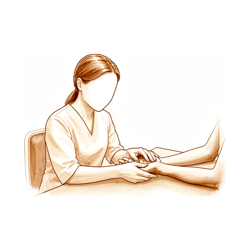
Overview
Assessment of strength and motion in upper extremity surgery is evolving, with a shift toward patient-reported outcomes as traditional measures of range of motion and strength are considered less reliable [1]. Nevertheless, functional range of motion remains a critical metric for directing surgical indications, guiding rehabilitation, and assessing treatment outcomes [2]. Digits require both sensation and freedom of motion to be used effectively, and effective surgery aims to restore biomechanical motions to achieve optimum use [26]. While age significantly affects postoperative range of motion and strength [7], specific pathologies present distinct profiles; for instance, patients with posttraumatic arthritis following distal radius fractures experience diminished range of motion and poorer general functioning, yet grip strength often remains unaltered [3]. Conversely, successful replantation for radiocarpal joint amputation is associated with major motion restriction, decreased strength, and moderate disability [19].
Objective physical examination and radiographic findings must remain the foundation for determining a patient’s abilities, as functional capacity evaluations should not replace the surgeon’s clinical judgment [23]. The optimal assessment for return to work relies on a competent physician’s understanding of residual impairments combined with knowledge of specific work requirements [23]. Measurement techniques are also advancing; new digital dynamometer features may influence grip scores in ways unaccounted for by analog devices [9], and surface electromyographic signals can distinguish maximal from submaximal efforts in upper extremity injuries [32]. Although a repeatable technique for measuring palmar thumb abduction strength exists, its clinical utility is limited by large inter-rater differences and low sample variability [17].
Rehabilitation strategies and surgical outcomes vary by procedure. Early active motion following flexor tendon repair may not yield better long-term range of motion than passive protocols [12], though the spiral linking technique modification offers a repair theoretically strong enough for active early mobilization [66]. The Latarjet procedure more commonly affects range of motion rather than shoulder and elbow strength [13], while cross-education effects on rotator muscle strength after stabilization surgery do not impact functional outcomes [5]. Distal radioulnar joint constrained arthroplasty allows patients to return to regular activities with excellent range of motion and restored lifting capacity [63]. In specific tendon injuries, a ruptured distal biceps tendon results in approximately a 30% loss in maximum flexion and a 50% loss in maximum supination strength [138].
Anatomy & Pathophysiology
Bony Anatomy and Kinematics
The skeleton of the hand and wrist comprises 27 bones, of which 19 are long bones [40]. The hand specifically contains 19 bones, 17 articulations, and 19 muscles situated entirely within the hand, along with a comparable number of tendons activated by forearm muscles [37]. The radioulno-carpal articulation possesses two axes of movement, to which a third axis—pronation and supination from the forearm—is added [40]. This configuration grants the wrist three axes of movement, permitting the hand to assume any spatial configuration and positioning it as needed for grasping [40]. In the sagittal plane, the thumb metacarpal forms an angle of approximately 45 degrees with the second metacarpal [40]. The transverse axis of the palm forms an acute angle of approximately 75 degrees with the longitudinal axis [40]. Metacarpal mobility varies by digit; the ring metacarpal exhibits about 10 degrees of mobility in flexion and extension [81], while the fifth metacarpal has a range of flexion–extension of approximately 20 degrees [81]. Epiphyseal plates are located at the proximal ends of the phalanges and the first metacarpal, whereas they are located at the distal ends of the other metacarpals [40]. Biomechanically, the middle finger is the most important contributor to grip strength [103].
Musculotendinous Anatomy
Extrinsic muscles originate outside the hand and insert on the hand or carpus, whereas intrinsic muscles have both origin and insertion within the hand [39]. The extrinsic extensors traverse six distinct fibroosseous retinacular compartments at the wrist level [39]: * First compartment: Contains the abductor pollicis longus and the extensor pollicis brevis [39]. * Second compartment: Contains the extensor carpi radialis longus and the extensor carpi radialis brevis [39]. * Third compartment: Contains the extensor pollicis longus, which turns abruptly radialward about Lister tubercle [39]. * Fourth compartment: Contains the extensor indicis proprius lying deep to the four tendons of the extensor digitorum communis [39]. * Fifth compartment: Contains the extensor digiti quinti [39]. * Sixth compartment: Contains the extensor carpi ulnaris tendon, which inserts at the base of the little finger metacarpal [39].
Sagittal band fibers insert onto the volar proximal phalanx and onto the lateral borders of the volar plate [39]. Rupture or attenuation of these fibers allows the extrinsic extensor tendon to sublux to the ulnar side of the metacarpal head, causing ulnar deviation of the finger [39]. The flexor digitorum profundus inserts on the proximal volar aspect of the distal phalanx [39]. The flexor digitorum superficialis tendon bifurcates around the flexor digitorum profundus at the beginning of the A2 pulley [85]. The A2 and A4 pulleys are the most essential structures in maintaining the mechanical advantage of the flexor tendons [85].
There are seven interosseous muscles, comprising four dorsal and three volar muscles [79]. The dorsal interossei function as abductors, while the volar interossei function as adductors [79]. The middle finger possesses two dorsal interossei (abductors) and no volar interossei (adductors) because the central axis of the hand lies within it [79]. The deep head of each dorsal interosseous muscle forms a lateral tendon, or lateral band, at the level of the metacarpophalangeal joint [79]. The volar interossei form the ulnar lateral band of the index finger and the radial lateral band of the ring and little fingers [79]. The abductor digiti quinti inserts onto the ulnar lateral tubercle at the base of the proximal phalanx of the little finger [79]. The opponens digiti quinti arises from the pisohamate ligament and the hook of the hamate and inserts onto the ulnar side of the diaphysis of the fifth metacarpal [79].
Soft Tissue and Vascular Anatomy
The dorsal skin is thin and lined by a horny layer that is only 0.02 mm thick [87]. Unlike the palm, the dorsal skin possesses a normal pilosebaceous system [87]. The superficial palmar fascia covers a triangular area of the central palm, with the proximal corner facing directly proximal [88]. There is no central band of the superficial palmar fascia for the thumb [88]. Fibers dorsal to the digital neurovascular bundle are collectively called Cleland ligament, while those palmar to the neurovascular bundle are called Grayson ligament [88]. The princeps pollicis artery is the terminal branch of the radial artery and runs along the ulnar side of the first metacarpal bone [82]. In anatomical studies, only 15% of dissections fall into the classical category for the layout of palmar arteries of the thumb [82]. The tenosynovial sheath of the flexor pollicis longus is continuous with the radial bursa [85]. The tenosynovial sheath to the little finger is continuous with the ulnar digital bursa [85].
Pathophysiology and Biomechanics
A fracture of the distal radius occurs within 3 cm of the distal end of this bone [10]. Intra-articular gaps caused by the fracture line result in significant joint incongruity that could possibly interfere with wrist and hand function [10]. Radial shortening results in loss of the volar tilt, interferes with the function of the distal radioulnar joint, and can lead to midcarpal instability [10]. Extra-articular malalignment is associated with pain and decreased grip strength [10]. Limited joint mobility of the thumb and index finger may cause temporal changes in precision grip force control, which can lead to reduced manual dexterity [21]. At increasing amounts of finger flexion, progressive metacarpal shortening produces proportionally greater loss of fingertip flexion force [107].
The intrinsic plus deformity is caused by excessive intrinsic muscle tension, in which the proximal interphalangeal joint cannot be flexed fully when the metacarpophalangeal joint is fully extended [97]. Flexor tenosynovitis may produce limitation of interphalangeal joint motion, so the range of active flexion of these joints is significantly less than passive flexion [97]. Individuals with hand osteoarthritis modulate grip force magnitude and temporal parameters but apply higher grip forces at liftoff and peak, and demonstrate longer latency compared to controls [99]. At a position of 30 degrees of wrist extension, maximal grip strength is achieved without significantly compromising grip endurance [109].
During thumb oppositional motion, internal rotation of the first metacarpal occurs, with the palmar base rotating primarily with respect to the dorsal base [119]. The position of the thumb interphalangeal joint affects movements of the trapeziometacarpal joint and thenar muscle activity during opposition [124]. Simultaneous activation of the flexor digitorum profundus and the intrinsic muscles results in an apparently more functional hand closing compared with flexor digitorum profundus activation alone because of altered kinematics and larger fingertip-to-palm distances [114]. The dynamic interaction of finger joints during cylinder grip shows specific patterns, with distal interphalangeal joints consistently initiating flexion last and synchronization increasing significantly by the end of motion compared to the beginning [101]. Grip-load force adjustments are not completely determined by the mechanics of object motion; nonmechanical factors related to movement performance, for instance perceptual factors, may affect the coupling [43]. Normal synovial fluid exhibits non-Newtonian, thixotropic properties [177]. The unique lubricating properties of synovial joints are not solely due to either synovial fluid or to the articular cartilage but to some complex interaction of both [177].
Classification
AO/OTA: Fractures in the locked volar plating study were classified using the OTA classification [170].
Bayne-Klug: The severity grading in radial dysplasia should be used together with the Bayne-Klug classification to provide precise information about functional capacity [141].
Gross Motor Function Classification System (GMFCS): Heterogeneity of subject participants in cerebral palsy resistance training studies includes severity levels, specifically GMFCS levels, and ages [172].
Other Considerations: Physical outcome measures are being changed for the use of patient reported outcomes [1]. Range of motion and strength are not as reliable measures as one would think [1]. The functional range of motion is important for directing indications for surgery and rehabilitation [2] and for assessing outcome of treatment [2]. Patients with posttraumatic arthritis following distal radius fractures experienced diminished range of motion [3] and poorer patient-reported outcomes regarding general functioning and satisfaction [3], but did not experience altered grip strength [3]. Assessments using range-of-motion measurements or a questionnaire are not sufficient to evaluate a patient's movement disorder after distal radius fracture [51]; kinematic analysis should be conducted for quantitative assessments of movement disorder after distal radius fracture [51].
Intra-articular gaps caused by distal radius fracture lines result in significant joint incongruity that could possibly interfere with wrist and hand function [10]. Chondral lesions and increased pressure on the articular surface following distal radius fracture interfere with sagittal motion of the carpal bones [10], which restricts the range of wrist movement that could be attained [10]. Failure to achieve normal anatomical configuration following extra-articular distal radius fracture is associated with a number of problems [10]. Radial shortening results in loss of the volar tilt [10], interferes with the function of the distal radioulnar joint [10], and can lead to midcarpal instability [10]. Improved understanding of carpal motion patterns may lead to changes in functional rehabilitation following injury [11] and surgical management [11].
Accurate diagnosis and management of hand and carpal fractures and dislocations are predicated on a thorough physical examination and appropriate imaging [14], aiming to limit joint stiffness while preserving mobility and function [14]. Standard manual strength-testing of the intrinsic muscles of the hand was not diagnostically sensitive [15]. Unless handheld dynamometry is widely adopted or a better grading system is developed and well validated, the MRC will continue to be used [55].
The term "Stiffness" of a structure always requires an exact description of the load configuration [134] and the exact localization and kind of deformation measured [134]. Directionally coupled motion patterns in the CMC joint are similar in men and women [146], though CMC adduction is difficult to measure due to poor reliability [150]. Knowing frequently used grasp types is essential for assessing grasp rehabilitation processes [152] and for developing hand prostheses [152].
Limited joint mobility of the thumb and index finger may cause temporal changes in precision grip force control [21], which can lead to reduced manual dexterity [21]. Grip-load force adjustments are not completely determined by the mechanics of object motion [43]; nonmechanical factors related to movement performance, such as perceptual factors, may affect grip-load force coupling [43]. Kinematic analysis results can be used to inform selection of tasks for kinematic evaluation [25] and provide expected variability for comparison to patient populations [25].
Isolated flexion of the metacarpophalangeal joint was not possible to the full extent in any finger [165]. Full metacarpophalangeal-joint flexion could be achieved with 45 degrees or less of proximal interphalangeal-joint flexion in only five fingers [165]. In fingers where full MCP flexion required more than 45 degrees of PIP flexion, the amount of associated PIP flexion at the end of the test movement exceeded 45 degrees [165]. The nearly normal pattern of metacarpophalangeal-joint flexion was seen in only twelve fingers (15 per cent) [165]. The pattern where metacarpophalangeal-joint flexion was associated with proximal interphalangeal flexion even during the early stages of the movement was more common (sixty-eight fingers [85 per cent]) [165].
Strength of the thumb is affected in all types of triphalangeal thumb [58], although thumb strength in triphalangeal thumb is apparently sufficient in daily life for the investigated group [58]. Simulated PIP joint arthrodesis at 40° resulted in an average decrease in grip strength ratio of 7.0% [59]. Simulated PIP joint arthrodesis at 20° resulted in an average decrease in grip strength ratio of 8.9% [59]. Simulated PIP joint arthrodesis at 0° resulted in an average decrease in grip strength ratio of 12.3% [59]. The average decrease in grip strength ratio was significantly different between simulated arthrodesis of the PIP joint at 40° and 20° [59], and between 40° and 0° [59].
Functional results were significantly better in the brace treated group at 8, 13 and 26 weeks following Colles' fracture treatment [155]. Grip strength was better in the brace group at splint removal and at 8 weeks following Colles' fracture treatment [155], but this advantage was lost by 13 weeks [155]. Pinch strength in the brace group remained significantly better than the control group throughout most of the treatment and follow-up period following Colles' fracture treatment [155].
An exercise regimen for carpometacarpal osteoarthritis was developed to preserve CMC joint range of motion [31] and to increase the strength of the stabilizing muscles of the thumb [31]. The increase in passive range of motion of a stiff joint is directly proportional to the length of time the joint is held at its end range [34]. EAM rehabilitation may not lead to better range of motion long-term than passive motion protocols following flexor tendon repair [12].
Resistance training has been shown to produce strength gains in children and adolescents with cerebral palsy [172]. Muscle plasticity, including muscle hypertrophy, has been demonstrated in youth with cerebral palsy in response to resistance training [172], as have neural adaptations [172]. The degree of strength gains is somewhat variable due to the heterogeneity of subject participants [172], which includes severity levels (i.e., Gross Motor Function Classification System [GMFCS] levels) and ages [172], as well as types of exercises, targeted muscle groups, and varying “dosing” parameters of the intervention [172].
Measurement Tools: A novel finger grip dynamometer system can measure each finger's grip strength at one time and record the time course of grip motion [4], quantifying a patient's symptoms easily and objectively [4]. New features in digital dynamometers, unavailable with the analog Jamar dynamometer and unaccounted for in existing clinical guidelines, could potentially influence grip scores [9]. The Multiple Angle Testing Method can assess the strength of various muscle groups following disease or surgery [8], determine the effectiveness of therapy [8], and detect old muscle injuries that have escaped detection by existing methods of diagnosis [8].
Oxford Manual Muscle Test (MMT): MMT was graded using the Oxford Manual muscle test for isometric strength [178]. Ability to maintain a position against gravity is grade 3 in the Oxford Manual muscle test [178]. Minimal resistance is grade 3+ in the Oxford Manual muscle test [178]. Less than moderate resistance is grade 4- in the Oxford Manual muscle test [178]. Moderate resistance is grade 4 in the Oxford Manual muscle test [178]. Maximal resistance is grade 5 in the Oxford Manual muscle test [178].
JAMAR Dynamometry Protocol: Mass grip strength was measured using a JAMAR hand dynamometer in the locked volar plating study [170]. The second grip handle was used for all patients in the locked volar plating study in accordance with guidelines for the use of the JAMAR dynamometer issued by the American Society for Surgery of the Hand [170]. The mean of three successive trials was used on both injured and un-injured hands and recorded as percentages of the unaffected side [170]. Grip strengths were adjusted by 10% for the non-dominant hand for analysis [170]. Chuck, key and pinch grip strengths were also measured in the locked volar plating study [170].
Clinical Presentation
General Principles and Assessment
A careful physical examination is essential to direct care and future testing, as patients often have difficulty accurately describing symptoms and may incorrectly attribute pathology to perceived deficits [20]. Clinical evaluation of the injured or dysfunctional hand and wrist requires a systematic method to approach the physical examination, as painless and full hand function requires seamless integration of joints, muscles, and nerves [20]. Accurate diagnosis and management of hand and carpal fractures and dislocations are predicated on a thorough physical examination and appropriate imaging to limit joint stiffness while preserving mobility and function [14]. Functional range of motion is important for directing indications for surgery and rehabilitation, as well as for assessing the outcome of treatment [2].
Physical outcome measures such as range of motion and strength are considered less reliable than previously thought, with a shift toward patient-reported outcomes [1]. The Medical Research Council (MRC) muscle strength grading system will continue to be used unless handheld dynamometry is widely adopted or a better grading system is developed and well validated [55]. The Multiple Angle Testing Method can assess the strength of various muscle groups following disease or surgery, determine the effectiveness of therapy, and detect old muscle injuries that have escaped detection by existing methods of diagnosis [8].
Grip Strength Measurement
Hand grip strength has predictive potential regarding short and long-term mortality and morbidity [27]. Grip strength measurement is a simple test with predictive value that can provide information regarding the overall health of the patient and foreshadow potential postoperative problems [56]. For use as a diagnostic tool, standardization and device-specific cutoff points for handgrip dynamometry are needed [48]. Providers can use the strongest grip strength values obtained in clinical practice to compare with reported dominant hand grip strength values [106]. The test-retest reliability of grip strength measurement in full elbow extension in healthy participants is excellent [49]. However, rapid, repeated measurement of grip strength is not a reliable discriminator of true and faked hand weakness [29].
The availability of visual feedback on digital dynamometers may influence grip scores, a factor not accounted for in existing clinical guidelines for analog dynamometers [9]. In patients with ulnar and median nerve paralysis, only relatively large changes in intrinsic hand strength can be confidently detected using a hand-held dynamometer [47]. Greater scapular dyskinesis is associated with lower hand grip strength [57]. Multifinger performance is not predictable from single-finger strengths, and this information may help clinically identify and isolate muscle pathology [112]. A novel finger grip dynamometer system can quantify a patient's symptoms easily and objectively by measuring each finger's grip strength and recording the time course of grip motion [4]. A novel instrument for quantifying hand forces during a jar-opening task has been validated for use in studying hand kinetics and joint protection strategies [45].
Simulated arthrodesis of the proximal interphalangeal joint at 40° results in an average decrease in grip strength ratio of 7.0% across different dynamometer settings [59]. Simulated arthrodesis of the proximal interphalangeal joint at 0° results in an average decrease in grip strength ratio of 12.3% [59]. The average decrease in grip strength ratio is significantly different between simulated arthrodesis angles of 40° and 20°, with a difference of 1.9% [59]. The average decrease in grip strength ratio is significantly different between simulated arthrodesis angles of 40° and 0°, with a difference of 5.3% [59].
Range of Motion and Kinematics
Assessments using range-of-motion measurements or a questionnaire are not sufficient to evaluate a patient's movement disorder, necessitating kinematic analysis for quantitative assessments [51]. The motion analysis system provides useful data about actual anatomical deficits in injured fingers by recording dynamic changes in joint angles, though the evaluation is time-consuming [16]. Patients with stenosing tenosynovitis demonstrate a significant decrease in maximum velocity in slow fist tasks, highlighting the need for comprehensive assessment of functional limitations [108]. In the great majority of patients, idiopathic frozen shoulder is a self-limiting condition in which symptoms subside and full shoulder movement returns within a maximum of two years from the onset of symptoms [53, 54].
Specific Clinical Conditions
In the evaluation of hand and wrist pathology, a systematic physical examination is essential to pinpoint or narrow the scope of possible pathologic processes [20]. Most patients with carpal tunnel syndrome do not appear to have notable weakness of thumb abduction strength [22]. A repeatable technique for measuring palmar thumb abduction strength has been validated with excellent nonparametric reliability, though large differences between raters and a lack of variability in the sample limit clinical utility [17]. The forced small finger abduction test is a diagnostic test for assessing the strength of intrinsic muscles innervated by the ulnar nerve [52]. Pulley rupture or insufficiency is a diagnostic and therapeutic dilemma owing to the rarity of the injury and the lack of comparative clinical studies describing its diagnosis, treatment, and outcome [24].
The Mayo Modified Wrist Score includes range of motion and grip strength as components, each weighted at 25 points, with scoring based on percentage of normal [113]. In the physical examination for tetraplegia, manual muscle testing is performed on all muscle groups in the upper extremity to establish the patient’s International Classification for Surgery of the Hand in Tetraplegia (ICSHT) level [105]. The ICSHT classification assigns patients to groups based on the key muscle below the elbow that has grade 4 or higher strength, such as brachioradialis for Group 1 and extensor carpi radialis longus for Group 2 [105]. Distinguishing between ICSHT Group 2 and Group 3 injuries involves assessing whether only the extensor carpi radialis longus (Group 2) or both the extensor carpi radialis longus and extensor carpi radialis brevis (Group 3) have grade 4 or higher strength [105].
Passive range of motion examination is required to evaluate joint and muscle contractures along with the resting posture of the hand [105]. The tenodesis effect can be induced by passively flexing and extending the wrist while observing passive myostatic tensioning of the digital extensors and digital flexors [105]. Wrist extension is the primary fundamental movement in the "hierarchy of hand function," yielding tenodesis for grip and lateral pinch [105]. Lateral pinch is the second most essential movement, necessary to perform numerous activities of daily living such as holding an object, turning a key, or using a fork [105]. Grasp is the third essential motion, allowing the holding of objects [105]. Digital opening for object acquisition is placed lowest on the functional hierarchy because wrist flexion yields passive digital opening, which is often adequate for object procurement [105]. Synchronous digital opening is difficult to achieve via surgery because metacarpophalangeal joint extension is mainly an extrinsic function and interphalangeal joint extension is primarily an intrinsic function [105].
Mirror therapy significantly improved flexion-extension range of motion in patients with carpal tunnel syndrome compared to the treatment-as-usual group at both 1 and 2 weeks post-intervention [126]. There were no significant changes in wrist mobility for ulnar-radius deviation, pronosupination, or fatigue test flexion-extension after mirror therapy or treatment-as-usual application [126].
Investigations
General Principles and Reliability
Physical outcome measures are shifting toward patient-reported outcomes, as range of motion and strength are considered less reliable than previously thought [1]. A careful physical examination remains essential to direct care and future testing, because diagnostic tests such as imaging and laboratory studies can be expensive, time-consuming, and nonspecific [20].
Range of Motion Assessment
The functional range of motion is important for directing indications for surgery and rehabilitation, and for assessing the outcome of treatment [2]. Intra-articular gaps caused by distal radius fractures result in significant joint incongruity that could interfere with wrist and hand function [10]. Chondral lesions and increased pressure on the articular surface following distal radius fractures interfere with sagittal motion of the carpal bones, restricting the range of wrist movement [10]. Successful replantation for a radiocarpal joint amputation is associated with major restriction of motion [19]. Video motion capture provides new insight into the dynamics of hand movement during lateral and pulp pinches, providing a basis for subsequent evaluations of movement patterns in activities of daily living [46].
Strength Assessment
A novel finger grip dynamometer system that measures each finger's grip strength simultaneously and records the time course of grip motion can quantify a patient's symptoms easily and objectively [4]. The availability of visual feedback on a digital dynamometer may influence grip strength scores, a factor not accounted for in existing clinical guidelines for analog dynamometers [9]. For individual patients, the Standard Error of Measurements and Smallest Detectable Differences for hand-held dynamometry indicate that only relatively large changes in strength can be confidently detected [47]. Extra-articular malalignment following distal radius fractures is associated with decreased grip strength [10]. Successful replantation for a radiocarpal joint amputation is associated with decreased strength [19]. More advanced radiographic trapeziometacarpal osteoarthritis severity is associated with lower grip strength [128]. No border digit proximal interphalangeal joint arthrodesis angle was superior for grip and pinch strength [171]. Isolated gracilis tendon harvesting is not associated with loss of strength [6].
Correlation with Outcomes and Radiology
Patients with posttraumatic arthritis following distal radius fractures had poorer patient-reported outcomes regarding general functioning and satisfaction [3]. Restoration of 'normal' radiographic parameters may not be necessary to achieve a satisfactory functional outcome for patients with distal radius fractures [136]. Better final radiological and objective physical results were associated with a better patient-perceived outcome, as measured by the DASH score, in patients with displaced distal radius fractures [144]. Only patients with volar tilt and ulnar variance restored after distal radius fractures had a good functional outcome [167]. The dorsal intercarpal ligament capsulodesis does not consistently prevent radiographic deterioration and the development of arthrosis in the long-term, yet functionality and patient satisfaction remained relatively high in 58% of patients, suggesting a lack of correlation between radiographic findings and functional outcomes [157]. Most studies find no relationship between radiographic subsidence of an average of 5 millimeters and levels of strength, capability, comfort, or satisfaction after trapeziometacarpal arthroplasty [159]. Wrist alignment was maintained over time following corrective osteotomy for distal radius malunion, but 13 patients presented mild to moderate symptomatic wrist arthritis [77].
Treatment
Rehabilitation and Motion Protocols
Early controlled passive motion in animal models of closed extra-articular metacarpal fractures reduces dorsal angulation and improves resistance to 4-point bending loads during the first 28 days without increasing total callus area [137]. In flexor tendon repair, large registry data suggest that early active motion rehabilitation may not yield better long-term range of motion than passive motion protocols [12]. For extensor tendon repairs, the Relative Motion concept and associated orthoses are widely embraced by hand therapists and surgeons, supported by considerable evidence [154]. A randomized clinical trial demonstrated earlier hand function, total active motion, and higher orthotic satisfaction with a relative motion extension program for zones V and VI extensor tendon repairs compared to other early active motion programs [71]. Success in Zone II combined flexor digitorum superficialis and flexor digitorum profundus repair relies on atraumatic technique, preservation of pulleys, multistrand repair, and controlled early rehabilitation to minimize rupture and stiffness [129]. The six-strand Tsuge cross short technique demonstrated a lower failure rate compared with the Tsuge normal technique, and both techniques provided sufficient strength for early active motion [158].
Non-pharmacological adjuncts include movement representation techniques, which increase pain relief, functional performance, and range of motion in post-orthopaedic surgery individuals compared with conventional rehabilitation [61]. Graded motor imagery provides beneficial effects for controlling pain, improving grip strength, and increasing upper extremity functions in patients with distal radius fracture [140]. Functional taping of 5th metacarpal fractures results in quicker recovery, with a significant difference in extension deficit seen after one week compared to casting [70]. Kinesiotaping alone resulted in significant improvement in symptoms and function compared to a control group in the first two months for carpal tunnel syndrome, but this improvement did not continue at the third month [130]. An exercise regimen for carpometacarpal osteoarthritis was developed to preserve CMC joint range of motion and increase the strength of the stabilizing muscles of the thumb [31]. In the management of complex regional pain syndrome, the mainstay of therapy should include active and passive joint range of motion, stress-loading activities, transcutaneous electrical nerve stimulation, desensitization techniques, or sensory reeducation [92].
Surgical Outcomes and Strength Preservation
Approach and Technique: The volar approach for proximal interphalangeal joint surface replacement arthroplasty offers the advantages of maintaining the integrity of the extensor mechanism and allowing early postoperative motion [160]. Volar locking plating enhances earlier recovery in range of motion and grip strength than intrafocal pinning for dorsally angulated, unstable distal radius fractures in elderly patients [33].
Functional Outcomes: The arthroscopic Latarjet procedure more commonly affects range of motion rather than shoulder and elbow strength [13]. Although there is some reduction in thumb abduction from 3.5 to 13 years after abductor pollicis longus tendon interposition for first carpo-metacarpal joint arthrosis, other gains after surgery are retained and in some instances slightly improved [35]. Published clinical results for the contralateral C7 transfer have demonstrated significant improvements in upper limb function, confirming the procedure's safety and efficacy, with donor site morbidity that is typically mild and transient [132]. A simplified follow-up program is recommended for pollicization to identify cases where additional surgeries to enhance strength should be considered during the growth of the child [65].
Assessment and Measurement Considerations
Reliability and Validity: Physical outcome measures are being changed for the use of patient reported outcomes, and range of motion and strength are not as reliable measures as one would think [1]. The functional range of motion is important for directing indications for surgery and rehabilitation, and assessing outcome of treatment [2]. Quantitative outcome measures are becoming increasingly important to assess treatment efficacy and effectiveness, especially when submitting evidence to the US Food and Drug Administration for treatment or device approval [64].
Instrumentation and Protocol: Findings regarding digital dynamometers suggest the need to consider how new features, unavailable with the analog Jamar dynamometer and unaccounted for in existing clinical guidelines, could potentially influence grip scores [9]. Therapists need to be clear about why they wish to measure a person's grip strength and consider how an accurate and useful measurement can best be obtained [139]. The identification of the metrological qualities of a standardized protocol of muscle strength assessment by hand-held dynamometry should encourage and promote the optimal use of manual dynamometry [151]. The study validates a repeatable technique for measuring palmar thumb abduction strength with excellent nonparametric reliability, though large differences between raters and a lack of variability in the sample limit clinical utility and require further study with a larger, more diverse population [17].
Clinical Correlates: The use of low profile splints to limit wrist range of motion demonstrated a correlation between restricted wrist motion and functional ability [163]. Patients with arthritis following distal radius fractures experienced diminished range of motion and poorer patient-reported outcomes regarding general functioning and satisfaction, but no altered grip strength [3].
Complications
Joint Incongruity and Motion Restriction: Intra-articular distal radius fractures cause joint incongruity that can interfere with wrist and hand function [10]. Chondral lesions and increased articular pressure following these fractures interfere with sagittal motion of the carpal bones [10]. This interference with sagittal carpal motion restricts the range of wrist movement [10].
Malalignment Consequences: Failure to restore radial length, volar tilt, and radial inclination in extra-articular distal radius fractures is associated with a number of problems [10]. Radial shortening results in loss of volar tilt, interferes with distal radioulnar joint function, and can lead to midcarpal instability [10]. Extra-articular malalignment of the distal radius is associated with pain and decreased grip strength [10].
Repair Site Integrity: A gap at the repair site of more than three millimeters prevents the accrual of strength and stiffness that normally occurs with time [73]. However, a gap at the repair site of more than three millimeters does not increase the prevalence of adhesions or impair the range of motion [73].
Other Considerations: Successful replantation for a radiocarpal joint amputation is associated with moderate disability on functional outcome assessments [19]. The long-term impact of mid-frequency electrical muscle stimulation during immobilization on functional outcomes after arthroscopic rotator cuff repair remains unclear [28]. There is some reduction in thumb abduction from 3.5 to 13 years after abductor pollicis longus tendon interposition for first carpo-metacarpal joint arthrosis [35]. Other functional gains after abductor pollicis longus tendon interposition are retained and in some instances slightly improved [35]. Fifty-eight percent of patients had good hand function results following trapeziectomy with ligamentoplasty stabilisation [74]. The spherical grip is the most difficult to restore following trapeziectomy with ligamentoplasty stabilisation [74].
Recovery
General Principles and Prognosis: Physical outcome measures such as range of motion and strength are considered less reliable than patient-reported outcomes in orthopaedic research [1]. Age is likely to have a significant effect on postoperative range of motion and strength [7]. Hand grip strength has predictive potential for short- and long-term mortality and morbidity [27]. Bilateral handgrip strength assessments may guide rehabilitation strategies after rotator cuff repair [30]. The K-force grip dynamometer can be used to monitor progress over time but cannot be used to compare results against normative data [184]. Hand strength declines after 30 to 44 years in line with normal aging processes involving muscle fibers and reduced daily activities [183].
Shoulder: In the great majority of patients, idiopathic frozen shoulder is a self-limiting condition where symptoms subside and full shoulder movement returns within a maximum of two years from onset [53, 54]. Cross-education effects on shoulder rotator muscle strength and function after shoulder stabilization surgery have no effect on functional outcomes [5]. Joint stability and range of motion after a Latarjet are not impacted by the location of the subscapularis muscle split [153]. Mid-frequency electrical muscle stimulation during immobilization may prevent early deltoid muscle atrophy and promote early strength recovery after arthroscopic rotator cuff repair, though its long-term impact on functional outcomes remains unclear [28]. Ninety-six percent of shoulders had a good clinical result after distal release of deltoid muscle contracture [76].
Hand and Wrist: The functional range of motion is important for directing indications for surgery and rehabilitation and for assessing treatment outcome [2]. A motion analysis system provides useful data about actual anatomical deficits in injured fingers by recording dynamic changes in joint angles, though the evaluation is time-consuming [16]. The increase in passive range of motion of a stiff joint is directly proportional to the length of time the joint is held at its end range, or total end range time [34]. Long-term improvement in patients with diabetes remained after carpal tunnel release to the same extent as for patients without diabetes [67]. The conclusion of improved functional results with a new functional brace for Colles' fractures is not valid in the long run (13 weeks onwards), as there is no significant difference in pinch strength between brace and control groups at 13 and 26 weeks [68]. Fifty-eight percent of patients had good hand function results following trapeziectomy with ligamentoplasty stabilisation, with the spherical grip being the most difficult to restore [74].
Fingers and Tendons: A large registry study supports the hypothesis that early active motion rehabilitation may not lead to better range of motion long-term than passive motion protocols following flexor tendon repair [12]. A gap at the repair site of more than three millimeters does not increase the prevalence of adhesions or impair the range of motion but does prevent the accrual of strength and stiffness that normally occurs with time [73]. Although there is some reduction in thumb abduction from 3.5 to 13 years after abductor pollicis longus tendon interposition for arthrosis of the first carpo-metacarpal joint, other gains after surgery are retained and in some instances slightly improved [35].
Key Evidence
- [Paper] Physical outcome measures are being changed for the use of patient reported outcomes, and range of motion and strength are not as reliable measures as one would think. [1] (10.1016/j.injury.2019.11.017)
- [L4] The functional range of motion is important for directing indications for surgery and rehabilitation, and assessing outcome of treatment. [2] (10.1177/1753193414533754)
- [L3] Patients with arthritis experienced diminished range of motion and poorer patient-reported outcomes regarding general functioning and satisfaction, but no altered grip strength. [3] (10.1007/s00402-018-3046-2)
- [L4] This new system that measures each finger's grip strength at one time and records the time course of grip motion could quantify a patient's symptoms easily and objectively, which may contribute to the evaluation of hand function. [4] (10.1186/s13018-020-01773-9)
- [L1] However, it has no effect on functional outcomes. [5] (10.1016/j.jse.2023.10.037)
- [L3] Additionally, good functional outcome as well as excellent knee-specific subjective outcome was found. [6] (10.1007/s00167-019-05790-y)
- [L4] Age will most likely have a significant effect on postoperative ROM and strength. [7] (10.1016/j.jseint.2022.08.016)
- [L4] It can assess the strength of various muscle groups following disease or surgery, determine the effectiveness of therapy, and detect old muscle injuries that have escaped detection by existing methods of diagnosis. [8] (10.2106/00004623-196345010-00011)
- [L3] These findings suggest the need to consider how new features, unavailable with the analog Jamar dynamometer and unaccounted for in existing clinical guidelines, could potentially influence grip scores. [9] (10.1016/j.jht.2011.01.004)
- [L3] [10] (10.1177/1758998315574352)
- [L5] Improved understanding of this motion pattern may lead to changes in functional rehabilitation following injury and in surgical management. [11] (10.5435/00124635-201001000-00007)
- [L4] This large registry study supports the hypothesis that EAM rehabilitation may not lead to better range of motion long-term than passive motion protocols. [12] (10.1016/j.jhsa.2024.08.003)
- [L4] The findings of this study suggest that the Latarjet procedure more commonly affects range of motion rather than shoulder and elbow strength. [13] (10.1177/17585732231165227)
- [L3] Standard manual strength-testing of the intrinsic muscles of the hand was not diagnostically sensitive. [15] (10.2106/00004623-199703000-00013)
- [L4] The motion analysis system provides useful data about actual anatomical deficits in injured fingers by recording dynamic changes in joint angles, though the evaluation is time-consuming. [16] (10.1054/jhsb.1999.0344)
- [L5] The study validates a repeatable technique for measuring palmar thumb abduction strength with excellent nonparametric reliability, though large differences between raters and a lack of variability in the sample limit clinical utility and require further study with a larger, more diverse population. [17] (10.1016/j.jht.2018.09.010)
- [L4] Successful replantation for a radiocarpal joint amputation is associated with major restriction of motion, decreased strength, and moderate disability on functional outcome assessments. [19] (10.1016/j.jhsa.2014.10.017)
- [L2] Limited joint mobility of the thumb and index finger may cause temporal changes in precision grip force control, which can lead to reduced manual dexterity. [21] (10.1016/j.jht.2013.05.007)
- [L3] Most patients with CTS do not appear to have notable weakness of thumb abduction strength. [22] (10.1016/j.jhsa.2007.04.007)
- [L5] Functional capacity evaluations should not replace the surgeon's determination of a patient's abilities based on objective physical examination and radiographic findings; the best form of assessing the ability to return to work is based on a competent physician's understanding of residual impairments combined with knowledge of the patient's work requirements. [23] (10.1016/j.jhsa.2015.11.008)
- [L5] Pulley rupture or insufficiency is a diagnostic and therapeutic dilemma owing to the overall rarity of the injury as well as the lack of comparative clinical studies describing its diagnosis, treatment, and outcome. [24] (10.1016/j.jhsa.2012.07.021)
- [L4] Results can be used to inform selection of tasks for kinematic evaluation and provide expected variability for comparison to patient populations. [25] (10.1016/j.jht.2018.10.002)
- [L5] Ideally, digits need sensation and freedom of motion to enable patients to use them effectively, and effective surgery restores biomechanical motions so patients have optimum use. [26] (10.1016/j.hcl.2013.08.003)
- [L4] Numerous clinical and epidemiological studies have shown the predictive potential of hand grip strength regarding short and long-term mortality and morbidity. [27] (10.1016/j.clnu.2010.09.010)
- [L3] However, its long-term impact on functional outcomes remains unclear and warrants further investigation. [28] (10.1002/ksa.70303)
- [L3] Rapid, repeated measurement of grip strength is not a reliable discriminator of true and faked hand weakness. [29] (10.1054/jhsb.2000.0433)
- [L2] These findings highlight the potential clinical utility of bilateral handgrip strength assessments in guiding rehabilitation strategies after rotator cuff repair. [30] (10.1016/j.jse.2025.07.010)
- [L4] The exercise regimen was developed in accordance with recommendations of the American College of Sports Medicine guidelines for the development of individualized exercise prescriptions to preserve CMC joint range of motion and increase the strength of the stabilizing muscles of the thumb. [31] (10.1016/j.jht.2012.03.008)
- [L4] The study aimed to identify the specific amount of motion allowed within four different immobilization devices and the level of function allowed within each device to establish data for evidence-based decisions. [32] (10.1016/j.jht.2009.07.011)
- [L2] VLP enhances earlier recovery in range of motion and grip strength than IFP. [33] (10.1016/j.jhsa.2007.07.005)
- [L2] The increase in passive range of motion of a stiff joint is directly proportional to the length of time the joint is held at its end range, or total end range time (TERT). [34] (10.1016/j.jht.2011.12.003)
- [L3] Although there is some reduction in thumb abduction from 3.5 to 13 years after surgery, other gains after surgery are retained and in some instances slightly improved. [35] (10.1186/s12891-016-0910-5)
- [L3] The measurement of extension grip strength is a useful objective tool to aid in the diagnosis of lateral epicondylitis. [41] (10.1016/j.jhsa.2007.04.010)
- [L4] Grip-load force adjustments are not completely determined by the mechanics of object motion; nonmechanical factors related to movement performance, for instance perceptual factors, may affect the coupling. [43] (10.1197/j.jht.2007.06.002)
- [L4] Our findings support that we have presented a tool which can be used in future study within this population to better understand the hand kinetics associated with the highly problematic task of jar-opening and joint protection strategies intended to reduce hand loads. [45] (10.1016/j.jht.2021.04.012)
- [L4] The results provide new insight to the dynamics of hand movement as well as a basis for subsequent evaluations of movement patterns performed in ADLs and instrumental ADLs. [46] (10.1007/s11552-013-9517-6)
- [L3] However, for the results of individual patients, the calculated Standard Error of Measurements and the Smallest Detectable Differences indicate that only relatively large changes in strength can be confidently detected with this technique. [47] (10.1054/jhsb.2000.0415)
- [L3] For use as a diagnostic tool, standardization and device-specific cutoff points for handgrip dynamometry are needed. [48] (10.1371/journal.pone.0270132)
- [L4] The test-retest reliability of grip strength measurement in full elbow extension in healthy participants is excellent, demonstrating its potential for use in clinical practice. [49] (10.1177/1753193412449804)
- [L3] Since assessments using range-of-motion measurements or a questionnaire are not sufficient to evaluate a patient's movement disorder, a kinematic analysis should be conducted for quantitative assessments. [51] (10.1177/1558944720949952)
- [L3] [52] (10.1177/17531934241310728)
- [L4] In the great majority of patients idiopathic frozen shoulder is a self-limiting condition, in which symptoms subside and full shoulder movement returns within a maximum of two years from the onset of symptoms. [53] (10.2106/00004623-197860040-00030)
- [L4] In the great majority of patients idiopathic frozen shoulder is a self-limiting condition, in which symptoms subside and full shoulder movement returns within a maximum of two years from the onset of symptoms. [54] (10.2106/00004623-197860040-00029)
- [L5] Unless handheld dynamometry is widely adopted or a better grading system is developed and well validated, the MRC will continue to be used. [55] (10.1016/j.jhsa.2006.11.008)
- [L5] Grip strength measurement is a simple, straightforward test with predictive value that can provide helpful information regarding the overall health of the patient and foreshadow potential postoperative problems. [56] (10.5435/jaaosglobal-d-20-00196)
- [L4] This suggests greater the scapular dyskinesis, lower will be the hand grip strength. [57] (10.1177/17585732241309088)
- [L4] Strength of the thumb is affected in all types of triphalangeal thumb, although it is apparently sufficient in daily life for the investigated group. [58] (10.1177/1753193412438195)
- [L4] [59] (10.1007/s00402-021-04317-w)
- [L1] Compared with conventional rehabilitation, movement representation techniques increase pain relief, functional performance and range of motion. [61] (10.1186/s12891-025-08496-0)
- [L4] Patient satisfaction, grip strength, and return to work were high despite limitations such as the lack of preoperative outcome measures and no control group. [62] (10.1177/1753193419889297)
- [L4] Patients were able to return to regular activities with excellent range of motion and restored lifting capacity, reporting no pain and utmost satisfaction with the prosthesis. [63] (10.1016/j.hcl.2012.08.023)
- [L5] Quantitative outcome measures are becoming increasingly important to assess treatment efficacy and effectiveness, especially when submitting evidence to the US Food and Drug Administration for treatment or device approval. [64] (10.1016/j.jhsa.2018.10.006)
- [L4] We recommend a simplified follow-up program to identify cases where additional surgeries to enhance strength should be considered during growth of the child. [65] (10.1016/j.jhsa.2021.05.023)
- [L5] This modification provides an elegant, flexible repair that is theoretically strong enough for active early mobilization and appears easier and quicker to carry out. [66] (10.1177/1753193410390759)
- [L2] Long-term improvement in patients with diabetes remained after carpal tunnel release to the same extent as for patients without diabetes. [67] (10.1016/j.jhsa.2014.01.012)
- [L5] The author argues that the conclusion of improved functional results with the brace is not valid in the long run (13 weeks onwards), as Table VII shows no significant difference in pinch strength between brace and control groups at 13 and 26 weeks. [68] (10.1016/s0020-1383(97)83189-0)
- [L1] [70] (10.1016/s0020-1383(97)00106-x)
- [L1] [71] (10.1016/j.jht.2018.10.003)
- [L5] A gap at the repair site of more than three millimeters does not increase the prevalence of adhesions or impair the range of motion but does prevent the accrual of strength and stiffness that normally occurs with time. [73] (10.2106/00004623-199907000-00010)
- [L4] Fifty-eight percent of patients had good hand function results, with the spherical grip being the most difficult to restore. [74] (10.1016/j.otsr.2009.03.015)
- [L3] Forty-seven (96 per cent) of the forty-nine shoulders had a good clinical result after distal release of the contracture. [76] (10.2106/00004623-199802000-00010)
- [L4] Wrist alignment was maintained over time but 13 patients presented mild to moderate symptomatic wrist arthritis. [77] (10.1177/1753193409357373)
- [L3] Individuals with hand OA modulate grip force magnitude and temporal parameters but apply higher grip forces at liftoff and peak, and demonstrate longer latency compared to controls. [99] (10.1016/j.jht.2011.06.002)
- [L4] The dynamic interaction of finger joints during cylinder grip shows specific patterns, with DIP joints consistently initiating flexion last and synchronization increasing significantly by the end of motion compared to the beginning. [101] (10.1177/1753193412444399)
- [L4] The middle finger was the most important contributor to grip strength. [103] (10.1016/j.jhsa.2014.06.121)
- [L4] Providers can use the strongest grip strength values obtained in clinical practice to compare with reported dominant hand grip strength values. [106] (10.1016/j.jht.2024.08.027)
- [L5] At increasing amounts of finger flexion, progressive metacarpal shortening produces proportionally greater loss of fingertip flexion force. [107] (10.1177/1753193412461589)
- [L3] Those subjects demonstrate a significant decrease in maximum velocity in slow fist tasks, highlighting the need for comprehensive assessment to ascertain the full extent of functional limitations that can occur in the setting of hand pathology. [108] (10.1177/1558944717729218)
- [L4] At a position of 30 of wrist extension, maximal grip strength is achieved without significantly compromising grip endurance. [109] (10.1016/j.jhsa.2016.07.100)
- [L4] Multifinger performance was not predictable from single-finger strengths, and the information derived may be helpful to clinically identify and isolate muscle pathology. [112] (10.1197/j.jht.2008.02.002)
- [L3] [113] (10.1016/j.jhsa.2007.10.003)
- [L5] Simultaneous activation of the FDP and the intrinsic muscles results in an apparently more functional hand closing compared with FDP activation alone because of altered kinematics and larger fingertip-to-palm distances. [114] (10.1016/j.jhsa.2013.08.099)
- [L5] During thumb oppositional motion, internal rotation of the first metacarpal occurred, with the palmar base rotating primarily with respect to the dorsal base. [119] (10.1016/j.jhsa.2017.07.028)
- [L3] The study showed significant differences between grip strength in flexion and extension in patients with lateral epicondylitis, providing additional objective evidence implicating lateral epicondylitis as the cause of pain. [123] (10.1197/j.jht.2007.10.014)
- [L4] The position of the thumb interphalangeal joint affects movements of the trapeziometacarpal joint and thenar muscle activity during opposition. [124] (10.1177/17531934211065879)
- [L1] [126] (10.1016/j.jht.2024.02.007)
- [L3] More advanced radiographic trapeziometacarpal OA severity is associated with lower grip strength and poorer self-reported functional outcomes. [128] (10.1016/j.jht.2021.10.001)
- [L5] Success relies on atraumatic technique, preservation of pulleys, multistrand repair, and controlled early rehabilitation to minimize rupture and stiffness. [129] (10.1016/j.jhsa.2010.06.024)
- [L2] Kinesiotaping alone resulted in significant improvement in symptoms and function compared to the control group in the first two months, but this improvement did not continue at the 3rd month and was not superior to other treatment groups. [130] (10.5152/tftrd.2014.24993)
- [L5] Published clinical results have demonstrated significant improvements in upper limb function, confirming the procedure's safety and efficacy, with donor site morbidity that is typically mild and transient. [132] (10.1177/17531934251314640)
- [Paper] The term "Stiffness" of a structure always requires an exact description of the load configuration and the exact localization and kind of deformation measured. [134] (10.1016/s0020-1383(00)80040-6)
- [L2] Restoration of 'normal' radiographic parameters may not be necessary to achieve a satisfactory functional outcome for the patient. [136] (10.1302/0301-620x.99b3.35819)
- [L5] During the initial 28 days after the fracture, the early controlled passive motion protocol led to a clinical and statistical significant reduction in fracture dorsal angulation and improvement in the fracture's ability to resist and bear 4-point bending loads without increasing the total callus area. [137] (10.1016/j.jhsa.2006.11.004)
- [L4] Patients with a ruptured distal biceps tendon can expect approximately a 30% loss in maximum flexion and a 50% loss in maximum supination strength. [138] (10.1016/j.jse.2009.06.001)
- [L5] Therapists need to be clear about why they wish to measure a person's grip strength and consider how an accurate and useful measurement can best be obtained. [139] (10.1177/175899830501000101)
- [L1] GMI appears to provide beneficial effects to control pain, improve grip strength, and increase upper extremity functions in patients with distal radius fracture. [140] (10.1016/j.jht.2017.09.004)
- [L4] It should be used together with the Bayne-Klug classification to provide precise information about functional capacity. [141] (10.1177/1753193413518709)
- [L3] Better final radiological and objective physical results were associated with a better patient-perceived outcome, as measured by the DASH score, in this patient group. [144] (10.1197/j.jht.2007.06.001)
- [L4] Directionally coupled motion patterns in the CMC joint are similar in men and women. [146] (10.1007/s11999-013-3063-y)
- [L4] The study provides a means of thumb movement measurement and establishes a range for each joint movement, noting that CMC adduction is difficult to measure due to poor reliability. [150] (10.1007/s11552-013-9492-y)
- [L4] The identification of the now known metrological qualities of such a protocol should encourage and promote the optimal use of manual dynamometry. [151] (10.1186/s12891-023-06400-2)
- [L4] Knowing these frequently used grasp types is essential for assessing grasp rehabilitation processes and developing hand prostheses. [152] (10.1016/j.jht.2014.04.002)
- [L5] Joint stability and range of motion after a Latarjet are not impacted by the location of the subscapularis muscle split. [153] (10.1177/2325967119s00202)
- [L5] The Relative Motion concept and orthoses have evolved from the 1980s to be widely embraced by hand therapists and surgeons worldwide for managing extensor and flexor tendon repairs, with considerable supportive evidence now available. [154] (10.1016/j.jht.2023.03.006)
- [L1] [155] (10.1016/0020-1383(95)00111-l)
- [L4] The DILC does not consistently prevent radiographic deterioration and the development of arthrosis in the long-term; however, the level of functionality and patient satisfaction remained relatively high in 58% of our patients, suggesting a lack of correlation between the radiographic findings and development of arthrosis and the functional outcomes and patient satisfaction. [157] (10.1016/j.jhsa.2007.07.016)
- [L5] The Tsuge cross short technique demonstrated a lower failure rate compared with the Tsuge normal technique; nevertheless, both the Tsuge cross and Tsuge cross short techniques had sufficient strength for early active motion. [158] (10.1177/17531934251403976)
- [L1] The observation that most studies find no relationship between radiographic subsidence of an average of 5 millimeters and levels of strength, capability, comfort, or satisfaction after TMC arthroplasty suggests that primary surgeries may not benefit from a focus on limiting subsidence and revision arthroplasty ought not be offered based on this radiographic measure. [159] (10.5435/jaaos-d-23-01264)
- [L4] The volar approach offers the advantages of maintaining the integrity of the extensor mechanism and allowing early postoperative motion. [160] (10.1016/j.jhsa.2011.03.003)
- [L3] The use of low profile splints to limit wrist range of motion did demonstrate a correlation between restricted wrist motion and functional ability. [163] (10.1016/j.jht.2009.07.002)
- [L4] [165] (10.2106/00004623-197961040-00010)
- [L4] Only patients with volar tilt and ulnar variance restored had a good functional outcome. [167] (10.1016/j.injury.2014.10.018)
- [L4] [170] (10.1016/j.injury.2009.08.024)
- [L4] No border digit PIP joint arthrodesis angle was superior for grip and pinch strength. [171] (10.1016/j.jhsa.2019.11.008)
- [L4] [172] (10.1016/j.jht.2014.08.003)
- [L3] [178] (10.1258/ht.2011.011010)
- [L4] After 30 to 44 years, hand strength declines in line with the normal process of aging that also entails muscle fibers and the reduction of daily activities in older adults. [183] (10.1016/j.jht.2021.05.004)
- [L4] K-force can be used to monitor progress over time but cannot be used to compare results against normative data. [184] (10.1177/17589983231152958)
See Also
References
[1] Physical outcome measures: The role of strength and range of motion in orthopaedic research. Injury. 2020. DOI: 10.1016/j.injury.2019.11.017
[2] The functional range of motion of the finger joints. Journal of Hand Surgery (European Volume). 2014. DOI: 10.1177/1753193414533754
[3] Prevalence of posttraumatic arthritis following distal radius fractures in non-osteoporotic patients and the association with radiological measurements, clinician and patient-reported outcomes. Archives of Orthopaedic and Trauma Surgery. 2018. DOI: 10.1007/s00402-018-3046-2
[4] Assessment of grip-motion characteristics in carpal tunnel syndrome patients using a novel finger grip dynamometer system. Journal of Orthopaedic Surgery and Research. 2020. DOI: 10.1186/s13018-020-01773-9
[5] Cross-education effects on shoulder rotator muscle strength and function after shoulder stabilization surgery: a randomized controlled trial. Journal of Shoulder and Elbow Surgery. 2024. DOI: 10.1016/j.jse.2023.10.037
[6] Isolated gracilis tendon harvesting is not associated with loss of strength and maintains good functional outcome. Knee Surgery, Sports Traumatology, Arthroscopy. 2019. DOI: 10.1007/s00167-019-05790-y
[7] Impact of age on shoulder range of motion and strength. JSES International. 2022. DOI: 10.1016/j.jseint.2022.08.016
[8] The Multiple Angle Testing Method for the Evaluation of Muscle Strength. The Journal of Bone & Joint Surgery. 1963. DOI: 10.2106/00004623-196345010-00011
[9] A Test Case: Does the Availability of Visual Feedback Impact Grip Strength Scores When Using a Digital Dynamometer?. Journal of Hand Therapy. 2011. DOI: 10.1016/j.jht.2011.01.004
[10] Pathomechanics of the wrist following fractures of the distal radius. Hand Therapy. 2015. DOI: 10.1177/1758998315574352
[11] The Advantage of Throwing the First Stone: How Understanding the Evolutionary Demands of Homo sapiens Is Helping Us Understand Carpal Motion. Journal of the American Academy of Orthopaedic Surgeons. 2010. DOI: 10.5435/00124635-201001000-00007
[12] Range of Motion Following Flexor Tendon Repair: Comparing Active Flexion and Extension With Passive Flexion Using Rubber Bands Followed by Active Extension. The Journal of Hand Surgery. 2024. DOI: 10.1016/j.jhsa.2024.08.003
[13] Strength after the arthroscopic Latarjet procedure: Are shoulder internal rotation, elbow flexion & supination strength decreased?. Shoulder & Elbow. 2023. DOI: 10.1177/17585732231165227
[14] Chapter 29 Hand/Carpal Fractures and Dislocations. 2021.
[15] Side-to-Side Confrontational Strength-Testing for Weakness of the Intrinsic Muscles of the Hand. The Journal of Bone & Joint Surgery*. 1997. DOI: 10.2106/00004623-199703000-00013
[16] The Use of the Motion Analysis System for Evaluation of Loss of Movement in the Finger. Journal of Hand Surgery. 2000. DOI: 10.1054/jhsb.1999.0344
[17] Clinical relevance commentary in response to: Intrarater and inter-rater reliability of a handheld dynamometric technique to quantify palmar thumb abduction strength in individuals with and without carpal tunnel syndrome. Journal of Hand Therapy. 2018. DOI: 10.1016/j.jht.2018.09.010
[19] Functional Outcomes of Replantation Following Radiocarpal Amputation. The Journal of Hand Surgery. 2015. DOI: 10.1016/j.jhsa.2014.10.017
[20] Orthopaedic Knowledge Update 13 Ebook Without Multimedia. Anatomy, Evaluation, Clinical Examination, and Imaging > Evaluation and Clinical Examination: Current Concepts.
[21] The effect of finger joint hypomobility on precision grip force. Journal of Hand Therapy. 2013. DOI: 10.1016/j.jht.2013.05.007
[22] Thumb Abduction Strength Measurement in Carpal Tunnel Syndrome. The Journal of Hand Surgery. 2007. DOI: 10.1016/j.jhsa.2007.04.007
[23] Functional Capacity Evaluation in Hand Surgery. The Journal of Hand Surgery. 2016. DOI: 10.1016/j.jhsa.2015.11.008
[24] A2 Pulley Insufficiency. The Journal of Hand Surgery. 2013. DOI: 10.1016/j.jhsa.2012.07.021
[25] Assessing kinematic variability during performance of Jebsen-Taylor Hand Function Test. Journal of Hand Therapy. 2020. DOI: 10.1016/j.jht.2018.10.002
[26] Biomechanics of the Hand. Hand Clinics. 2013. DOI: 10.1016/j.hcl.2013.08.003
[27] Hand grip strength: Outcome predictor and marker of nutritional status. Clinical Nutrition. 2011. DOI: 10.1016/j.clnu.2010.09.010
[28] Mid‐frequency electrical muscle stimulation during immobilization may prevent early deltoid muscle atrophy and promote early strength recovery after arthroscopic rotator cuff repair. Knee Surgery, Sports Traumatology, Arthroscopy. 2026. DOI: 10.1002/ksa.70303
[29] Rapid Repeat Testing of Grip Strength for Detection of Faked Hand Weakness. Journal of Hand Surgery. 2000. DOI: 10.1054/jhsb.2000.0433
[30] Prognostic value of baseline muscle strength for functional recovery after rotator cuff repair: an observational study. Journal of Shoulder and Elbow Surgery. 2026. DOI: 10.1016/j.jse.2025.07.010
[31] An Exercise Program for Carpometacarpal Osteoarthritis Based on Biomechanical Principles. Journal of Hand Therapy. 2012. DOI: 10.1016/j.jht.2012.03.008
[32] Using the Surface Electromyographic Signal to Identify Maximal versus Submaximal Efforts in People with Upper Extremity Injuries. Journal of Hand Therapy. 2009. DOI: 10.1016/j.jht.2009.07.011
[33] A Comparative Study of Clinical and Radiological Outcomes of Dorsally Angulated, Unstable Distal Radius Fractures in Elderly Patients: Intrafocal Pinning Versus Volar Locking Plating. The Journal of Hand Surgery. 2007. DOI: 10.1016/j.jhsa.2007.07.005
[34] Effect of Total End Range Time on Improving Passive Range of Motion. Journal of Hand Therapy. 2012. DOI: 10.1016/j.jht.2011.12.003
[35] Abductor pollicis longus tendon interposition for arthrosis of the first carpo-metacarpal joint. Long-term results. BMC Musculoskeletal Disorders. 2016. DOI: 10.1186/s12891-016-0910-5
[37] Exam Of The Hand Wrist 2Ed. INTRODUCTION.
[39] A Lange Medical Book Current Diagnosis Treatment In Orthopedics Fifth Edition. 9Hand Surgery > Image DISORDERS OF THE MUSCULATURE OF THE HAND.
[40] Exam Of The Hand Wrist 2Ed. 1.1 SKELETON OF THE HAND > The osseous skeleton.
[41] Effect of Elbow Position on Grip Strength in the Evaluation of Lateral Epicondylitis. The Journal of Hand Surgery. 2007. DOI: 10.1016/j.jhsa.2007.04.010
[43] Similar Motion of a Hand-held Object may Trigger Nonsimilar Grip Force Adjustments. Journal of Hand Therapy. 2007. DOI: 10.1197/j.jht.2007.06.002
[45] Reliability and validity of a novel instrument for the quantification of hand forces during a jar opening task. Journal of Hand Therapy. 2022. DOI: 10.1016/j.jht.2021.04.012
[46] Identification of Three Movement Phases of the Hand during Lateral and Pulp Pinches Using Video Motion Capture. HAND. 2013. DOI: 10.1007/s11552-013-9517-6
[47] Strength of the Intrinsic Muscles of the Hand Measured with a Hand-Held Dynamometer: Reliability in Patients with Ulnar and Median Nerve Paralysis. Journal of Hand Surgery. 2000. DOI: 10.1054/jhsb.2000.0415
[48] Validity and reliability of handgrip dynamometry in older adults: A comparison of two widely used dynamometers. PLOS ONE. 2022. DOI: 10.1371/journal.pone.0270132
[49] Test–retest reliability of grip strength measurement in full elbow extension to evaluate maximum grip strength. Journal of Hand Surgery (European Volume). 2012. DOI: 10.1177/1753193412449804
[51] Kinematic Analyses Using Finger-Tapping Task for Patients After Surgery With Distal Radius Fracture at Acute Phase. HAND. 2020. DOI: 10.1177/1558944720949952
[52] The forced small finger abduction test for assessing the strength of intrinsic muscles innervated by the ulnar nerve. Journal of Hand Surgery (European Volume). 2025. DOI: 10.1177/17531934241310728
[53] Brief Note The Natural History of 'Idiopathic' Frozen Shoulder. The Journal of Bone & Joint Surgery. 1978. DOI: 10.2106/00004623-197860040-00030
[54] The natural history of 'idiopathic' frozen shoulder.. The Journal of Bone & Joint Surgery. 1978. DOI: 10.2106/00004623-197860040-00029
[55] Use of the Medical Research Council Muscle Strength Grading System in the Upper Extremity. The Journal of Hand Surgery. 2007. DOI: 10.1016/j.jhsa.2006.11.008
[56] Letter to the Editor: Do Not Forget to Measure Grip Strength. JAAOS: Global Research and Reviews. 2021. DOI: 10.5435/jaaosglobal-d-20-00196
[57] Correlation of scapular Dyskinesia and hand grip strength in students with smartphone addiction pursuing a bachelor's degree in healthcare profession education. Shoulder & Elbow. 2025. DOI: 10.1177/17585732241309088
[58] Thumb strength in all types of triphalangeal thumb. Journal of Hand Surgery (European Volume). 2012. DOI: 10.1177/1753193412438195
[59] The impact of various simulated arthrodesis angles of the proximal interphalangeal joint of the ring and middle finger on grip strength. Archives of Orthopaedic and Trauma Surgery. 2022. DOI: 10.1007/s00402-021-04317-w
[61] Evaluating the impact of movement representation techniques on recovery outcomes in post-orthopaedic surgery individuals: a systematic review and meta-analysis. BMC Musculoskeletal Disorders. 2025. DOI: 10.1186/s12891-025-08496-0
[62] The effect of knot position in Adelaide flexor tendon repairs. Journal of Hand Surgery (European Volume). 2019. DOI: 10.1177/1753193419889297
[63] Distal Radioulnar Joint Constrained Arthroplasty. Hand Clinics. 2013. DOI: 10.1016/j.hcl.2012.08.023
[64] Assessment of Upper Extremity Function. The Journal of Hand Surgery. 2019. DOI: 10.1016/j.jhsa.2018.10.006
[65] Outcome After Pollicization for Congenital Thumb Deficiency: A Cohort Study of Cases in a Single Unit, 1987 to 2016. The Journal of Hand Surgery. 2022. DOI: 10.1016/j.jhsa.2021.05.023
[66] Optimal configuration of the spiral linking technique for tendon repair. Biomechanical evaluation. Journal of Hand Surgery (European Volume). 2010. DOI: 10.1177/1753193410390759
[67] Carpal Tunnel Release in Patients With Diabetes: A 5-Year Follow-Up With Matched Controls. The Journal of Hand Surgery. 2014. DOI: 10.1016/j.jhsa.2014.01.012
[68] A new functional brace for the treatment of Colles' fractures. Injury. 1996. DOI: 10.1016/s0020-1383(97)83189-0
[70] Functional taping of fractures of the 5th metacarpal results in a quicker recovery. Injury. 1998. DOI: 10.1016/s0020-1383(97)00106-x
[71] A randomized clinical trial comparing early active motion programs: Earlier hand function, TAM, and orthotic satisfaction with a relative motion extension program for zones V and VI extensor tendon repairs. Journal of Hand Therapy. 2020. DOI: 10.1016/j.jht.2018.10.003
[73] The Effect of Gap Formation at the Repair Site on the Strength and Excursion of Intrasynovial Flexor Tendons. AN EXPERIMENTAL STUDY ON THE EARLY STAGES OF TENDON-HEALING IN DOGS. The Journal of Bone & Joint Surgery*. 1999. DOI: 10.2106/00004623-199907000-00010
[74] Prognostic factors for the recovery of hand function following trapeziectomy with ligamentoplasty stabilisation. Orthopaedics & Traumatology: Surgery & Research. 2009. DOI: 10.1016/j.otsr.2009.03.015
[76] Contracture of the Deltoid Muscle. The Journal of Bone and Joint Surgery (American Volume). 1998. DOI: 10.2106/00004623-199802000-00010
[77] Long-Term Outcomes of Corrective Osteotomy for the Treatment of Distal Radius Malunion. Journal of Hand Surgery (European Volume). 2010. DOI: 10.1177/1753193409357373
[79] Green S Operative Hand Surgery. Interosseous and Hypothenar Muscles.
[81] Exam Of The Hand Wrist 2Ed. The arches of the hand > The metacarpal arch.
[82] Exam Of The Hand Wrist 2Ed. Techniques of investigation of the arterial supply by J P Melki > Vascularization of the thumb > Palmar aspect.
[85] A Lange Medical Book Current Diagnosis Treatment In Orthopedics Fifth Edition. 9Hand Surgery > FLEXOR TENDON INJURY.
[87] Exam Of The Hand Wrist 2Ed. The dorsal skin.
[88] Green S Operative Hand Surgery. PERTINENT ANATOMY.
[92] Green S Operative Hand Surgery. A Practical Guide for Complex Regional Pain Syndrome in the Acute Stage and Late Stage > Hand Therapy.
[97] Campbell S Operative Orthopaedics 4 Volume Set. TRANSFER OF THE EXTENSOR INDICIS PROPRIUS AND EXTENSOR DIGITI QUINTI PROPRIUS > FINGER DEFORMITIES CAUSED BY RHEUMATOID ARTHRITIS.
[99] Grip Force Control in Individuals with Hand Osteoarthritis. Journal of Hand Therapy. 2011. DOI: 10.1016/j.jht.2011.06.002
[101] Dynamic aspects during the cylinder grip — flexion sequence of the finger joints analyzed using a sensor glove. Journal of Hand Surgery (European Volume). 2012. DOI: 10.1177/1753193412444399
[103] Comparison of Grip Strength Among 6 Grip Methods. The Journal of Hand Surgery. 2014. DOI: 10.1016/j.jhsa.2014.06.121
[105] Green S Operative Hand Surgery. Physical Examination.
[106] GRIP STRENGTH MEASUREMENTS TAKEN IN OUTPATIENT PHYSICAL THERAPY PRACTICE COMPARED TO ESTABLISHED NORMS: A RETROSPECTIVE OBSERVATIONAL STUDY. Journal of Hand Therapy. 2025. DOI: 10.1016/j.jht.2024.08.027
[107] The effect of metacarpal shortening on digital flexion force. Journal of Hand Surgery (European Volume). 2012. DOI: 10.1177/1753193412461589
[108] Dynamic Functional Assessment of Hand Motion Using an Animation Glove: The Effect of Stenosing Tenosynovitis. HAND. 2017. DOI: 10.1177/1558944717729218
[109] The Effect of Wrist Position on Grip Endurance and Grip Strength. The Journal of Hand Surgery. 2016. DOI: 10.1016/j.jhsa.2016.07.100
[112] Multifinger Ab- and Adduction Strength and Coordination. Journal of Hand Therapy. 2008. DOI: 10.1197/j.jht.2008.02.002
[113] Predictors of Wrist Function and Health Status After Operative Treatment of Fractures of the Distal Radius. The Journal of Hand Surgery. 2008. DOI: 10.1016/j.jhsa.2007.10.003
[114] Intrinsic Hand Muscle Function, Part 1: Creating a Functional Grasp. The Journal of Hand Surgery. 2013. DOI: 10.1016/j.jhsa.2013.08.099
[119] In Vivo 3-Dimensional Kinematics of Thumb Carpometacarpal Joint During Thumb Opposition. The Journal of Hand Surgery. 2018. DOI: 10.1016/j.jhsa.2017.07.028
[123] Effect_of_Elbow_Position_on_Grip_Strength_in_the_Evaluation_of_Lateral_Epicondyl_S0894113007002153. Journal of Hand Therapy. 2008. DOI: 10.1197/j.jht.2007.10.014
[124] Effect of the position of the interphalangeal joint on movements of the trapeziometacarpal joint during thumb opposition. Journal of Hand Surgery (European Volume). 2022. DOI: 10.1177/17531934211065879
[126] A randomized controlled trial on the effectiveness of mirror therapy in improving strength, range of movement and muscle activity, in people with carpal tunnel syndrome. Journal of Hand Therapy. 2024. DOI: 10.1016/j.jht.2024.02.007
[128] Pain, function, and radiographic disease in trapeziometacarpal osteoarthritis. Journal of Hand Therapy. 2023. DOI: 10.1016/j.jht.2021.10.001
[129] Zone II Combined Flexor Digitorum Superficialis and Flexor Digitorum Profundus Repair Distal to the A2 Pulley. The Journal of Hand Surgery. 2010. DOI: 10.1016/j.jhsa.2010.06.024
[130] Efficacy of Kinesiotaping on Symptoms, Hand Functions, and Hand Grip Strength in Carpal Tunnel Syndrome: A Single-Blind and Randomized Controlled Study. Türkiye Fiziksel Tip ve Rehabilitasyon Dergisi. 2014. DOI: 10.5152/tftrd.2014.24993
[132] Harnessing the uninjured hemisphere for treatment of the stroke or brain-injured patient – evolution of the contralateral C7 transfer. Journal of Hand Surgery (European Volume). 2025. DOI: 10.1177/17531934251314640
[134] Stiffness — an unknown world of mechanical science?. Injury. 2000. DOI: 10.1016/s0020-1383(00)80040-6
[136] Do radiological and functional outcomes correlate for fractures of the distal radius?. The Bone & Joint Journal. 2017. DOI: 10.1302/0301-620x.99b3.35819
[137] Early Controlled Passive Motion Improves Early Fracture Alignment and Structural Properties in a Closed Extra-Articular Metacarpal Fracture in a Rabbit Model. The Journal of Hand Surgery. 2007. DOI: 10.1016/j.jhsa.2006.11.004
[138] Elbow strength and endurance in patients with a ruptured distal biceps tendon. Journal of Shoulder and Elbow Surgery. 2010. DOI: 10.1016/j.jse.2009.06.001
[139] What can Handgrip Strength tell the Therapist about Hand Function?. The British Journal of Hand Therapy. 2005. DOI: 10.1177/175899830501000101
[140] Effectiveness of the graded motor imagery to improve hand function in patients with distal radius fracture: A randomized controlled trial. Journal of Hand Therapy. 2018. DOI: 10.1016/j.jht.2017.09.004
[141] Severity grading in radial dysplasia. Journal of Hand Surgery (European Volume). 2014. DOI: 10.1177/1753193413518709
[144] Patient-perceived Outcome after Displaced Distal Radius Fractures. Journal of Hand Therapy. 2007. DOI: 10.1197/j.jht.2007.06.001
[146] In Vivo Kinematics of the Thumb Carpometacarpal Joint During Three Isometric Functional Tasks. Clinical Orthopaedics & Related Research. 2014. DOI: 10.1007/s11999-013-3063-y
[150] The Range of Movement of the Thumb. HAND. 2013. DOI: 10.1007/s11552-013-9492-y
[151] Psychometric properties of a standardized protocol of muscle strength assessment by hand-held dynamometry in healthy adults: a reliability study. BMC Musculoskeletal Disorders. 2023. DOI: 10.1186/s12891-023-06400-2
[152] An introductory study of common grasps used by adults during performance of activities of daily living. Journal of Hand Therapy. 2014. DOI: 10.1016/j.jht.2014.04.002
[153] Effect of the location of the split of the subscapularis on range of motion and stability of the glenohumeral joint following Latarjet or Trillat procedures. Orthopaedic Journal of Sports Medicine. 2019. DOI: 10.1177/2325967119s00202
[154] The ongoing evolution of a clinical innovation: Relative motion concept and orthoses. Journal of Hand Therapy. 2023. DOI: 10.1016/j.jht.2023.03.006
[155] A new functional brace for the treatment of Colles' fractures. Injury. 1995. DOI: 10.1016/0020-1383(95)00111-l
[157] Long-Term Outcomes of Dorsal Intercarpal Ligament Capsulodesis for Chronic Scapholunate Dissociation. The Journal of Hand Surgery. 2007. DOI: 10.1016/j.jhsa.2007.07.016
[158] The six-strand Tsuge cross technique prevents early gap formation in flexor tendon repair. Journal of Hand Surgery (European Volume). 2025. DOI: 10.1177/17531934251403976
[159] Subsidence after Trapeziometacarpal Arthroplasty. Journal of the American Academy of Orthopaedic Surgeons. 2024. DOI: 10.5435/jaaos-d-23-01264
[160] Surface Replacement Arthroplasty of the Proximal Interphalangeal Joint Using a Volar Approach: Case Series. The Journal of Hand Surgery. 2011. DOI: 10.1016/j.jhsa.2011.03.003
[163] Functional_Disability_of_the_Wrist_Direct_Correlation_with_Decreased_Wrist_Motio_S0894113009000799. Journal of Hand Therapy. 2009. DOI: 10.1016/j.jht.2009.07.002
[165] Movement patterns of interosseus-minus fingers.. The Journal of Bone & Joint Surgery. 1979. DOI: 10.2106/00004623-197961040-00010
[167] Is it really necessary to restore radial anatomic parameters after distal radius fractures?. Injury. 2014. DOI: 10.1016/j.injury.2014.10.018
[170] Locked volar plating for unstable distal radial fractures: Clinical and radiological outcomes. Injury. 2010. DOI: 10.1016/j.injury.2009.08.024
[171] The Optimal Position for Arthrodesis of the Proximal Interphalangeal Joints of the Border Digits. The Journal of Hand Surgery. 2020. DOI: 10.1016/j.jhsa.2019.11.008
[172] Addressing muscle performance impairments in cerebral palsy: Implications for upper extremity resistance training. Journal of Hand Therapy. 2015. DOI: 10.1016/j.jht.2014.08.003
[177] Lubrication in Synovial Joints: A Theoretical Analysis: A RHEOLOGICAL APPROACH TO THE PROBLEMS OF JOINT MOVEMENTS AND JOINT LUBRICATION.. The Journal of Bone and Joint Surgery. American Volume. 1963.
[178] The intra- and inter-rater reliability of manual muscle testing and a hand-held dynamometer for measuring wrist strength in symptomatic and asymptomatic subjects. Hand Therapy. 2011. DOI: 10.1258/ht.2011.011010
[183] Flexion-extension strength of the index-thumb system in Italian population. A cross-sectional study to gather normative data. Journal of Hand Therapy. 2023. DOI: 10.1016/j.jht.2021.05.004
[184] Reliability and validity of the K-force grip dynamometer in healthy subjects: do we need to assess it three times?. Hand Therapy. 2023. DOI: 10.1177/17589983231152958