Clinicians › Elbow
Pronator, Lacertus and Anterior Interosseous Nerve Syndromes
High median nerve compression: pronator syndrome (sensory, thenar/palm sparing distinguishes from CTS) and AIN palsy (pure motor, OK-sign), with observation-first management and decompression for refractory cases.
For patients: a plain-language version of this topic is available. See the patient guide.
Overview
Pronator syndrome, more accurately termed superficialis-pronator syndrome, involves median nerve compression in the proximal forearm, while lacertus syndrome represents a distinct postexertional compartment syndrome that is more common and surgically separate [2, 5]. Diagnosis remains challenging due to overlapping symptoms with carpal tunnel syndrome and a lack of objective pathophysiology, supported only by subjective operative findings [3, 10]. Clinicians must distinguish these entities through careful workup, as electrodiagnostic, ultrasound, and MRI are not helpful when clinical evaluation is the reference standard [5, 16]. Concurrent carpal tunnel and pronator syndromes are rarely considered, leading to overlooked proximal compression sites [11].
Surgical decompression is rarely indicated for median or anterior interosseous nerve compression, with a prolonged nonsurgical approach warranted in most cases [21]. Indications for operative intervention include persistent symptoms for >6 months in pronator syndrome and a minimum of 12 months with no signs of motor improvement in anterior interosseous nerve syndrome [1]. An isolated anterior interosseous nerve injury may not by itself be an indication for urgent surgery [23]. For pronator syndrome associated with a persistent median artery, resection of a segment of the artery is the recommended treatment [14]. In cases complicating Monteggia lesions, surgical decompression appears to be the treatment of choice [50].
Outcomes for minimally invasive and endoscopic techniques are promising, with pronator teres symptoms disappearing in 93% of cases following a mini-invasive technique [4]. Endoscopically assisted approaches have been shown to adequately decompress all anatomical points of compression and improve DASH scores [19]. The morphology of the ulnar head of the pronator teres, which is fibrotic or fibromuscular in 71.5% of cases, is critical for surgical planning [12]. While most publications on these uncommon compression syndromes are small retrospective series or case reports, standardized outcome measures and comprehensive rehabilitation are important for optimizing nerve transfer outcomes [13, 36].
Anatomy & Pathophysiology
Median Nerve Compression Sites
The median nerve is susceptible to compression at the elbow and proximal forearm by the lacertus fibrosus, the fibrotendinous origin of flexor digitorum superficialis, the humeral head of pronator teres, or a supracondylar process [88]. A topographic study identified six potentially compressive structures neighboring or crossing the median nerve in this region, with precise proximal-to-distal locations varying by 0.5 to 1.5 cm [37]. The ligament of Struthers, a variant anatomical structure arising from the supracondylar process to attach to the medial epicondyle, serves as an additional potential site of median nerve compression [54, 55]. Anatomically, the median nerve and brachial artery are located just superficial to the anteromedial joint capsule and brachialis muscle [65]. The flexor-pronator mass consists of the pronator teres, flexor carpi radialis, palmaris longus, flexor carpi ulnaris, and flexor digitorum superficialis [54, 55]. The medial epicondyle serves as the origin for both the flexor-pronator muscle group and the medial collateral ligament [62].
Anterior Interosseous Nerve Pathophysiology
Anterior interosseous nerve palsy is characterized by objective weakness and electrophysiological abnormalities, distinguishing it from pronator syndrome, which lacks objective diagnostic tests [32]. Fascicular constrictions above the elbow typify anterior interosseous nerve syndrome [6]. In a study of fascicular constrictions, no extrinsic compression of the median or anterior interosseous nerves was identified in the arm or forearm [6]. Consequently, compression of the anterior interosseous nerve by normal surrounding tissues may not exist, making such compression very rare [9]. Anterior interosseous nerve palsy is a rare complication of open capsular release for elbow stiffness, likely caused by traction or mass effect from packing [34]. Additionally, fluid extravasation from shoulder arthroscopy causing increased pressure in the upper arm and forearm is hypothesized to be the cause of mixed neuropathy presenting as anterior interosseous nerve palsy [106].
Lacertus Syndrome Pathophysiology
Lacertus syndrome is a postexertional compartment syndrome that is more common than pronator syndrome [5]. It presents with increasing pain and fullness in the flexor-pronator muscles following continued high-level activity, with symptoms resolving 8 hours to 4 days after cessation of activity [42]. The dynamic nature of lacertus syndrome, which presents after exertional activity, distinguishes it from anterior interosseous nerve and pronator syndrome [42]. Lacertus syndrome can include intermittent median nerve neuropathy [42]. Baseball pitchers and American football quarterbacks are the most common position players to present with lacertus syndrome due to repetitive stress mechanisms [42]. An intact lacertus fibrosus contributes to elbow strength [84].
Pronator Syndrome Pathophysiology
Pronator syndrome symptoms may result from dynamic compression of the median nerve as it passes between the heads of pronator teres or through the humeral head of the muscle [88]. The term "pronator syndrome" should be replaced with "superficialis-pronator syndrome" to clearly distinguish it from lacertus syndrome as a distinct surgical target [2]. Pronator syndrome due to a schwannoma has been reported [27].
General Elbow Anatomy & Biomechanics
The elbow is a trocho-ginglymoid joint with trochoid motion through the radiocapitellar and proximal radioulnar joints and ginglymoid motion through the ulnohumeral joint [64]. The articular surface of the distal humerus is angled 30 degrees anterior to the humeral shaft axis [59, 60]. In the coronal plane, the medial column of the distal humerus diverges approximately 45 degrees from the humeral shaft, while the lateral column diverges approximately 20 degrees [64]. The normal range of elbow flexion/extension is 0 to 150 degrees, and the normal range of forearm pronosupination is 80 to 85 degrees in each direction [59, 60]. The functional range of motion for the elbow is 30 to 130 degrees for flexion/extension and 50 degrees for pronosupination [59, 60]. The normal valgus carrying angle of the elbow is 5 to 10 degrees for men and 10 to 15 degrees for women [59, 60]. In full extension, 60% of axial load is transmitted through the radiocapitellar joint [59, 60].
The anterior bundle of the medial collateral ligament is the primary restraint to valgus stress within functional elbow range of motion, while the posterior bundle is the primary restraint to valgus stress with the elbow in maximal flexion [59, 60]. The ulnar nerve passes through the cubital tunnel at the medial column of the elbow and enters the anterior forearm by traveling between the two heads of the flexor carpi ulnaris [61]. The radial nerve splits into the superficial sensory branch and the posterior interosseous nerve on the anterolateral side of the elbow [65]. The posterior interosseous nerve is just superficial to the anterior joint capsule at the level of the radiocapitellar joint and courses longitudinally along the medial side of the capitellum, where it may come in direct contact with the joint capsule at the level of the radial neck [65].
The brachialis is the strongest elbow flexor and attaches to the coronoid 11 mm distal to the tip [54, 55]. The biceps brachii inserts at the ulnar margin of the radial tuberosity and acts as a powerful supinator of the forearm, while the triceps is the primary elbow extensor and inserts on the olecranon process [54, 55]. The throwing elbow is a common source of nerve injuries due to the unique combination of anatomy, high forces, and repetition associated with throwing sports [29].
Classification
Terminology Standardization: The term "pronator syndrome" should be replaced with "superficialis-pronator syndrome" to clearly distinguish it from lacertus syndrome as a separate surgical target [2]. Consequently, lacertus syndrome and superficialis-pronator syndrome are distinct targets for surgical release [2]. The term "lacertus syndrome" is recommended to refer specifically to intermittent median nerve compression at the level of the lacertus fibrosus [22]. Proximal median nerve entrapment at the level of the lacertus fibrosus should be referred to as lacertus tunnel syndrome (LTS) to distinguish it from other levels of median nerve entrapment [33].
Bennett's Syndrome: The pathology originally described by Bennett should be termed chronic exertional compartment syndrome of the pronator teres or "Bennett's syndrome" to resolve terminology confusion [22].
Anterior Interosseous Nerve Syndrome (AINS): Anterior interosseous nerve syndrome (AINS) in the majority of cases is not a surgically treatable entrapment neuropathy but a multifocal mononeuropathy selectively involving the motor fascicles within the main trunk of the median nerve [44]. No extrinsic compression of the median or anterior interosseous nerves was identified in the arm or forearm in cases typified by fascicular constrictions above the elbow [6]. Compression to the anterior interosseous nerve is very rare, and compression by normal surrounding tissues may not exist [9].
Other Considerations: The ulnar head of the pronator teres is fibrotic or fibromuscular in 71.5% of cases, a morphology important for surgical treatment of pronator syndrome [12].
Clinical Presentation
Pronator Syndrome
Pronator syndrome presents with vague proximal volar forearm pain, often accompanied by paresthesia and numbness in the median nerve distribution [40]. Weakness is typically absent in this condition [40]. Because median nerve entrapment in the forearm without motor paralysis is a clinical diagnosis, it relies on pain drawings, pain quality, and provocative tests rather than objective deficits [46]. Clinicians must thoroughly understand median nerve anatomy, potential compression sites, and characteristic findings to ensure a reliable diagnosis [8]. Patients presenting with median nerve symptoms require assessment at both the wrist and proximally to exclude compression elsewhere [15]. Concurrent carpal tunnel syndrome and pronator syndrome are rarely considered together, leading to easily overlooked proximal compression sites [11].
Diagnostic imaging and electrodiagnostics offer limited utility in this context. With clinical evaluation as the reference standard, electrodiagnostic, ultrasound, and MRI studies are not helpful in diagnosing pronator syndrome concurrent with carpal tunnel syndrome [16]. Nerve conduction studies are usually normal and thus unhelpful for proximal median nerve entrapment [78]. Ultrasonography may be difficult to use for diagnosis in the absence of a mass or haematoma, as fibrous bands or scar tissue may be too small to visualize [78]. However, ultrasonography provides real-time, dynamic assessment of the median nerve and surrounding structures, remaining a valuable supplementary tool for diagnosing nerve compression pathologies [77].
Anatomical variations significantly influence presentation and surgical planning. The ulnar head of the pronator teres is fibrotic or fibromuscular in 71.5% of cases, with its morphology and location relative to the median nerve being critical for surgical treatment [12]. Pronator syndrome can be associated with a persistent median artery, for which resection of a segment of the artery is the recommended treatment [14]. Additionally, pronator syndrome can be caused by a schwannoma [27].
Lacertus Syndrome
Lacertus syndrome is a postexertional compartment syndrome that is more common than pronator syndrome and can be distinguished with a careful workup [5]. Terminology must be precise to distinguish distinct surgical targets. The term "lacertus syndrome" should refer to intermittent median nerve compression at the level of the lacertus fibrosus, while the pathology originally described by Bennett should be termed chronic exertional compartment syndrome of the pronator teres or "Bennett's syndrome" [22]. Proximal median nerve entrapment at the level of the lacertus fibrosus should be referred to as lacertus tunnel syndrome to distinguish it from other levels of median nerve entrapment [33]. Furthermore, the term "pronator syndrome" should be replaced with "superficialis-pronator syndrome" to distinguish it from lacertus syndrome, and lacertus syndrome and superficialis-pronator syndrome should be clearly presented as distinct targets for surgical release [2].
Acute compression of the median nerve from the proximally displaced and tethered lacertus fibrosus causes dynamic compression of the median nerve [30]. Diagnostic challenges persist due to low specificity of physical findings. Local tenderness at the lacertus fibrosus is found in nearly one-fifth of asymptomatic forearms, indicating that local tenderness alone lacks diagnostic specificity for lacertus syndrome [79]. Consequently, contralateral limb comparison is unreliable for diagnosing lacertus syndrome based on local tenderness [79]. Negative ultrasonography findings cannot rule out the diagnosis, as median nerve compression in the forearm is often mild and may not produce positive US findings [77].
Anterior Interosseous Nerve Syndrome
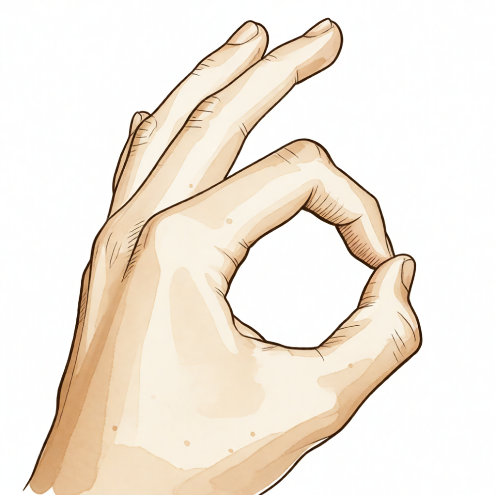
Anterior interosseous nerve palsy is an uncommon disease of unknown etiology and pathophysiology [32]. It creates objective weakness and electrophysiological abnormalities [32]. Clinical suspicion should arise in the presence of isolated paralysis of the AIN-supplied muscles [92]. The clinical manifestations of proximal entrapment of the median nerve include aching pain in the anterior forearm, loss of key and tip pinch strength, inability to make an 'OK' sign, and loss of fine motor skills [78]. Paraesthesias in the median nerve innervated region of the hand similar to a carpal tunnel syndrome may also be present [78].
The pathophysiology is often non-compressive. In the majority of cases, anterior interosseous nerve syndrome is not a surgically treatable entrapment neuropathy but a multifocal mononeuropathy selectively involving the motor fascicles that continue distally to form the anterior interosseous nerve [44]. Compression to the anterior interosseous nerve is very rare, and compression by the normal tissues surrounding it may not exist [9]. The specific muscles involved in anterior interosseous nerve palsy reflect variations in the distribution of the nerve and the ease with which each muscle may be specifically tested [25].
General Diagnostic Considerations
The work-up for nerve injuries about the elbow must include an evaluation of the entire length of the nerve and awareness of potential double crush syndromes [43]. The throwing elbow is a common source of nerve injuries due to the unique combination of anatomy, high forces, and sheer repetition associated with throwing sports [29]. Entrapment neuropathy of the lateral antebrachial cutaneous nerve should be considered in the differential diagnosis of elbow pain [93]. Most publications on uncommon compression syndromes of the radial, ulnar, and median nerves are small retrospective series or case reports, and treatment decisions are not typically based on high levels of evidence [13].
Investigations
Diagnostic Challenges and Terminology: Treatment decisions for uncommon compression syndromes are not typically based on high levels of evidence [13]. To resolve current terminology confusion, the pathology originally described by Bennett should be termed chronic exertional compartment syndrome of the pronator teres or "Bennett's syndrome" [22]. The term "lacertus syndrome" is recommended to refer specifically to intermittent median nerve compression at the level of the lacertus fibrosus [22].
MRI: MRI determination of posterior interosseous nerve (PIN) position is reliable and consistent with prior cadaveric study [41].
Other Considerations: Surgeons are encouraged to bear anomalies such as bifid median nerves in mind when assessing patients with symptoms of median nerve compression [97]. An adequate preoperative evaluation of the level of compression enables the most suitable surgical approach for radial nerve entrapment around the elbow [99].
Treatment
Non-Operative
Conservative management for pronator syndrome and anterior interosseous nerve syndrome includes avoidance of aggravating activities, rest, and anti-inflammatory medication [18]. The treatment of pronator syndrome is largely nonsurgical, consisting of activity modification, anti-inflammatory medication, corticosteroid injections, stretching, and periods of splinting [40]. A prolonged nonsurgical approach is warranted in most cases of median nerve or anterior interosseous nerve compression in the forearm [21]. The majority of peripheral nerve injuries around the elbow respond well to nonsurgical care [20].
Operative
Indications: Surgery is indicated for pronator syndrome when conservative therapy fails [40].
Surgical Approach / Technique: The mini-invasive technique for pronator teres syndrome resulted in symptom disappearance in 93% of cases [4]. Endoscopically assisted, minimally invasive decompression for pronator syndrome adequately and safely decompressed all anatomical points of compression and improved DASH scores [19]. Endoscopic proximal median nerve decompression enables complete decompression of the median nerve in the distal aspect of the arm and proximal aspect of the forearm through a small incision [40].
Other Considerations: Lacertus release significantly reduces pain and numbness with markedly improved hand grip and pinch strength [100]. Surgical release was the most effective mode of treatment for bilateral radial nerve compression syndrome in an elite swimmer, evidenced by full recovery and resumption of normal sports activity at 9-year follow-up [109].
Complications
Vascular injury: The presence of a persistent median artery and superficial brachial artery within the lacertus tunnel poses a risk of injury during surgical decompression or infiltration anesthesia for lacertus tunnel syndrome [105]. Injury to the persistent median artery during lacertus tunnel decompression may lead to serious complications due to the artery's contribution to hand blood supply [105].
Nerve palsy: Median nerve palsy is a rare but possibly devastating complication of arthroscopic elbow contracture release [35]. Acute median nerve compression secondary to lacertus fibrosis syndrome can occur following distal biceps tendon reconstruction [17].
Recovery
Non-Operative Management: Surgical treatment is appropriate for pronator syndrome and anterior interosseous nerve syndrome if symptoms persist despite conservative management [18].
Surgical Indications: Surgical indications for nerve decompression in pronator syndrome include persistent symptoms for more than 6 months [1].
Operative Techniques and Outcomes: Pronator teres symptoms disappeared in 93% of cases following a mini-invasive decompression technique [4]. An author proponent of the endoscopic technique for pronator syndrome foresees its wide indication in the future due to minimal damage [7]. Surgical release of the lacertus fibrosus is a well-treatment for acute compression of the median nerve caused by a proximally displaced and tethered lacertus fibrosus [30].
Postoperative Complications: Delayed-onset anterior interosseous nerve palsy can develop one week after open elbow contracture release, likely due to a stretch injury [112].
Key Evidence
- [Paper] Surgical indications for nerve decompression include persistent symptoms for >6 months in patients with pronator syndrome or for a minimum of 12 months with no signs of motor improvement in those with anterior interosseous nerve syndrome. [1] (10.5435/jaaos-21-05-268)
- [L5] Lacertus syndrome and superficialis-pronator syndrome should be clearly presented as distinct targets for surgical release, and the term pronator syndrome should be replaced with superficialis-pronator syndrome. [2] (10.1177/17531934211024092)
- [L5] The diagnosis of pronator syndrome lacks objective pathophysiology and is supported only by subjective operative findings and relief after surgery, with a paucity of controlled trials demonstrating that operative treatment is more effective than other treatments or sham surgery. [3] (10.1016/j.jhsa.2011.02.014)
- [L4] Pronator teres symptoms disappeared in 93% of cases following the mini-invasive technique. [4] (10.1016/j.jhsa.2012.05.033)
- [L5] Lacertus syndrome is a postexertional compartment syndrome that is more common than pronator syndrome and can be distinguished with a careful workup. [5] (10.1016/j.csm.2020.03.004)
- [L4] No extrinsic compression of the median or anterior interosseous nerves was identified in the arm or forearm. [6] (10.1002/mus.26768)
- [L4] The author is a proponent of the endoscopic technique with minimal damage and foresees its wide indication in the future for pronator syndrome. [7] (10.1177/17531934221076298)
- [L5] It is essential for clinicians to have a thorough understanding of median nerve anatomy, possible sites of compression, and characteristic clinical findings of pronator syndrome to provide a reliable diagnosis and treat their patients. [8] (10.1016/j.jhsa.2020.07.006)
- [L5] [9] (10.1177/17531934221074903)
- [L4] The diagnostic process to differentiate pronator syndrome from carpal tunnel syndrome remains a challenge due to overlapping symptoms and limited reliable information in the literature; this review provides a comprehensive clinical comparison to aid in establishing appropriate diagnosis and treatment. [10] (10.3390/diagnostics12102433)
- [L4] Concurrent carpal tunnel syndrome and pronator syndrome are rarely considered and proximal compression sites are easily overlooked. [11] (10.1016/j.otsr.2016.10.009)
- [L5] The ulnar head of the pronator teres is fibrotic or fibromuscular in 71.5% of cases, and its morphology and location relative to the median nerve are important for surgical treatment of pronator syndrome. [12] (10.1016/j.otsr.2016.08.016)
- [L4] This article reviews uncommon compression syndromes of the radial, ulnar, and median nerves, noting that most publications are small retrospective series or case reports and treatment decisions are not typically based on high levels of evidence. [13] (10.1016/j.hcl.2013.04.014)
- [Case_report] Resection of a segment of the persistent median artery is the recommended treatment for pronator syndrome associated with this anomaly. [14] (10.2106/00004623-198769020-00026)
- [L4] The case demonstrates that patients presenting with median nerve symptoms must be assessed both at the wrist and proximally to exclude compression elsewhere. [15] (10.1111/j.1758-5740.2010.00051.x)
- [L4] With clinical evaluation as the reference standard, electrodiagnostic, ultrasound, and MRI are not helpful in making a diagnosis of pronator syndrome concurrent with carpal tunnel syndrome. [16] (10.1016/j.jhsa.2020.06.006)
- [Case_report] This case presents a unique instance of median nerve symptoms secondary to lacertus fibrosus syndrome after distal biceps tendon repair. [17] (10.1016/j.xrrt.2024.08.009)
- [L5] Conservative management, including avoidance of aggravating activities, rest, and anti-inflammatory medication, is recommended for pronator syndrome and anterior interosseous nerve syndrome; surgery is appropriate if symptoms persist. [18] (10.1016/j.jhsa.2009.10.017)
- [L4] The endoscopically assisted, minimally invasive approach to treat pronator syndrome adequately and safely decompressed all anatomical points of compression and improved DASH scores. [19] (10.1016/j.jhsa.2012.02.023)
- [Paper] Peripheral nerve injuries account for less than 0.5% of all sports-related injuries, with the majority of conditions responding well to nonsurgical care. [20] (10.1016/j.csm.2020.02.006)
- [L5] Surgical decompression of the median nerve or the AIN in the forearm is rarely indicated; a prolonged nonsurgical approach is warranted in most cases. [21] (10.5435/jaaos-d-16-00010)
- [L5] The authors recommend that the term 'lacertus syndrome' refer to intermittent median nerve compression at the level of the lacertus fibrosus, while the pathology originally described by Bennett should be termed chronic exertional compartment syndrome of the pronator teres or 'Bennett's syndrome' to resolve current terminology confusion. [22] (10.1177/17531934231170347)
- [L4] An isolated anterior interosseous nerve injury may not by itself be an indication for urgent surgery. [23] (10.2106/jbjs.n.00136)
- [L5] [25] (10.1016/s0020-1383(84)80015-7)
- [L5] [27] (10.1007/s12593-013-0115-1)
- [L5] The throwing elbow is a common source of nerve injuries due to the unique combination of anatomy, high forces, and sheer repetition associated with throwing sports. [29] (10.1016/j.csm.2004.04.012)
- [L4] Acute compression of the median nerve from the proximally displaced and tethered lacertus fibrosus appears to cause the resultant dynamic compression of the median nerve and is well treated by surgical release of the lacertus fibrosus. [30] (10.1016/j.jse.2006.04.005)
- [L5] [32] (10.1016/j.jhsa.2010.08.018)
- [L4] Proximal median nerve entrapment at the level of the lacertus fibrosus should be referred to as lacertus tunnel syndrome, LTS, to distinguish it from other levels of median nerve entrapment. [33] (10.1007/s11552-012-9483-4)
- [L4] Anterior interosseous nerve palsy is a rare complication of open capsular release for elbow stiffness, likely caused by traction or mass effect from packing. [34] (10.1016/j.jhsa.2008.10.019)
- [L4] We report a rare but possibly devastating complication of median nerve palsy after arthroscopic elbow contracture release. [35] (10.1016/j.jse.2019.06.003)
- [L3] The review highlights the importance of standardized outcome measures, early intervention, and comprehensive rehabilitation for optimizing supercharged end-to-side anterior interosseous nerve to ulnar nerve transfer outcomes. [36] (10.1016/j.jhsg.2024.06.003)
- [Paper] The study inventoried and mapped 6 potentially compressive structures neighboring or crossing the median nerve, showing precise proximal-to-distal locations with variations of 0.5 to 1.5 cm. [37] (10.1016/j.otsr.2021.102813)
- [L4] [40] (10.1016/j.jhsg.2021.03.006)
- [L4] MRI determination of PIN position is reliable and consistent with prior cadaveric study. [41] (10.1016/j.arthro.2020.12.118)
- [L5] [42] (10.1016/j.radcr.2021.02.022)
- [L5] This article presents a review of the cause, pathology, work-up, and treatment options for nerve injuries about the elbow in athletes, emphasizing that the work-up must include an evaluation of the entire length of the nerve and awareness of potential double crush syndromes. [43] (10.1016/j.csm.2010.06.001)
- [L4] Our data strongly support that AINS in the majority of cases is not a surgically treatable entrapment neuropathy but a multifocal mononeuropathy selectively involving, within the main trunk of the median nerve, the motor fascicles that continue distally to form the anterior interosseous nerve. [44] (10.1212/wnl.0000000000000128)
- [L4] Median nerve entrapment in the forearm without motor paralysis is a clinical diagnosis supported by pain drawings, pain quality, and provocative tests. [46] (10.1177/1558944719874137)
- [L4] Surgical decompression of the nerves appears to be the treatment of choice. [50] (10.2106/00004623-197153070-00022)
- [L4] [77] (10.1177/17531934251346595)
- [L4] [78] (10.1177/1753193417726214)
- [L5] [79] (10.1177/17531934251382852)
- [L3] An intact lacertus fibrosus contributes to elbow strength and should be preserved in distal biceps tendon repair. [84] (10.1007/s00167-019-05673-2)
- [L4] [88] (10.1054/jhsb.1998.0177)
- [L5] Clinical suspicion should arise in the presence of isolated paralysis of the AIN-supplied muscles. [92] (10.1016/j.ijscr.2016.02.021)
- [L4] Entrapment neuropathy of the LABCN should be considered in the differential diagnosis of elbow pain. [93] (10.1016/j.jhsa.2004.06.011)
- [L5] We encourage surgeons to bear such anomalies in mind when assessing patients with symptoms of median nerve compression. [97] (10.1177/17531934251401431)
- [L5] An adequate preoperative evaluation of the level of compression enables the most suitable surgical approach, aiming to treat the entrapment with minimal damage to the soft tissues. [99] (10.1016/j.jisako.2024.03.001)
- [L3] Lacertus release appears to significantly reduce pain and numbness with markedly improved hand grip and pinch strength. [100] (10.1016/j.jhsg.2023.03.001)
- [L5] The presence of a persistent median artery (PMA) and superficial brachial artery within the lacertus tunnel poses a risk of injury during surgical decompression or infiltration anesthesia for lacertus tunnel syndrome, which may lead to serious complications due to the artery's contribution to hand blood supply. [105] (10.1007/s11552-013-9525-6)
- [L4] The authors hypothesize that fluid extravasation from arthroscopy causing increased pressure in the upper arm and forearm is the singular cause of these neuropathies. [106] (10.1016/j.jse.2016.04.037)
- [L4] Surgical release was the most effective mode of treatment in this elite athlete, as evidenced by her 9-year follow-up visit and successful outcome with full recovery and resumption of normal sports activity. [109] (10.1177/03635465020300042301)
- [Case_report] The authors describe a case of delayed-onset anterior interosseous nerve palsy developing one week after open elbow contracture release, likely due to a stretch injury. [112] (10.5397/cise.2022.00899)
References
[1] Pronator Syndrome and Anterior Interosseous Nerve Syndrome. Journal of the American Academy of Orthopaedic Surgeons. 2013. DOI: 10.5435/jaaos-21-05-268
[2] Median nerve compression: lacertus syndrome versus superficialis-pronator syndrome. Journal of Hand Surgery (European Volume). 2021. DOI: 10.1177/17531934211024092
[3] Pronator Syndrome. The Journal of Hand Surgery. 2011. DOI: 10.1016/j.jhsa.2011.02.014
[4] New Mini-invasive Decompression for Pronator Teres Syndrome. The Journal of Hand Surgery. 2012. DOI: 10.1016/j.jhsa.2012.05.033
[5] The Lacertus Syndrome of the Elbow in Throwing Athletes. Clinics in Sports Medicine. 2020. DOI: 10.1016/j.csm.2020.03.004
[6] Fascicular constrictions above elbow typify anterior interosseous nerve syndrome. Muscle & Nerve. 2019. DOI: 10.1002/mus.26768
[7] Re: Tang JB. Median nerve compression: lacertus syndrome versus superficialis-pronator syndrome. J Hand Surg Eur. 2021, 46: 1017–22. Journal of Hand Surgery (European Volume). 2022. DOI: 10.1177/17531934221076298
[8] Proximal Median Nerve Compression: Pronator Syndrome. The Journal of Hand Surgery. 2020. DOI: 10.1016/j.jhsa.2020.07.006
[9] Compression to the anterior interosseous nerve is very rare: compression by the normal tissues surrounding it may not exist. Journal of Hand Surgery (European Volume). 2022. DOI: 10.1177/17531934221074903
[10] How to Differentiate Pronator Syndrome from Carpal Tunnel Syndrome: A Comprehensive Clinical Comparison. Diagnostics. 2022. DOI: 10.3390/diagnostics12102433
[11] Concurrent carpal tunnel syndrome and pronator syndrome: A retrospective study of 21 cases. Orthopaedics & Traumatology: Surgery & Research. 2017. DOI: 10.1016/j.otsr.2016.10.009
[12] Morphology and morphometry of the ulnar head of the pronator teres muscle in relation to median nerve compression at the proximal forearm. Orthopaedics & Traumatology: Surgery & Research. 2016. DOI: 10.1016/j.otsr.2016.08.016
[13] Uncommon Upper Extremity Compression Neuropathies. Hand Clinics. 2013. DOI: 10.1016/j.hcl.2013.04.014
[14] Pronator syndrome associated with a persistent median artery. A case report.. The Journal of Bone & Joint Surgery. 1987. DOI: 10.2106/00004623-198769020-00026
[15] Median Nerve Compression Secondary to a High Insertion of Pronator Teres. Shoulder & Elbow. 2010. DOI: 10.1111/j.1758-5740.2010.00051.x
[16] Clinical, Radiological, and Electrodiagnostic Diagnosis of Pronator Syndrome Concurrent With Carpal Tunnel Syndrome. The Journal of Hand Surgery. 2020. DOI: 10.1016/j.jhsa.2020.06.006
[17] Acute median nerve compression secondary to lacertus fibrosis syndrome following distal biceps tendon reconstruction: a case report. JSES Reviews, Reports, and Techniques. 2025. DOI: 10.1016/j.xrrt.2024.08.009
[18] Unusual Compression Neuropathies of the Forearm, Part II: Median Nerve. The Journal of Hand Surgery. 2009. DOI: 10.1016/j.jhsa.2009.10.017
[19] Endoscopically Assisted Decompression for Pronator Syndrome. The Journal of Hand Surgery. 2012. DOI: 10.1016/j.jhsa.2012.02.023
[20] Conditions and Injuries Affecting the Nerves Around the Elbow. Clinics in Sports Medicine. 2020. DOI: 10.1016/j.csm.2020.02.006
[21] Ulnar Tunnel Syndrome, Radial Tunnel Syndrome, Anterior Interosseous Nerve Syndrome, and Pronator Syndrome. Journal of the American Academy of Orthopaedic Surgeons. 2017. DOI: 10.5435/jaaos-d-16-00010
[22] Lacertus syndrome: one term – two different pathologies. Journal of Hand Surgery (European Volume). 2023. DOI: 10.1177/17531934231170347
[23] Supracondylar Humeral Fractures with Isolated Anterior Interosseous Nerve Injuries: Is Urgent Treatment Necessary?. The Journal of Bone and Joint Surgery-American Volume. 2014. DOI: 10.2106/jbjs.n.00136
[25] Anterior interosseous nerve palsy and dislocation of the elbow. Injury. 1984. DOI: 10.1016/s0020-1383(84)80015-7
[27] Pronator Syndrome Due to Schwannoma. Journal of Hand and Microsurgery. 2015. DOI: 10.1007/s12593-013-0115-1
[29] Nerve injuries in the throwing elbow. Clinics in Sports Medicine. 2004. DOI: 10.1016/j.csm.2004.04.012
[30] Acute compression of the median nerve at the elbow by the lacertus fibrosus. Journal of Shoulder and Elbow Surgery. 2007. DOI: 10.1016/j.jse.2006.04.005
[32] Anterior Interosseous Nerve Syndrome. The Journal of Hand Surgery. 2010. DOI: 10.1016/j.jhsa.2010.08.018
[33] Clinical Diagnosis and Wide-Awake Surgical Treatment of Proximal Median Nerve Entrapment at the Elbow: A Prospective Study. HAND. 2013. DOI: 10.1007/s11552-012-9483-4
[34] Anterior Interosseous Nerve Palsy After Open Capsular Release for Elbow Stiffness: Report of 2 Cases. The Journal of Hand Surgery. 2009. DOI: 10.1016/j.jhsa.2008.10.019
[35] High median nerve injury after arthroscopic elbow contracture release with complete recovery at 6 months. Journal of Shoulder and Elbow Surgery. 2019. DOI: 10.1016/j.jse.2019.06.003
[36] Exploring Outcomes and Mediating Factors Following Supercharged End-to-Side Anterior Interosseous Nerve to Ulnar Nerve Transfer: A Scoping Review With Expert Insight. Journal of Hand Surgery Global Online. 2024. DOI: 10.1016/j.jhsg.2024.06.003
[37] Topographic anatomy of structures liable to compress the median nerve at the elbow and proximal forearm. Orthopaedics & Traumatology: Surgery & Research. 2021. DOI: 10.1016/j.otsr.2021.102813
[40] Endoscopic Proximal Median Nerve Decompression: An Alternative Treatment for Pronator Syndrome. Journal of Hand Surgery Global Online. 2021. DOI: 10.1016/j.jhsg.2021.03.006
[41] Implications of Posterior Interosseus Nerve Distance from the Radial Tuberosity: A Radiologic Study. Arthroscopy: The Journal of Arthroscopic & Related Surgery. 2021. DOI: 10.1016/j.arthro.2020.12.118
[42] Lacertus syndrome: Use of pre- and post-exercise MRI to aid in diagnosis and treatment. Radiology Case Reports. 2021. DOI: 10.1016/j.radcr.2021.02.022
[43] Nerve Injuries About the Elbow. Clinics in Sports Medicine. 2010. DOI: 10.1016/j.csm.2010.06.001
[44] Anterior interosseous nerve syndrome. Neurology. 2014. DOI: 10.1212/wnl.0000000000000128
[46] Median Nerve Compression in the Forearm: A Clinical Diagnosis. HAND. 2019. DOI: 10.1177/1558944719874137
[50] Nerve Injuries Complicating Monteggia Lesions. The Journal of Bone & Joint Surgery. 1971. DOI: 10.2106/00004623-197153070-00022
[54] Miller S Review Of Orthopaedics. SECTION 16 PATELLAR TRACKING IN TOTAL KNEE ARTHROPLASTY > TABLE 2.3 Shoulder Spaces.
[55] Miller S Review Of Orthopaedics. Genetics of musculoskeletal conditions and abnormalities are summarized in Table 1.27 > TABLE 2.3 Shoulder Spaces.
[59] Miller S Review Of Orthopaedics. ELBOW.
[60] Miller S Review Of Orthopaedics. SECTION 16 PATELLAR TRACKING IN TOTAL KNEE ARTHROPLASTY > ELBOW.
[61] A Lange Medical Book Current Diagnosis Treatment In Orthopedics Fifth Edition. 2Musculoskeletal Trauma Surgery > INJURIES AROUND THE ELBOW.
[62] Orthopaedic Knowledge Update Trauma. Distal Humerus Fracture > Anatomy.
[64] Rockwood And Green S Fractures In Adults. 29: Principles of Nonunion and Bone Defect Treatment > Pathoanatomy and Applied Anatomy Related to Distal Humerus Fractures.
[65] Green S Operative Hand Surgery. PERTINENT ANATOMY AND PORTALS.
[77] Incidence of local tenderness at the lacertus fibrosus in healthy people. Journal of Hand Surgery (European Volume). 2025. DOI: 10.1177/17531934251346595
[78] The scratch collapse test in the diagnosis of compression of the median nerve in the proximal forearm. Journal of Hand Surgery (European Volume). 2017. DOI: 10.1177/1753193417726214
[79] Re: Fang J, Zhang LQ, Tang JB. Incidence of local tenderness at the lacertus fibrosus in healthy people. Journal of Hand Surgery (European Volume). 2025. DOI: 10.1177/17531934251382852
[84] An intact lacertus fibrosus improves strength after reinsertion of the distal biceps tendon. Knee Surgery, Sports Traumatology, Arthroscopy. 2019. DOI: 10.1007/s00167-019-05673-2
[88] Microscopic Anatomy of the Posterior Interosseous and Median Nerves at Sites of Potential Entrapment in the Forearm. Journal of Hand Surgery. 1999. DOI: 10.1054/jhsb.1998.0177
[92] Anterior interosseous nerve syndrome diagnosis and intraoperative findings: A case report. International Journal of Surgery Case Reports. 2016. DOI: 10.1016/j.ijscr.2016.02.021
[93] Painful entrapment of the lateral antebrachial cutaneous nerve at the elbow. The Journal of Hand Surgery. 2004. DOI: 10.1016/j.jhsa.2004.06.011
[97] Bifid median nerve entrapment by forearm musculature – a case report and systematic literature review. Journal of Hand Surgery (European Volume). 2025. DOI: 10.1177/17531934251401431
[99] Current concepts of surgical approach for radial nerve entrapment around the elbow. Journal of ISAKOS. 2024. DOI: 10.1016/j.jisako.2024.03.001
[100] Lacertus Syndrome: an Outcome Analysis After Lacertus Release. Journal of Hand Surgery Global Online. 2023. DOI: 10.1016/j.jhsg.2023.03.001
[105] Median Nerve Decompression at Lacertus Tunnel: Possible Complications. HAND. 2013. DOI: 10.1007/s11552-013-9525-6
[106] Mixed neuropathy presenting clinically as an anterior interosseous nerve palsy following shoulder arthroscopy: a report of four cases. Journal of Shoulder and Elbow Surgery. 2016. DOI: 10.1016/j.jse.2016.04.037
[109] Bilateral Radial Nerve Compression Syndrome in an Elite Swimmer. The American Journal of Sports Medicine. 2002. DOI: 10.1177/03635465020300042301
[112] Anterior interosseous nerve palsy in the early postoperative period after open capsular release for elbow stiffness: a case report. Clinics in Shoulder and Elbow. 2023. DOI: 10.5397/cise.2022.00899