Clinicians › Hand
Flaps and Grafts
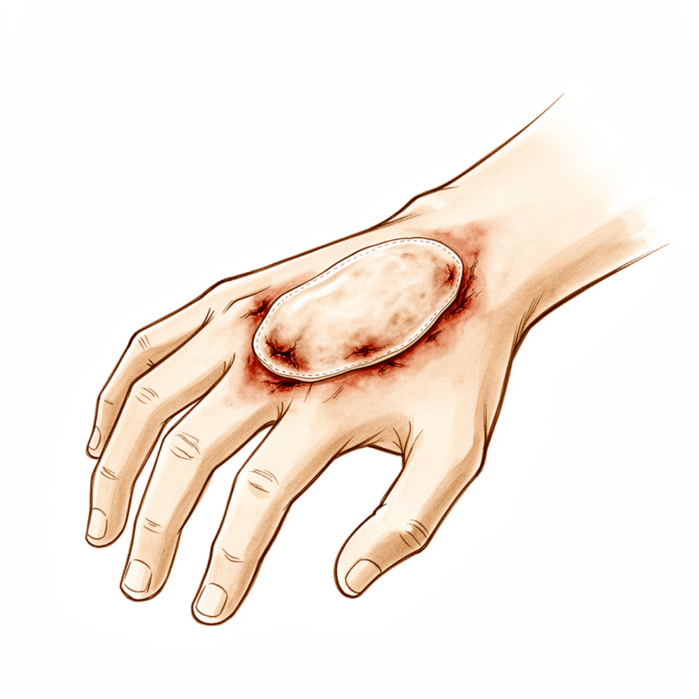
Hand reconstruction: flap and graft options for soft tissue defects, bone loss, and complex trauma—local to free flaps.
Overview
Reconstructive surgery for complex hand defects relies on efficient vascular anastomoses performed outside the zone of injury to ensure success in both replantation and free flap procedures [1]. Free flaps demonstrate the highest versatility and are among the first choices for treating mutilated hands, offering critical advantages over non-microsurgical techniques such as early mobilization and appropriate coverage with acceptable donor morbidity [17, 18]. These procedures improve cosmetic appearance and augment function [5]. While local hand flaps offer excellent coverage by replacing like with like and frequently result in optimum functional and aesthetic outcomes while sparing patients from more complicated repair methods [6], they are primarily considered for small or moderate defects [10]. Vascularized free flaps are reserved for large defects of 3 cm or larger [10].
A framework exists for assessing defects and selecting the best-suited reconstructive option using representative flaps and case examples [3]. Optimal functional and aesthetic outcomes depend on optimizing flap selection based on patient body habitus, anatomic location, and defect characteristics [27]. Utilizing adjuncts like suprafascial harvest and meticulous inset helps minimize donor site morbidity [27]. The need for refinement and secondary surgery should be taken into consideration during the initial flap selection process [21]. Experience dictates that basic principles and indications of replantation surgery must be adhered to for successful outcomes [49]. Hand transplantation and prosthetic reconstruction should not be viewed as competing options [16].
Microsurgeons continue to push the limits of reconstructive and replantation feasibility despite complications such as flap necrosis or loss [4]. Five described free vascularized flaps reconstruct complex soft tissue losses of the hand without harvesting major arteries and can be quite reliable with appropriate planning and microsurgical expertise [5]. In a clinical series of 9 cases, the reverse flow shunt restricted arterialized venous free flap resulted in high flap survival rates with satisfactory functional outcomes [7]. Venous flaps offer a versatile and well-tolerated reconstructive option [8] and are an optimal candidate for hand and digit reconstruction when conventional flaps are limited or unavailable [32]. Venous flaps offer advantages such as ease of design, thinness, and lack of donor site morbidity [32], although clinical application remains under further investigation due to inconsistent survival in some cases [32]. A series of 79 free flaps using the ulnar artery distal cutaneous descending branch provided satisfactory reconstruction of hand wounds with 100% flap survival and acceptable aesthetic and functional results [9].
Anatomy & Pathophysiology
Skeletal Architecture
The skeleton of the hand and wrist comprises 27 bones, 19 of which are long bones [44]. The hand skeleton is organized into five rays, each forming a polyarticulated chain of metacarpals and phalanges [44]. The thumb ray (first ray) is the shortest, consisting of a metacarpal and two phalanges, while the four finger rays consist of a metacarpal and three phalanges [44]. In the sagittal plane, the thumb metacarpal makes an angle of about 45 degrees with the second metacarpal, and the transverse axis of the palm forms an acute angle of approximately 75 degrees with the longitudinal axis [44].
Mobility varies among the metacarpals; the index metacarpal is the most firmly fixed, whereas the fifth metacarpal has a range of flexion–extension of approximately 20 degrees and the ring metacarpal has about 10 degrees of mobility in flexion and extension [72]. The metacarpophalangeal joints serve as the keystones of the longitudinal arches of the hand, with volar plates preventing hyperextension at these joints [72].
Musculotendinous Anatomy
Extrinsic muscles originate outside the hand and insert on the hand or carpus, whereas intrinsic muscles have both origin and insertion within the hand [43]. The extrinsic extensors traverse six fibroosseous retinacular compartments at the wrist level [43]: * First compartment: Contains the abductor pollicis longus and extensor pollicis brevis [43]. * Second compartment: Contains the extensor carpi radialis longus and extensor carpi radialis brevis [43]. * Third compartment: Contains the extensor pollicis longus, which turns abruptly radialward about Lister tubercle [43]. * Fourth compartment: Contains the extensor indicis proprius, lying deep to the four tendons of the extensor digitorum communis [43]. * Fifth compartment: Contains the extensor digiti quinti [43]. * Sixth compartment: Contains the extensor carpi ulnaris [43].
Sagittal bands stabilize the digital extensor tendons over the midline of the metacarpophalangeal joint; rupture or attenuation of these fibers can cause the extrinsic extensor tendon to sublux to the ulnar side of the metacarpal head [43]. The flexor digitorum profundus inserts on the proximal volar aspect of the distal phalanx [43]. The flexor digitorum superficialis inserts via radial and ulnar slips into the proximal metaphysis of the middle phalanx, with its tendon bifurcating around the flexor digitorum profundus at the beginning of the A2 pulley [76]. The A2 and A4 pulleys are the most essential in maintaining the mechanical advantage of the flexor tendons [76].
There are seven interosseous muscles: four dorsal and three volar [70]. The dorsal interossei act as abductors, while the volar interossei act as adductors [70]. The middle finger possesses two dorsal interossei and no volar interossei because the central axis of the hand lies within it [70]. The deep head of each dorsal interosseous muscle forms a lateral tendon, or lateral band, at the level of the metacarpophalangeal joint, and the terminal tendon inserts at the base of the distal phalanx to extend it [70].
Vascular Anatomy
The "princeps pollicis" artery is the terminal branch of the radial artery that crosses the first intermetacarpal space, although in only 15% of anatomical dissections do the palmar arteries of the thumb follow the classical layout [73]. The radial artery originates from the brachial artery and travels in a relatively superficial course in the forearm, giving branches to a plexus of vessels in the overlying deep fascia that supplies the skin of the anterior and radiodorsal surfaces of the forearm [85]. It is accompanied by two or more venae comitantes, and multiple anastomoses between these veins permit reversal of flow without valvular obstruction [85].
The posterior interosseous artery flap provides stable coverage while preserving both the radial and ulnar arteries [13]. The flow-through venous flap relies on the subcutaneous venous network for backflow, with hypothesized mechanisms for venous skin flap survival including arteriovenous shunting, reverse flow, and capillary bypass [68].
Cutaneous Anatomy and Functional Units
The dorsal skin of the hand is thin, with a horny layer only 0.02 mm thick, and possesses a normal pilosebaceous system unlike the palm [78]. Flexion of the fingers produces significant lengthening of the dorsal skin, with an average increase of 3 cm in the middle finger from extension to full flexion [78].
The palm forms a cutaneous unit extending from the distal transverse crease of the wrist up to the transverse crease at the base of the digits [69]. The skin of the radial portion of the palm is relatively well vascularized and mobile, while the skin of the ulnar and distal portion has poor mobility [69]. The central triangular part of the palm is fixed and poorly vascularized, covering almost directly the superficial palmar aponeurosis [69]. The dorsal covering of the interphalangeal articulations forms a unique cutaneous unit characterized by a considerable excess of skin when the digits are in extension [69]. The dorsal integument of the distal phalanx is distinct due to the nail bed with its matrix [69].
Web spaces are formed from the union of two nonsymmetrical cutaneous surfaces [69]. The dorsal slope of the web space has a gradual incline and supple skin that is not adherent to the subjacent region, whereas the palmar surface is flat and precipitously interrupted, with skin densely adherent to the commissural skeleton [69].
Specific dimensions for skin coverage are as follows: * Thumb distal to MCP: Approximately 9 cm wide and 8 cm long [71]. * Thumb and first metacarpal loss: 13 cm wide and 12 cm long [71]. * Palmar and dorsal hand surfaces: 12 cm by 10 cm [71]. * Each finger (palmar and dorsal): 7 cm by 10 cm [71]. * Both sides of hand and digits: Requires 20 cm by 20 cm of skin for grafting or flaps [71].
Pathophysiology of Injury
Severe hand injuries are complex traumas characterized by the simultaneous involvement of tendons, bones, joints, and neurovascular structures, frequently involving soft tissue loss that results in exposure of critical components [13]. Closed degloving of the thumb is caused by crushing and elongation forces that detach soft tissues without skin disruption [128].
Wounds with distally attached flaps may have enough skin for primary closure but not enough venous drainage for the skin to survive [89]. Deficient venous drainage causes engorgement, venous distention, and finally thrombosis and necrosis [89]. The color of a flap with venous congestion changes from a deep blue to purple and then to black [89]. A retrograde flap on the dorsal surface of the hand or forearm is less likely to survive than one on the palm [89].
The fingertip is defined as the portion of the finger distal to the insertion of the flexor and extensor tendons [66]. The unique anatomy and specialized structure of the fingertip make it critical for functions such as sensation, fine handling, and gripping [66].
Classification
Merle: The Merle classification is the commonly used system for categorizing thumb amputations [20].
Urbaniak: The Urbaniak classification was the first to classify ring degloving injuries [141]. Kay et al. produced a modified classification of ring degloving injuries [141]. Both Urbaniak and Kay grouped complete degloving in the severest category, along with amputation [141].
Allen: Allen’s classification is used to categorize fingertip amputations [142]. It includes types 2, 3, and 4 for fingertip injuries [142].
Tubiana: Tubiana’s classification system categorizes the total passive extension deficit of each ray in 4 stages [151]. Stage I corresponds to a passive extension deficit of 0–45° [151]. Stage II corresponds to a passive extension deficit of 46–90° [151]. Stage III corresponds to a passive extension deficit of 91–135° [151]. Stage IV corresponds to a passive extension deficit of 136–180° [151].
Banff Working Classification: The Banff Working Classification provides a standardized grading scheme for acute rejection in composite tissue allografts [101].
Chen: Chen's classification lacks utility for type III venous flaps [84].
Woo et al.: The Woo et al. classification is proposed for better clinical description and design of venous flaps [84].
Clinical Presentation
Severe hand injuries constitute complex traumas characterized by the simultaneous involvement of tendons, bones, joints, and neurovascular structures [13]. These injuries are frequently accompanied by soft tissue loss that results in the exposure of these critical components [13]. Such damage may lead to devastating functional outcomes, including partial or complete loss of digits or the limb [13]. Patients with firework-related hand injuries often require reconstruction rather than repair, and many will require secondary procedures [31].
The main goal of reconstruction is to achieve normal or near-normal mobility [34]. When conventional reconstruction is not feasible for complex tissue injuries, hand transplantation serves as a valuable treatment option [48]. This procedure is the most common form of modern composite tissue allotransplantation [50].
Reconstructive strategy depends on defect size and specific anatomical requirements. Local flaps are primarily considered for small or moderate defects, while vascularized free flaps are reserved for large defects (3 cm or larger) [10]. For thumb reconstruction, the maximal age for complex procedures is around 60 to 65 years old [20]. An optimal thumb reconstruction should meet three main objectives: * Restore thumb function [20] * Recreate its aesthetics [20] * Minimize donor-site morbidity [20]
Investigations
CT: Preoperative computed tomography angiography (CTA) and computed tomography densitometry (CDS) are accurate methods for assessing flap location and course [157].
MRI: Magnetic resonance imaging is probably most useful for identifying additional pathology, such as flexor tendon bowstringing, in the context of Dupuytren's disease [82]. MRI may also provide a quantitative noninvasive measure of cellularity in affected areas, serving as an index of biologic activity [82]. Visualization of digital vessels via magnetic resonance angiography (MRA) remains difficult in Dupuytren's disease, even with 3D reconstruction [82]. In the context of autologous osteochondral mosaicplasty, MRI indicates that the donor site is resurfaced with fibrous tissue [132].
Doppler: An 8-MHz Doppler tone assessment may be used to identify superficially displaced neurovascular bundles when Dupuytren cords lie beneath soft fleshy prominences, though false-negatives are possible [82]. Doppler imaging is a promising improvement for identifying palmar structures, though higher resolution imaging technology is needed [82].
Plain radiography: Follow-up radiography results demonstrate the feasibility of the induced membrane technique and structural treatment with a cylindrical bone graft for segmental bone defects of the metacarpal or phalanx [179].
Treatment
General Principles and Planning
Reconstructive surgery for the upper extremity continues to expand its feasibility, with microsurgeons pushing the limits of replantation and reconstruction despite risks such as flap necrosis or loss [4]. The primary goal in managing the mangled upper extremity is to preserve life, adhering to the "life before limb" priority [150]. An early assessment of limb viability is essential to determine whether to attempt salvage or proceed with early complete amputation [150]. The surgeon responsible for limb reconstruction must be involved in surgical planning from the initial débridement [150]. For acute burned upper extremities, the surgical plan prioritizes key functional recovery over the range of motion of all individual joints [58]. Treatment goals for burns and frostbite focus on wound coverage and closure, infection prevention, motion maintenance, and early transition to functional rehabilitation [152]. In firework-related hand injuries, patients often require reconstruction rather than simple repair, with many necessitating secondary procedures [31].
Local and Regional Flaps
Local hand flaps provide excellent soft tissue coverage by replacing like with like, often yielding optimum functional and aesthetic outcomes while avoiding more complex repair methods [6]. These flaps are used to protect against contractures and facilitate mobility within the hand [177]. For expendable zones such as the dorsum of the hand or forearm, traditional management with flaps or skin grafts remains appropriate [172]. Specific local options include the homodigital unipedicle neurovascular island flap, which provides satisfactory long-term patient-reported and objective outcomes [2]. However, for fingertip injuries with bone exposure, the parallelogram flap is a better choice than the homodigital island flap [22]. The heterodigital reversed flow neurovascular island flap serves as an alternative to microsurgical reconstruction for extensive pulp defects where other flaps are not feasible [107].
Regional flaps offer reliable coverage for larger defects. The posterior interosseous artery (PIA) flap is a reliable option for dorsal hand and distal forearm defects, providing stable coverage while preserving both the radial and ulnar arteries [13]. The reversed vascularized pedicled forearm flap provides adequate soft tissue and good functional outcomes for salvage of complete degloved digits compared with other salvage procedures [12]. The dorso-ulnar osteocutaneous reverse flow flap is useful for distal osteocutaneous defects of the thumb when more complex procedures are not feasible or considered excessive [35]. The volar cross-finger flap using the index finger for transverse and dorsal-oblique amputations of the distal thumb has resulted in 100% survival of flaps and skin grafts, with no postoperative flap ischemia, venous congestion, or donor site complications [122].
Free Flaps
Five described free vascularized flaps reconstruct complex soft tissue losses of the hand without harvesting major arteries, improving cosmetic appearance and augmenting function with high reliability when appropriate planning and microsurgical expertise are applied [5]. Free-style flaps can be raised reliably and safely, as demonstrated by experience over the past 10 years [11]. The anterolateral thigh free flap is encouraged for hand defects where locoregional flaps would result in unacceptable cosmetic deformity [26]. The modified reverse flow shunt restricted arterialized venous free flap resulted in high flap survival rates and satisfactory functional outcomes in a clinical series of 9 cases [7].
Specific applications include the microsurgical free lateral great toe flap for revision of residual deformities after primary surgery for Wassel-Flatt IV-D thumb duplication, which resulted in all flaps surviving with satisfactory appearance at 8–12 months follow-up [14]. Microsurgical reconstruction of partial thumb defects using free flaps achieved 100% flap survival and functional improvement with near normal appearance [54]. The free 'mini' groin flap for digital resurfacing had no flap losses in a series of 10 cases and is recommended for moderate to large dorsal defects, proximal volar defects, circumferential skin loss, and multiple digit injuries [104]. The pedicled osteo-onychocutaneous island flap combined with growth-limiting procedures for finger macrodactyly achieved satisfactory aesthetic and functional outcomes at 1 year [56].
Venous Flaps
Venous flaps are an optimal candidate for hand and digit reconstruction when conventional flaps are limited or unavailable, offering advantages such as ease of design, thinness, and lack of donor site morbidity [32]. However, the clinical application of venous flaps remains under further investigation due to inconsistent survival in some cases [32].
Bone Grafts and Vascular Conduits
A nonvascularized iliac crest bone graft is supported for use when soft tissues are preserved or as a second stage after flap reconstruction and bone temporizing measures [15]. Nonvascularized autogenous bone graft is capable of restoring good hand function in severe injuries with substantial phalangeal bone loss [52]. Pedicle bone grafts united to the recipient bed earlier than non-pedicle bone grafts, and the rate of non-union of the pedicle bone grafts was lower than that of the non-pedicle grafts [173]. Intermediate-term outcomes for arterial grafts in upper extremity vascular reconstruction are promising, with patency rates up to 100% reported, though no long-term outcomes studies exist [36].
Adjunctive and Alternative Techniques
Dermal skin substitutes can be used to cover resultant defects following adequate surgical debridement of upper extremity wounds [155]. Meshed dermal substitutes are advocated to fill voids, which decreases the possibility of recurrent skin contractures and improves the final aesthetic appearance of the grafted site [155]. Dermal regeneration templates require either single-staged or 2-staged reconstructions, with the latter involving application of the dermal matrix followed by coverage with a skin graft 5 to 7 days later [155].
External tissue expansion has emerged as an attractive alternative to conventional skin grafting and flap-based reconstruction for complex extremity wounds, offering a technically simple and low-morbidity option for durable soft tissue coverage [168]. The combined rib-latissimus dorsi flap is a useful option for composite defects of the upper extremity for the practicing non-microvascular orthopaedic surgeon [170]. The fasciocutaneous transposition flap for coverage of defects of the lower extremity proved to be safe and simple [100]. The combined salvage microsurgical procedures with non-microsurgical methods, such as hand-to-hand finger transfer associated with hallux transfer, provided a satisfactory functional result in a patient with severe frostbite injury who refused any cosmetic prosthesis [166]. Large-caliber processed nerve allografts (4- to 5-mm-diameter) have an overall success rate that suggests their continued incorporation into repair algorithms for upper extremity nerve injuries is acceptable and reasonable [99].
Replantation and Transplantation
Experience dictates that satisfactory functional results in replantation can be achieved only if the basic principles and indications of replantation surgery are adhered to [49]. While microsurgery has made replantation a routine procedure with reliable viability rates, achieving a satisfactory functional result is difficult and depends on a variety of factors [53]. Hand transplantation is a valuable treatment option for patients with complex tissue injuries where conventional reconstruction is not feasible, though long-term high-dose immunosuppression remains a limiting obstacle [48]. Bilateral hand transplantation is justified in cases of bilateral amputations with strict indications, while transplantation for the loss of one hand is not indicated [95].
Ectopic banking of amputated parts may be performed rapidly with skin and vessel repairs only, with the most beneficial location being the contralateral radial artery level of the forearm [25]. When harvesting an ectopically banked part, the previously performed arterial venous anastomosis should be preserved if possible, using the entire length of the ectopic recipient artery and vein as harvested conduits for transfer to the amputation stump [25]. The contralateral C7 transfer procedure has demonstrated significant improvements in upper limb function with donor site morbidity that is typically mild and transient [106].
Complications
Flap Survival and Necrosis: Flap necrosis and loss are recognized complications in microsurgical upper extremity reconstruction [4]. Survival rates vary by technique and indication. A series of 79 free flaps using the ulnar artery distal cutaneous descending branch achieved 100% flap survival [9]. In a series of 9 cases using a reverse flow shunt restricted arterialized venous free flap, the technique resulted in high flap survival rates [7]. For free lateral great toe flaps for thumb pulp defects, 4 patients incurred flap necrosis and 20 survived, resulting in an 83% survival rate [47]. Smoking was identified as a potential risk factor for flap necrosis in this specific reconstruction (P < .05) [47]. In a series of 10 patients undergoing revision of thumb duplication with a microsurgical free lateral great toe flap, all flaps survived [14]. In a series of 9 arterialized venous toenail flaps, one flap suffered 25% necrosis and the nail was deformed [64]. In a series of 10 patients using a modified pivot flap for fingertip defects, venous congestion was observed in two flaps, with one developing superficial partial flap necrosis that healed in 10 days [108].
Traumatic Tibial Wound Reconstruction: Complete flap survival in reconstruction of traumatic tibial wounds has been reported to range from 91% to 93% [156]. Total flap failure in free tissue transfer for these wounds has been reported to be 1% to 3% [156]. Elevated creatinine level was an independent risk factor for flap necrosis in patients with critical limb ischemia undergoing free tissue transfer [156]. The experience of the microsurgeon and the volume of the surgeon and hospital are factors that can contribute to complication rate and overall success in free tissue transfer [156]. History of diabetes mellitus, injury to the posterior tibial artery, and the nature of the injury are patient-related factors that influence the rate of amputation after attempted free-flap reconstruction for limb salvage [156].
Sensory and Functional Outcomes: In cases where an oblique triangular flap for fingertip reconstruction was advanced more than 12 mm, sensory disturbance of the fingertip occurred and did not subside [45]. Reported two-point discrimination (2-PD) data of flaps in digit-tips in the previous 6 years in English literature should be generally considered insufficiently reliable [65]. In a series of 10 patients undergoing secondary thumb reconstruction via ectopic banking, examinations focused on infection, range of motion, bone union, and signs of osseous resorption [29]. The macroscopic appearance of banked bone at the time of harvest looked normal [29].
Donor Site and Vascular Complications: Donor site complications occurred at similar rates among patients undergoing osteocutaneous and fasciocutaneous radial forearm free flap harvest [28]. In a series of 10 patients using a reverse posterior interosseous flap based on an exteriorized pedicle, there were no vascular complications and wrist mobility was normal at 6 months [57]. Intermediate-term outcomes for arterial grafts in upper extremity vascular reconstruction show patency rates up to 100%, though no long-term outcome studies exist [36]. The use of both true flow-through flap and flow-through conduit methods is useful in cases with few vascular resources [19].
Other Considerations: Reconstructive surgery was required in 15% of patients during the 10-year follow-up period after hand burns [59]. Failure in digit replantation is defined as necrosis of the replanted finger, requiring revision amputation or a flap to cover the bone [137]. Despite a high complication rate, free vascularized fibular graft reconstruction offers a reliable treatment of large skeletal defects after tumor resection without increased risk of limb loss, local recurrence, or tumor metastasis [119]. It is possible to achieve good results with acceptable complications when full-thickness skin grafts are used as part of a modification of the Flatt technique for syndactyly release [125]. The drawbacks of free tissue transfer include increased surgical time, donor site morbidity, and the need for microvascular expertise [156]. The use of traumatized tissue to cover a traumatic wound is fraught with potential complications [156]. In a series of 79 free flaps for hand reconstruction, acceptable aesthetic and functional results were achieved [9]. In a series of 10 patients undergoing secondary thumb reconstruction via ectopic banking, all wraparound flap transfers remained viable and no emergent re-explorations were required [29]. No bone graft infection was observed in this series [29]. One patient was readmitted 3 weeks after discharge because of donor toe skin necrosis [29]. The bone cortex was visually thinner in 2 patients with a banked duration of more than 1 month [29].
Recovery
General Principles and Planning: Microsurgical reconstruction should always be undertaken regardless of the class of injury to ensure excellent survival and good functional outcome [60].
Flap Survival and Complications: The five described free vascularized flaps reconstruct complex soft tissue losses of the hand without harvesting major arteries, improve cosmetic appearance, augment function, and can be quite reliable with appropriate planning and microsurgical expertise [5]. In a clinical series of 9 cases, the modified reverse flow shunt restricted arterialized venous free flap technique resulted in high flap survival rates with satisfactory functional outcomes [7]. All flaps survived in the revision of residual deformities after primary surgery for Wassel-Flatt IV-D thumb duplication using a microsurgical free lateral great toe flap, with patients followed up for 8–12 months showing satisfactory appearance [14]. By using arterialized venous free flaps having 2 parallel veins, the technique of arterialized venous flaps was simplified [23]. All wraparound flap transfers remained viable in secondary thumb reconstruction via ectopic banking of bony phalanges, and no emergent re-explorations were required [29]. No bone graft infection was observed in any of the patients undergoing secondary thumb reconstruction via ectopic banking of bony phalanges [29]. One patient undergoing secondary thumb reconstruction via ectopic banking of bony phalanges was readmitted 3 weeks after discharge because of donor toe skin necrosis [29]. Flap survival was 100% and functional improvement with near normal appearance was obtained in the reconstructed thumbs following microsurgical reconstruction of partial thumb defects [54]. The single-stage technique using a pedicled osteo-onychocutaneous island flap combined with growth-limiting procedures achieved satisfactory aesthetic and functional outcomes at 1 year for finger macrodactyly [56]. There were no vascular complications, flap survival was complete, and wrist mobility was normal at 6 months following a reverse posterior interosseous flap based on an exteriorized pedicle to cover digital skin defects [57]. The aesthetic and functional outcomes of the reconstructed thumbs and fingers significantly improved with the vascularized half–big toenail flap, and donor site functional morbidity was minimum [62]. In a series of arterialized venous toenail flaps for treating nail loss in the fingers, nine flaps survived completely and one flap suffered 25% necrosis with a deformed nail [64]. The 2-point discrimination data of flaps in digit-tips in the previous 6 years in English literature should be generally considered insufficiently reliable [65]. The rate of complete composite graft survival in a paediatric population is low, although the long-term function of these patients is good [147]. Following revascularization, the skin from a completely degloved finger will survive in approximately two cases out of three [181].
Specific Outcomes and Follow-up: At a long-term follow-up, the patient-reported outcome measures and objective outcomes of the homodigital unipedicle neurovascular island flap are satisfactory and it is a safe and reliable flap [2]. The report supports the use of a nonvascularized iliac crest bone graft when soft tissues are preserved or as a second stage after flap reconstruction and bone temporizing measures [15]. The cross-finger flap is a simple, reliable, long-lasting reconstruction technique for fingertip amputations [37]. This is the first report of a one-stage combined reconstruction of an amputated thumb with application of a free neurovascular flap and an iliac-bone graft in the English literature [39]. Follow-up assessments show that the majority of zone I replantations led to satisfactory function [114]. Delayed and suspended replantations demonstrate results comparable to immediate replantation regarding graft survival and clinical outcome [180]. Hand transplant is an effective treatment in selected patients with continual improvements in function seen for up to 5 years [182]. The procedure of split-thickness skin grafting has been successfully used in four cases with follow-up up to two years, and the grafts have proved serviceable and stable [183]. Prophylactic antibiotics were continued for 1 week following secondary thumb reconstruction via ectopic banking of bony phalanges [29]. Kirschner wires were left in place until evidence of fusion was noted on radiographic examination, which was usually seen after approximately 6 weeks in secondary thumb reconstruction via ectopic banking of bony phalanges [29]. Examinations of the reconstructed thumb focused on infection, range of motion of the IP and metacarpophalangeal joints, bone union of the graft, and signs of osseous resorption [29]. The macroscopic appearance of banked bone at the time of harvest from the subcutaneous pocket looked normal in secondary thumb reconstruction via ectopic banking of bony phalanges [29]. In 2 patients with a banked duration of more than 1 month, the bone cortex was visually thinner in secondary thumb reconstruction via ectopic banking of bony phalanges [29]. All surgical wounds at the banking site healed without any complications in secondary thumb reconstruction via ectopic banking of bony phalanges [29].
Key Evidence
- [L5] Efficient surgery with carefully performed vascular anastomoses outside the zone of injury is paramount to the success of both replantation and free flap surgery. [1] (10.1016/j.hcl.2015.01.008)
- [L4] At a long-term follow-up, the patient-reported outcome measures and objective outcomes of this flap are satisfactory and it is a safe and reliable flap. [2] (10.1177/17531934231172081)
- [L5] This review provides a framework for assessing defects and selecting the best-suited reconstructive option using representative flaps and case examples. [3] (10.1016/j.jhsa.2016.04.020)
- [L5] Despite complications such as flap necrosis or loss, microsurgeons continue to push the limits of reconstructive and replantation feasibility. [4] (10.1016/j.hcl.2010.01.007)
- [L5] The 5 described free vascularized flaps reconstruct complex soft tissue losses of the hand without harvesting major arteries, improve cosmetic appearance, augment function, and can be quite reliable with appropriate planning and microsurgical expertise. [5] (10.1016/j.hcl.2017.04.011)
- [L5] Local hand flaps offer excellent coverage of soft tissue defects by replacing like with like, frequently resulting in optimum functional and aesthetic outcomes while sparing patients from more complicated repair methods. [6] (10.1016/j.hcl.2013.12.004)
- [L4] In this clinical series of 9 cases, the modified technique resulted in high flap survival rates with satisfactory functional outcomes. [7] (10.1016/j.jhsa.2018.02.023)
- [L2] Venous flaps offer a versatile and well-tolerated reconstructive option. [8] (10.1177/1753193417712879)
- [L4] The series of 79 free flaps provided satisfactory reconstruction of hand wounds with 100% flap survival and acceptable aesthetic and functional results. [9] (10.1016/j.injury.2009.04.009)
- [L5] Local flaps are primarily considered for small or moderate defects, while vascularized free flaps are reserved for large defects (3 cm or larger). [10] (10.1177/17531934211051303)
- [Paper] Experience over the past 10 years has consistently demonstrated that free-style flaps can reliably and safely be raised. [11] (10.1016/j.injury.2008.05.020)
- [L4] This flap provides adequate soft tissue and good functional outcome compared with other salvage procedures. [12] (10.1016/j.jhsa.2012.01.032)
- [L4] [13] (10.1016/j.injury.2026.113151)
- [L4] All flaps survived and patients were followed up for 8–12 months with satisfactory appearance of the reconstructed thumbs. [14] (10.1177/17531934231222400)
- [L4] The report supports the use of a nonvascularized iliac crest bone graft when soft tissues are preserved or as a second stage after flap reconstruction and bone temporizing measures. [15] (10.1177/1753193420970162)
- [L5] Hand transplantation and prosthetic reconstruction should not be viewed as competing options. [16] (10.1016/j.jht.2013.10.007)
- [L5] Free flaps demonstrate the highest versatility and are among the first choices in treating mutilated hands. [17] (10.1016/s0749-0712(02)00128-2)
- [L4] Free flap reconstructions offer critical advantages over non-microsurgical techniques, including early mobilization and appropriate coverage with acceptable donor morbidity. [18] (10.1016/j.injury.2013.01.021)
- [L4] The use of both true flow-through flap and flow-through conduit methods is very useful, especially in cases with few vascular resources. [19] (10.1016/j.jhsg.2024.02.017)
- [L4] [20] (10.1177/1753193417723310)
- [L5] The need for refinement and secondary surgery should be taken into consideration during the initial flap selection process. [21] (10.1016/j.hcl.2014.01.004)
- [L2] This method is a better choice for reconstruction of fingertip injury. [22] (10.1186/s13018-022-03214-1)
- [L4] By using these flaps, the authors were able to simplify the technique of arterialized venous flaps. [23] (10.1016/j.jhsa.2008.08.001)
- [L4] [25] (10.1016/j.jhsa.2011.09.003)
- [L4] The authors encourage hand surgeons to consider this flap for defects where locoregional flaps would result in unacceptable cosmetic deformity. [26] (10.1054/jhsb.2002.0863)
- [L5] Optimal functional and aesthetic outcomes depend on optimizing flap selection based on patient body habitus, anatomic location, and defect characteristics, while utilizing adjuncts like suprafascial harvest and meticulous inset to minimize donor site morbidity. [27] (10.1016/j.hcl.2014.01.005)
- [L3] Donor site complications occurred at similar rates among patients undergoing osteocutaneous and fasciocutaneous flap harvest. [28] (10.1016/j.jhsa.2016.07.027)
- [L4] [29] (10.1016/j.jhsa.2022.06.027)
- [L4] The presented flap provided a simple yet favourable alternative for reconstruction of the thumb tip, with thick and durable tissue requiring minimum dissection and donor side morbidity. [30] (10.1177/1753193418778447)
- [L4] Patients often require reconstruction rather than repair, and many will require secondary procedures, making prevention the best treatment. [31] (10.1016/j.jhsa.2014.08.041)
- [L4] Venous flaps are an optimal candidate for hand and digit reconstruction when conventional flaps are limited or unavailable, offering advantages such as ease of design, thinness, and lack of donor site morbidity, though their clinical application remains under further investigation due to inconsistent survival in some cases. [32] (10.1007/s00402-010-1107-2)
- [L5] The main goal of reconstruction is to achieve normal or near-normal mobility. [34] (10.1016/j.hcl.2019.09.002)
- [L4] Even though the indications are rare, this flap is useful for the reconstruction of distal osteocutaneous defects of the thumb where more complex procedures are not feasible or considered as excessive. [35] (10.1054/jhsb.2001.0584)
- [L4] Intermediate-term outcomes for arterial grafts in upper extremity vascular reconstruction are promising, with patency rates up to 100% reported, though no long-term outcomes studies exist. [36] (10.1016/j.jhsa.2012.12.009)
- [L4] This is a simple, reliable, long-lasting reconstruction technique. [37] (10.1016/j.otsr.2016.03.006)
- [Case_report] This is the first report of a one-stage combined reconstruction of an amputated thumb with application of a free neurovascular flap and an iliac-bone graft in the English literature. [39] (10.2106/00004623-197961080-00024)
- [L3] In cases where the flap was advanced more than 12 mm, sensory disturbance of the fingertip occurred and did not subside. [45] (10.1016/j.jhsa.2008.02.022)
- [L4] [47] (10.1016/j.jhsa.2020.10.004)
- [L5] Hand transplantation is a valuable treatment option for patients with complex tissue injuries where conventional reconstruction is not feasible, though long-term high-dose immunosuppression remains a limiting obstacle. [48] (10.1016/j.hcl.2011.08.009)
- [L5] Experience dictates that this can be achieved only if the basic principles and indications of replantation surgery are adhered to. [49] (10.1054/jhsb.2001.0595)
- [L4] Hand transplantation is the most common form of modern composite tissue allotransplantation, with success depending on proper patient selection, technical success, rehabilitation, and immunotherapy protocols that prevent rejection while minimizing morbidity. [50] (10.1016/j.jhsa.2011.09.001)
- [Case_report] The technique is capable of restoring good hand function in severe injuries with substantial phalangeal bone loss. [52] (10.1016/j.jhsa.2008.04.025)
- [L5] While microsurgery has made replantation a routine procedure with reliable viability rates, achieving a satisfactory functional result is difficult and depends on a variety of factors. [53] (10.5435/00124635-199803000-00004)
- [L4] Flap survival was 100% and functional improvement with near normal appearance was obtained in the reconstructed thumbs. [54] (10.1054/jhsb.1998.0176)
- [L4] The presented single-stage technique using a pedicled osteo-onychocutaneous island flap combined with growth-limiting procedures achieved satisfactory aesthetic and functional outcomes at 1 year. [56] (10.1016/j.jhsa.2021.04.021)
- [L4] There were no vascular complications, flap survival was complete, and wrist mobility was normal at 6 months. [57] (10.1054/jhsb.2000.0392)
- [L5] The surgical plan should prioritize key functional recovery over range of motion of all individual joints. [58] (10.1016/j.jhsa.2025.02.020)
- [L4] Reconstructive surgery was required in 15% of patients during the 10-year follow-up period after hand burns. [59] (10.1016/j.jhsa.2017.02.006)
- [L4] Microsurgical reconstruction should always be undertaken regardless of the class of injury to ensure excellent survival and good functional outcome. [60] (10.1177/1753193408089052)
- [L4] The aesthetic and functional outcomes of the reconstructed thumbs and fingers significantly improved, and donor site functional morbidity was minimum. [62] (10.1016/j.jhsg.2020.05.005)
- [L4] [64] (10.1054/jhsb.1999.0254)
- [L4] The 2-PD data of flaps in digit-tips in the previous 6 years in English literature should be generally considered insufficiently reliable. [65] (10.1177/17531934211017047)
- [L5] [66] (10.1016/j.jhsa.2015.02.010)
- [L4] [68] (10.1016/j.injury.2015.11.037)
- [Letter] The authors comment on the terminology and classification of venous flaps, arguing that the term 'venous flap' is misleading and that Chen's classification lacks utility for type III flaps, proposing instead the Woo et al. classification for better clinical description and design. [84] (10.1177/1753193417730524)
- [L5] Bilateral hand transplantation is justified in cases of bilateral amputations with strict indications, while transplantation for the loss of one hand is not indicated. [95] (10.1054/jhsb.2001.0674)
- [L4] The overall success rate suggests that the continued incorporation of 4- to 5-mm-diameter allografts into repair algorithms is acceptable and reasonable. [99] (10.1177/1558944716646782)
- [L4] Coverage with a fasciocutaneous transposition flap proved to be safe and simple. [100] (10.2106/00004623-198971070-00005)
- [L5] The Banff Working Classification provides a standardized grading scheme for acute rejection in composite tissue allografts. [101] (10.1016/j.hcl.2011.08.006)
- [L4] [104] (10.1177/1753193408101464)
- [L5] Published clinical results have demonstrated significant improvements in upper limb function, confirming the procedure's safety and efficacy, with donor site morbidity that is typically mild and transient. [106] (10.1177/17531934251314640)
- [L4] This new procedure is indicated for extensive pulp defects in fingers in which reconstruction cannot be done using other flaps and as an alternative to microsurgical reconstruction. [107] (10.1054/jhsb.1999.0164)
- [L4] [108] (10.1177/1753193420956320)
- [L4] Follow-up assessments show that the majority of zone I replantations led to satisfactory function. [114] (10.1016/j.jhsa.2008.05.005)
- [L4] [122] (10.1177/1753193414554752)
- [L4] The study demonstrates that it is possible to achieve good results with acceptable complications when full-thickness skin grafts are used as part of this modification. [125] (10.1177/1753193414523247)
- [L4] Closed degloving of the thumb is a rare condition caused by crushing and elongation forces that detaches soft tissues without skin disruption. [128] (10.1016/j.jhsa.2007.06.016)
- [L4] However, magnetic resonance imaging indicates that the donor site is resurfaced with fibrous tissue. [132] (10.1177/0363546507306465)
- [L3] [137] (10.2106/jbjs.17.00494)
- [L4] [141] (10.1054/jhsb.1999.0199)
- [L4] [142] (10.1016/j.jhsa.2013.03.032)
- [L3] The rate of complete composite graft survival in a paediatric population is low, although the long-term function of these patients is good. [147] (10.1177/1753193415613667)
- [Textbook] [151] (10.1007/978-3-642-22697-7_29)
- [L5] [155] (10.1016/j.hcl.2014.02.001)
- [L4] Preoperative CTA and CDS are accurate methods for assessing the flap location and course. [157] (10.1186/s13018-020-01733-3)
- [L4] The reconstruction procedure provided a satisfactory functional result in a very active patient who refused any cosmetic prosthesis, demonstrating the usefulness of combining salvage microsurgical procedures with non-microsurgical methods. [166] (10.1054/jhsb.1999.0225)
- [L4] External tissue expansion has emerged as an attractive alternative to conventional skin grafting and flap-based reconstruction for complex extremity wounds, offering a technically simple and low-morbidity option for durable soft tissue coverage. [168] (10.1016/j.jhsa.2021.07.039)
- [L4] In composite defects of the upper extremity, it is a useful option for the practicing non-microvascular orthopaedic surgeon. [170] (10.1016/j.jse.2010.08.010)
- [L4] For expendable zones like the dorsum of the hand or forearm, traditional management with flaps or skin grafts remains appropriate. [172] (10.1016/j.injury.2013.01.025)
- [L4] The purpose of this review was to present an overview of local and regional flaps commonly used for soft tissue reconstruction within the hand, focusing on a systematic approach to treating challenging conditions with optimal soft tissue reconstruction that protects against contractures and facilitates mobility. [177] (10.1016/j.jhsa.2013.09.027)
- [L4] The feasibility of the induced membrane technique and structural treatment with a cylindrical bone graft for segmental bone defects of the metacarpal or phalanx is demonstrated by follow-up radiography results. [179] (10.1186/s12891-023-06519-2)
- [L4] Delayed and suspended replantations demonstrate results comparable to immediate replantation regarding graft survival and clinical outcome. [180] (10.1016/j.jhsa.2015.01.006)
- [L4] Following revascularization, the skin from a completely degloved finger will survive in approximately two cases out of three. [181] (10.1177/1753193417724680)
- [L4] Hand transplant is an effective treatment in selected patients with continual improvements in function seen for up to 5 years. [182] (10.1177/17531934251325654)
See Also
- Reconstructive Surgery
- Fingertip Injuries
- Dupuytren's Disease
References
[1] Management of Complications with Flap Procedures and Replantation. Hand Clinics. 2015. DOI: 10.1016/j.hcl.2015.01.008
[2] Long-term patient-reported outcome measures of fingertip coverage with a homodigital unipedicle neurovascular island flap. Journal of Hand Surgery (European Volume). 2023. DOI: 10.1177/17531934231172081
[3] Soft Tissue Coverage of the Hand and Upper Extremity: The Reconstructive Elevator. The Journal of Hand Surgery. 2016. DOI: 10.1016/j.jhsa.2016.04.020
[4] Microsurgical Complications in the Upper Extremity. Hand Clinics. 2010. DOI: 10.1016/j.hcl.2010.01.007
[5] Technical Points of 5 Free Vascularized Flaps for the Hand Repairs. Hand Clinics. 2017. DOI: 10.1016/j.hcl.2017.04.011
[6] Local Flaps of the Hand. Hand Clinics. 2014. DOI: 10.1016/j.hcl.2013.12.004
[7] Reverse Flow Shunt Restricted Arterialized Venous Free Flap. The Journal of Hand Surgery. 2018. DOI: 10.1016/j.jhsa.2018.02.023
[8] Venous flaps for coverage of traumatic soft tissue defects of the hand: a systematic review. Journal of Hand Surgery (European Volume). 2017. DOI: 10.1177/1753193417712879
[9] Ulnar artery distal cutaneous descending branch as free flap in hand reconstruction. Injury. 2009. DOI: 10.1016/j.injury.2009.04.009
[10] Soft and tissue repair of the hand and digital reconstruction. Journal of Hand Surgery (European Volume). 2021. DOI: 10.1177/17531934211051303
[11] Free-style free flap. Injury. 2008. DOI: 10.1016/j.injury.2008.05.020
[12] Salvage of Complete Degloved Digits With Reversed Vascularized Pedicled Forearm Flap: A New Technique. The Journal of Hand Surgery. 2012. DOI: 10.1016/j.jhsa.2012.01.032
[13] Posterior interosseous artery flap for severe hand injuries: Outcomes of reconstruction combined with local and regional flaps. Injury. 2026. DOI: 10.1016/j.injury.2026.113151
[14] Revision of residual deformities after primary surgery for Wassel-Flatt IV-D thumb duplication using a microsurgical free lateral great toe flap. Journal of Hand Surgery (European Volume). 2024. DOI: 10.1177/17531934231222400
[15] Iliac crest bone grafting for metacarpal bone defects using bridging bone block. Journal of Hand Surgery (European Volume). 2020. DOI: 10.1177/1753193420970162
[16] Upper extremity limb loss: Functional restoration from prosthesis and targeted reinnervation to transplantation. Journal of Hand Therapy. 2014. DOI: 10.1016/j.jht.2013.10.007
[17] Soft tissue coverage in devastating hand injuries. Hand Clinics. 2003. DOI: 10.1016/s0749-0712(02)00128-2
[18] Microsurgical reconstruction of soft-tissue defects in digits. Injury. 2013. DOI: 10.1016/j.injury.2013.01.021
[19] Flow-Through Procedure in Sequela After Complex Injuries of the Hand With Fingers’ Amputation. Journal of Hand Surgery Global Online. 2025. DOI: 10.1016/j.jhsg.2024.02.017
[20] Microsurgical thumb repair and reconstruction. Journal of Hand Surgery (European Volume). 2017. DOI: 10.1177/1753193417723310
[21] Refinements and Secondary Surgery After Flap Reconstruction of the Traumatized Hand. Hand Clinics. 2014. DOI: 10.1016/j.hcl.2014.01.004
[22] Parallelogram flap versus homodigital island flap in the treatment of fingertip defects with bone exposure: a prospective controlled study. Journal of Orthopaedic Surgery and Research. 2022. DOI: 10.1186/s13018-022-03214-1
[23] Finger Soft Tissue Reconstruction Using Arterialized Venous Free Flaps Having 2 Parallel Veins. The Journal of Hand Surgery. 2008. DOI: 10.1016/j.jhsa.2008.08.001
[25] Ectopic Banking of Amputated Parts: A Clinical Review. The Journal of Hand Surgery. 2011. DOI: 10.1016/j.jhsa.2011.09.003
[26] Anterolateral Thigh Free Flap for Complex Soft Tissue Hand Reconstructions. Journal of Hand Surgery. 2003. DOI: 10.1054/jhsb.2002.0863
[27] Optimizing Functional and Aesthetic Outcomes of Upper Limb Soft Tissue Reconstruction. Hand Clinics. 2014. DOI: 10.1016/j.hcl.2014.01.005
[28] Donor Site Outcomes with Osteocutaneous versus Fasciocutaneous Radial Forearm Free Flap Harvest. The Journal of Hand Surgery. 2016. DOI: 10.1016/j.jhsa.2016.07.027
[29] Secondary Thumb Reconstruction via Ectopic Banking of Bony Phalanges From a Nonreplantable Amputated Thumb: A Follow-Up Study. The Journal of Hand Surgery. 2024. DOI: 10.1016/j.jhsa.2022.06.027
[30] Reconstruction of extensive pulp defects of the thumb with a radial-based pedicled flap from the index finger. Journal of Hand Surgery (European Volume). 2018. DOI: 10.1177/1753193418778447
[31] Firework-Related Injuries of the Hand. The Journal of Hand Surgery. 2015. DOI: 10.1016/j.jhsa.2014.08.041
[32] Clinical applications of venous flaps in the reconstruction of hands and fingers. Archives of Orthopaedic and Trauma Surgery. 2010. DOI: 10.1007/s00402-010-1107-2
[34] Soft Tissue Coverage of the Digits and Hand. Hand Clinics. 2020. DOI: 10.1016/j.hcl.2019.09.002
[35] Dorso-Ulnar Osteocutaneous Reverse Flow Flap of the Thumb. Journal of Hand Surgery. 2001. DOI: 10.1054/jhsb.2001.0584
[36] Arterial Conduits for Distal Upper Extremity Bypass. The Journal of Hand Surgery. 2013. DOI: 10.1016/j.jhsa.2012.12.009
[37] Cross-finger flap for reconstruction of fingertip amputations: Long-term results. Orthopaedics & Traumatology: Surgery & Research. 2016. DOI: 10.1016/j.otsr.2016.03.006
[39] Reconstruction of an amputated thumb in one stage. Case report--free neurovascular-flap transfer with iliac-bone graft.. The Journal of Bone & Joint Surgery. 1979. DOI: 10.2106/00004623-197961080-00024
[43] A Lange Medical Book Current Diagnosis Treatment In Orthopedics Fifth Edition. 9Hand Surgery > Image DISORDERS OF THE MUSCULATURE OF THE HAND.
[44] Exam Of The Hand Wrist 2Ed. 1.1 SKELETON OF THE HAND > The osseous skeleton.
[45] Relationship Between Sensory Recovery and Advancement Distance of Oblique Triangular Flap for Fingertip Reconstruction. The Journal of Hand Surgery. 2008. DOI: 10.1016/j.jhsa.2008.02.022
[47] Reconstruction of Thumb Pulp Defects Using Free Lateral Great Toe Flaps. The Journal of Hand Surgery. 2021. DOI: 10.1016/j.jhsa.2020.10.004
[48] Hand Transplantation. Hand Clinics. 2011. DOI: 10.1016/j.hcl.2011.08.009
[49] Indications and Selection for Digital Amputation and Replantation. Journal of Hand Surgery. 2001. DOI: 10.1054/jhsb.2001.0595
[50] The Current State of Hand Transplantation. The Journal of Hand Surgery. 2011. DOI: 10.1016/j.jhsa.2011.09.001
[52] Nonvascularized Autogenous Bone Graft for Extensive Phalangeal Bone Loss: Case Report. The Journal of Hand Surgery. 2008. DOI: 10.1016/j.jhsa.2008.04.025
[53] Amputations of the Fingers and Hand: Indications for Replantation. Journal of the American Academy of Orthopaedic Surgeons. 1998. DOI: 10.5435/00124635-199803000-00004
[54] Microsurgical Reconstruction of Partial Thumb Defects. Journal of Hand Surgery. 1999. DOI: 10.1054/jhsb.1998.0176
[56] Pedicled Osteo-Onchyocutaneous Island Flap for Finger Macrodactyly: A Review of Literature. The Journal of Hand Surgery. 2022. DOI: 10.1016/j.jhsa.2021.04.021
[57] Reverse Posterior Interosseous Flap Based on an Exteriorized Pedicle to Cover Digital Skin Defects. Journal of Hand Surgery. 2000. DOI: 10.1054/jhsb.2000.0392
[58] The Acute Burned Upper Extremity: Evolution of Practices and Management. The Journal of Hand Surgery. 2025. DOI: 10.1016/j.jhsa.2025.02.020
[59] Indications and Predictors for Reconstructive Surgery After Hand Burns. The Journal of Hand Surgery. 2017. DOI: 10.1016/j.jhsa.2017.02.006
[60] Ring Avulsion Injury with Rupture of Both Digital Arteries Despite a Completely Intact Skin Envelope. Journal of Hand Surgery (European Volume). 2008. DOI: 10.1177/1753193408089052
[62] Aesthetic Reconstruction of Fingers and Thumbs With the Vascularized Half–Big Toenail Flap With Minimum Donor Site Morbidity. Journal of Hand Surgery Global Online. 2020. DOI: 10.1016/j.jhsg.2020.05.005
[64] Aterialized Venous Toenail Flaps for Treating Nail Loss in the Fingers. Journal of Hand Surgery. 1999. DOI: 10.1054/jhsb.1999.0254
[65] Reported sensory function after digit-tip defect repair with flaps lacks sufficient details. Journal of Hand Surgery (European Volume). 2021. DOI: 10.1177/17531934211017047
[66] Reconstruction of Fingertip Injuries: Surgical Tips and Avoiding Complications. The Journal of Hand Surgery. 2015. DOI: 10.1016/j.jhsa.2015.02.010
[68] Use of giant-sized flow-through venous flap for simultaneous reconstruction of dual or multiple major arteries in salvage therapy for complex upper limb traumatic injury. Injury. 2016. DOI: 10.1016/j.injury.2015.11.037
[69] Exam Of The Hand Wrist 2Ed. Functional cutaneous units.
[70] Green S Operative Hand Surgery. Interosseous and Hypothenar Muscles.
[71] Exam Of The Hand Wrist 2Ed. Planning skin cover of the hand and forearm.
[72] Exam Of The Hand Wrist 2Ed. The arches of the hand > The metacarpal arch.
[73] Exam Of The Hand Wrist 2Ed. Techniques of investigation of the arterial supply by J P Melki > Vascularization of the thumb > Palmar aspect.
[76] A Lange Medical Book Current Diagnosis Treatment In Orthopedics Fifth Edition. 9Hand Surgery > FLEXOR TENDON INJURY.
[78] Exam Of The Hand Wrist 2Ed. The dorsal skin.
[82] Dupuytren S Disease And Related Hyperproliferative Disorders. 54. The Future of Dupuytren’s Research and Treatment > 54.4 Mechanical Measurements and Procedures > 54.4.4 Imaging.
[84] Letter about a Published Paper. Journal of Hand Surgery (European Volume). 2018. DOI: 10.1177/1753193417730524
[85] Green S Operative Hand Surgery. Radial Artery Forearm Flap.
[89] Campbell S Operative Orthopaedics 4 Volume Set. RESULTS OF SUTURE OF THE SCIATIC NERVE > SKIN GRAFTS.
[95] Bilateral Hand Transplantation – Indication and Rationale. Journal of Hand Surgery. 2001. DOI: 10.1054/jhsb.2001.0674
[99] A Preliminary Assessment of the Utility of Large-Caliber Processed Nerve Allografts for the Repair of Upper Extremity Nerve Injuries. HAND. 2016. DOI: 10.1177/1558944716646782
[100] A fasciocutaneous transposition flap for coverage of defects of the lower extremity.. The Journal of Bone & Joint Surgery. 1989. DOI: 10.2106/00004623-198971070-00005
[101] Acute and Chronic Rejection in Upper Extremity Transplantation: What Have We Learned?. Hand Clinics. 2011. DOI: 10.1016/j.hcl.2011.08.006
[104] Free ‘Mini’ Groin Flap for Digital Resurfacing. Journal of Hand Surgery (European Volume). 2009. DOI: 10.1177/1753193408101464
[106] Harnessing the uninjured hemisphere for treatment of the stroke or brain-injured patient – evolution of the contralateral C7 transfer. Journal of Hand Surgery (European Volume). 2025. DOI: 10.1177/17531934251314640
[107] The Heterodigital Reversed Flow Neurovascular Island Flap for Fingertip Injuries. Journal of Hand Surgery. 1999. DOI: 10.1054/jhsb.1999.0164
[108] Fingertip defect reconstruction with a modified pivot flap. Journal of Hand Surgery (European Volume). 2020. DOI: 10.1177/1753193420956320
[114] Reconstruction of Circulation in the Fingertip Without Vein Repair in Zone I Replantation. The Journal of Hand Surgery. 2008. DOI: 10.1016/j.jhsa.2008.05.005
[119] Free_Vascularized_Fibular_Graft_Reconstruction_of_Large_00003086-201002000-00042. 2010.
[122] Reconstruction of the transverse and dorsal-oblique amputations of the distal thumb with volar cross-finger flap using the index finger. Journal of Hand Surgery (European Volume). 2014. DOI: 10.1177/1753193414554752
[125] Results of syndactyly release using a modification of the Flatt technique. Journal of Hand Surgery (European Volume). 2014. DOI: 10.1177/1753193414523247
[128] Closed Degloving of the Thumb. The Journal of Hand Surgery. 2007. DOI: 10.1016/j.jhsa.2007.06.016
[132] Donor Site Evaluation after Autologous Osteochondral Mosaicplasty for Cartilaginous Lesions of the Elbow Joint. The American Journal of Sports Medicine. 2007. DOI: 10.1177/0363546507306465
[137] Patency Test of Vascular Anastomosis with Assistance of High-Speed Video Recording in Digit Replantation. Journal of Bone and Joint Surgery. 2018. DOI: 10.2106/jbjs.17.00494
[141] Primary Reconstruction of a Degloved Middle Finger with a Temporoparietal Free Flap. Journal of Hand Surgery. 1999. DOI: 10.1054/jhsb.1999.0199
[142] Fingertip Reconstruction With Simultaneous Flaps and Nail Bed Grafts Following Amputation. The Journal of Hand Surgery. 2013. DOI: 10.1016/j.jhsa.2013.03.032
[147] The outcomes of digital tip amputation replacement as a composite graft in a paediatric population. Journal of Hand Surgery (European Volume). 2015. DOI: 10.1177/1753193415613667
[150] Chapter 33 Hand and Wrist Reconstruction: Microsurgery and Replantation. 2020.
[151] 29. The “Jacobsen Flap” for the Treatment of Stage III–IV Dupuytren’s Disease at Little Finger: Our Review of 123 Cases. Dupuytren’s Disease and Related Hyperproliferative Disorders. 2012. DOI: 10.1007/978-3-642-22697-7_29
[152] Chapter 33 Burns and Frostbite. 2019.
[155] Dermal Skin Substitutes for Upper Limb Reconstruction. Hand Clinics. 2014. DOI: 10.1016/j.hcl.2014.02.001
[156] Chapter 16 Extremity Soft-Tissue Reconstruction Associated With Fracture. 2021.
[157] Free superficial circumflex iliac artery perforator flap with a single-pedicle bilobed design for pediatric multi-digit defect reconstruction. Journal of Orthopaedic Surgery and Research. 2020. DOI: 10.1186/s13018-020-01733-3
[166] Pinch Reconstruction by Hand o Hand Finger Transfer Associated with Hallux Transfer after a Severe Frostbite Injury. Journal of Hand Surgery. 1999. DOI: 10.1054/jhsb.1999.0225
[168] External Tissue Expansion in Complex Extremity Reconstruction. The Journal of Hand Surgery. 2021. DOI: 10.1016/j.jhsa.2021.07.039
[170] The use of a combined rib-latissimus dorsi flap for elbow arthrodesis and soft-tissue coverage. Journal of Shoulder and Elbow Surgery. 2011. DOI: 10.1016/j.jse.2010.08.010
[172] A new classification to aid the selection of revascularization techniques in major degloving injuries of the upper limb. Injury. 2013. DOI: 10.1016/j.injury.2013.01.025
[173] Bone grafts with microvascular anastomoses of vascular pedicles: an experimental study in dogs.. The Journal of Bone and Joint Surgery. American Volume. 1977.
[177] Local and Regional Flaps for Hand Coverage. The Journal of Hand Surgery. 2014. DOI: 10.1016/j.jhsa.2013.09.027
[179] Feasibility evaluation of the induced membrane technique with structural autologous strip bone graft management of phalangeal and metacarpal segmental defects using radiography. BMC Musculoskeletal Disorders. 2023. DOI: 10.1186/s12891-023-06519-2
[180] Delayed and Suspended Replantation for Complete Amputation of Digits and Hands. The Journal of Hand Surgery. 2015. DOI: 10.1016/j.jhsa.2015.01.006
[181] Techniques and survival incidence for revascularization of degloved fingers. Journal of Hand Surgery (European Volume). 2017. DOI: 10.1177/1753193417724680
[182] UK Hand and Upper Limb Transplant Service, functional outcomes of the first six patients: A case series. Journal of Hand Surgery (European Volume). 2025. DOI: 10.1177/17531934251325654
[183] Although the resulting split-thickness skin graft may be considered an end result, it is possible at any time in the future to remove it and to have a clean surface for subsequent reconstruction.. 1951.