Clinicians › Wrist
Intersection Syndrome
Proximal (and distal) intersection syndrome: differentiation from de Quervain's, imaging, and conservative-first management with rare release.
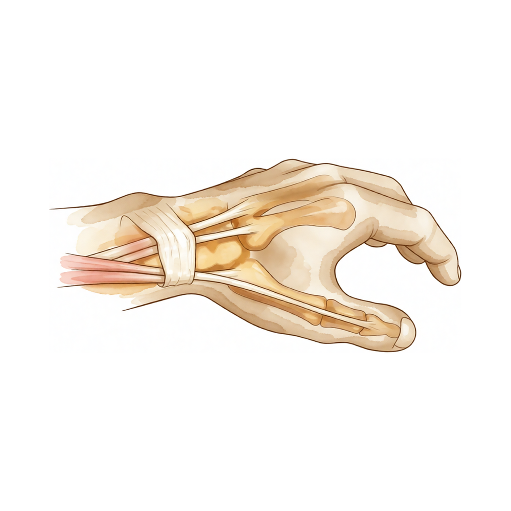
For patients: a plain-language version of this topic is available. See the patient guide.
Overview
Intersection syndrome is a condition of the distal forearm that requires clinical familiarity due to its potential for progressive morbidity. Prolonged pain associated with the condition may lead to tendon attrition, a development that indicates a need for surgical treatment [1]. Understanding this trajectory is essential for orthopaedic surgeons managing patients with persistent symptoms in this region.
The condition represents a significant portion of the orthopaedic surgical workload. Approximately one of every ten patients who are referred for orthopaedic services presents with a hand or wrist condition [9]. Among this specific cohort, nearly half of patients referred for orthopaedic services with a hand or wrist condition will require surgery [9]. These statistics underscore the high volume of cases where intersection syndrome and related pathologies must be considered in the differential diagnosis and treatment planning.
Anatomy & Pathophysiology
Osseous Anatomy
The wrist constitutes the anatomic region between the forearm and hand, encompassing the distal radioulnar, radiocarpal, and ulnocarpal joints along with eight carpal bones [12]. The proximal row comprises the scaphoid, lunate, triquetrum, and pisiform, while the distal row includes the trapezium, trapezoid, capitate, and hamate [12]. The distal radius features two concave articular facets for the scaphoid and lunate, separated by the scapholunate ridge, and a sigmoid notch along the ulnar border that articulates with the ulnar head [19]. The distal ulna is covered by hyaline cartilage on its dorsal, lateral, palmar, and distal surfaces, with the ulnar styloid projecting distally to serve as the insertion site for the triangular fibrocartilaginous complex at its base [19].
Specific carpal bones possess distinct morphological and functional characteristics. The scaphoid receives its primary vascular supply from a radial artery branch at the dorsal ridge, with smaller vessels supplying the distal 30% via the palmar tubercle [19]. The lunate exhibits both dorsal and palmar vascular supply in 80% of wrists, whereas only a palmar supply is present in 20% [19]. The pisiform acts as a sesamoid bone within the flexor carpi ulnaris tendon and serves as the origin for the abductor digiti minimi [19]. The hamate consists of a body and a hook (hamulus) that attaches the transverse carpal ligament and provides origins for the flexor digiti minimi and opponens digiti minimi [19]. The trapezium features a saddle-shaped articulation with the thumb metacarpal base and a palmar groove for the flexor carpi radialis [19].
Radiographic alignment of the distal radius includes an average volar tilt of 11 degrees in the sagittal plane and an average radial inclination of 23 degrees in the frontal plane [16]. Radial length, measured from the radial styloid tip to the ulnar articular surface, averages 13 mm [16].
Ligamentous Anatomy
Extrinsic carpal ligaments connect the radius or ulna to the carpus, with volar ligaments generally stronger than dorsal counterparts [17]. The scapholunate interosseous ligament is C-shaped in the sagittal plane, with the dorsal third being the thickest and strongest portion [19]. The volar portion of the lunotriquetral ligament is the thickest [19]. The triangular fibrocartilage complex (TFCC) comprises the central meniscus homolog, dorsal and volar radioulnar ligaments, the floor of the extensor carpi ulnaris tendon sheath, and volar ulnocarpal ligaments [19]. It arises from the radial border of the distal radius and inserts into the base of the ulnar styloid and distal ulna via the ligamentum subcruentum [19]. The dorsal and volar radioulnar ligaments serve as the primary stabilizers of the distal radioulnar joint [19].
The space of Poirier is a ligament-free area adjacent to the proximal capitate, situated ulnar to the radioscaphocapitate ligament and radial to the long radiolunate in the carpal tunnel floor [17]. This weak area is vulnerable to instability, allowing the distal carpal row to separate from the lunate during perilunate dislocation [17]. The dorsal radiocarpal ligament has a trapezoidal shape, extending from the dorsal rim of the distal radius to the lunate and triquetrum [17]. The dorsal intercarpal ligament passes from the dorsal tubercle of the triquetrum to the distal pole of the scaphoid [17].
Vascular Anatomy
Extraosseous blood supply to the carpus is provided by terminal branches of the radial, ulnar, and anterior interosseous arteries through three dorsal and three palmar transverse arterial arches with longitudinal connections [22]. The dorsal radiocarpal arch is located at the radiocarpal joint and supplies the lunate and triquetrum [22]. The dorsal intercarpal arch, the largest of the dorsal arches, lies between the carpal rows and supplies the distal row [22]. The basal metacarpal arch is located at the metacarpal bases, is the most variable, and supplies the distal carpal row [22].
On the palmar side, the radiocarpal arch is located at the radiocarpal joint level on the palmar surfaces of the lunate and triquetrum [22]. The intercarpal arch is located between the carpal rows, is the most variable, and does not contribute to nutrient vessels in the carpus [22]. The deep palmar arch is located at the metacarpal bases, is consistent, and communicates with the dorsal basal metacarpal arch and palmar metacarpal arteries [22].
Biomechanics and Kinematics
The wrist functions as a two-joint system linking the hand to the forearm around the highly mobile proximal carpal row [20]. The two principal articulations are the radiocarpal and midcarpal joints, situated proximal and distal to this mobile segment [20]. The proximal carpal row lacks muscular or tendinous attachments, classifying it as an intercalary segment [19]. Under axial loading through a neutral wrist, approximately 80% of forces transmit through the distal radius and 20% through the distal ulna [19]. The distal radius normally bears about 80% of distal radioulnar joint load, while the distal ulna bears 20% [26]. Ulnar load bearing increases with ulnar lengthening and decreases with ulnar shortening [26].
Motion distribution varies with wrist position. With wrist flexion, 60% of motion occurs at the midcarpal joint and 40% at the radiocarpal joint [19]. With wrist extension, 33% of motion is midcarpal and 66% is radiocarpal [19]. During ulnar deviation, the proximal row extends relative to the forearm and distal row, whereas during radial deviation, the proximal row flexes relative to these structures [19]. Normal wrist range of motion includes 65 degrees of flexion, 55 degrees of extension, 15 degrees of radial deviation, and 35 degrees of ulnar deviation [26]. Functional wrist range of motion includes 10 degrees of flexion, 35 degrees of extension, 10 degrees of radial deviation, and 15 degrees of ulnar deviation [26].
Soft Tissue Anatomy
The dorsal surface of the wrist contains six compartments housing wrist and digital extensor tendons [16]. The volar surface contains the carpal tunnel, which includes nine flexor tendons and the median nerve [16]. Guyon's canal is bounded by the volar carpal ligament and transverse carpal ligament, the hook of the hamate radially, and the pisiform ulnarly, and contains the ulnar artery and nerve [16]. The interosseous membrane connects the radial and ulnar shafts, with a thickened central portion critical for force transmission between the bones [16].
Palpation landmarks for specific tendons are distinct. The extensor carpi radialis tendons are palpable only for a short distance proximal to the bases of the second and third metacarpals during resisted wrist extension [24]. The extensor carpi ulnaris tendon is palpable during resisted extension and adduction, immediately distal to the ulnar styloid until the fifth metacarpal [24]. The flexor carpi radialis and palmaris longus tendons are palpable and visible in resisted wrist flexion just proximal to the distal wrist flexion crease [24].
Classification
Distal Intersection Syndrome: Distal intersection syndrome is a distinct clinical entity that physicians should be familiar with [1]. Prolonged pain associated with distal intersection syndrome may lead to tendon attrition [1]. Tendon attrition in the context of distal intersection syndrome indicates a need for surgical treatment [1].
Clinical Presentation
Prolonged pain in distal intersection syndrome may lead to tendon attrition [1]. Tendon attrition in distal intersection syndrome indicates a need for surgical treatment [1].
Investigations
Plain radiography: Four standard views are required for wrist imaging: posteroanterior with the wrist in ulnar deviation, lateral, semi-pronated oblique, and semi-supinated oblique [30]. An anteroposterior view with the fist clenched is added when scapholunate injury is suspected [30]. On the lateral view, the axes of the radius, lunate, capitate, and third metacarpal must be co-linear, with the scaphoid projecting at an angle of approximately 45 degrees to this line [30]. Dorsal intercalated segmental instability (DISI) presents with the lunate tilting backwards and the scaphoid tilting volarwards [30]. Volar intercalated segment instability (VISI) is characterized by the lunate and scaphoid tilting volarwards, with the capitate and metacarpals lying anterior to the radius [30]. If initial X-rays are normal, 10–15% of scaphoid fractures remain invisible [30]. In such cases, repeated X-rays are necessary two weeks later to detect undisplaced scaphoid fractures if MRI is unavailable [30].
MRI: MRI is the modality of choice for imaging radiographically occult fractures of the hand and wrist [18]. Its primary advantages over CT and radiography include improved tissue characterization of soft tissues such as ligaments and synovium, and the absence of ionizing radiation [18]. Modern imaging is generally performed at 1.5T or 3T, with 3T preferred for small fields of view [18]. A static magnetic field strength of at least 1.5 T using a dedicated wrist coil is recommended for analyzing interosseous, intrinsic, and extrinsic ligament insertions [27]. The volar extrinsic, scapholunate interosseous, dorsal intercarpal, and lunotriquetral ligaments are best visualized using 1 mm slices with no interslice gap in the coronal plane [27]. Oblique axial views along the longitudinal axes of ligaments allow further analysis when injury is suspected [27]. The distal radiocarpal and intercarpal ligaments are best viewed on both coronal and sagittal images [27].
MRI determination of posterior interosseous nerve (PIN) position is reliable and consistent with prior cadaveric study [5]. Magnetic resonance neurography provides anatomical correlation and pinpoints nerve pathology, enhancing diagnostic confidence and guiding treatment planning [2]. MRI with contrast enhancement is most commonly used to determine whether soft-tissue lesions are solid or cystic [18]. MR arthrography can be performed for evaluation of triangular fibrocartilage and intercarpal ligament tears, but this is generally unnecessary with the increasing availability of high-field MRI [18]. The addition of arthrographic contrast improves the visualization of scapholunate and lunotriquetral ligaments on MR images [29].
In the wrist, a common indication for MRI is evaluation of the intrinsic carpal ligaments [29]. Perforations in the triangular fibrocartilage complex (TFCC) appear as linear defects or gaps filled with hyperintense fluid on coronal gradient-echo or T2-weighted pulse sequences [29]. MRI is useful in detecting additional marrow abnormalities in osteonecrosis, as seen in the lunate in Kienböck disease or in the scaphoid after fracture [29]. Asymmetry of marrow signal in proximal and distal fragments of a fractured scaphoid is suggestive of proximal pole ischemia [29]. MRI has a limited role in the evaluation of carpal tunnel syndrome, which remains a clinical diagnosis [29]. However, axial imaging with T2 weighting can display masses within the confines of the carpal tunnel, as well as edema and swelling of the median nerve [29]. MRI provides earlier detection of synovitis and erosive bone changes associated with rheumatoid arthritis than radiographs [29]. Tenosynovitis and tendon injuries in the wrist and hand can be assessed with MRI [29].
Early MRI reduces uncertainty and streamlines care for suspected scaphoid fractures [30]. The MRI examination should be directed at solving a specific clinical problem or question [29]. MRI has an expanding role in the evaluation of pathologic conditions of the elbow and wrist, requiring high-resolution images best obtained with surface coil technique and high-field systems [29]. Concomitant cartilage-sensitive imaging is integrative to influence the assessment and surgical management, especially in the setting of scapholunate advanced collapse (SLAC) wrist [27]. Real-time MRI has been used to investigate dynamic instabilities, although its routine use in clinical practice is yet to be further determined [27].
MRI has been used to evaluate dorsal wrist pain, including the diagnosis of occult dorsal wrist ganglions [15]. It has been used to evaluate ulnar impaction [15], traumatic and overuse injuries of the wrist and hand in athletes [15], and Kienböck disease with histologic correlations [15]. MRI has been used to evaluate the triangular fibrocartilage complex [15], the anterior radiocarpal ligaments [15], and to establish the carpal contents/canal ratio [15]. It has been used to evaluate rupture of the distal biceps tendon [15], sports and occupational injuries of the elbow [15], and overuse syndromes and injuries involving the elbow [15]. MRI has been used to evaluate ulnar collateral ligament injury in the throwing athlete with saline-enhanced MR arthrography [15] and preoperative ulnar collateral ligament status by magnetic resonance imaging and computed tomography arthrography [15].
MRI has been used to evaluate detection of occult wrist fractures [15] and low-field scaphoid fracture [15]. It has been used to evaluate magnetic resonance imaging of the wrist and elbow in clinical sports medicine [15], the musculoskeletal system specifically the elbow [15], and the efficacy of magnetic resonance imaging of the elbow [15]. MRI has been used to evaluate magnetic resonance imaging of the elbow in baseball pitchers [15], the wrist and hand at 7 T [15], and 3-Tesla imaging of the wrist and hand [15]. It has been used to evaluate a practical guide to diagnostic imaging of the upper extremity [15] and magnetic resonance imaging of trauma: elbow and wrist [15]. MRI has been used to evaluate magnetic resonance imaging of the wrist [15], the wrist in Orthopedics [15], and the elbow in Orthopedics [15]. It has been used to evaluate magnetic resonance imaging of the elbow: part I. Normal anatomy, imaging technique, and osseous abnormalities [15], and part II. Abnormalities of the ligaments, tendons, and nerves [15]. MRI has been used to evaluate magnetic resonance imaging in Kienbock disease [15].
MRI has been used to evaluate magnetic resonance imaging of the wrist and hand: techniques and spectrum of disease [14]. It has been used to evaluate MR and CT arthrography of the wrist [14] and imaging of ulnar-sided wrist pain [14]. MRI has been used to evaluate cone-beam CT in diagnosis of scaphoid fractures [14] and wrist injuries in young adults: the diagnostic impact of CT and MRI [14]. It has been used to evaluate distal radioulnar joint: functional anatomy, including pathomechanics [14] and distal radioulnar joint stress radiography for detecting radioulnar ligament injury [14]. MRI has been used to evaluate cine MRI: a new approach to the diagnosis of scapholunate dissociation [14] and comparison of radiographic stress views for scapholunate dynamic instability in a cadaver model [14]. It has been used to evaluate imaging of radial wrist pain. I. Imaging modalities and anatomy [14].
MRI has been used to evaluate intrinsic ligament and triangular fibrocartilage complex tears of the wrist: comparison of MDCT arthrography, conventional 3-T MRI, and MR arthrography [14]. It has been used to evaluate cone-beam computed tomography arthrography: an innovative modality for the evaluation of wrist ligament and cartilage injuries [14]. MRI has been used to evaluate MRI of wrist ligaments [14] and 3D analysis of the wrist [14]. It has been used to evaluate normal and variant anatomy of the wrist and hand on MR imaging [14] and use of hybrid SPECT/CT for diagnosis of radiographic occult fractures of the wrist [14].
CT: CT scanning enables 3D analysis of carpal dysfunction [27].
Other Considerations: Dynamic fluoroscopy shows abnormal motion between the scaphoid and lunate and changes in the kinematics of the midcarpal joint [27]. Arthroscopy is considered by many to be the diagnostic intervention of choice for determining the degree of injury to the wrist and assessing cartilage condition [27]. A staged version of the Finkelstein test is reliable, easy, and reproducible for diagnosing de Quervain's tendonitis while causing minimal discomfort compared to traditional descriptions [4]. MRI has been used to evaluate efficacy of magnetic resonance imaging and clinical tests in diagnostics of wrist ligament injuries: a systematic review [21]. It has been used to evaluate accuracy of simple plain radiographic signs and measures to diagnose acute scapholunate ligament injuries of the wrist [21]. MRI has been used to evaluate physical examination of the wrist: useful provocative maneuvers [21] and the wrist insufflation test: a confirmatory test for detecting intercarpal ligament and triangular fibrocartilage complex tears [21]. It has been used to evaluate radiographic clues for determining carpal instability and treatment protocol for scaphoid fractures [21] and the role of magnetic resonance imaging in scaphoid fractures [21]. MRI has been used to evaluate examination of the wrist: ulnar-sided pain due to ligamentous injury [21] and the role of imaging in diagnosing diseases of the distal radioulnar joint, triangular fibrocartilage complex, and distal ulna [21]. It has been used to evaluate ulnar-sided wrist pain, part I: anatomy and physical examination [21] and physical examination of the wrist [21].
Treatment
Non-Operative
The provided evidence does not detail specific conservative management protocols such as weight loss, physical therapy, NSAIDs, or injections for intersection syndrome.
Operative
Indications: Prolonged pain in distal intersection syndrome may lead to tendon attrition, indicating a need for surgical treatment [1].
Other Considerations: Stiffness and contracture are the most common sequela of pyogenic flexor tenosynovitis and benefit from early motion and hand therapy [3].
Complications
Stiffness / Arthrofibrosis: Stiffness and contracture are the most common sequela of pyogenic flexor tenosynovitis [3].
Key Evidence
- [L4] Physicians should be familiar with distal intersection syndrome, as prolonged pain may lead to tendon attrition, indicating a need for surgical treatment. [1] (10.1016/j.jhsg.2021.04.005)
- [Paper] Its main benefit is providing anatomical correlation and pinpointing nerve pathology, thereby enhancing diagnostic confidence and guiding appropriate treatment planning. [2] (10.1177/23259671251400763)
- [L5] Stiffness and contracture are the most common sequela and benefit from early motion and hand therapy. [3] (10.2106/jbjs.rvw.26.00015)
- [L4] The authors describe a staged version of the Finkelstein test that is reliable, easy, and reproducible for diagnosing de Quervain's tendonitis while causing minimal discomfort compared to traditional descriptions. [4] (10.1016/j.jhsa.2010.05.022)
- [L4] MRI determination of PIN position is reliable and consistent with prior cadaveric study. [5] (10.1016/j.arthro.2020.12.118)
- [L3] Approximately one of every ten patients who are referred for orthopaedic services has a hand or wrist condition, and nearly half will require surgery. [9] (10.2106/00004623-200401000-00009)
See Also
- Carpal Tunnel and Nerve Compression
- Scaphoid Fracture
- Wrist Ligament Injuries
References
[1] Distal Intersection Syndrome Combined With Partial Attritional Changes of the Extensor Carpi Radialis Brevis in Tennis Players. Journal of Hand Surgery Global Online. 2021. DOI: 10.1016/j.jhsg.2021.04.005
[2] Magnetic Resonance Neurography Findings in Clinically Suspected Posterior Interosseous Neuropathy: Response. Orthopaedic Journal of Sports Medicine. 2026. DOI: 10.1177/23259671251400763
[3] Management of Pyogenic Flexor Tenosynovitis. JBJS Reviews. 2026. DOI: 10.2106/jbjs.rvw.26.00015
[4] Staged Description of the Finkelstein Test. The Journal of Hand Surgery. 2010. DOI: 10.1016/j.jhsa.2010.05.022
[5] Implications of Posterior Interosseus Nerve Distance from the Radial Tuberosity: A Radiologic Study. Arthroscopy: The Journal of Arthroscopic & Related Surgery. 2021. DOI: 10.1016/j.arthro.2020.12.118
[9] Utilization of Orthopaedic Services for Hand and Wrist Conditions in a Capitated Population. The Journal of Bone and Joint Surgery-American Volume. 2004. DOI: 10.2106/00004623-200401000-00009
[12] Campbell S Operative Orthopaedics 4 Volume Set. NERVE INJURIES AT THE LEVEL OF THE HAND AND WRIST > ANATOMY.
[14] Campbell S Operative Orthopaedics 4 Volume Set. ANATOMIC RECONSTRUCTION OF THE DISTAL RADIOULNAR LIGAMENTS > RADIOGRAPHIC TECHNIQUES.
[15] Campbell S Operative Orthopaedics 4 Volume Set. ELBOW, WRIST, AND HAND.
[16] A Lange Medical Book Current Diagnosis Treatment In Orthopedics Fifth Edition. 2Musculoskeletal Trauma Surgery > FRACTURES AND DISLOCATIONS OF THE DISTAL AND MID-FOREARM.
[17] Aaos Comprehensive Orthopaedic Review 3. Carpal Instability* > II. Anatomy and Biomechanics (See Chapter 92).
[18] Orthopaedic Knowledge Update 13 Ebook Without Multimedia. Anatomy, Evaluation, Clinical Examination, and Imaging > Imaging: Advances in Imaging of the Hand and Upper Extremity > Magnetic Resonance Imaging.
[19] Aaos Comprehensive Orthopaedic Review 3. Anatomy of the Hand and Wrist > VII. The Wrist.
[20] Green S Operative Hand Surgery. WRIST BIOMECHANICS > Carpal Kinematics.
[21] Campbell S Operative Orthopaedics 4 Volume Set. ANATOMIC RECONSTRUCTION OF THE DISTAL RADIOULNAR LIGAMENTS > DIAGNOSIS AND EVALUATION.
[22] Campbell S Operative Orthopaedics 4 Volume Set. NERVE INJURIES AT THE LEVEL OF THE HAND AND WRIST > CIRCULATION.
[24] Exam Of The Hand Wrist 2Ed. 2.3 EXAMINATION OF THE MUSCULOTENDINOUS APPARATUS.
[26] Miller S Review Of Orthopaedics. Genetics of musculoskeletal conditions and abnormalities are summarized in Table 1.27 > Elbow biomechanics > Arthrodesis.
[27] Green S Operative Hand Surgery. Advanced Imaging.
[29] Campbell S Operative Orthopaedics 4 Volume Set. WRIST AND ELBOW.
[30] Apley And Solomon S Concise System Of Orthopaedics And Trauma. FRACTURES OF THE DISTAL RADIUS IN CHILDREN > Imaging.