Clinicians › Wrist
Distal Radius Malunion (and Corrective Osteotomy)
Symptomatic distal radius malunion: deformity patterns and DRUJ consequences, planning (CT/3D guides), opening/closing-wedge corrective osteotomy and outcomes.
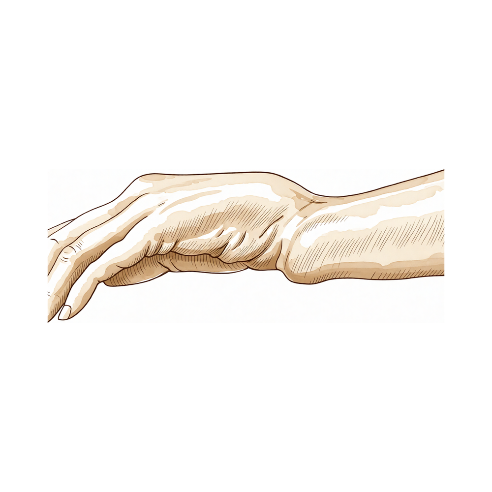
For patients: a plain-language version of this topic is available. See the patient guide.
Overview
Distal radius fractures are among the most common injuries presenting to the emergency department, with increased risk in elderly patients with osteoporosis during low-energy falls [54]. The primary goals of treatment are to optimize comfort and function, with surgical indications determined by patient infirmity, functional demands, tolerance of deformity, and personal preferences [54]. Specific radiographic criteria for surgical intervention include ulnar variance of 5 mm or more positive, dorsal articular tilt of ≥15°, loss of radial inclination >10°, or an articular gap or step of 2 mm or more [54]. Surgery is also indicated for unstable volar extra-articular fractures, associated neurovascular or intercarpal ligament injuries, and displaced radial styloid fractures with intra-articular displacement greater than 2 mm [54]. Multiple trauma, such as bilateral distal radius fractures or the need for crutches due to leg injury, constitutes a relative indication for surgical treatment [54].
Corrective osteotomy is an effective method for treating symptomatic malunions of the distal radius, yielding good functional outcomes and high patient satisfaction in both adults and children [1, 2, 3]. This procedure is particularly valuable for dorsally angulated malunions, where the severity of the initial fracture and time from injury to surgery correlate with the ability to correct radiographic parameters [5]. Radiological results correlate with functional outcomes, and successful osteotomy can improve wrist function even in complex intra- and extra-articular malunions [6, 11]. While surgical correction remains a challenging problem with unpredictable clinical outcomes, new technologies such as 3-dimensional modeling and computer-generated osteotomy guides are likely to have a positive impact on results [7]. Outcomes following corrective osteotomy after unsuccessful internal fixation are comparable to those reported after initial nonoperative treatment [8].
To obtain optimal clinical outcomes, distal radius osteotomy must correct the rotational component of the malunion, and surgery on the ulna to restore distal radioulnar joint anatomy is needed in some cases [26]. Corrective osteotomy with fixed-angle volar plate fixation provides satisfactory union rates and clinical and radiological outcomes even without bone graft, irrespective of osteotomy type, size, or location [28]. Early corrective osteotomies should be considered in young patients with intra-articular distal radius malunions before considering salvage procedures such as partial or complete wrist arthrodesis [34]. The Lift-Off Screw technique offers a reproducible method to plan the amount of sagittal plane correction during corrective osteotomy surgery for dorsally angulated distal radius fracture malunions [37].
Anatomy & Pathophysiology
Bony Anatomy & Normal Alignment
The distal radius articular surface is biconcave, featuring distinct scaphoid and lunate facets [78]. The distal radioulnar joint (DRUJ) articulates with the ulna at the sigmoid notch [78]. Lister's tubercle, a small dorsal prominence, serves as a landmark for the dorsal approach to the wrist and is a cause of attritional rupture of the extensor pollicis longus following distal radius fracture [78]. The distal radial metaphysis possesses thin cortex, rendering it vulnerable to bending forces [78]. Additionally, the brachioradialis insertion on the radial styloid acts as a deforming force in distal radius fractures [78]. In a normal wrist with neutral ulnar variance, the distal radius bears 80% of the axial load [78].
Normal radiographic alignment of the distal radius is defined by specific parameters: an average radial inclination of 22 to 23 degrees [40], an average radial height of 11 to 12 mm [40], an average volar tilt of 11 to 12 degrees [40], and an ulnar variance of ± 2 mm [40]. This alignment is often summarized by the "11:11:22 rule," which describes normal wrist alignment as radial height of 11 mm, volar tilt of 11 degrees, and radial inclination of 22 degrees [78].
The carpus consists of two rows of eight bones that bridge the forearm and hand, providing movement at the wrist joint while retaining stability [44]. The proximal carpal row includes the scaphoid, lunate, and triquetrum, which act as a key intercalated segment between the forearm and the distal row [44]. The distal carpal row includes the trapezium, trapezoid, capitate, and hamate [44]. The scaphoid is an S-shaped tubular bone located in the proximal carpal row on the radial aspect of the wrist [71]. It articulates with the trapezium/trapezoid, radius, capitate, and lunate [71]. Due to its articular cartilage coverage, the scaphoid has a reduced capacity for periosteal healing and an increased tendency for delayed union and nonunion [71].
Ligamentous Anatomy
Extrinsic ligaments of the carpus connect the carpal bones to the forearm bones proximally and the metacarpals distally [74]. The extrinsic palmar radiocarpal ligaments include the transverse carpal, radioscaphocapitate, radioscapholunate, radial collateral, long radiolunate, and short radiolunate ligaments [74]. The extrinsic ulnocarpal ligaments include the ulnotriquetral, ulnolunate, and ulnocapitate ligaments [74]. Strong oblique extrinsic palmar radial ligaments prevent the carpus from translating medially on the angulated slope of the distal radius [74]. The space of Poirier is a V-shaped interligamentous sulcus over the capitolunate articulation that represents an interval of capsular weakness [74]. The arcuate ligament is formed from the interdigitation of transverse fibers of the radioscaphocapitate, ulnocapitate, triquetrocapitate, and volar scaphotriquetral ligaments [74]. This structure forms a support sling for the midcarpal region, particularly the head of the capitate [74].
Intrinsic ligaments connect individual carpal bones to one another and include palmar midcarpal, proximal interosseous, and distal interosseous ligaments [74]. The scapholunate ligament is an intrinsic ligament that connects the scaphoid and lunate [83]. The lunotriquetral ligament is an intrinsic ligament that connects the lunate and triquetrum [83]. The triangular fibrocartilage complex (TFCC) is responsible for the normal function of the distal radioulnar joint and the ulnar compartment of the wrist [120]. Radio-ulno-carpal joint stability relies on the proper function of eight different anatomical structures and is modified by the degree of forearm rotation [107]. The flexor carpi ulnaris and extensor carpi ulnaris muscles serve as dynamic stabilizers of the distal radioulnar joint [87].
Pathophysiology of Malunion
Malunion is a common complication after distal radius fractures, occurring in up to 46% of cases [10]. It occurs commonly in distal radius fractures, particularly in elderly patients managed nonoperatively [32]. Fracture characteristics and initial treatment contribute to the development of malunion, including failure to achieve or maintain accurate reduction or inadequate immobilization [40]. Reduction is most difficult to obtain and maintain in fractures with marked comminution, severe osteoporosis, or disruption of the distal radioulnar ligaments [40]. Older patients have a higher incidence of malunion compared to younger patients, with a mean age of 60 years for those with malunions versus 51 years for those without [40].
Malunions of the distal radius may involve extraarticular deformities, intraarticular malalignment, distal radioulnar joint incongruity or instability, or a combination of these features [40]. Extraarticular deformities include shortening and excessive dorsal or volar tilt of the distal radial articular surface [40]. Angulations of more than 20° in either the palmar or dorsal directions result in significant changes in load mechanics across the proximal wrist joint [10]. Radial inclination of less than 10° results in significant changes in load mechanics across the proximal wrist joint [10]. Malunion can result in functional deficits in the long term, including decreased grip strength, limitations in range of motion, pain, and cosmetic deformity [32].
Dorsally angulated distal radius fractures are associated with type 4 carpal instability nondissociative [32]. The distal radioulnar joint is often incongruous in dorsally displaced distal radius malunions due to dorsal angulation and shortening [32]. More than 10 degrees of dorsal tilt leads to decreased wrist flexion [40]. 6 mm of radial shortening causes dysfunction of the distal radioulnar joint [40]. Fractures with more than 25 or 30 degrees of angulation in the frontal or sagittal plane are likely to become symptomatic [40]. Fractures with 6 mm or more of radial shortening are likely to become symptomatic [40]. Patients with constitutional joint laxity may develop midcarpal instability with a dorsal tilt of only 10 to 15 degrees [40].
Malunion of the distal radius is associated with significant changes to distal radioulnar joint mechanics and ligament lengthening [40]. 20 to 30 degrees of dorsal tilt alters the force distribution across the radiocarpal joint and should be considered a prearthritic condition [40]. Significant articular incongruity and radial shortening are more consistently correlated with the development of symptoms than other radiographic measurements [40]. Intraarticular incongruity in the radiocarpal joint of more than 2 mm is associated with poor functional outcome [40]. A 1- to 2-mm step-off at the distal radioulnar joint is associated with poor functional outcome [40]. Dorsal angulation of more than 20 degrees is associated with poor functional outcome [40]. Radial inclination of less than 10 degrees is associated with poor functional outcome [40]. Loss of sagittal tilt of 20 to 30 degrees is associated with poor functional outcome [40].
A malunion of the distal radius is defined by radial inclination <10 degrees, volar tilt >20 degrees or dorsal tilt >20 degrees, radial height <10 mm, ulnar variance >2+ mm, or intra-articular incongruity or diastasis >2 mm [32]. Some malunited radius fractures that appear shortened may have healed in malrotation rather than true shortening [21]. True shortening occurs due to volar comminution or loss of continuity of the volar cortex and is not corrected by pure derotational osteotomy [21]. Restoration of the hinge point on the volar cortex is critical to prevent shortening and incomplete correction of volar tilt during osteotomy [21]. Loss of radial inclination can compromise grip strength [21]. Warwick et al. failed to correlate radial tilt loss with clinical outcome after Colles' fractures [21]. Altissimi et al. found a negative effect of radial tilt loss only when it was greater than 20 degrees (less than 5 degrees radial tilt) [21].
Posttraumatic ulnar impaction syndrome usually occurs as a consequence of radial shortening in malunited distal radius fractures [22]. Malunion of the distal radius or forearm fractures can result in incongruence in the distal radioulnar joint and subsequent cartilage damage [22]. Decreased radial inclination significantly increases distal radioulnar joint stiffness when the ligament is intact or partially divided [51]. The distal radioulnar joint stiffness in dorsal translation decreases significantly with dorsal tilt of 10° and 20° in pronation [127]. The lengths of some carpal ligaments are substantially altered even by mild dorsal angulation of the distal radius [125]. Rotational malalignment of the wrist has significant effects on carpal, distal radial, and distal radioulnar joint measurements [69].
The contact area of the distal radioulnar joint increases during wrist flexion and decreases during wrist extension and ulnar deviation [90]. Damage to the ulnar collateral mechanism adversely affects the clinical result despite a good anatomical result in distal radius fractures [53]. Injuries to adjacent soft-tissue structures occur in approximately one-half of distal radius fractures and in almost all intra-articular fractures [89]. The most common associated soft-tissue injuries in distal radius fractures are TFCC, scapholunate ligament, and lunotriquetral ligament injuries [89]. Ulnar styloid fractures have little influence on functional outcome in distal radius fractures [89].
The forearm functions as a ring, making isolated radial shaft fractures uncommon in adults [48]. The conventional concept of the forearm as part of either the elbow or wrist is outdated; the forearm is a triarticular complex where proximal, middle, and distal radioulnar joints work together [109]. All wrists have similar loading across the distal ulna regardless of ulnar variance, while pronation relatively increases loading across the distal ulna [86]. Radial lengthening beyond the native length was not detrimental to radial loading and further reduced distal ulnar loading [102]. Achieving at least native ulnar variance is appropriate to restore normal biomechanical loading based on in vitro studies [102]. A progressive decrease in distal ulnar loads with generation of tensile loads is observed with sequential ulnar shortening [105]. The geometry of the distal metaphyseal ulnar shortening osteotomy and resultant change in the position of the ulnar head did not increase transverse joint reaction forces [115]. Casting a distal radius fracture decreases the forces and pressures in the radiocarpal joint [117].
The Aptis distal radioulnar joint arthroplasty considerably alters forearm kinematics [72]. Reconstructed animation from four-phase grip MRI demonstrates impairment of the articular disc and longitudinal instability of the distal radioulnar joint simultaneously in patients with ulnar-sided pain [103]. Dynamic CT imaging shows promise for evaluating the distal radioulnar joint during wrist motion [108]. The rotation axis of the scaphoid relative to the lunate is highly variable across subjects and positions during flexion-extension and radial-ulnar deviation motions [121]. Isolated midcarpal motion during radioulnar deviation can be approximated as a rotation in a plane of radiodorsal/ulnopalmar rotation of the wrist [99]. Constant radiocarpal and midcarpal congruence during radioulnar deviation in normal wrists is no longer possible with intercarpal kinematic modifications after arthrodeses [97].
Classification
Morphological Description: No formal classification system for distal radius malunion exists, a gap likely attributable to a lack of consensus regarding the definition of the condition [32]. Consequently, distal radius malunions are generally described by their morphological characteristics [32]. For simplicity of discussion, these malunions can be viewed as dorsally displaced, volarly displaced, or intra-articular [32]. Certain distal radius fractures exhibit characteristics of more than one type of malunion [32]. The typical malunion types include radial shortening, dorsal tilt, step-off at the articular surface of the radius, or a combination of these [63].
Fernandez Classification: The Fernandez classification defines various combinations of injuries to the distal radioulnar joint in distal radius fractures [155]. Type I injuries are stable with articular congruity and intact or minimally disrupted radioulnar ligaments [155]. Type II injuries are unstable despite adequate reduction of the distal radius [155]. Type III injuries have the potential to be unstable and include disruption of the joint surface at the sigmoid notch or the ulnar head [155].
Other Considerations: A recent article defines distal radius malunion using specific radiographic parameters: radial inclination <10 degrees [32], volar tilt >20 degrees or dorsal tilt >20 degrees [32], radial height <10 mm [32], ulnar variance >2+ mm [32], and intra-articular incongruity or diastasis >2 mm [32].
Clinical Presentation
Epidemiology and Risk Factors
Distal radius malunion is a frequent complication, occurring in up to 46% of cases [10]. Etiology involves failure to achieve or maintain accurate reduction, or inadequate duration or type of immobilization [40]. Older patients exhibit higher rates of malunion, with a mean age of 60 years for those with malunions compared to 51 years for those without [40]. Forearm malunions may arise following non-operative or operative treatment of acute fractures, as well as after deformity correction surgery [30].
Symptoms and Functional Deficits
Not all distal radial malunions are symptomatic; elderly patients with low functional demands often require no further treatment [40]. Conversely, posttraumatic wrist deformities in younger, active patients may be sufficiently disabling to warrant surgical correction [40]. Malunion is associated with higher arm-related disability regardless of age [40]. Significant changes in load mechanics across the proximal wrist joint occur with angulations exceeding 20° in either the palmar or dorsal directions, or radial inclination of less than 10° [10]. Long-term functional deficits are a recognized consequence of malunion [10]. Some patients with malunited radius fractures that appear shortened report deceptively severe symptoms despite moderate deformities on plain radiographs [21]. Ulnar impaction syndrome presents with ulnar-sided wrist pain increased by rotational or ulnar deviation loading and a long ulna relative to the radius [22]. Posttraumatic ulnar impaction syndrome usually results from radial shortening, which in adults is most frequently seen in malunited distal radius fractures [22].
Radiographic Parameters and Deformity
Malunions may involve extraarticular deformities, intraarticular malalignment, distal radioulnar joint incongruity or instability, or a combination of these features [40]. Normal alignment of an intact distal radius averages 22 to 23 degrees of radial inclination, 11 to 12 mm of radial height, 11 to 12 degrees of volar tilt, and ± 2 mm of ulnar variance [40]. Poor functional outcomes are likely associated with: * Intraarticular incongruity in the radiocarpal joint of more than 2 mm [40]. * A 1- to 2-mm step-off at the distal radioulnar joint [40]. * Dorsal angulation of more than 20 degrees and radial inclination of less than 10 degrees [40]. * Loss of sagittal tilt of 20 to 30 degrees [40].
Fractures with more than 25 or 30 degrees of angulation in the frontal or sagittal plane, or 6 mm or more of radial shortening, were likely to become symptomatic [40]. Significant articular incongruity and radial shortening correlate more consistently with symptom development than other measurements [40]. Twenty to 30 degrees of dorsal tilt alters force distribution across the radiocarpal joint and should be considered a prearthritic condition [40]. Loss of normal anterior tilt can lead to carpal malalignment, resulting in dorsiflexion of the lunate with compensatory flexion of the capitate [119]. Capitate shift is defined as displacement of the capitate either dorsal or anterior to the longitudinal axis of the radius [119]. Capitate shift consistently demonstrated the strongest relationship with dorsal tilt and was the only parameter not influenced by age or wrist position in acute distal radial fractures [119]. Accurate radiographic assessment of deformity requires comparison with the opposite uninjured wrist due to wide ranges of alignment parameter values [129]. Injuries and malalignments of the distal radioulnar joint, as well as rotational malunion of the distal radius, are more difficult to evaluate with standard radiographs [129].
Associated Pathology
Intra-articular malunions of the distal radius are time sensitive; delaying treatment for more than 3 months can result in irreversible cartilage damage [55]. Median neuropathy can occur in malunited fractures of the distal radius [38].
Investigations
Radiographic Assessment and Normal Parameters
Plain radiography: Standard evaluation of the distal radius requires posteroanterior (PA), lateral, and oblique views [98]. All measurements must be taken on a true lateral view with the radius and ulna superimposed to avoid errors caused by forearm rotation; a 5-degree change in forearm rotation results in a 1.6-degree change in volar tilt on the lateral view [98]. Normal parameters include an average volar tilt of 11 to 12 degrees (range 0 to 28 degrees), average radial inclination of 22 to 23 degrees (range 12 to 30 degrees), and average radial height of 11 to 12 mm (range 8–18 mm) [98]. Ulnar variance is measured as the vertical distance between lines drawn perpendicular to the long axis of the radius, one parallel to the medial corner of the radial articular surface and the other parallel to the most distal aspect of the ulnar head articular surface [98]. Carpal malalignment is assessed by drawing lines down the long axes of the capitate and radius, which should overlap or intersect within the carpus in normal alignment [98].
The teardrop angle is measured on the lateral view as the angle between the radial shaft and the central axis of the teardrop (the U-shaped outline of the volar lip of the distal radius), averaging 70 degrees in normal radii [98]. A teardrop angle less than 45 degrees is associated with articular gap and step-off on CT [98]. Anteroposterior (AP) distance is measured on the lateral view as the distance between the apices of the dorsal and volar rims of the lunate facet [98]. Angulations of more than 20° in either the palmar or dorsal directions, and radial inclination of less than 10°, result in significant changes in load mechanics across the proximal wrist joint [10]. Clinicians should assess deterioration in fracture alignment on radiographic follow-up to identify potential need for surgery to avoid malunion in cases showing early secondary displacement [153].
Advanced Imaging and 3D Modeling
CT: CT scan is used for preoperative planning of intraarticular fractures [46]. Measurements made on both plain radiographs and 3-dimensional computer bone models are accurate for evaluating the deformity in extraarticular distal radius malunions [58]. Malunited diaphyseal fractures of both forearm bones showed complex deformities, suggesting that 3-dimensional modeling may be a more effective method than standard computed tomography or radiographs [152]. Three-dimensionally planned and printed patient-tailored plates offer a reliable method for correcting even complex malunions of the distal radius and forearm [157].
MRI: MRI is used to rule out injuries to the carpal ligaments (e.g., lunotriquetral and scapholunate ligament) or to the triangular fibrocartilage complex [46].
Other Considerations: 3D imaging is useful in reconstructive procedures for malunions and nonunions of carpal injuries [84]. Dynamic CT is used by some for ligament injuries [84]. Wrist arthroscopy can be used as an aid to the diagnosis of ligament injuries and fracture displacement [84]. Ultrasound scanning (USS) provides an additional tool for the detection of carpal ligament injuries [84]. Live/video fluoroscopic evaluation of the wrist provides diagnostic clarity for dynamic instability with sensitivities reported between 86% and 95% and specificity between 80% and 97% for diagnosing scapholunate ligament injury [84].
Preoperative Planning and Surgical Strategy
Other Considerations: The surgical decision for corrective osteotomy is based on limitation of joint motion, grip strength, level of pain, degree of cosmetic deformity, and radiographic findings including pre-arthritic deformity, carpal malalignment, and an incongruent distal radioulnar joint [10]. Critical analysis of radiographic and clinical information allows for a systematic approach to the evaluation and treatment of the distal radioulnar joint (DRUJ) in cases of distal radius malunion [23]. To obtain optimal clinical outcomes, distal radius osteotomy must correct the rotational component of the malunion, and surgery on the ulna to restore DRUJ anatomy is needed in some cases [26]. Surgeons are influenced by radiographic deformity but do not agree on adequate alignment after reduction of a distal radial fracture [146]. A randomized controlled trial protocol exists comparing computer-assisted versus conventional preoperative planning for corrective osteotomy of distal radius malunions, though results regarding clinical outcomes are not yet reported in the cited document [9]. The severity of the initial fracture and time taken from injury to corrective osteotomy correlate with the ability to correct radiographic parameters in dorsally angulated malunions [5].
Treatment
Non-Operative
The provided evidence does not detail specific conservative management protocols such as weight loss, physical therapy, NSAIDs, or injections for distal radius malunion.
Operative
Indications: Treatment of distal radius malunion should focus on the symptomatic patient rather than radiographic appearance [32]. Operative indications for correction include decreased grip strength, limitations in range of motion, pain, and cosmetic deformity [32]. In cases of intra-articular malunion, early treatment may be warranted before there is evidence of degeneration [32]. Recent data support intervention as early as 6 weeks, resulting in a technically less challenging case and a shorter overall period of patient disability [32]. Insufficient evidence exists to make rigorous comparison between patients who underwent "early" (before fracture consolidation) versus "late" (following fracture healing) corrective osteotomy [32].
Surgical Approach / Technique: Corrective osteotomy of the distal radius to reconstruct the natural anatomy is the foundation of treatment of malunion [20]. The type of osteotomy selected depends on surgeon preference, experience, and the unique deformity of the distal radius [20]. Corrective osteotomies of distal radius malunions can be performed via either a dorsal or palmar approach [18]. A simplified technique of distraction corrective osteotomy after distal radius malunion results in improved clinical and radiographic outcomes [4]. A less invasive technique for a dorsal single-cut open-wedge osteotomy is described to correct extrarticular rotational malunion of the distal radius [10]. Minimally invasive osteotomy of the distal radius for extra-articular malunion is feasible with similar results to the literature, without the need for bone grafting or complex preoperative planning [15]. For patients with symptomatic malunion of the distal radius, corrective osteotomy with 90-90 plate fixation is an effective treatment option for improving pain and restoring function for both volarly and dorsally angulated malunions, including those with an intraarticular component [12]. New technologies such as 3-dimensional modeling and computer-generated osteotomy guides are likely to have a positive impact on the outcomes of surgical treatment [7]. The protocol for a randomized controlled trial comparing computer-assisted versus conventional preoperative planning for corrective osteotomy of distal radius malunions has been described, but results and conclusions regarding clinical outcomes are not yet reported in that document [9].
Outcomes and Functional Results: The results of operative correction of a distal radius malunion following an unsuccessful internal fixation can be achieved with outcomes comparable to those reported after initial nonoperative treatment [8]. Corrective osteotomy is an effective method of treating symptomatic distal radius malunions with good long-term functional results, measured with the DASH and PRWE score, and improvement in radiographic parameters and pain scores [24]. Operative treatment of inadequately or imperfectly treated fractures of the distal radius can improve wrist and hand function substantially, but rarely restores the limb to normal [25]. Surgical procedures designed to correct malunions of the distal radius rarely result in a normal wrist [32]. In comparison to the contralateral limb, deficits are often still appreciated after surgical correction, particularly with respect to range of motion and grip strength, which rarely exceeds 70% of the contralateral limb [32]. Corrective osteotomy in adults with malunited forearm fractures leads to better function and improvement in range of motion, with good outcomes achievable even with a long delay between injury and surgery [27].
Other Considerations: In severe malunions where it is impossible to restore radial length, corrective osteotomy can be performed to correct the sagittal plane deformity while an ulnar shortening osteotomy is performed to achieve congruency of the DRUJ [32]. Radial corrective osteotomy and Sauvé–Kapandji surgeries have similar clinical and functional outcomes in patients aged 60 years [41]. Corrective osteotomy without carpal tunnel release is a sufficient treatment for neuropathy in malunited distal radius fractures [38]. Intra-operative arthroscopy has been described for improving the accuracy of intra-articular distal radius malunion correction [32].
Complications
Overall Outcomes: A complication rate of nearly 50% was observed in distal radius osteotomies [136]. In comparison to the contralateral limb, deficits are often still appreciated after corrective osteotomy, particularly with respect to range of motion and grip strength, which rarely exceeds 70% of the contralateral limb [32].
Hardware and Reoperation: Surgeons should be fully aware of the increased risks when using dorsal plate fixation after corrective osteotomy for dorsally angulated distal radial malunions [36]. One in 3 patients will undergo a reoperation after ulna shortening osteotomy, most often due to hardware irritation or nonunion of osteotomy [43].
Neurological: Delayed-onset ulnar neuropathy at the wrist can occur 12 to 30 years after conservatively treated distal radius fractures with malunion and distal radioulnar joint arthritis [64].
Other Considerations: Adverse outcomes such as radial overgrowth should be discussed when considering surgical options for Kienböck disease in the skeletally immature patient [143]. The technique of pure derotational osteotomy will not correct the loss of radial tilt, which can be considered a major drawback as the loss of radial inclination can compromise grip strength [21]. True shortening, that occurs due to volar comminution or when the continuity of the volar cortex is lost, will not be corrected by pure derotational osteotomy [21]. Removal of the plate before carrying out the osteotomy can leave the distal segment unstable, making the reduction more difficult, and can jeopardise secure screw fixation [21]. Shortening of more than four millimeters does preclude a satisfactory functional result in comminuted fractures of the distal end of the radius treated by ulnar pinning [60]. In severe malunions with marked radial shortening, it is often impossible to restore length [32].
Recovery
Functional Outcomes: Corrective osteotomy for symptomatic distal radius malunions yields good functional outcomes and high levels of patient satisfaction [3]. This procedure is an effective treatment method that provides good long-term functional results, demonstrated by improvements in DASH and PRWE scores, radiographic parameters, and pain scores [24]. Conversely, the presence of a distal radius fracture malunion exerts a negative impact on a wide range of daily activities and other aspects of daily life [59].
Specific Populations and Techniques: For patients with symptomatic malunion, corrective osteotomy with 90-90 plate fixation effectively improves pain and restores function for both volarly and dorsally angulated malunions, including those with an intraarticular component [12]. Minimally invasive osteotomy is a feasible option for extra-articular malunion, achieving results similar to established literature without the need for bone grafting or complex preoperative planning [15]. In cases of intra-articular malunion, 3-dimensional-guided corrective osteotomies have shown excellent patient-reported outcomes and no clinically relevant progression of osteoarthritis at intermediate-term follow-up [159]. Additionally, multiple-level osteotomies of the radius and ulna with rigid internal fixation allow early range of motion and result in near-normal forearm rotation at 12-month followup [160].
Timing and Prognostic Factors: The clinical results of early and late reconstruction of malunited fractures of the distal end of the radius are comparable [65].
Associated Pathologies and Complications: Delayed-onset ulnar neuropathy at the wrist can occur 12 to 30 years after conservatively treated distal radius fractures with malunion and DRUJ arthritis [64]. While surgical correction of distal radius malunion remains a challenging problem with unpredictable clinical outcomes, new technologies such as 3-dimensional modeling and computer-generated osteotomy guides are likely to have a positive impact on the outcomes of surgical treatment [7].
Key Evidence
- [L3] An accompanying ulnar styloid nonunion in patients with distal radius malunion has no apparent adverse effect on outcome or function after corrective radial osteotomy. [1] (10.1016/j.jhsa.2012.12.006)
- [L4] Corrective osteotomy is an effective method to treat symptomatic malunions of the distal radius in children with good functional outcomes. [2] (10.1055/s-0039-3402082)
- [L3] Corrective osteotomy for malunion of the distal radius can result in good functional outcomes and high levels of patient satisfaction. [3] (10.1302/0301-620x.102b11.bjj-2020-0848.r3)
- [L4] This simplified technique of distraction corrective osteotomy after distal radius malunion results in improved clinical and radiographic outcomes for patients. [4] (10.1055/s-0041-1726293)
- [L4] The severity of the initial fracture and time taken from injury to corrective osteotomy correlate with the ability to correct radiographic parameters in dorsally angulated malunions of the distal radius. [5] (10.1016/j.jhsg.2023.06.008)
- [L3] The study demonstrates that the radiological result correlates with the functional outcome for malunited fractures of the distal end of the radius treated with corrective osteotomy. [6] (10.1054/jhsb.2001.0693)
- [L5] Surgical correction of distal radius malunion is a challenging problem with unpredictable clinical outcomes, but new technologies such as 3-dimensional modeling and computer-generated osteotomy guides are likely to have a positive impact on the outcomes of surgical treatment. [7] (10.1016/j.jhsa.2020.02.008)
- [L4] The results of this study suggest that the operative correction of a distal radius malunion following an unsuccessful internal fixation can be achieved with outcomes comparable to those reported after initial nonoperative treatment. [8] (10.1007/s00402-013-1779-5)
- [L2] This manuscript describes the protocol for a randomized controlled trial comparing computer-assisted versus conventional preoperative planning for corrective osteotomy of distal radius malunions; results and conclusions regarding clinical outcomes are not yet reported in this document. [9] (10.1186/1471-2474-11-282)
- [L4] [10] (10.1177/1753193414551910)
- [L4] A successful corrective osteotomy for the treatment of complex intra- and extra-articular distal radius malunions can improve wrist function. [11] (10.1016/j.jhsa.2012.07.013)
- [L4] For patients with symptomatic malunion of the distal radius, corrective osteotomy with 90-90 plate fixation is an effective treatment option for improving pain and restoring function for both volarly and dorsally angulated malunions, including malunions with an intraarticular component. [12] (10.1016/j.jhsa.2016.10.012)
- [L4] Ulna shortening osteotomy has proven efficacy in ulnar impaction syndrome with 96% of patients considering themselves cured or improved. [13] (10.1016/j.otsr.2021.102970)
- [L4] Good clinical results observed in patients 10 years after radial shortening osteotomy are likely to remain stable at 20 years after surgery. [14] (10.1016/j.jhsa.2025.04.018)
- [L4] The short series confirms the feasibility of minimally invasive osteotomy of the distal radius for extra-articular malunion with similar results to those of the literature, without the need for bone grafting or complex preoperative planning. [15] (10.1016/j.otsr.2015.07.016)
- [L3] Distal radial osteotomy is a reliable technique for correction of the deformity at the distal end of the radius and carpal malalignment. [16] (10.1055/s-0034-1384823)
- [L2] It is also a successful technique for stabilization of corrective osteotomy of the distal radius for malunion. [17] (10.1016/j.hcl.2005.04.006)
- [L3] Corrective osteotomies of distal radius malunions can be done in either way. [18] (10.1055/s-0032-1326725)
- [L5] Operative correction through the malunion has become a well-accepted reconstructive procedure for symptomatic malunited extra-articular fractures of the distal radius. [19] (10.1097/01.bth.0000126572.28568.88)
- [L5] [20] (10.5435/00124635-200701000-00004)
- [L4] [21] (10.1177/1753193408097324)
- [L4] [22] (10.1007/bf00434545)
- [L5] Critical analysis of radiographic and clinical information allows for a systematic approach to the evaluation and treatment of the DRUJ in cases of distal radius malunion. [23] (10.1016/s0749-0712(21)00161-x)
- [L3] Corrective osteotomy is an effective method of treating symptomatic distal radius malunions with good long-term functional results, measured with the DASH and PRWE score, and improvement in radiographic parameters and pain scores. [24] (10.1016/j.injury.2017.01.045)
- [L4] Operative treatment of inadequately or imperfectly treated fractures of the distal radius can improve wrist and hand function substantially, but rarely restores the limb to normal. [25] (10.1097/01.blo.0000152442.66083.ff)
- [L4] To obtain optimal clinical outcomes, distal radius osteotomy must correct the rotational component of the malunion, and surgery on the ulna to restore DRUJ anatomy is needed in some cases. [26] (10.1016/j.otsr.2015.12.010)
- [L4] Corrective osteotomy in adults with malunited forearm fractures leads to better function and improvement in range of motion, with good outcomes achievable even with a long delay between injury and surgery. [27] (10.1177/1753193410380585)
- [L4] Corrective osteotomy and fixed-angle volar plate fixation for distal radius malunion provide satisfactory union rates and clinical and radiological outcomes even without bone graft, irrespective of the osteotomy type, size, or location. [28] (10.1016/j.jhsg.2024.09.001)
- [L4] Distal radial osteotomy is a reliable technique for correction of the deformity at the distal end of the radius and both radiocarpal and midcarpal malalignment. [29] (10.1016/j.jhsa.2009.09.017)
- [L4] [30] (10.1007/s11552-014-9635-9)
- [L4] Early corrective osteotomies should be considered in young patients with intra-articular distal radius malunions before considering salvage procedures such as partial or complete wrist arthrodesis. [34] (10.1177/1558944718793972)
- [L1] Surgeons should be fully aware of the increased risks when using dorsal plate fixation after corrective osteotomy for dorsally angulated distal radial malunions. [36] (10.1177/17531934241254962)
- [L4] The LOS technique is a reproducible method to plan the amount of sagittal plane correction during corrective osteotomy surgery for dorsally angulated distal radius fracture malunions. [37] (10.1016/j.jhsa.2024.07.025)
- [L3] Corrective osteotomy without carpal tunnel release is a sufficient treatment for neuropathy in malunited distal radius fractures. [38] (10.1007/s00402-013-1803-9)
- [L2] Radial corrective osteotomy and SK surgeries have similar clinical and functional outcomes in patients aged 60 years. [41] (10.1016/j.jhsa.2024.06.007)
- [L3] One in 3 patients will undergo a reoperation after ulna shortening osteotomy, most often due to hardware irritation or nonunion of osteotomy. [43] (10.1177/1558944719828004)
- [L5] Decreased radial inclination significantly increases distal radioulnar joint stiffness when the ligament is intact or partially divided. [51] (10.1177/1753193418761266)
- [L4] [53] (10.1016/s0020-1383(75)80004-0)
- [L5] Intra-articular malunions of the distal radius are time sensitive, as delaying treatment for more than 3 months can result in irreversible cartilage damage. [55] (10.1016/j.hcl.2017.07.004)
- [L4] Despite small differences, measurements made on both plain radiographs and 3-dimensional computer bone models are accurate for evaluating the deformity in extraarticular distal radius malunions. [58] (10.1016/j.jhsa.2020.03.009)
- [L4] The malunion of a distal radius fracture has a negative impact on a wide range of daily activities, as well as other aspects of daily life. [59] (10.1080/09638288.2018.1561954)
- [L4] [60] (10.2106/00004623-195234030-00019)
- [L3] [63] (10.1177/17531934231193849)
- [L4] Delayed-onset ulnar neuropathy at the wrist can occur 12 to 30 years after conservatively treated distal radius fractures with malunion and DRUJ arthritis. [64] (10.1016/j.jhsa.2009.11.005)
- [L3] The results of early and late reconstruction of malunited fractures of the distal end of the radius are comparable. [65] (10.2106/00004623-199605000-00014)
- [L4] Rotational malalignment of the wrist has significant effects on carpal, distal radial and distal radioulnar joint measurements. [69] (10.1177/1753193408090393)
- [L4] The Aptis distal radioulnar joint arthroplasty considerably alters forearm kinematics, which can have clinical implications. [72] (10.1177/17531934241274142)
- [L5] The results show that all wrists have similar loading across the distal ulna regardless of ulnar variance, while pronation relatively increases loading across the distal ulna. [86] (10.1016/j.jhsa.2014.10.001)
- [L4] The flexor carpi ulnaris and extensor carpi ulnaris muscles serve as dynamic stabilizers of the distal radioulnar joint. [87] (10.1177/17531934231168299)
- [L4] The contact area of the DRUJ increases during wrist flexion and decreases during wrist extension and ulnar deviation. [90] (10.1016/j.jhsa.2015.07.027)
- [L5] The study confirms that constant radiocarpal and midcarpal congruence during radioulnar deviation in normal wrists is no longer possible with intercarpal kinematic modifications after these arthrodeses. [97] (10.1177/17531934231176004)
- [L5] Isolated midcarpal motion during radioulnar deviation could be approximated to be a rotation in a plane of a radiodorsal/ulnopalmar rotation of the wrist, which may coincide with a motion plane of one of the most essential human wrist motions, known as the dart-throwing motion. [99] (10.1016/j.jhsa.2004.04.010)
- [L5] Radial lengthening beyond the native length was not detrimental to radial loading and further reduced distal ulnar loading; achieving at least native ulnar variance seems to be appropriate to restore normal biomechanical loading based on this in vitro study. [102] (10.1016/j.jhsa.2019.03.017)
- [L4] Reconstructed animation from four-phase grip MRI demonstrated impairment of the articular disc and longitudinal instability of the distal radioulnar joint simultaneously and should be of value in investigating dynamic pathophysiology causing ulnar wrist pain. [103] (10.1177/1753193413476979)
- [L5] A progressive decrease in distal ulnar loads with generation of tensile loads was observed with sequential ulnar shortening. [105] (10.1016/j.jhsa.2019.04.007)
- [L5] The radio-ulno-carpal joint (RUCJ) is a complex multifactorial phenomenon where stability rests on the proper function of eight different anatomical structures and is modified by the degree of forearm rotation, rather than relying solely on the TFCC. [107] (10.1177/17531934211042316)
- [L4] Dynamic CT imaging shows promise for evaluating the distal radioulnar joint during wrist motion. [108] (10.1177/17531934251397297)
- [L5] The conventional concept of the forearm as part of either the elbow or wrist is outdated; a more relevant concept describes the forearm as a triarticular complex where the proximal, middle, and distal radioulnar joints work together to provide stability, mobility, and load transfer. [109] (10.1177/1753193410396976)
- [L5] The geometry of the osteotomy and resultant change in the position of the ulnar head did not increase transverse joint reaction forces. [115] (10.1016/j.jhsa.2013.06.038)
- [L5] Casting a distal radius fracture decreases the forces and pressures in the radiocarpal joint. [117] (10.1016/j.jhsa.2021.06.007)
- [L4] [119] (10.1177/17531934231159786)
- [L4] The FCC is responsible for the normal function of the distal radioulnar joint (DRUJ) and the ulnar compartment of the wrist. [120] (10.1016/s0749-0712(21)00023-8)
- [L4] The rotation axis of the scaphoid relative to the lunate is highly variable across subjects and positions during both flexion-extension and radial-ulnar deviation motions. [121] (10.1016/j.jhsa.2019.05.001)
- [L4] The lengths of some ligaments are substantially altered even by mild dorsal angulation of the distal radius, while the lengths of most other ligaments are not substantially affected. [125] (10.1177/1753193413517070)
- [L5] The distal radioulnar joint stiffness in dorsal translation decreased significantly with dorsal tilt of 10° and 20° in pronation. [127] (10.1177/1753193412473036)
- [L5] [129] (10.1016/j.hcl.2005.04.005)
- [L4] A complication rate of nearly 50% was observed in distal radius osteotomies. [136] (10.1016/j.jhsa.2018.12.013)
- [L4] Adverse outcomes such as radial overgrowth should be discussed when considering surgical options. [143] (10.1016/j.jhsa.2018.02.029)
- [L4] Surgeons are influenced by radiographic deformity, but do not agree on adequate alignment after reduction of a distal radial fracture. [146] (10.2106/jbjs.20.00482)
- [L4] Malunited diaphyseal fractures of both forearm bones showed complex deformities, which suggests that 3-dimensional modeling may be a more effective method than standard computed tomography or radiographs. [152] (10.1016/j.jhsa.2013.03.052)
- [L3] Clinicians should assess deterioration in fracture alignment on radiographic follow-up and be aware of the potential need for surgery to avoid malunion in cases showing early secondary displacement. [153] (10.1177/17531934221146063)
- [L5] [155] (10.1016/j.hcl.2021.02.011)
- [L4] Three-dimensionally planned and printed patient-tailored plates offer a reliable method for correcting even complex malunions of the distal radius and forearm. [157] (10.1016/j.jhsa.2022.06.021)
- [L4] The intermediate-term outcome of 3-dimensional-guided corrective osteotomies for distal radius intra-articular malunions showed excellent patient-reported outcomes and no clinically relevant progression of osteoarthritis. [159] (10.1016/j.jhsa.2021.07.018)
- [L4] Multiple-level osteotomies of the radius and ulna with rigid internal fixation allowed early range of motion and resulted in near-normal forearm rotation at 12-month followup. [160] (10.1097/blo.0b013e31805c7405)
See Also
References
[1] Nonunion of the Ulnar Styloid Associated With Distal Radius Malunion. The Journal of Hand Surgery. 2013. DOI: 10.1016/j.jhsa.2012.12.006
[2] Functional Outcomes after Corrective Osteotomy of Symptomatic Distal Radius Malunions in Children. Journal of Wrist Surgery. 2082. DOI: 10.1055/s-0039-3402082
[3] Patient-reported outcomes after corrective osteotomy for a symptomatic malunion of the distal radius. The Bone & Joint Journal. 2020. DOI: 10.1302/0301-620x.102b11.bjj-2020-0848.r3
[4] Simplifying the Volar Distraction Osteotomy for Distal Radius Malunion Repair. Journal of Wrist Surgery. 2021. DOI: 10.1055/s-0041-1726293
[5] Carpal Joint Malalignment With Distal Radius Malunion and Factors in Correction After Distal Radius Osteotomy. Journal of Hand Surgery Global Online. 2023. DOI: 10.1016/j.jhsg.2023.06.008
[6] Outcome after Corrective Osteotomy for Malunited Fractures of the Distal End of the Radius. Journal of Hand Surgery. 2002. DOI: 10.1054/jhsb.2001.0693
[7] Distal Radius Malunion. The Journal of Hand Surgery. 2020. DOI: 10.1016/j.jhsa.2020.02.008
[8] Corrective osteotomy of the distal radius following failed internal fixation. Archives of Orthopaedic and Trauma Surgery. 2013. DOI: 10.1007/s00402-013-1779-5
[9] Computer-assisted versus non-computer-assisted preoperative planning of corrective osteotomy for extra-articular distal radius malunions: a randomized controlled trial. BMC Musculoskeletal Disorders. 2010. DOI: 10.1186/1471-2474-11-282
[10] A less invasive distal osteotomy of the radius for malunited dorsally displaced extra-articular fractures. Journal of Hand Surgery (European Volume). 2014. DOI: 10.1177/1753193414551910
[11] Corrective Osteotomy for Combined Intra- and Extra-articular Distal Radius Malunion. The Journal of Hand Surgery. 2012. DOI: 10.1016/j.jhsa.2012.07.013
[12] Orthogonal Plate Fixation With Corrective Osteotomy for Treatment of Distal Radius Fracture Malunion. The Journal of Hand Surgery. 2017. DOI: 10.1016/j.jhsa.2016.10.012
[13] Ulna shortening osteotomy for ulnar impaction syndrome: Impact of distal radioulnar joint morphology on clinical outcome. Orthopaedics & Traumatology: Surgery & Research. 2021. DOI: 10.1016/j.otsr.2021.102970
[14] Long-Term Outcomes of Radial Shortening Osteotomy for Kienböck Disease: Minimum 20-Year Follow-Up Study. The Journal of Hand Surgery. 2026. DOI: 10.1016/j.jhsa.2025.04.018
[15] Minimally invasive osteotomy for distal radius malunion: A preliminary series of 9 cases. Orthopaedics & Traumatology: Surgery & Research. 2015. DOI: 10.1016/j.otsr.2015.07.016
[16] Carpal Malalignment in Malunion of the Distal Radius and the Effect of Corrective Osteotomy. Journal of Wrist Surgery. 2014. DOI: 10.1055/s-0034-1384823
[17] Non-spanning External Fixation of the Distal Radius. Hand Clinics. 2005. DOI: 10.1016/j.hcl.2005.04.006
[18] Opening Wedge Osteotomy for Distal Radius Malunion: Dorsal or Palmar Approach?. Journal of Wrist Surgery. 2013. DOI: 10.1055/s-0032-1326725
[19] Corrective Osteotomy of the Distal Radius Through Volar Approach. Techniques in Hand & Upper Extremity Surgery. 2004. DOI: 10.1097/01.bth.0000126572.28568.88
[20] Malunion of the Distal Radius. Journal of the American Academy of Orthopaedic Surgeons. 2007. DOI: 10.5435/00124635-200701000-00004
[21] Sagittal Rotational Malunions of the Distal Radius: The Role of Pure Derotational Osteotomy. Journal of Hand Surgery (European Volume). 2009. DOI: 10.1177/1753193408097324
[22] Mar shortening osteotomy in posttraumatic ulnar impaction syndrome. Archives of Orthopaedic and Trauma Surgery. 1996. DOI: 10.1007/bf00434545
[23] A SYSTEMATIC APPROACH TO HANDLING THE DISTAL RADIO-ULNAR JOINT IN CASES OF MALUNITED DISTAL RADIUS FRACTURES. Hand Clinics. 1998. DOI: 10.1016/s0749-0712(21)00161-x
[24] Corrective osteotomy is an effective method of treating distal radius malunions with good long-term functional results. Injury. 2017. DOI: 10.1016/j.injury.2017.01.045
[25] Treatment of the Neglected Distal Radius Fracture. Clinical Orthopaedics & Related Research. 2005. DOI: 10.1097/01.blo.0000152442.66083.ff
[26] Distal radius fracture malunion: Importance of managing injuries of the distal radio-ulnar joint. Orthopaedics & Traumatology: Surgery & Research. 2016. DOI: 10.1016/j.otsr.2015.12.010
[27] Corrective osteotomy in forearm fracture malunion improves functional outcome in adults. Journal of Hand Surgery (European Volume). 2010. DOI: 10.1177/1753193410380585
[28] Clinical and Radiographic Outcomes of Gap Osteotomy Versus Dorsal Opening Wedge Osteotomy for Extra-Articular Distal Radius Malunion and Internal Fixation Using a Volar Locking Plate Without Bone Graft. Journal of Hand Surgery Global Online. 2025. DOI: 10.1016/j.jhsg.2024.09.001
[29] Evaluation of Corrective Osteotomy of the Malunited Distal Radius on Midcarpal and Radiocarpal Malalignment. The Journal of Hand Surgery. 2010. DOI: 10.1016/j.jhsa.2009.09.017
[30] Reconstruction of Malunited Diaphyseal Fractures of the Forearm. HAND. 2014. DOI: 10.1007/s11552-014-9635-9
[32] Rockwood And Green S Fractures In Adults. 42: Fractures of the Distal Radius and Ulna > Malunion.
[34] Early Correction of Distal Radius Partial Articular Malunion Leads to Good Long-term Functional Recovery at Mean Follow-up of 4 Years. HAND. 2018. DOI: 10.1177/1558944718793972
[36] A meta-analysis comparing complications of anterior versus dorsal osteotomy and plating for distal radial malunions. Journal of Hand Surgery (European Volume). 2024. DOI: 10.1177/17531934241254962
[37] Validation of the Lift-Off Screw Technique in Patients Undergoing Corrective Osteotomy for Malunited Distal Radius Fractures. The Journal of Hand Surgery. 2025. DOI: 10.1016/j.jhsa.2024.07.025
[38] Median neuropathy in malunited fractures of the distal radius. Archives of Orthopaedic and Trauma Surgery. 2013. DOI: 10.1007/s00402-013-1803-9
[40] Campbell S Operative Orthopaedics 4 Volume Set. RESECTION OF PROXIMAL PART OF RADIAL SHAFT > DISTAL RADIUS.
[41] Comparative Analysis of Radial Corrective Osteotomy and Sauvé–Kapandji Procedure for Malunited Distal Radius Fractures in Older Adults. The Journal of Hand Surgery. 2024. DOI: 10.1016/j.jhsa.2024.06.007
[43] Nonunion and Reoperation After Ulna Shortening Osteotomy. HAND. 2019. DOI: 10.1177/1558944719828004
[44] Rockwood And Green S Fractures In Adults. 42: Fractures of the Distal Radius and Ulna > Pathoanatomy and Applied Anatomy Relating to Carpal Fractures and Dislocations.
[46] A Lange Medical Book Current Diagnosis Treatment In Orthopedics Fifth Edition. 2Musculoskeletal Trauma Surgery > 1. Distal Radius and Ulna Fracture (ICD-9:813.40).
[48] Orthopaedic Knowledge Update Trauma. Fractures of the Forearm and Distal Radius > Forearm Fractures > Galeazzi Fractures.
[51] Effect of decrease in radial inclination of distal radius fractures on distal radioulnar joint stability: a biomechanical study. Journal of Hand Surgery (European Volume). 2018. DOI: 10.1177/1753193418761266
[53] The importance of the ulnar side of the wrist in fractures of the distal end of the radius. Injury. 1975. DOI: 10.1016/s0020-1383(75)80004-0
[54] Aaos Comprehensive Orthopaedic Review 3. Wrist Fractures and Dislocations, Carpal Dissociation, and Distal Radius Fractures > III. Fractures of the Distal Radius.
[55] Arthroscopic Management of Intra-articular Malunion in Fractures of the Distal Radius. Hand Clinics. 2017. DOI: 10.1016/j.hcl.2017.07.004
[58] Three-Dimensional Versus Radiographic Measurements for Analyzing Extra-Articular Distal Radius Malunion. The Journal of Hand Surgery. 2020. DOI: 10.1016/j.jhsa.2020.03.009
[59] Life has become troublesome – my wrist bothers me around the clock: an interview study relating to daily life with a malunited distal radius fracture. Disability and Rehabilitation. 2019. DOI: 10.1080/09638288.2018.1561954
[60] COMMINUTED FRACTURES OF THE DISTAL END OF THE RADIUS TREATED BY ULNAR PINNING. The Journal of Bone & Joint Surgery. 1952. DOI: 10.2106/00004623-195234030-00019
[63] Distal radial fractures: a nationwide register study on corrective osteotomies after malunion. Journal of Hand Surgery (European Volume). 2023. DOI: 10.1177/17531934231193849
[64] Delayed-Onset Ulnar Neuropathy at the Wrist Associated With Distal Radioulnar Joint Arthritis After Radius Malunion: Report of Two Cases. The Journal of Hand Surgery. 2010. DOI: 10.1016/j.jhsa.2009.11.005
[65] A Comparison of Early and Late Reconstruction of Malunited Fractures of the Distal End of the Radius. The Journal of Bone & Joint Surgery*. 1996. DOI: 10.2106/00004623-199605000-00014
[69] The Effect of Rotational Malalignment on X-rays of the Wrist. Journal of Hand Surgery (European Volume). 2009. DOI: 10.1177/1753193408090393
[71] Rockwood And Green S Fractures In Adults. 42: Fractures of the Distal Radius and Ulna > Pathoanatomy and Applied Anatomy Related to Scaphoid Fractures.
[72] Performance of the Aptis distal radioulnar joint implant: kinematic and geometric analysis. Journal of Hand Surgery (European Volume). 2024. DOI: 10.1177/17531934241274142
[74] Rockwood And Green S Fractures In Adults. 42: Fractures of the Distal Radius and Ulna > Extrinsic Ligaments.
[78] Miller S Review Of Orthopaedics. DISTAL RADIUS FRACTURES.
[83] Rockwood And Green S Fractures In Adults. 42: Fractures of the Distal Radius and Ulna > Extrinsic Ligaments of the Carpus.
[84] Rockwood And Green S Fractures In Adults. 42: Fractures of the Distal Radius and Ulna > Secondary Imaging Methods.
[86] Force Variations in the Distal Radius and Ulna: Effect of Ulnar Variance and Forearm Motion. The Journal of Hand Surgery. 2015. DOI: 10.1016/j.jhsa.2014.10.001
[87] Stability of the distal radioulnar joint with and without activation of forearm muscles. Journal of Hand Surgery (European Volume). 2023. DOI: 10.1177/17531934231168299
[89] Orthopaedic Knowledge Update Trauma. Fractures of the Forearm and Distal Radius > Distal Radius Fractures > Associated Injuries.
[90] In Vivo Contact Characteristics of Distal Radioulnar Joint With Malunited Distal Radius During Wrist Motion. The Journal of Hand Surgery. 2015. DOI: 10.1016/j.jhsa.2015.07.027
[97] The effect of intercarpal arthrodeses on wrist kinematics during radial and ulnar deviation: a cadaveric study using four-dimensional CT. Journal of Hand Surgery (European Volume). 2023. DOI: 10.1177/17531934231176004
[98] Rockwood And Green S Fractures In Adults. 42: Fractures of the Distal Radius and Ulna > Imaging and Other Diagnostic Studies for Fractures of the Distal Radius and Ulna > Radiography.
[99] Capitate-based kinematics of the midcarpal joint during wrist radioulnar deviation: an in vivo three-dimensional motion analysis. The Journal of Hand Surgery. 2004. DOI: 10.1016/j.jhsa.2004.04.010
[102] Effect of Radial Lengthening on Distal Forearm Loading Following Simulated In Vitro Radial Shortening During Simulated Dynamic Wrist Motion. The Journal of Hand Surgery. 2019. DOI: 10.1016/j.jhsa.2019.03.017
[103] Reconstructed animation from four-phase grip MRI of the wrist with ulnar-sided pain. Journal of Hand Surgery (European Volume). 2013. DOI: 10.1177/1753193413476979
[105] An In Vitro Study to Determine the Effect of Ulnar Shortening on Distal Forearm Loading During Wrist and Forearm Motion: Implications in the Treatment of Ulnocarpal Impaction. The Journal of Hand Surgery. 2019. DOI: 10.1016/j.jhsa.2019.04.007
[107] Ligaments and muscles stabilizing the radio-ulno-carpal joint. Journal of Hand Surgery (European Volume). 2021. DOI: 10.1177/17531934211042316
[108] Dynamic CT-based assessment of ulnar-sided wrist kinematics in healthy participants using automated 3-D analysis. Journal of Hand Surgery (European Volume). 2025. DOI: 10.1177/17531934251397297
[109] The middle radioulnar joint and triarticular forearm complex. Journal of Hand Surgery (European Volume). 2011. DOI: 10.1177/1753193410396976
[115] Biomechanical Analysis of the Distal Metaphyseal Ulnar Shortening Osteotomy. The Journal of Hand Surgery. 2013. DOI: 10.1016/j.jhsa.2013.06.038
[117] Casting Position for Distal Radius Fractures Changes Radiocarpal Joint Forces: A Cadaveric Study. The Journal of Hand Surgery. 2022. DOI: 10.1016/j.jhsa.2021.06.007
[119] Change in capitate shift after osteotomy for distal radial fracture malunion. Journal of Hand Surgery (European Volume). 2023. DOI: 10.1177/17531934231159786
[120] ARTHROSCOPIC REPAIR OF RADIAL-SIDED LESIONS OF THE FIBROCARTILAGE COMPLEX. Hand Clinics. 1995. DOI: 10.1016/s0749-0712(21)00023-8
[121] Differences in the Rotation Axes of the Scapholunate Joint During Flexion-Extension and Radial-Ulnar Deviation Motions. The Journal of Hand Surgery. 2019. DOI: 10.1016/j.jhsa.2019.05.001
[125] In Vivo changes in the lengths of carpal ligaments after mild dorsal angulation of distal radius fractures. Journal of Hand Surgery (European Volume). 2013. DOI: 10.1177/1753193413517070
[127] The effects of dorsally angulated distal radius fractures on distal radioulnar joint stability: a biomechanical study. Journal of Hand Surgery (European Volume). 2013. DOI: 10.1177/1753193412473036
[129] Extra-articular Distal Radial Fracture Malunion. Hand Clinics. 2005. DOI: 10.1016/j.hcl.2005.04.005
[136] Complications of Corrective Osteotomies for Extra-Articular Distal Radius Malunion. The Journal of Hand Surgery. 2019. DOI: 10.1016/j.jhsa.2018.12.013
[143] Kienböck Disease in the Skeletally Immature Patient. The Journal of Hand Surgery. 2018. DOI: 10.1016/j.jhsa.2018.02.029
[146] Satisfaction with Alignment After Reduction of a Displaced Distal Radial Fracture. Journal of Bone and Joint Surgery. 2021. DOI: 10.2106/jbjs.20.00482
[152] 3-Dimensional Deformity Analysis of Malunited Forearm Diaphyseal Fractures. The Journal of Hand Surgery. 2013. DOI: 10.1016/j.jhsa.2013.03.052
[153] Marginal secondary displacement in fractures of the distal radius at follow-up – an important predictor for late displacement and malunion. Journal of Hand Surgery (European Volume). 2023. DOI: 10.1177/17531934221146063
[155] Distal Radius Fracture and the Distal Radioulnar Joint. Hand Clinics. 2021. DOI: 10.1016/j.hcl.2021.02.011
[157] Three-Dimensionally Planned and Printed Patient-Tailored Plates for Corrective Osteotomies of the Distal Radius and Forearm. The Journal of Hand Surgery. 2024. DOI: 10.1016/j.jhsa.2022.06.021
[159] Intermediate-Term Outcome of 3-Dimensional Corrective Osteotomy for Malunited Distal Radius Fractures With a Mean Follow-Up of 6 Years. The Journal of Hand Surgery. 2022. DOI: 10.1016/j.jhsa.2021.07.018
[160] CASE REPORT: Plastic Deformation of the Forearm in an Adult. Clinical Orthopaedics & Related Research. 2007. DOI: 10.1097/blo.0b013e31805c7405