Clinicians › Hand
Tumors
Hand tumors: diagnosis & management of benign (ganglia, glomus) and malignant lesions (sarcomas), prioritizing functional preservation.
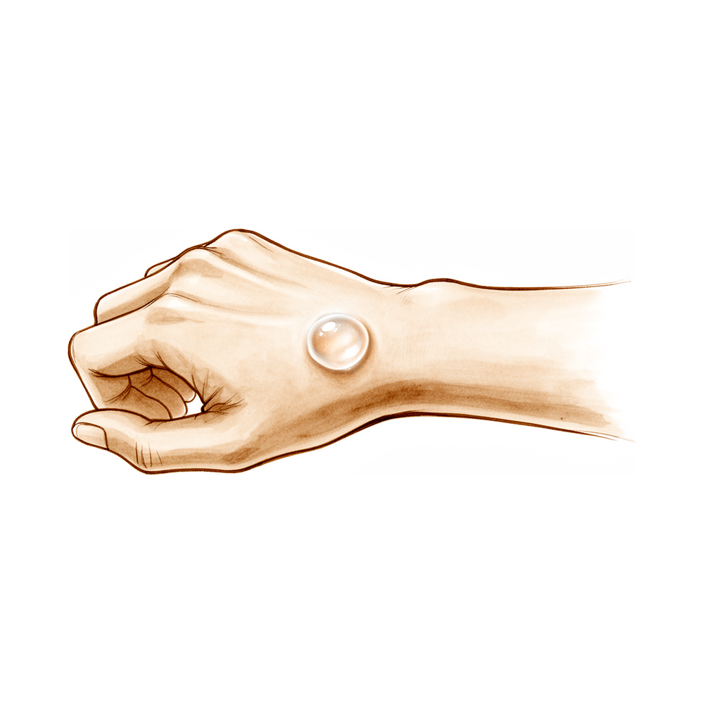
Overview¶
Tumors of the hand and upper extremity encompass a heterogeneous spectrum of benign and malignant neoplasms, ranging from nerve sheath tumors and enchondromas to soft-tissue sarcomas and skin malignancies. Definitive diagnosis requires pathologic evaluation, as MRI findings alone are not conclusive [1]. For brachial plexus tumors, biopsies are indicated when malignancy is suspected prior to definitive treatment, while asymptomatic benign lesions warrant observation [2]. The evidence base for many hand and upper extremity skin tumors relies heavily on case reports and retrospective reviews rather than controlled trials [6]. Surgical management of benign tumors balances the risk of recurrence against postoperative functionality, with most lesions indicated for surgery based on patient-rated outcomes [64, 137]. Improvement varies by pathology and location, but not by size [137].
Treatment strategies are dictated by tumor type and histology. Schwannomas treated by excision typically result in few long-term deficits, whereas neurofibroma excision carries a higher risk of nerve dysfunction and pain worsening [1]. Preoperative MRI is essential for schwannomas to guide surgical approach and minimize complications [20]. For malignant entities, complete surgical excision with clear margins is the only curative treatment for malignant peripheral nerve sheath tumours, synovial sarcoma, and costal chondrosarcoma [26, 49, 50]. Adjuvant radiation is utilized for larger or deeper synovial sarcomas, while the role of chemotherapy remains controversial [50]. Prognosis for malignant peripheral nerve sheath tumours remains poor, particularly in patients with neurofibromatosis [49].
Long-term follow-up is critical across the spectrum. Soft-tissue sarcomas of the hand require ten years of clinical follow-up even after adequate oncologic treatment [21]. Costal chondrosarcoma necessitates rigorous and prolonged surveillance [26]. For enchondromas in Ollier disease, early surgical treatment may limit lesion growth, but long-term follow-up is needed to monitor for recurrence and malignant transformation [7]. The absence of disease on MRI should not be the sole criterion for deciding against repeat resection after marginal excision of unsuspected soft tissue sarcomas [19]. Biopsy planning must be as rigorous as definitive surgery, with referral to specialized centers if local resources are insufficient [55, 59]. High-resolution MRI is recommended for glomus tumours to ensure complete excision and reduce recurrence [56].
Anatomy & Pathophysiology¶
General Principles¶
Benign peripheral nerve tumors encompass a wide range of neoplasms and non-neoplastic tumor-like lesions [3]. While some benign peripheral nerve lesions occur sporadically, others are encountered in the setting of genetic syndromes [3]. Although classified as benign, these tumors exhibit different levels of malignant potential [3]. The treatment of musculoskeletal tumors is based on the biologic activity and potential morbidity of each lesion [63]. The important biologic aspects of musculoskeletal tumors are the risk of local recurrence and metastasis [63]. Surgical margins are designed to reduce the risk of local recurrence [63].
Surgical margin types are defined by the plane of dissection relative to the tumor: * Intralesional: involves a plane of dissection that enters into the tumor [63]. * Marginal: involves a plane of dissection through the reactive zone at the edge of the tumor [63]. * Wide: involves removing the entire tumor with a cuff of normal tissue [63]. * Radical: involves removing the entire compartment that the tumor occupies [63].
Chemotherapy commonly works by inducing programmed cell death (apoptosis) [63]. Chemotherapeutic agents can directly damage DNA via alkylating agents, platinum compounds, and anthracyclines [63]. They can deplete cellular building blocks via antifolates, cytidine analogs, and 5-fluoropyrimidines [63]. Additionally, chemotherapeutic agents can interfere with microtubule function via vinca alkaloids and taxanes [63]. Radiation therapy causes DNA damage through the production of free radicals or direct genetic damage [63]. The rationale for radiation therapy is the treatment of microscopic satellite disease that may extend beyond the surgical margin [22]. Radiation therapy doses range from approximately 50 to 65 Gy and are given in multiple small fractions over several weeks [22]. A break of several weeks between radiation and surgery is instituted to decrease the rates of adverse wound reactions [22]. Preoperative radiation therapy is performed with a smaller dose and treatment field than postoperative radiation [22]. The rate of wound complications is significantly higher with preoperative radiation therapy than with postoperative radiation [22]. Postoperative radiation therapy has fewer wound complications but a higher rate of soft-tissue fibrosis [22].
Soft Tissue Sarcomas¶
Soft tissue sarcomas are relatively rare in the hand and forearm [123]. Approximately 12,000 new cases of soft tissue sarcoma are diagnosed each year in the United States [123]. Only 15% of soft tissue sarcoma cases occur in the upper extremity [123]. The majority of soft tissue sarcomas share a common primitive mesenchymal origin [123]. Specific cell origin or tumor differentiation of soft tissue sarcoma has limited value in predicting the behavior of a tumor [123]. The biologic behavior of soft tissue sarcomas is more accurately predicted by the lesion’s histologic grade [123]. Histologic grade takes into account cell mitotic index, cellularity, tumor necrosis, and anaplasia [123]. The most common histologic subtypes of soft tissue sarcoma seen in the hand are epithelioid sarcoma, synovial sarcoma, and malignant fibrous histiocytoma [123]. Histologic diagnosis can be useful in assessing the propensity for regional lymphatic spread [123].
Patients exposed to radiation and forestry workers exposed to herbicides may have a higher incidence of soft tissue sarcoma [123]. Genetic syndromes implicated in soft tissue sarcoma include neurofibromatosis and Li-Fraumeni syndrome [123]. Most patients with soft tissue sarcoma note a painless mass that may have been present for a long time with recent growth [123]. Epithelioid sarcoma is notable for manifestation as an ulcerating nodule that can be mistakenly treated for infection [123]. Soft tissue sarcomas in the hand are frequently misdiagnosed as infection, ganglion, and lipoma [123]. Incisional biopsies or biopsies in which the tumor is inadvertently violated result in contamination of all tissues included in the exposure [123]. Poorly planned biopsies may compromise the ability to perform limb-sparing surgery [123]. Transverse, oblique, and zigzag (Brunner) incisions should specifically be avoided for biopsy of soft tissue sarcomas [123].
Benign Nerve Tumors¶
A schwannoma is a benign nerve tumor composed almost entirely of Schwann cells [204]. Schwannomas consist of hypercellular (Antoni A cell) and hypocellular (Antoni B cell) areas [204]. The nuclei of spindle cells in schwannomas have a palisading arrangement referred to as Verocay bodies [204]. Most schwannomas are asymptomatic, but some can cause neurologic deficits resulting from compression of nerve fibers [204]. A schwannoma typically is well circumscribed and eccentrically located on a peripheral nerve [204]. The fascicles of the nerve do not enter a schwannoma but are splayed over it [204]. Malignant transformation of a schwannoma is extremely rare [204].
Neurofibromas contain perineural cells, fibroblasts, and mucoid material [204]. Neurofibromas are more likely to be associated with multiple lesions, a condition referred to as neurofibromatosis or von Recklinghausen's disease [204]. A neurofibroma is intimately connected with the nerve fascicles, and it is usually not possible to separate the two surgically [204]. Nerve sheath ganglions are rare lesions which present most commonly in the peroneal nerve [46]. Cases of nerve sheath ganglions have been reported in the median and ulnar nerve [46]. The cause of nerve sheath ganglions is thought to be a degenerative process [46]. A traumatic origin has been suggested for nerve sheath ganglions [46]. Nerve sheath ganglions may present as an asymptomatic swelling or be associated with nerve dysfunction [46]. On surgical approach to a nerve sheath ganglion, a cystic lesion is noted within the nerve [46]. In nerve sheath ganglions, the nerve bundles are displaced and are not found entering the lesion [46]. Recurrence of nerve sheath ganglions may be associated with incomplete excision [46].
Giant Cell Tumors of Tendon Sheath¶
Giant cell tumors of tendon sheath (GCTTS) are the second most common benign proliferative tumours in the upper extremities after ganglion cysts [199]. GCTTS are usually slow-growing, painless, benign and consist of soft tissue [199]. GCTTS are most commonly found in the fingers and among women in their fourth and fifth decades [199]. GCTTS originates from the synovial membrane, tendon sheath or synovial bursa [199]. Inflammation resulting from reactive or regenerative hyperplasia is the generally accepted theory of pathogenesis for GCTTS [199]. Genetic factors have been observed in previous studies regarding GCTTS [199]. GCTTS are usually asymptomatic; however, as the tumour grows some patients may present with swelling, pain and limitation of movement [199].
Giant-cell tumors of the tendon sheath correspond to a localized form of pigmented villonodular synovitis [202]. GCTTS typically occur in the hand where they represent the second most common type of soft tissue tumors after synovial ganglions [202]. The etiology of giant cell tumors of the tendon sheath is unknown [202]. Pathogenetic theories for GCTTS have included inflammatory process, trauma, immune mechanisms, and neoplasia [202]. Giant cell tumors usually present as a solitary and firm slowgrowing nodular lesion, which affects the volar aspect of the hand [202].
Bone Tumors and Cysts¶
Unicameral bone cysts are common lesions of childhood more consistent with a developmental or reactive lesion than a true tumor [69]. Eighty-five percent of unicameral bone cysts occur in the first two decades with a 2:1 male predominance [69]. Unicameral bone cysts are most common in the proximal humerus and femur [69]. In adults, unicameral bone cysts are more common in the ilium and calcaneus [69]. Unicameral bone cysts are often asymptomatic unless a pathologic fracture has occurred [69]. Two thirds of patients with unicameral bone cysts present with fractures that can stimulate the cyst to heal [69]. The most widely accepted theory for the pathogenesis of unicameral bone cysts is that a focal defect in metaphyseal remodeling blocks interstitial fluid drainage [69]. This leads to focal bone necrosis and accumulation of fluid in unicameral bone cysts [69]. Cyst fluid in unicameral bone cysts contains prostaglandins, oxygen-free radicals, interleukins, cytokines, and metalloproteinases [69].
The hand is a common location of exostoses development for patients with multiple hereditary exostoses [190]. For low-grade chondrosarcomas of the hands and feet, surgical management should aim to preserve function [79]. Grade 2 and high-grade chondrosarcomas of the hands and feet require more aggressive approaches [79].
Other Benign Lesions¶
Dupuytren’s contracture is classified as a benign superficial fibromatosis but has some histological features of a neoplasm [189]. Dupuytren’s contracture is characterized by marked fibroblast proliferation within the palmar nodules [189]. Like fibrosarcoma, Dupuytren’s disease arises from mesenchymal cells and is characterized by infiltrative growth, proliferation, lack of apoptosis and a tendency towards recurrence [189]. Unlike sarcoma, Dupuytren’s contracture never metastasizes [189]. Fibroblasts derived from Dupuytren tissue show in vitro properties similar to those of solid tumours [189]. Fibroblasts derived from Dupuytren tissue exhibit leukaemia-like chromosomal abnormalities [189]. Fibroblasts derived from Dupuytren tissue exhibit the capability to bind monoclonal antibodies derived from human sarcomata [189]. Atypical lipomas of the hand and forearm may have a more benign behavior than atypical lipomas of other anatomical sites [223].
Classification¶
General Principles and Diagnostic Requirements¶
Definitive diagnosis of nerve tumors requires pathologic evaluation, as MRI is not definitive [1]. Benign peripheral nerve tumors encompass a wide range of neoplasms and non-neoplastic tumor-like lesions, some of which occur sporadically while others are associated with genetic syndromes [3]. Although classified as benign, some peripheral nerve tumors possess different levels of malignant potential, requiring awareness to avoid pitfalls with devastating outcomes [3]. Soft-tissue tumors in children are a heterogeneous group of lesions that may be benign or malignant [5]. Tumor-like conditions of the hand and upper extremity typically present as masses and can be confused with more serious conditions [8]. Diagnosis of tumor-like conditions of the hand and upper extremity often relies on clinical examination and imaging, with excisional biopsy needed for definitive diagnosis to guide treatment [8]. Benign bone tumors represent a diverse group of pathological and clinical entities that vary greatly in aggressiveness and clinical behavior [159]. Fibro-osseous and cystic tumors of bone are common lesions with distinct clinical, radiologic, and pathologic features that aid in their classification [14]. Nonmelanotic malignant skin tumors represent the most common type of cancer to afflict mankind and are the most common malignancies of the hand [71]. Vascular tumors of the hand are classified into benign, congenital, and malignant categories [142]. In a review of 516 vascular tumors of the upper extremity, 90% were classified as either hemangiomas or malformations [24]. Soft-tissue sarcomas comprise a heterogeneous and rare group of tumors that display a wide spectrum of local growth patterns and metastatic potential [81]. Malignant primary bone tumors are rare and varied, requiring management tailored to specific tumor types [54].
Staging Systems for Malignant Tumors¶
Enneking Staging System: This system classifies primary bone tumors according to whether they are high or low grade, whether metastases are present, and whether the tumor has grown out of its original compartment [132]. A compartment is defined as an enclosed tissue space, such as a bone, a joint space, or a muscle group confined by its fascial envelope [132]. Stage 1A describes a low-grade, intracompartmental tumor [132]. Stage 1B describes a low-grade, extracompartmental tumor [132]. Stage 2A describes a high-grade, intracompartmental tumor [132]. Stage 2B describes a high-grade, extracompartmental tumor [132]. Stage 3 describes any of the above stages with metastases [132].
TNM Staging System: For primary sarcomas of bone, this system takes into consideration histological subtype, size, continuity, grade, and local and distant spread [132]. In soft-tissue sarcomas, size is dichotomized as small (<5 cm) or large (>5 cm) [132]. In bone sarcomas, size is dichotomized as small (<8 cm) or large (>8 cm) [132]. Stage 1A describes a low-grade, small tumor with no metastases [132]. Stage 1B describes a low-grade, large tumor with no metastases [132]. Stage 2A describes an intermediate- or high-grade, small tumor with no metastases [132]. Stage 2B describes an intermediate-grade, large tumor with no metastases [132]. Stage 3 describes a high-grade, large tumor with no metastases [132]. Stage 4 describes any tumor with metastases [132].
Musculoskeletal Tumor Society and AJCC Systems: The Musculoskeletal Tumor Society system remains intuitive for surgeons, while the American Joint Committee on Cancer staging system has widespread acceptance due to its multidisciplinary nature and ability to facilitate more nuanced prediction of prognosis [138].
Chondrosarcoma Staging: Chondrosarcomas are staged according to the Enneking system, with nonmetastatic low-grade chondrosarcomas considered stage I neoplasms [78]. Nonmetastatic intermediate- and high-grade chondrosarcomas are considered stage II [78]. Metastatic chondrosarcomas are stage III [78]. Chondrosarcomas are subclassified as stage A or stage B based on whether they are located within the bone or extend outside the bone [78]. A low-grade intramedullary chondrosarcoma without metastases is classified as stage IA [78]. A high-grade nonmetastatic chondrosarcoma with an associated soft-tissue mass is classified as stage IIB [78].
Staging and Classification of Benign Tumors¶
Enneking Benign Tumor Staging: Benign bone lesions are classified into three stages: stage 1 (latent), stage 2 (active), and stage 3 (locally aggressive) [77]. In the Enneking staging system for benign tumors, a stage 1 tumor is latent, meaning it does not progress or heals spontaneously [78]. A stage 2 tumor is active, meaning it progresses but respects natural barriers such as the bone cortex [78]. A stage 3 tumor is aggressive, meaning it progresses and will ultimately destroy natural barriers [78]. Enchondromas are usually stage 1 but are occasionally stage 2 [78].
Campanacci Classification: This classification grades giant cell tumors (GCT) into three grades based on radiographic appearance [51]. Grade 1 GCT lesions are latent, characterized by a well-defined margin and an intact cortex [51]. Grade 2 GCT lesions are active, characterized by a relatively well-defined margin, no radiopaque rim, and a thinned and moderately expanded cortex [51]. Grade 3 GCT lesions are aggressive, characterized by indistinct borders and cortical destruction [51]. The Campanacci grading system for GCT is similar to the Enneking classification for benign bone tumors overall [51].
Enneking Clinico-Radiological Classification: This classification for benign bone tumors includes stage 1 (latent), stage 2 (active), stage 3 (aggressive), and stage IV (malignant) [51]. Stage 1 refers to a lesion confined totally by bone, asymptomatic, inactive on bone scan, and histologically benign [51]. Stage 2 refers to an expanded cortex with no breakthrough, symptomatic status, active bone scan, and histologically benign status [51]. Stage 3 refers to a rapidly growing mass with cortical perforation, soft tissue mass, potential for metastasis, symptomatic status, extensive bone scan activity, and histologically benign status [51]. Stage IV refers to a sarcomatous lesion contiguous with a benign giant cell tumor [51].
Other Considerations: A novel GCT grading system demonstrates excellent interrater agreement and classified more nonrecurrent curetted tumors as low grade compared with the Campanacci classification [151]. Jaffe et al. classified giant cell tumors as benign, aggressive, and malignant based on the histological appearance of stromal cells and the number of giant cells and mitoses [51]. The prognostic value of the Jaffe histological staging system for giant cell tumors has been disputed [51].
Specific Tumor Classifications¶
Vascular Bone Tumor Classification: A proposed classification scheme for vascular bone tumors includes hemangioma, hemangioendothelioma, angiosarcoma, and their epithelioid variants [33].
PUMCH Classification: The PUMCH classification for extremity and pelvic hemophilic pseudotumors is based on anatomic pathology and surgical strategy [150].
Molecular Classification: Confirming PIK3CA mutation status allows for more accurate diagnosis and classification of isolated macrodactyly based on its molecular pathogenesis [152].
Cartilage Tumor Analysis: Separating cartilage tumors based on histological and biochemical parameters may provide a system for analysis that offers information regarding chemical abnormalities and adds a new approach in the assessment of malignancy [158].
WHO Classification: The 2020 WHO Classification of Tumors of Soft Tissue provides an updated classification scheme and essential diagnostic criteria, including newly recognized entities, subtypes, and reclassified tumors [36].
Benign Hand Bone Tumor Grouping: Benign hand bone tumors are grouped largely according to tissue type: cartilage tumors, cystic lesions, osteogenic tumors, and miscellaneous lesions [77].
Clinical Presentation¶
History and General Presentation¶
Patients with musculoskeletal tumors typically present with pain, a palpable mass, or an abnormal radiographic finding detected during evaluation for an unrelated problem [101]. Bone tumors most frequently manifest as pain, which in malignancy is often progressive and occurs at rest and at night [101]. Benign bone tumors may cause activity-related pain if the lesion is large enough to weaken the bone, while osteoid osteoma may initially present with night pain [101]. Soft-tissue tumors rarely cause pain; patients more often report a mass, though nerve sheath tumors are an exception and may present with pain or neurologic signs [101]. Soft tissue sarcomas typically present as enlarging painless masses greater than 5 cm, and the presence or absence of pain is not indicative of malignancy in soft-tissue tumors [89, 45]. A nerve sheath ganglion may present as an asymptomatic swelling or be associated with nerve dysfunction [46]. Melanoma in the hand often presents at a later stage with a poorer prognosis [117].
Age is the most important historical information because most benign and malignant musculoskeletal neoplasms occur within specific age ranges [101]. Family history is helpful in cases of multiple hereditary exostosis and neurofibromatosis, both of which have autosomal dominant inheritance [101]. The physical diagnosis must include a complete history that elicits the duration of symptoms, as well as accurate identification of both size and location of a particular mass [37].
Diagnostic Evaluation and Imaging¶
A thorough history, physical examination, and baseline plain x-rays are the foundation of accurate diagnosis for benign tumors of the hand [93]. Plain radiographs are frequently all that are necessary to determine the diagnosis and appropriate treatment of benign-appearing hand and finger tumors [114, 126]. Plain radiographs are the most useful modality to determine whether a mass is arising in the soft tissues or in bone [108]. Magnetic resonance imaging (MRI) gives the best resolution to characterize the lesion and define anatomical features [108]. MRI is often helpful for soft-tissue tumors, bone masses with a soft-tissue component, and any malignant tumor [45]. Lipomas can be identified through clinical history, physical examination, and imaging such as MRI [15].
Sonography generally conveys less detail concerning precise anatomic relationships and tumor margins than CT and MR images [41]. However, sonography can easily differentiate deep, fluid-filled lesions such as ganglion cysts from solid tumors, although echo patterns of solid masses on sonography are nonspecific [41]. A soft-tissue mass that is five centimeters in diameter or larger on physical examination, especially if deep to the fascia, should be referred to an orthopaedic oncologist without additional evaluation or biopsy due to the high probability of malignancy [94]. If clinical and radiographic information favors a diagnosis of malignant or aggressive benign bone tumor, the patient should be referred to an experienced orthopaedic oncologist without performing additional diagnostic tests or a biopsy [94].
Most benign bone tumors in children have characteristic clinical and radiographic presentations that allow for diagnosis without biopsy [10]. Many benign tumors have characteristics that enable the clinician to arrive at a diagnosis without biopsy [93]. Diagnosis of tumor-like conditions often relies on clinical examination and imaging, with excisional biopsy needed for definitive diagnosis to guide treatment [8]. A thorough history and physical examination, followed by appropriate imaging studies, can establish a correct diagnosis for soft-tissue tumors in children [5]. Fibro-osseous and cystic tumors of bone have distinct clinical, radiologic, and pathologic features that aid in their classification [14]. Most benign bone tumors have characteristic features that suggest their specific diagnosis, allowing practicing orthopaedic surgeons to follow with imaging or refer to an orthopaedic oncologist [39]. The vast majority of patients with musculoskeletal tumors can be diagnosed without invasive testing using radiographs and judicious use of advanced imaging modalities [45]. When clinical presentation and imaging fail to provide an answer, biopsy often with molecular testing is an extremely useful tool [45].
Biopsy and Referral Protocols¶
Biopsies should be performed for tumors suspected to be malignant before starting definitive treatment [2]. Diagnostic delay or inappropriate treatment may result if patients do not receive appropriate followup, biopsy (usually open), or referral whenever the diagnosis is in doubt [42]. A solitary bone lesion in a patient with or without a history of cancer should be biopsied to obtain an accurate diagnosis [140]. Presuming a solitary lesion is a bone metastasis in an older patient may lead to the wrong operation, cause extensive contamination, and potentially compromise the life or limb of the patient if the lesion is actually a primary sarcoma of bone [140]. In individuals with recent and known metastatic disease to bone, a new biopsy of bone is not necessary [140]. If the patient has a remote history of cancer or no known metastasis to bone, a biopsy should be performed to confirm that the lesion is not a primary sarcoma [140].
A needle biopsy is usually definitive when differentiating a carcinoma from a sarcoma [140]. Specific immunohistochemical staining may allow determination of the primary site of origin of a carcinoma, most commonly from the lung, breast, thyroid, kidney, or prostate [140]. If a needle biopsy is nondiagnostic or unable to be done, a careful incisional biopsy should be performed using oncologic principles so as not to preclude subsequent definitive surgical treatment [140]. The biopsy should be as small as possible, in a longitudinal fashion in line with the extremity, and performed with excellent hemostasis [140]. Tissues contaminated by a postbiopsy hematoma must be considered contaminated by tumor [140]. Cultures should always be sent at the time of biopsy to rule out infection, which can appear radiographically similar to a tumor [140]. If a definitive diagnosis of metastatic disease can be made on an intraoperative frozen section, surgical treatment of the pathologic fracture can be performed at the same operative setting [140]. If the frozen section is nondiagnostic, it is best to wait for the permanent sections before definitively treating the tumor and fracture [140].
Diagnostic Challenges and Suspicion¶
Diagnosis of malignant peripheral nerve sheath tumour (MPNST) can be challenging due to the rarity of these tumours and unreliable imaging, making a high index of suspicion paramount for early diagnosis and treatment [103]. Awareness of tumor characteristics may help physicians with diagnosis and treatment [32]. Awareness of tumor features may help physicians with diagnosis, and awareness of recurrence rates is important when counseling patients [105]. The continued importance of clinical suspicion is highlighted in glomus tumor diagnosis, particularly when not diagnosed on MRI [110]. A lower threshold for further workup in patients with atypical signs and symptoms allows for the potential to identify potential malignancy earlier [90]. Awareness of uncommon tumors such as intraosseous myofibroma in adult patients is essential to prevent mistaken diagnosis and subsequent extensive workup [98]. The diagnosis of Bizarre Parosteal Osteochondromatous Proliferation (Nora Lesion) in pediatric cases can be challenging due to presenting symptoms and radiographic findings [107]. Angiomatoid fibrous histiocytoma is a rare tumor that is difficult to diagnose, often leading to misdiagnosis and inadequate treatment by referring physicians [118]. Nerve tumors are rare and often overlooked in the differential diagnosis of upper extremity masses [111]. Glomus tumor should be included in the differential diagnosis in neurofibromatosis patients who present with a painful lesion of the hand or finger [99].
Signs of Malignancy and Management¶
Signs of malignancy, such as rapid growth, increasing pain, or other atypical features, mandate a thorough workup, and surgical management should be considered [93]. If a lesion increases in size or becomes symptomatic, or if the physical or radiographic appearance suggests an aggressive lesion, appropriate staging studies including a tissue diagnosis must be obtained [41]. Some benign peripheral nerve tumors have different levels of malignant potential, and treating physicians should be aware of this to avoid possible pitfalls with devastating outcomes [3]. Patients with giant cell tumour (GCT) should be followed indefinitely, and referred promptly if new symptoms, particularly pain, emerge [87]. Benign tumors that are asymptomatic should be observed [2]. Certain lesions with a characteristic appearance and benign features may be managed nonsurgically with observation alone [93]. Vigilance in monitoring for signs of malignancy is encouraged, even for seemingly benign tumors [93].
Investigations¶
General Diagnostic Principles¶
Asymptomatic benign tumors warrant observation, whereas biopsies are mandatory for tumors suspected to be malignant prior to definitive treatment [2]. A thorough history and physical examination, followed by appropriate imaging, establishes the correct diagnosis and guides treatment recommendations for soft-tissue tumors in children [5]. Most benign bone tumors in children present with characteristic clinical and radiographic features that allow diagnosis without biopsy, though some may simulate malignancy and require referral to an orthopaedic oncologist [10]. In patients with an underlying history of malignancy, a high level of suspicion and histological study are required [141]. Similarly, a high index of suspicion and thorough evaluation including radiographs and biopsy are essential to avoid misdiagnosis in patients with known metastatic disease [170]. For undiagnosed hand tumors, MRI and expeditious incisional biopsy are important, as initial incorrect diagnoses can lead to misguided treatments [171].
Imaging Modalities¶
Plain radiography: Longitudinal radiographs allow observation of the timing of first appearance and potential for resolution for each tumor type in asymptomatic children [23]. Plain radiographs may fail to show a peripheral rim of ossification in soft-tissue recurrence of giant-cell tumor, which may not be recognized without thorough physical examination and magnetic resonance imaging [166].
MRI: MRI is the preferred modality for staging soft tissue tumors and evaluating patients for tumor recurrence [120]. Although no major differences between MRI and CT were demonstrable in preoperative evaluation of soft-tissue tumors, MRI is preferred due to better spatial orientation and sharper contrast between tumor and adjacent tissues [133]. MR imaging is the modality of choice for evaluating synovial tumors and tumor-like lesions, as a presumptive diagnosis can be made in most cases [129]. In the coronal plane, magnetic resonance imaging accurately defines intramedullary abnormality and provides greater interpretive certainty than computerized tomography or scintigraphy in defining the true proximal and distal extent of a tumor [62]. The combination of ultrasound and MRI findings provides features that narrow the differential diagnosis for ancient schwannoma, helping to differentiate it from malignant tumors [29]. High-resolution MRI is recommended for glomus tumors to ensure complete excision and decrease recurrence rates [56]. High resolution MR imaging can indicate the location of a tumor before operation, supporting the hypothesis that a new tumor had developed rather than a recurrence due to incomplete excision [149]. MRI may be more useful than plain x-rays to identify bone and soft tissue extensions in patients with underlying malignancy [141]. Preoperative diagnosis by MRI and selection of the appropriate surgical approach are important in cases of schwannoma to give clinicians the best chance to separate the nerve from the tumor and decrease the risk of postsurgical complications [20]. However, there is considerable variability and inaccuracy in MRI-based measurements of musculoskeletal tumors [169]. Underestimation of tumor length occurred on MRI in 24 patients, but translesional resection would happen only if the underestimation was greater than the minimum 3-5 cm of tumor-free bone necessary for resection with wide margins [153]. The absence of disease on MRI should not be used as the sole criterion in determining whether a repeat resection should be performed [19].
CT: CT scanning with thin sectioning should be the preferred advanced imaging modality for diagnosing osteoid osteoma in children, as MRI may be misleading and lead to misdiagnosis [168].
Other Considerations: For all tumor types, MRI, CT, and plain radiographs were frequently rated as helpful studies [48]. MRI should be used as a complementary method, while angiography remains necessary for specific cases involving highly vascularized tumors [134]. Management of metaphyseal fibrous defects ranges from serial radiographic observation for asymptomatic lesions to biopsy, curettage, and bone grafting for large, symptomatic lesions or those with atypical features [164].
Treatment¶
General Principles and Diagnostic Prerequisites¶
Effective management of musculoskeletal tumors requires a foundational understanding of diagnosis, staging, biopsy, resection, and amputation principles [13]. Distinguishing between soft-tissue and bone tumors is the critical starting point for treatment planning [45]. Plain radiographs serve as an excellent initial diagnostic tool for most bone tumors [45]. Biopsy procedures must be executed with the same rigor as definitive surgery, as poor biopsy technique can lead to devastating patient outcomes [45, 55]. For spine tumors, proper biopsy diagnosis is the essential first step in devising treatment, and primary spinal tumors are best managed with en bloc resection to minimize local and distant metastases [72]. Care for spine tumor patients mandates a multidisciplinary team approach with continued follow-up [72]. In schwannomas, preoperative MRI diagnosis and appropriate surgical approach selection are vital for separating the nerve from the tumor and reducing postsurgical complications [20].
Treatment at specialized cancer centers improves overall survival and reduces local recurrences for adipose tumors [122]. Patients treated for primary malignant bone tumors at high-volume facilities experience slightly better overall survival than those at low-volume facilities [144]. Evaluation, diagnosis, management, and follow-up of soft-tissue sarcoma patients should preferably occur at high-volume centers involving trained pathologists, radiologists, medical oncologists, radiation oncologists, and surgeons [81]. Coordinated multidisciplinary teamwork is mandatory for optimal care in soft-tissue sarcomas [81].
Benign Tumors¶
Surgical Excision and Curettage: Surgical excision is the primary treatment for symptomatic perionychium tumors, with rare recurrence following complete removal [4]. For leiomyoma of the hand, complete surgical excision is uniformly curative, as metastasis has never been reported [135]. Surgical excision remains the primary treatment for invasive skin tumors of the hand, though nonsurgical options exist for unresectable or low-grade cancers [136]. Curettage is the standard of care for symptomatic enchondromas of the hand, with most patients returning to full function [156]. Curettage also appears to be the treatment of choice for chondromyxoid fibroma, proving curative in all cases with adequate follow-up [30]. For tumor-induced osteomalacia associated with extremity tumors, tumor resection is preferred over curettage [53].
Recurrence and Malignant Transformation: Reported recurrence rates for solitary enchondroma of the phalanx vary from 2% to 15%, with the largest published series reporting a 4.5% rate [180]. Inadequate curettage is the predominant cause of recurrence in these cases [180]. Malignant degeneration of isolated enchondroma to chondrosarcoma is extremely rare but described [180]. Clinical features suggesting chondrosarcoma include rapid growth, soft tissue mass, and pain in a previously asymptomatic digit [180]. Radiographic evidence of cortical destruction with soft tissue extension suggests a more aggressive neoplasm [180]. Longer-term follow-up is needed after early surgical treatment of enchondromas in Ollier disease to confirm the absence of recurrence and malignant transformation [7].
Conservative Management and Specific Lesions: Unicameral bone cysts are most active during skeletal growth and usually heal spontaneously at maturity [69]. In flat bones, these cysts are usually asymptomatic, found incidentally, and rarely fracture [69]. Fibrous dysplasia symptoms may resolve with conservative treatment, and lesions may remain radiographically stable [69]. Fibromatoses and related tumors of the hand in children are benign and do not metastasize; proper clinical, radiologic, and pathologic assessment is mandatory to avoid mutilating surgery [163]. The prognosis for osteochondrolipoma matches that of benign lipoma, with tumors usually not recurring after complete excision [27]. Benign hand tumors in the distal upper limb have a clear indication for surgery based on patient-rated outcome measures [74].
Other Considerations: Cure rates for non-melanotic skin tumors of the upper extremity vary by tumor type, size, and histologic subtype [17]. Recurrence rates for non-neoplastic tumors of the hand and upper extremity vary by lesion type and treatment modality [18]. Specific characteristics of squamous cell carcinoma of the hand may increase recurrence chances, while excision technique did not have a major role in outcome [52]. Although complete tumor resection confers a good prognosis in tumor-induced osteomalacia, surveillance for recurrence and metastasis is necessary [61]. The role of percutaneous sclerotherapy for benign bone tumors is highlighted in recent literature [157].
Malignant Tumors¶
Surgical Principles and Margins: Wide tumor resection aimed at achieving R0 surgical margins is the cornerstone of management for adult soft-tissue sarcomas of the extremities [81]. The goal of surgery is to achieve an acceptable margin to minimize local recurrence while maintaining reasonable function [83]. Limb salvage procedures are performed in approximately 90% of patients with soft-tissue sarcomas [83]. Sarcomas exhibit a centripetal growth pattern, surrounded by a reactive zone containing edema, fibrous tissue, inflammatory cells, and tumor cells [83]. "Shelling out" a sarcoma involves excising it through the reactive zone, which usually leaves tumor cells behind [83]. Standard oncologic techniques include using a tourniquet without exsanguination, excising any open biopsy tract, and placing drains distal, close, and in line with the tumor [83].
Adjuvant Therapies: Radiation therapy, either preoperatively or postoperatively, is recommended for high-grade, large, and deep-seated soft-tissue sarcoma lesions [81]. Chemotherapy has marginal efficacy in localized soft-tissue sarcoma disease but is considered in high-risk patients with large, deep, and high-grade tumors [81]. For high-grade malignant primary bone lesions, neoadjuvant therapies and surgery are typical, whereas low-grade lesions are usually managed with surgery alone [54]. Multidrug neoadjuvant chemotherapy is usually initiated after histologic diagnosis and staging for malignant bone tumors [174]. Chemotherapy helps control systemic disease by attacking micrometastases, increasing overall survival rates [174]. Neoadjuvant therapy "sterilizes" the reactive zone by destroying microscopic disease at the periphery, facilitating resection [174]. The effectiveness of chemotherapy regimens correlates with the extent of tumor necrosis [119].
Specific Tumor Management: Complete surgical excision with clear margins is the only curative treatment for malignant peripheral nerve sheath tumours, though prognosis remains poor, particularly in neurofibromatosis [49]. Wide surgical excision is the standard of care for synovial sarcoma, with adjuvant radiation utilized for larger and deeper lesions [50]. In nonmetastatic angiosarcoma of the upper extremity, amputation is the treatment of choice, though survival rates are uniformly poor [128]. Surgical resection is the main treatment for intraperitoneal extraosseous osteosarcoma, with a poor prognosis [131]. Grade 2 and high-grade chondrosarcomas of the hands and feet require more aggressive surgical approaches [79]. For infantile fibrosarcoma of the hand, multidisciplinary management combining neoadjuvant targeted oral chemotherapy with staged conservative surgical resection led to successful tumor eradication and near full hand function at 2-year follow-up [172].
Staging and Classification: Chondrosarcomas are staged according to the Enneking system [78]. Nonmetastatic low-grade chondrosarcomas are stage I neoplasms, while nonmetastatic intermediate- and high-grade chondrosarcomas are stage II [78]. Chondrosarcomas are subclassified as stage A (within bone) or stage B (extending outside bone) [78]. Enchondromas may be staged using the Enneking system for benign tumors: stage 1 is latent (no progress or spontaneous healing), stage 2 is active (progresses but respects natural barriers like the cortex), and stage 3 is aggressive (progresses and destroys natural barriers) [78].
Limb Salvage and Reconstruction: There are four basic principles for limb-sparing procedures for malignant bone tumors: local recurrence should be no greater and survival no worse than with amputation; the procedure should not delay adjuvant therapy; reconstruction should be enduring with few local complications; and limb function should approach that obtained by amputation [174]. Before considering limb preservation, patients must be appropriately staged and assessed through a multidisciplinary approach [174]. Biological reconstructions combining autologous or vascularized bone with tendon repair are preferable to prostheses for pediatric malignant tumors to ensure longevity and functional hand placement [82].
Other Considerations: The optimum treatment for Grade III giant-cell tumors of bone and patients with local recurrence is unclear [139]. Checkpoint inhibition may be a sensible treatment alternative for chordomas, particularly in inoperable, recurrent, or metastasized cases, though clinical trials are needed to confirm efficacy and determine optimal drug combinations [146]. Despite marked improvements in survival for localized Ewing sarcoma disease over the past 40 years, lesser improvements have been seen in metastatic or recurrent disease [60]. Recent literature highlights the importance of aggressive surgical resection for non-sacral pelvic Ewing sarcoma and the prognostic value of preoperative imaging in osteosarcoma and chondrosarcoma [157]. The article provides an update on current management of primary malignant and metastatic tumors of the hand, emphasizing optimal biopsy, negative surgical margins, and the roles of adjuvant radiotherapy and chemotherapy [57].
Desmoid Tumors¶
Management Strategy: Despite the benign nature of desmoid tumors, multidisciplinary care is needed to provide combined treatment options [44]. Upfront surgery is not advantageous compared to more conservative treatments such as observation or medical treatment [154]. Patients treated with local modalities for desmoid tumors did not experience improved event-free survival compared with those treated without local modalities [58].
Adjuvant Therapy: Adjuvant radiation therapy is probably beneficial for extra-abdominal desmoid tumors, but the precise indication for its use is not well defined [160].
Complications¶
Nerve and Soft-Tissue Tumors¶
Malignant Potential and Recurrence: Benign peripheral nerve tumors possess varying levels of malignant potential, necessitating awareness to avoid pitfalls with devastating outcomes [3]. Prognosis following resection of benign nerve tumours of the hand is good, characterized by a low risk of postoperative complications and recurrence [70]. However, recurrent symptoms from digital glomus tumors may stem from small, synchronous, satellite lesions missed during initial surgery rather than inadequate resection [182]. Upper extremity desmoid tumors exhibit a high recurrence rate after surgical excision, with 5-year disease-free intervals ranging from 53% to 72% in previous studies and 57% in the cited study [193]. Achieving adequate surgical margins for these desmoid tumors in the upper extremity is associated with patient morbidity due to the removal of nerves, tendons, and other critical structures [193].
Pediatric and Diagnostic Considerations: Lipofibromatosis in pediatric patients carries a high rate of regrowth or persistent disease, with 72% of patients experiencing these outcomes over a median follow-up of 6 years and 7 months [184]. No tumor in the cited study of pediatric lipofibromatosis has given rise to metastases [184]. Diagnostic delay or inappropriate treatment may result if patients with tumors masquerading as hematomas do not receive appropriate follow-up, biopsy, or referral whenever the diagnosis is in doubt [42].
Bone Tumors¶
Malignant Transformation and Recurrence: A secondary malignant tumor can develop as long as twenty-five years after the initial presentation of a benign giant-cell tumor [75]. Nodules of recurrent giant-cell tumor within a surgical scar may occur long after excision of the primary bone lesion [167]. The risk of malignant transformation for solitary osteochondromas is most likely <1% [191]. Patients with hereditary multiple exostoses (HME) may have a 2% to 4% chance of developing chondrosarcoma [191]. Patients with Ollier disease and Maffucci syndrome have an estimated risk of malignant transformation ranging from 10% to 40% [191]. One study suggested that malignancy is a near certainty in persons with Maffucci syndrome [191]. The risk of malignant transformation for solitary enchondromas is estimated to be well below 1% per patient year of observation [191]. A case report suggests the possibility of malignant transformation of an aneurysmal bone cyst into a malignant fibrous histiocytoma [186].
Management and Prognosis: The best results of treatment for secondary malignant giant-cell tumors of bone were obtained with early ablation [188]. All but one of nineteen patients with secondary malignant giant-cell tumors had received therapeutic irradiation four to thirty-nine years earlier [188]. Fibrosarcoma occurred three times as frequently as osteosarcoma in secondary malignant giant-cell tumors of bone [188]. Early surgical treatment of enchondromas in Ollier disease may limit the growth of large lesions and facilitate future surgery, though longer-term follow-up is needed to confirm the absence of recurrence and malignant transformation [7].
Malignant Tumors and Sarcomas¶
Epidemiology and Diagnosis: The ratio of benign to malignant soft-tissue masses is 100:1 [83]. Sixty percent of sarcomas affect the extremities, with lower extremities more commonly affected than upper extremities [83]. Eighty-five percent of sarcomas occur in individuals older than 15 years [83]. The diagnostic appearance of most sarcomas on MRI is indeterminate, requiring a biopsy [83]. Nonmelanotic malignant skin tumors are the most common malignancies of the hand [71].
Surgical Indications and Outcomes: Amputation is indicated when necessary to resect the entire tumor, when major nerves cannot be saved, in some locally recurrent sarcomas, or when the patient has significant comorbidities that preclude limb-sparing surgery [83]. After aggressive surgical management with negative margins for epithelioid sarcoma of the hand, 71% of patients were alive without evidence of disease at the last follow-up, with a 5- and 10-year survivorship of 85% [67]. Survival for malignant giant cell tumors of bone was poorest among older patients and those with evidence of distant metastases at time of diagnosis [73]. Age and stage at diagnosis were strongly associated with long-term survival for malignant giant cell tumors of bone [73].
Follow-up and Specific Tumor Behaviors: Even with adequate oncologic treatment, long-term clinical follow-up of 10 years is advised for soft-tissue sarcomas of the hand [21]. Merkel cell carcinoma is characterized by a high incidence of local recurrence, distant metastases, regional nodal metastases, and high mortality [192]. A history of extensive recurrent disease well removed from the primary tumor in cutaneous metastatic breast carcinoma is associated with a poor prognosis [68]. Wide surgical resection is the only demonstrated curative treatment for costal chondrosarcoma, necessitating rigorous and prolonged follow-up [26]. Recurrence of extraskeletal soft tissue chondrosarcoma has been reported as early as two months and as late as fifteen years after the primary operation [80].
Recovery¶
Light activity (weeks): The evidence base does not specify a typical week range for the resumption of desk work, driving, or light activities of daily living.
Full activity (months): The evidence base does not specify a typical month range for the return to manual work, sport, or full range of motion and strength.
Complete recovery / outcome plateau (months): The evidence base does not specify a month range for the stabilization of pain, strength, or final functional outcomes.
Rehabilitation protocol: The provided evidence does not detail specific physiotherapy phasing, immobilisation durations, weight-bearing or range-of-motion progressions, or timing for sling and brace removal.
Functional milestones: Specific validated patient-reported outcome measure trajectories are limited. In patients undergoing more aggressive local treatment for desmoid tumors, PROMIS Function Scores are lower [58]. One patient with epithelioid sarcoma in the median nerve of the hand achieved tactile and pain sensory function recovery, a QuickDASH score of 20, and grip strength at 60% of the normal side [28].
Other Considerations: Long-term surveillance is critical for many hand tumors. Long-term clinical follow-up of 10 years is advised for soft-tissue sarcoma of the hand, and patients should not be deceived by the smaller size of the tumor regarding the need for this duration of monitoring [21]. All patients with extraskeletal soft tissue chondrosarcoma require careful follow-up due to the potential for late recurrence [80]. Wide surgical resection for costal chondrosarcoma necessitates rigorous and prolonged follow-up [26]. A multi-disciplinary approach with long-term follow-up is recommended to monitor for local recurrence of intraosseous myoepithelioma in the hand [65]. Surveillance for recurrence and metastasis is necessary after complete tumor resection for tumor-induced osteomalacia associated with phosphaturic mesenchymal tumors [61].
Prognosis varies significantly by tumor type. Osteochondrolipomas usually do not recur after complete excision, and their prognosis is the same as with benign lipoma [27]. Curettage has proved curative in all cases of chondromyxoid fibroma in which adequate follow-up studies have been made [30]. For epithelioid sarcoma of the hand, aggressive surgical management with negative margins results in 71% of patients being alive without evidence of disease at the last follow-up, with 5-year and 10-year survivorship at 85% [67]. Ray amputation with tumour-free excision margins for hand sarcoma yields satisfactory long-term functional results, is associated with a low risk of lower local recurrences, allows for early return to daily activities, and is associated with good survival rates [165].
Malignant and metastatic lesions carry distinct risks. A secondary malignant tumor can develop twenty-five years after the initial presentation of a benign giant-cell tumor [75]. The presence of a metastatic lesion in the hand or skin carries an ominous prognosis with a median survival of a few months [210]. A history of extensive recurrent disease well removed from the primary tumor is associated with a poor prognosis for cutaneous metastatic breast carcinoma of the hand and upper extremity [68]. In a series of Ewing’s Sarcoma of the Hand, there were no local recurrences, but two patients developed metastases and died, while three patients were alive and free of disease at last follow-up [212]. Despite marked improvements in survival during the past 40 years for patients with localized Ewing Sarcoma Family of Tumors, lesser improvements have been seen in patients with metastatic or recurrent disease [60]. Chondrosarcoma has a much better prognosis, with three of seven patients in a 1957 series surviving five years [208]. Neurofibroma excision carries a higher risk of nerve dysfunction and pain worsening [1].
Management strategies influence outcomes. Referring the patient to a specialized cancer center has direct benefits on the patient's recurrence-free survival for soft tissue tumors of the musculoskeletal system and reduces time lost during the diagnostic and treatment phases [214]. Early surgical treatment of enchondromas of the hand during childhood in Ollier Disease may limit the growth of large lesions and facilitate future surgery [7]. With both patients at 5-year follow-up for Merkel Cell Tumor of the hand, recurrence is unlikely [66]. A patient with extraskeletal osteosarcoma of the hand achieved negative margins and maintained good functional outcomes at 7-month follow-up despite residual high-grade sarcoma at the surgical site [175]. The short follow-up of patients treated with ethanol sclerotherapy for symptomatic musculoskeletal hemangiomas requires additional long-term studies to assess the duration of the results [177].
Key Evidence¶
- [L5] Definitive diagnosis of nerve tumors requires pathologic evaluation as MRI is not definitive; schwannomas treated by excision typically have few long-term deficits, whereas neurofibroma excision carries a higher risk of nerve dysfunction and pain worsening. [1] (10.1016/j.jhsa.2010.07.001)
- [L4] Benign tumors that are asymptomatic should be observed, while biopsies should be performed for tumors suspected to be malignant before starting definitive treatment. [2] (10.2106/jbjs.rvw.24.00019)
- [L5] [3] (10.1016/j.injury.2019.10.054)
- [L5] Surgical excision is the primary treatment for symptomatic tumors, with recurrence rates being rare following complete removal. [4] (10.1016/s0749-0712(02)00070-7)
- [L5] Soft-tissue tumors in children are a heterogeneous group of lesions that may be benign or malignant; a thorough history and physical examination, followed by appropriate imaging studies, can establish a correct diagnosis and help determine appropriate treatment recommendations. [5] (10.5435/00124635-200309000-00006)
- [L4] Early surgical treatment may limit the growth of large lesions and facilitate future surgery, though longer-term follow-up is needed to confirm the absence of recurrence and malignant transformation. [7] (10.1016/j.jhsa.2018.02.010)
- [L5] Tumor-like conditions of the hand and upper extremity typically present as masses and can be confused with more serious conditions; diagnosis often relies on clinical examination and imaging, with excisional biopsy needed for definitive diagnosis to guide treatment. [8] (10.1016/j.jhsa.2017.09.012)
- [L5] Most benign bone tumors in children have characteristic clinical and radiographic presentations that allow for diagnosis without biopsy, though some may simulate malignancy and require referral to an orthopaedic oncologist. [10] (10.5435/00124635-199911000-00004)
- [L5] It is imperative to understand the principles of diagnosis, staging, biopsy, resection, and amputation before treating malignant tumors of the hand. [13] (10.5435/jaaos-d-20-00333)
- [L5] Fibro-osseous and cystic tumors of bone are common lesions with distinct clinical, radiologic, and pathologic features that aid in their classification. [14] (10.1016/j.mpdhp.2020.07.003)
- [L5] Lipomas are among the most common solid cellular hand tumors and can be identified through clinical history, physical examination, and imaging such as MRI. [15] (10.1016/j.hcl.2004.03.013)
- [L5] Treatment options include surgical excision, destructive techniques, and medical therapies, with cure rates varying by tumor type, size, and histologic subtype. [17] (10.1016/j.hcl.2004.03.018)
- [L5] Treatment options range from observation and nonsurgical methods to surgical excision, with recurrence rates varying by lesion type and treatment modality. [18] (10.1016/s0749-0712(21)00039-1)
- [L3] The absence of disease on MRI should not be used as the sole criterion in determining whether a repeat resection should be performed. [19] (10.1016/j.jhsa.2010.05.009)
- [Case_report] Preoperative diagnosis by MRI and selection of the appropriate surgical approach are important in these cases because they give clinicians the best chance to separate the nerve from the tumor and decrease the risk of postsurgical complications. [20] (10.1016/j.jhsa.2008.05.033)
- [L4] Even with adequate oncologic treatment, long-term clinical follow-up (10 years) is advised as they should not be deceived by their smaller size. [21] (10.5435/jaaos-d-20-00434)
- [L4] Longitudinal radiographs allowed observation of the timing of the first appearance and the potential for resolution for each tumor type. [23] (10.2106/jbjs.20.00999)
- [L4] This review summarizes 17 years' experience with 516 vascular tumors of the upper extremity, classifying 90% as either hemangiomas or malformations. [24] (10.1016/s0749-0712(21)00050-0)
- [L4] The absence of neurofibroma and NF2 criteria makes schwannomatosis a diagnosis of exclusion. [25] (10.1016/j.hansur.2017.07.001)
- [L4] Wide surgical resection is the only demonstrated curative treatment, necessitating rigorous and prolonged follow-up. [26] (10.1016/j.otsr.2008.08.001)
- [L4] The prognosis is the same as with benign lipoma, and the tumors usually do not recur after complete excision. [27] (10.1016/j.jhsa.2021.05.024)
- [Case_report] The patient presents tactile and pain sensory function recovery with a QuickDASH score of 20 and grip strength at 60% of the normal side. [28] (10.1177/15589447211030689)
- [L4] The combination of ultrasound and MRI findings provides features that narrow the differential diagnosis for ancient schwannoma, helping to differentiate it from malignant tumors. [29] (10.1016/j.jhsa.2011.08.009)
- [L4] Awareness and understanding of tumor characteristics may help physicians with diagnosis and treatment. [32] (10.1177/1558944720928499)
- [L4] The authors propose a classification scheme for vascular bone tumors that includes hemangioma, hemangioendothelioma, angiosarcoma, and their epithelioid variants. [33] (10.1007/s00256-012-1510-6)
- [L5] This new edition provides an updated classification scheme and essential diagnostic criteria, including newly recognized entities, subtypes, and reclassified tumors. [36] (10.1097/pap.0000000000000284)
- [L5] Physical diagnosis must include a complete history that elicits duration of symptoms, as well as accurate identification of both size and location of a particular mass. [37] (10.1016/s0749-0712(21)00042-1)
- [L5] [41] (10.1016/s0749-0712(21)00048-2)
- [L4] Diagnostic delay or inappropriate treatment may result if patients do not receive appropriate followup, biopsy (usually open), or referral whenever the diagnosis is in doubt. [42] (10.1097/blo.0b013e31815953a7)
- [L5] Despite the benign nature of these tumors, multidisciplinary care is needed to provide combined treatment options. [44] (10.5435/00124635-200804000-00002)
- [L5] [46] (10.1016/s0749-0712(21)00043-3)
- [L4] For all tumor types, MRI, CT, and plain radiographs were frequently rated as helpful studies. [48] (10.5435/jaaosglobal-d-25-00015)
- [L5] Complete surgical excision with clear margins is the only curative treatment for malignant peripheral nerve sheath tumours, though prognosis remains poor, particularly for patients with neurofibromatosis. [49] (10.1177/17531934241238739)
- [L4] Wide surgical excision remains the standard of care for definitive treatment with adjuvant radiation utilized for larger and deeper lesions, while the role of chemotherapy remains controversial with survival benefits observed in certain populations. [50] (10.3390/curroncol28030177)
- [Paper] [51] (10.1051/sicotj/2017041)
- [L4] Specific characteristics of the tumor may increase chances of recurrence, while the technique of tumor excision did not have a major role in outcome. [52] (10.1016/j.jhsa.2013.08.090)
- [L4] Other than tumor curettage surgery, tumor resection is the preferred options for these tumors. [53] (10.1186/s12891-015-0496-3)
- [L3] The Society suggests that the biopsy should be planned as carefully as definitive surgery and that patients should be referred to a treating center prior to biopsy if the institution is not prepared to perform accurate diagnostic studies or proceed with definitive treatment. [55] (10.1097/01.blo.0000229299.36969.b5)
- [Case_report] They recommend using high-resolution MRI and ensuring complete excision to decrease recurrence rates. [56] (10.1111/sae.12041)
- [L5] The article provides an update on current management of primary malignant and metastatic tumors of the hand, emphasizing the importance of optimal biopsy, negative surgical margins, and the roles of adjuvant radiotherapy and chemotherapy. [57] (10.1016/j.jhsa.2010.08.014)
- [L3] Patients treated with local modalities did not experience improved event-free survival compared with those treated without local modalities. [58] (10.1097/corr.0000000000000918)
- [L4] The authors suggest that the biopsy should be planned as carefully as definitive surgery and that patients should be referred to a treating center prior to biopsy if the institution is not prepared to perform accurate diagnostic studies or proceed with definitive treatment. [59] (10.2106/00004623-198264080-00002)
- [L5] Despite marked improvements in survival during the past 40 years for patients with localized disease, lesser improvements have been seen in patients with metastatic or recurrent disease. [60] (10.5435/00124635-201002000-00004)
- [L4] Although complete tumor resection confers a good prognosis in most patients, surveillance for recurrence and metastasis is necessary. [61] (10.1186/s12891-017-1756-1)
- [L4] Magnetic resonance imaging in the coronal plane accurately defined the intramedullary abnormality in every patient and provided greater interpretive certainty than computerized tomography or scintigraphy in defining the true proximal and distal extent of a tumor. [62] (10.2106/00004623-198668060-00003)
- [L4] Emphasis is placed on balancing tumor recurrence risk and postoperative functionality within the context of histologic diagnoses and surgical approaches. [64] (10.5435/jaaosglobal-d-21-00207)
- [L4] A multi-disciplinary approach with long-term follow-up is recommended to monitor for local recurrence. [65] (10.1177/1753193416676229)
- [L4] With both patients at 5-year follow-up, recurrence is unlikely. [66] (10.1016/j.jhsa.2007.12.003)
- [L4] After aggressive surgical management with negative margins, 71% of the patients were alive without evidence of disease at the last followup, with a 5- and 10-year survivorship of 85%. [67] (10.1097/01.blo.0000150317.50594.96)
- [Case_report] A history of extensive recurrent disease well removed from the primary tumor is associated with a poor prognosis. [68] (10.1016/j.jhsa.2006.10.022)
- [L4] The prognosis after resection is good, with a low risk of postoperative complications and recurrence. [70] (10.1007/s00402-015-2327-2)
- [L5] Nonmelanotic malignant skin tumors represent the most common type of cancer to afflict mankind and are the most common malignancies of the hand. [71] (10.1016/s0749-0712(21)00047-0)
- [L4] Survival was poorest among older patients and those with evidence of distant metastases at time of diagnosis, while age and stage at diagnosis were strongly associated with long-term survival. [73] (10.4081/rt.2009.e52)
- [L4] According to the results of PROM, benign hand tumors located within the distal upper limb have a clear indication for surgery. [74] (10.1016/j.jhsa.2015.06.078)
- [Case_report] This case demonstrates that a secondary malignant tumor can develop as long as twenty-five years after the initial presentation of a benign giant-cell tumor, a latency period longer than previously recorded. [75] (10.2106/00004623-198971050-00019)
- [L5] [77] (10.5435/jaaos-d-23-01020)
- [L5] [78] (10.5435/00124635-200009000-00003)
- [L3] For low-grade CSs of the hands and feet, surgical management should aim to preserve function, while grade 2 and high-grade tumours require more aggressive approaches. [79] (10.1302/0301-620x.103b3.bjj-2020-1337.r1)
- [L5] [81] (10.2106/jbjs.21.00196)
- [L4] Biological reconstructions combining autologous or vascularized bone with tendon repair are preferable to prostheses for pediatric malignant tumors to ensure longevity and functional hand placement. [82] (10.1530/eor-2025-0149)
- [L4] Patients with GCT should be followed indefinitely, and referred promptly if new symptoms, particularly pain, emerge. [87] (10.1302/0301-620x.104b12.bjj-2022-0401.r1)
- [Case_report] It shows that a lower threshold for further workup in patients with atypical signs and symptoms allows for the potential to identify potential malignancy earlier. [90] (10.1177/15589447231151260)
- [L5] [93] (10.1016/j.jhsa.2010.08.015)
- [L5] [94] (10.2106/00004623-199304000-00019)
- [L5] Awareness of this uncommon tumor in adult patients is essential to prevent mistaken diagnosis and subsequent extensive workup. [98] (10.1007/s11552-014-9729-4)
- [L3] Glomus tumor should be included in the differential diagnosis in neurofibromatosis patients who present with a painful lesion of the hand or finger. [99] (10.1016/j.jhsa.2013.05.025)
- [L4] Diagnosis of MPNST can be challenging due to the rarity of these tumours and unreliable imaging, making a high index of suspicion paramount for early diagnosis and treatment. [103] (10.1177/1753193408092039)
- [L4] Awareness of tumor features may help physicians with diagnosis, and awareness of recurrence rates is important when counseling patients. [105] (10.1177/1558944720922921)
- [L4] The diagnosis in pediatric cases can be challenging due to presenting symptoms and radiographic findings. [107] (10.1016/j.jhsa.2020.05.002)
- [L5] [108] (10.2106/00004623-199601000-00018)
- [L4] These findings highlight the continued importance of clinical suspicion in glomus tumor diagnosis. [110] (10.1016/j.jhsa.2014.12.002)
- [L5] Nerve tumors are rare and often overlooked in the differential diagnosis of upper extremity masses; inappropriate surgery can result in irreversible loss of function, necessitating strict tumor surgery techniques and careful preoperative planning. [111] (10.1016/j.hcl.2004.03.003)
- [L5] Melanoma in the hand poses additional diagnostic and management challenges, often presenting at a later stage with a poorer prognosis. [117] (10.1177/17531934241245028)
- [L4] AFH is a rare tumor that is difficult to diagnose, often leading to misdiagnosis and inadequate treatment by referring physicians. [118] (10.1186/s12891-017-1390-y)
- [L5] The effectiveness of the chemotherapy regimen correlates with the extent of tumor necrosis. [119] (10.5435/jaaos-21-08-480)
- [L4] Magnetic resonance imaging is the preferred modality for the staging of soft tissue tumors and for evaluating patients for tumor recurrence. [120] (10.1016/0730-725x(88)90397-9)
- [L5] Treatment at a specialized cancer center has been proven to be effective as it improves overall survival and reduces local recurrences. [122] (10.1016/j.otsr.2021.103162)
- [Case_report] In the setting of nonmetastatic disease, amputation is the treatment of choice; however, this aggressive tumor uniformly has a poor survival rate. [128] (10.1177/1558944717702466)
- [L4] MR imaging has become the modality of choice in evaluating synovial tumors and tumor-like lesions, because with it a presumptive diagnosis can be made in most cases. [129] (10.1007/s003300000759)
- [Case_report] Surgical resection is the main treatment and the prognosis is poor. [131] (10.1186/s12891-020-03429-5)
- [L2] Although no major differences between MRI and CT were demonstrable in the preoperative evaluation of soft-tissue tumors, MRI is to be preferred in view of its better spatial orientation and the sharper contrast it gives between tumor and adjacent tissues. [133] (10.1007/bf00395801)
- [L4] MRI should be used as a complementary method, while angiography remains necessary for specific cases involving highly vascularized tumors. [134] (10.1007/bf00395793)
- [L5] Treatment is complete surgical excision, which is uniformly curative, as this tumor has never been reported to metastasize. [135] (10.1007/s11552-008-9143-x)
- [L5] Surgical excision remains the primary treatment for invasive tumors, though nonsurgical options exist for unresectable or low-grade cancers. [136] (10.5435/jaaos-d-14-00040)
- [L2] Most benign upper limb tumours are indicated for surgery, however, degree of improvement varies according to tumour pathology and location, but not to size. [137] (10.1016/j.bjps.2016.12.010)
- [L5] While the Musculoskeletal Tumor Society system remains intuitive for surgeons, the American Joint Committee on Cancer staging system has widespread acceptance due to its multidisciplinary nature and ability to facilitate more nuanced prediction of prognosis. [138] (10.5435/jaaos-d-17-00055)
- [L4] The optimum treatment for Grade III lesions and patients with local recurrence is unclear. [139] (10.1097/01.blo.0000160024.06739.ff)
- [L4] A high level of suspicion and histological study are required when there is an underlying history of malignancy, and MRI may be more useful than plain x-rays to identify bone and soft tissue extensions. [141] (10.1016/j.jhsa.2020.03.014)
- [L5] Vascular tumors of the hand are classified into benign, congenital, and malignant categories, with treatment ranging from observation and medical therapy to surgical excision or amputation depending on the specific lesion type and patient factors. [142] (10.1016/j.hcl.2004.03.009)
- [L3] Patients undergoing treatment for primary malignant bone tumors at high-volume facilities experience a slightly better overall survival than those receiving treatment at low-volume facilities. [144] (10.1097/corr.0000000000001034)
- [L4] Ultrasound is an acceptable imaging modality for diagnosis, and surgical excision is the treatment of choice. [145] (10.1016/j.jhsa.2016.10.003)
- [L5] Checkpoint inhibition may be a sensible treatment alternative for chordomas, particularly in inoperable, recurrent, or metastasized cases, though clinical trials are needed to confirm efficacy and determine optimal drug combinations. [146] (10.1097/corr.0000000000001626)
- [L3] Surgical resection of the primary site was associated with an overall survival advantage in patients with primary malignant bone tumors who presented with metastatic disease. [148] (10.1097/corr.0000000000001361)
- [L4] High resolution MR imaging indicated a new (palmar) location of the tumour before the last operation, supporting the hypothesis that a new tumour had developed rather than a recurrence due to incomplete excision. [149] (10.1054/jhsb.1998.0047)
- [L4] The PUMCH classification is based on the anatomic pathology and surgical strategy for HPTs. [150] (10.2106/jbjs.22.00781)
- [L4] The novel GCT grading system demonstrates excellent interrater agreement and classified more nonrecurrent curetted tumors as low grade, suggesting improved predictive performance compared with the Campanacci classification. [151] (10.5435/jaaosglobal-d-24-00321)
- [L4] Confirming PIK3CA mutation status allows for more accurate diagnosis and classification of the disease based on its molecular pathogenesis. [152] (10.1177/1753193418770366)
- [L4] Underestimation of tumor length occurred on MRI in 24 patients, but translesional resection would happen only if the underestimation was greater than the minimum 3-5 cm of tumor-free bone necessary for resection with wide margins. [153] (10.4103/0366-6999.217087)
- [L3] Upfront surgery is not advantageous compared to more conservative treatments such as observation or medical treatment for patients with desmoid tumors. [154] (10.1186/s12891-020-03897-9)
- [L4] Surgical management with curettage is the standard of care for symptomatic lesions, with most patients returning to full function after surgery. [156] (10.5435/jaaos-d-15-00452)
- [L5] The field of musculoskeletal tumor surgery continues to advance through multidisciplinary collaboration, with recent literature highlighting the importance of aggressive surgical resection for non-sacral pelvic Ewing sarcoma, the prognostic value of preoperative imaging in osteosarcoma and chondrosarcoma, and the role of percutaneous sclerotherapy for benign bone tumors. [157] (10.2106/jbjs.20.01510)
- [L4] The data suggest that separating tumors based on histological and biochemical parameters may provide a system for analysis that offers information regarding chemical abnormalities and potentially adds a new approach in the assessment of malignancy. [158] (10.2106/00004623-198062020-00003)
- [L5] Benign tumors of bone represent a diverse group of pathological and clinical entities that vary greatly in aggressiveness and clinical behavior, requiring a broad spectrum of treatment ranging from observation to complete en bloc excision followed by complex reconstruction. [159] (10.2106/00004623-199511000-00018)
- [L3] Adjuvant radiation therapy is probably beneficial, but the precise indication for its use is not well defined. [160] (10.2106/00004623-199606000-00007)
- [L5] It emphasizes that these lesions are benign and do not metastasize, and that proper clinical, radiologic, and pathologic assessment is mandatory to avoid mutilating surgery. [163] (10.1016/s0749-0712(21)00222-5)
- [L5] Management ranges from serial radiographic observation for asymptomatic lesions to biopsy, curettage, and bone grafting for large, symptomatic lesions or those with atypical features. [164] (10.5435/00124635-200403000-00004)
- [L3] Ray amputation with tumour-free excision margins allows for early return to daily activities and yields satisfactory long-term functional results, low risk of lower local recurrences and good survival rates. [165] (10.1177/1753193419885039)
- [L4] A soft-tissue recurrence of giant-cell tumor may not be recognized if a thorough physical examination is not performed and magnetic resonance imaging studies are not carried out, as plain radiographs may not show a peripheral rim of ossification. [166] (10.2106/00004623-199905000-00012)
- [L4] The development of nodules of recurrent tumor within a surgical scar may occur long after excision of the primary bone lesion. [167] (10.2106/00004623-196749020-00016)
- [L3] CT scanning with thin sectioning should be the preferred advanced imaging modality for diagnosing osteoid osteoma in children, as MRI may be misleading and lead to misdiagnosis. [168] (10.1097/01.blo.0000151426.55933.be)
- [L3] There is considerable variability and inaccuracy in MRI-based measurements of MSK tumors. [169] (10.1002/jor.25197)
- [Case_report] A high index of suspicion and thorough evaluation including radiographs and biopsy are essential to avoid misdiagnosis in patients with known metastatic disease. [170] (10.1177/15589447211028922)
- [Case_report] This case illustrates the importance of MRI and expeditious incisional biopsy in the setting of an undiagnosed hand tumor, as initial incorrect diagnoses led to misguided treatments. [171] (10.1016/j.jhsa.2014.01.033)
- [L4] Multidisciplinary management combining neoadjuvant modern targeted oral chemotherapy with staged conservative surgical resection led to successful tumor eradication and near full hand function at 2-year follow-up. [172] (10.1016/j.jhsg.2025.100887)
- [L4] [174] (10.5435/00124635-200301000-00005)
- [L4] This case highlights a unique telangiectatic histologic subtype and demonstrates that despite residual high-grade sarcoma at the surgical site, the patient achieved negative margins and maintained good functional outcomes at 7-month follow-up. [175] (10.1016/j.jhsa.2017.09.007)
- [L4] However, the short followup of our patients requires additional long-term studies to assess the duration of the results. [177] (10.1007/s11999-009-0919-2)
- [L4] [180] (10.1016/j.jhsa.2011.05.002)
- [L4] Recurrent symptoms might be attributed to small, synchronous, satellite lesions not detected during the original surgery rather than inadequate resection. [182] (10.1016/j.jhsa.2010.02.019)
- [L4] [184] (10.1097/00000478-200011000-00004)
- [L4] This case suggests the possibility of malignant transformation of an aneurysmal bone cyst into a malignant fibrous histiocytoma, as the long disease-free period and histological review make a missed malignant focus unlikely. [186] (10.2106/00004623-200201000-00016)
- [L4] [188] (10.2106/00004623-198668070-00016)
- [L3] [189] (10.1054/jhsb.2001.0652)
- [L4] The hand is a common location of exostoses development for MHE patients. [190] (10.1016/j.jhsa.2020.12.011)
- [L4] [191] (10.5435/00124635-201010000-00004)
- [L4] [192] (10.1177/1558944715616098)
- [L4] [193] (10.1016/j.jhsa.2014.06.015)
- [L4] [199] (10.1177/17531934231222401)
- [L5] [202] (10.1007/s12593-015-0185-3)
- [L5] [204] (10.5435/00124635-200303000-00007)
- [L4] Chondrosarcoma has a much better prognosis, with three of seven patients in this series surviving five years. [208] (10.2106/00004623-195739030-00009)
- [Case_report] The presence of a metastatic lesion in the hand or skin carries an ominous prognosis, with a median survival of a few months. [210] (10.1016/j.jhsa.2007.01.001)
- [L4] There were no local recurrences, but two patients developed metastases and died, while the other three were alive and free of disease at last follow-up. [212] (10.1177/1753193408094922)
- [L5] Referring the patient to a specialized cancer center has direct benefits on the patient's recurrence-free survival and reduces time lost during the diagnostic and treatment phases. [214] (10.1016/j.otsr.2017.05.031)
- [L4] The results of the current series and a review of the literature suggest that atypical lipomas of the hand and forearm may have a more benign behavior than atypical lipomas of other anatomical sites. [223] (10.1016/j.jhsa.2015.12.032)
References¶
[1] Nerve Tumors. The Journal of Hand Surgery. 2010. DOI: 10.1016/j.jhsa.2010.07.001
[2] Tumors of the Brachial Plexus. JBJS Reviews. 2024. DOI: 10.2106/jbjs.rvw.24.00019
[3] Benign peripheral nerve tumors. Injury. 2019. DOI: 10.1016/j.injury.2019.10.054
[4] Tumors of the perionychium. Hand Clinics. 2002. DOI: 10.1016/s0749-0712(02)00070-7
[5] Pediatric Soft-Tissue Tumors. Journal of the American Academy of Orthopaedic Surgeons. 2003. DOI: 10.5435/00124635-200309000-00006
[6] Skin Tumors of the Hand and Upper Extremity. 2021.
[7] Surgical Treatment of Enchondromas of the Hand During Childhood in Ollier Disease. The Journal of Hand Surgery. 2018. DOI: 10.1016/j.jhsa.2018.02.010
[8] Tumor-Like Conditions of the Hand and Upper Extremity. The Journal of Hand Surgery. 2017. DOI: 10.1016/j.jhsa.2017.09.012
[10] Benign Bone Tumors of Childhood. Journal of the American Academy of Orthopaedic Surgeons. 1999. DOI: 10.5435/00124635-199911000-00004
[13] Malignant Tumors of the Hand. Journal of the American Academy of Orthopaedic Surgeons. 2020. DOI: 10.5435/jaaos-d-20-00333
[14] Histopathology of fibro-osseous and cystic tumors of bone. Diagnostic Histopathology. 2020. DOI: 10.1016/j.mpdhp.2020.07.003
[15] Benign tumors of fibrous tissue and adipose tissue in the hand. Hand Clinics. 2004. DOI: 10.1016/j.hcl.2004.03.013
[16] Chapter 145 Benign Soft-Tissue Tumors and Reactive Lesions. 2019.
[17] Non-melanotic skin tumors of the upper extremity. Hand Clinics. 2004. DOI: 10.1016/j.hcl.2004.03.018
[18] NON-NEOPLASTIC TUMORS OF THE HAND AND UPPER EXTREMITY. Hand Clinics. 1995. DOI: 10.1016/s0749-0712(21)00039-1
[19] Predictive Value of Magnetic Resonance Imaging in Determining Presence of Residual Disease After Marginal Excision of Unsuspected Soft Tissue Sarcomas of the Hand. The Journal of Hand Surgery. 2010. DOI: 10.1016/j.jhsa.2010.05.009
[20] Posterior Interosseous Nerve Palsy Due to Schwannoma: Case Report. The Journal of Hand Surgery. 2008. DOI: 10.1016/j.jhsa.2008.05.033
[21] Soft-tissue Sarcoma of the Hand: Patient Characteristics, Treatment, and Oncologic Outcomes. Journal of the American Academy of Orthopaedic Surgeons. 2021. DOI: 10.5435/jaaos-d-20-00434
[22] Chapter 68 Soft-Tissue Tumors: Evaluation and Diagnosis. 2020.
[23] The Natural History of Benign Bone Tumors of the Extremities in Asymptomatic Children. Journal of Bone and Joint Surgery. 2021. DOI: 10.2106/jbjs.20.00999
[24] VASCULAR TUMORS IN CHILDREN. Hand Clinics. 1995. DOI: 10.1016/s0749-0712(21)00050-0
[25] Six cases of sporadic schwannomatosis: Topographic distribution and outcomes of peripheral nerve tumors. Hand Surgery and Rehabilitation. 2017. DOI: 10.1016/j.hansur.2017.07.001
[26] Costal chondrosarcoma. Report of five cases. Orthopaedics & Traumatology: Surgery & Research. 2009. DOI: 10.1016/j.otsr.2008.08.001
[27] Osteochondrolipoma of the Hand. The Journal of Hand Surgery. 2022. DOI: 10.1016/j.jhsa.2021.05.024
[28] Epithelioid Sarcoma in the Median Nerve of the Hand: A Case Report. HAND. 2021. DOI: 10.1177/15589447211030689
[29] Ancient Schwannoma of the Hand. The Journal of Hand Surgery. 2011. DOI: 10.1016/j.jhsa.2011.08.009
[30] CHONDROMYXOID FIBROMA OF BONE: Report of Two Cases.. The Journal of Bone and Joint Surgery. American Volume. 1953.
[32] Benign Hand Tumors (Part II): Soft Tissue Tumors. HAND. 2020. DOI: 10.1177/1558944720928499
[33] Vascular bone tumors: a proposal of a classification based on clinicopathological, radiographic and genetic features. Skeletal Radiology. 2012. DOI: 10.1007/s00256-012-1510-6
[36] The 2020 WHO Classification of Tumors of Soft Tissue: Selected Changes and New Entities. Advances in Anatomic Pathology. 2020. DOI: 10.1097/pap.0000000000000284
[37] BENIGN AND MALIGNANT MUSCLE TUMORS IN THE HAND AND FOREARM. Hand Clinics. 1995. DOI: 10.1016/s0749-0712(21)00042-1
[39] Chapter 66 Benign Bone Tumors. 2020.
[41] MALIGNANT BONY TUMORS OF THE UPPER EXTREMITY. Hand Clinics. 1995. DOI: 10.1016/s0749-0712(21)00048-2
[42] Tumors Masquerading as Hematomas. Clinical Orthopaedics & Related Research. 2007. DOI: 10.1097/blo.0b013e31815953a7
[44] Musculoskeletal Desmoid Tumors. Journal of the American Academy of Orthopaedic Surgeons. 2008. DOI: 10.5435/00124635-200804000-00002
[45] Chapter 65 Evaluation and Treatment of Musculoskeletal Tumors. 2020.
[46] BENIGN AND MALIGNANT NERVE TUMORS. Hand Clinics. 1995. DOI: 10.1016/s0749-0712(21)00043-3
[48] Rating the Efficacy of Diagnostic Imaging Studies Conducted for the Workup of Musculoskeletal Tumors. JAAOS: Global Research and Reviews. 2025. DOI: 10.5435/jaaosglobal-d-25-00015
[49] An algorithmic approach to the management of peripheral nerve tumours. Journal of Hand Surgery (European Volume). 2024. DOI: 10.1177/17531934241238739
[50] Synovial Sarcoma: A Clinical Review. Current Oncology. 2021. DOI: 10.3390/curroncol28030177
[51] Giant cell tumor of bone revisited. SICOT-J. 2017. DOI: 10.1051/sicotj/2017041
[52] Squamous Cell Carcinoma of the Hand: A 20-Year Review. The Journal of Hand Surgery. 2013. DOI: 10.1016/j.jhsa.2013.08.090
[53] Surgical treatment of tumor-induced osteomalacia: a retrospective review of 40 cases with extremity tumors. BMC Musculoskeletal Disorders. 2015. DOI: 10.1186/s12891-015-0496-3
[54] Chapter 67 Malignant Primary Bone Tumors. 2020.
[55] THE CLASSIC: The Hazards of Biopsy in Patients with Malignant Primary Bone and Soft-Tissue Tumors. Clinical Orthopaedics and Related Research. 2006. DOI: 10.1097/01.blo.0000229299.36969.b5
[56] A Glomus Tumour of the Elbow: A Case Report and Review of the Literature. Shoulder & Elbow. 2014. DOI: 10.1111/sae.12041
[57] Malignant and Metastatic Tumors of the Hand. The Journal of Hand Surgery. 2010. DOI: 10.1016/j.jhsa.2010.08.014
[58] PROMIS Function Scores Are Lower in Patients Who Underwent More Aggressive Local Treatment for Desmoid Tumors. Clinical Orthopaedics & Related Research. 2019. DOI: 10.1097/corr.0000000000000918
[59] The hazards of biopsy in patients with malignant primary bone and soft-tissue tumors.. The Journal of Bone & Joint Surgery. 1982. DOI: 10.2106/00004623-198264080-00002
[60] Ewing Sarcoma Family of Tumors. American Academy of Orthopaedic Surgeon. 2010. DOI: 10.5435/00124635-201002000-00004
[61] Treatment and outcomes of tumor-induced osteomalacia associated with phosphaturic mesenchymal tumors: retrospective review of 12 patients. BMC Musculoskeletal Disorders. 2017. DOI: 10.1186/s12891-017-1756-1
[62] Magnetic resonance imaging in planning limb-salvage surgery for primary malignant tumors of bone.. The Journal of Bone & Joint Surgery. 1986. DOI: 10.2106/00004623-198668060-00003
[63] Chapter 142 Principles of Treatment of Musculoskeletal Tumors. 2019.
[64] Surgical Management of Benign Tumors of the Proximal Fibula. JAAOS: Global Research and Reviews. 2021. DOI: 10.5435/jaaosglobal-d-21-00207
[65] Intraosseous myoepithelioma: a rare tumour in the hand. Journal of Hand Surgery (European Volume). 2016. DOI: 10.1177/1753193416676229
[66] Merkel Cell Tumor of the Hand: Report of Two Cases. The Journal of Hand Surgery. 2008. DOI: 10.1016/j.jhsa.2007.12.003
[67] Epithelioid Sarcoma of the Hand. Clinical Orthopaedics & Related Research. 2005. DOI: 10.1097/01.blo.0000150317.50594.96
[68] Extensive Cutaneous Metastatic Breast Carcinoma of the Hand and Upper Extremity: A Case Report. The Journal of Hand Surgery. 2007. DOI: 10.1016/j.jhsa.2006.10.022
[69] Benign Bone Tumors and Nonneoplastic Conditions Simulating Bone Tumors. 2020.
[70] Benign nerve tumours of the hand (excluding wrist). Archives of Orthopaedic and Trauma Surgery. 2015. DOI: 10.1007/s00402-015-2327-2
[71] NONMELANOTIC MALIGNANT SKIN TUMORS OF THE HAND. Hand Clinics. 1995. DOI: 10.1016/s0749-0712(21)00047-0
[72] Chapter 52 Concepts in Primary Benign, Primary Malignant, and Metastatic Tumors of the Spine. 2020.
[73] The epidemiology of malignant giant cell tumors of bone: an analysis of data from the Surveillance, Epidemiology and End Results Program (1975–2004). Rare Tumors. 2009. DOI: 10.4081/rt.2009.e52
[74] Benign Hand Tumors Have a Clear Indication for Surgery According to the Patient-rated Outcome Measures. The Journal of Hand Surgery. 2015. DOI: 10.1016/j.jhsa.2015.06.078
[75] Recurrence of a giant-cell tumor with malignant transformation to a fibrosarcoma twenty-five years after primary treatment. A case report.. The Journal of Bone & Joint Surgery. 1989. DOI: 10.2106/00004623-198971050-00019
[77] Benign Bone Tumors of the Hand: Beyond Enchondromas. Journal of the American Academy of Orthopaedic Surgeons. 2024. DOI: 10.5435/jaaos-d-23-01020
[78] Cartilage Tumors: Evaluation and Treatment. Journal of the American Academy of Orthopaedic Surgeons. 2000. DOI: 10.5435/00124635-200009000-00003
[79] Chondrosarcoma of the hands and feet. The Bone & Joint Journal. 2021. DOI: 10.1302/0301-620x.103b3.bjj-2020-1337.r1
[80] Chondrosarcoma of the Extraskeletal Soft Tissues: A REPORT OF SEVEN CASES AND REVIEW OF THE LITERATURE.. The Journal of Bone and Joint Surgery. American Volume. 1967.
[81] Adult Soft-Tissue Sarcomas of the Extremities. Journal of Bone and Joint Surgery. 2021. DOI: 10.2106/jbjs.21.00196
[82] The most common bone tumors of the upper extremity in childhood and adolescence and their treatment: a review of the current literature. EFORT Open Reviews. 2026. DOI: 10.1530/eor-2025-0149
[83] Chapter 146 Malignant Soft-Tissue Tumors. 2019.
[87] A systematic review of multicentric giant cell tumour with the presentation of three cases at long-term follow-up. The Bone & Joint Journal. 2022. DOI: 10.1302/0301-620x.104b12.bjj-2022-0401.r1
[89] MALIGNANT SOFT TISSUE TUMORS (SOFT TISSUE SARCOMAS). 2024.
[90] Cutaneous Paraneoplastic Syndrome Affecting the Hand in a Case of Underlying Waldenström Macroglobulinemia. HAND. 2023. DOI: 10.1177/15589447231151260
[93] Benign Bony and Soft Tissue Tumors of the Hand. The Journal of Hand Surgery. 2010. DOI: 10.1016/j.jhsa.2010.08.015
[94] Diagnostic strategy for bone and soft-tissue tumors.. The Journal of Bone & Joint Surgery. 1993. DOI: 10.2106/00004623-199304000-00019
[98] Solitary, Adult-Onset, Intraosseous Myofibroma of the Finger: Report of a Case and Review of Literature. HAND. 2014. DOI: 10.1007/s11552-014-9729-4
[99] The Association Between Glomus Tumors and Neurofibromatosis. The Journal of Hand Surgery. 2013. DOI: 10.1016/j.jhsa.2013.05.025
[101] Campbell S Operative Orthopaedics 4 Volume Set. ARTHROSCOPIC DEBRIDEMENT AND PARTIAL SYNOVECTOMY OF THE HIP IN AN ADULT > DIAGNOSTIC EVALUATION > GENERAL APPROACH TO MUSCULOSKELETAL NEOPLASMS.
[103] Re: Malignant peripheral nerve sheath tumour of the hand: challenges in diagnosis and management of a child. Journal of Hand Surgery (European Volume). 2008. DOI: 10.1177/1753193408092039
[105] Benign Hand Tumors (Part I): Cartilaginous and Bone Tumors. HAND. 2020. DOI: 10.1177/1558944720922921
[107] Bizarre Parosteal Osteochondromatous Proliferation (Nora Lesion) in Pediatric Phalanges. The Journal of Hand Surgery. 2021. DOI: 10.1016/j.jhsa.2020.05.002
[108] Instructional Course Lectures, The American Academy of Orthopaedic Surgeons - Evaluation, Diagnosis, and Classification of Benign Soft-Tissue Tumors†. The Journal of Bone & Joint Surgery*. 1996. DOI: 10.2106/00004623-199601000-00018
[110] Characteristics of Glomus Tumors in the Hand Not Diagnosed on Magnetic Resonance Imaging. The Journal of Hand Surgery. 2015. DOI: 10.1016/j.jhsa.2014.12.002
[111] Nerve tumors of the hand and upper extremity. Hand Clinics. 2004. DOI: 10.1016/j.hcl.2004.03.003
[114] Tumors and Tumorous Conditions of the Hand. 2020.
[117] Malignant melanoma in the hand: current evidence and recommendations. Journal of Hand Surgery (European Volume). 2024. DOI: 10.1177/17531934241245028
[118] Angiomatoid fibrous histiocytoma: a series of seven cases including genetically confirmed aggressive cases and a literature review. BMC Musculoskeletal Disorders. 2017. DOI: 10.1186/s12891-017-1390-y
[119] Sarcoma Chemotherapy. Journal of the American Academy of Orthopaedic Surgeons. 2013. DOI: 10.5435/jaaos-21-08-480
[120] Magnetic resonance imaging of soft tissue masses: An evaluation of fifty-three histologically proven tumors. Magnetic Resonance Imaging. 1988. DOI: 10.1016/0730-725x(88)90397-9
[122] Management of adipose tumors in the limbs. Orthopaedics & Traumatology: Surgery & Research. 2022. DOI: 10.1016/j.otsr.2021.103162
[123] Green S Operative Hand Surgery. BOX 59.1 Ganglions of the Hand and Wrist > MALIGNANT SOFT TISSUE LESIONS.
[126] Campbell S Operative Orthopaedics 4 Volume Set. TUMORS AND TUMOROUS CONDITIONS OF THE HAND > DIAGNOSIS.
[128] Case Report: Angiosarcoma in the Upper Extremity Related to a Nonfunctioning Arteriovenous Fistula. HAND. 2017. DOI: 10.1177/1558944717702466
[129] MR imaging of synovial tumors and tumor-like lesions. European Radiology. 2001. DOI: 10.1007/s003300000759
[131] Intraperitoneal extraosseous osteosarcoma: a case report and literatures review. BMC Musculoskeletal Disorders. 2020. DOI: 10.1186/s12891-020-03429-5
[132] Apley And Solomon S Concise System Of Orthopaedics And Trauma. STAGING.
[133] MRI and CT in the preoperative evaluation of soft-tissue tumors. Archives of Orthopaedic and Trauma Surgery. 1991. DOI: 10.1007/bf00395801
[134] Role of magnetic resonance imaging in the radiological management of musculoskeletal tumors. Archives of Orthopaedic and Trauma Surgery. 1991. DOI: 10.1007/bf00395793
[135] Leiomyoma of the Hand. HAND. 2008. DOI: 10.1007/s11552-008-9143-x
[136] Skin Cancer of the Hand. Journal of the American Academy of Orthopaedic Surgeons. 2015. DOI: 10.5435/jaaos-d-14-00040
[137] Patients with benign hand tumors are indicated for surgery according to patient-rated outcome measures. Journal of Plastic, Reconstructive & Aesthetic Surgery. 2017. DOI: 10.1016/j.bjps.2016.12.010
[138] Staging of Bone and Soft-tissue Sarcomas. Journal of the American Academy of Orthopaedic Surgeons. 2018. DOI: 10.5435/jaaos-d-17-00055
[139] Does Curettage without Adjuvant Therapy Provide Low Recurrence Rates in Giant-Cell Tumors of Bone?. Clinical Orthopaedics and Related Research. 2005. DOI: 10.1097/01.blo.0000160024.06739.ff
[140] Rockwood And Green S Fractures In Adults. 21: Psychosocial Aspects of Recovery After Trauma > Biopsy.
[141] Unusual Manifestation of Chronic Lymphocytic Leukemia in the Hand. The Journal of Hand Surgery. 2021. DOI: 10.1016/j.jhsa.2020.03.014
[142] Vascular tumors. Hand Clinics. 2004. DOI: 10.1016/j.hcl.2004.03.009
[144] Is Treatment at a High-volume Center Associated with an Improved Survival for Primary Malignant Bone Tumors?. Clinical Orthopaedics & Related Research. 2019. DOI: 10.1097/corr.0000000000001034
[145] Trigger Finger Caused by Extraskeletal Chondroma. The Journal of Hand Surgery. 2017. DOI: 10.1016/j.jhsa.2016.10.003
[146] CORR Insights®: Defective HLA Class I Expression and Patterns of Lymphocyte Infiltration in Chordoma Tumors. Clinical Orthopaedics & Related Research. 2021. DOI: 10.1097/corr.0000000000001626
[148] Is Surgical Resection of the Primary Site Associated with an Improved Overall Survival for Patients with Primary Malignant Bone Tumors Who Have Metastatic Disease at Presentation?. Clinical Orthopaedics & Related Research. 2020. DOI: 10.1097/corr.0000000000001361
[149] A Glomus Tumour with Four Recurrences. Journal of Hand Surgery. 1999. DOI: 10.1054/jhsb.1998.0047
[150] A Novel Surgical Classification for Extremity and Pelvic Hemophilic Pseudotumors. Journal of Bone and Joint Surgery. 2023. DOI: 10.2106/jbjs.22.00781
[151] A New Relevant Integrated Radiologic and Surgical Classification Scheme for Giant Cell Tumors of Bones. JAAOS: Global Research and Reviews. 2025. DOI: 10.5435/jaaosglobal-d-24-00321
[152] An investigation of PIK3CA mutations in isolated macrodactyly. Journal of Hand Surgery (European Volume). 2018. DOI: 10.1177/1753193418770366
[153] Magnetic Resonance Imaging for the Assessment of Long Bone Tumors. Chinese Medical Journal. 2017. DOI: 10.4103/0366-6999.217087
[154] Upfront surgery is not advantageous compared to more conservative treatments such as observation or medical treatment for patients with desmoid tumors. BMC Musculoskeletal Disorders. 2021. DOI: 10.1186/s12891-020-03897-9
[156] Enchondroma of the Hand: Evaluation and Management. Journal of the American Academy of Orthopaedic Surgeons. 2016. DOI: 10.5435/jaaos-d-15-00452
[157] What’s New in Musculoskeletal Tumor Surgery. Journal of Bone and Joint Surgery. 2020. DOI: 10.2106/jbjs.20.01510
[158] The biology of human chondrosarcoma. II. Variation in chemical composition among types and subtypes of benign and malignant cartilage tumors.. The Journal of Bone & Joint Surgery. 1980. DOI: 10.2106/00004623-198062020-00003
[159] Benign Bone Tumors. The Journal of Bone & Joint Surgery. 1995. DOI: 10.2106/00004623-199511000-00018
[160] Local Control of Extra-Abdominal Desmoid Tumors†. The Journal of Bone & Joint Surgery*. 1996. DOI: 10.2106/00004623-199606000-00007
[163] FIBROMATOSES AND RELATED TUMORS OF THE HAND IN CHILDREN. Hand Clinics. 2000. DOI: 10.1016/s0749-0712(21)00222-5
[164] Metaphyseal Fibrous Defects. Journal of the American Academy of Orthopaedic Surgeons. 2004. DOI: 10.5435/00124635-200403000-00004
[165] Extended ray resection for sarcoma of the hand: long-term survival and functional results. Journal of Hand Surgery (European Volume). 2019. DOI: 10.1177/1753193419885039
[166] Recurrent Giant-Cell Tumor Presenting as a Soft-Tissue Mass. A Report of Four Cases. The Journal of Bone & Joint Surgery*. 1999. DOI: 10.2106/00004623-199905000-00012
[167] Soft-Tissue Recurrence of Giant-Cell Tumor of Bone after Irridiation and Excision. The Journal of Bone & Joint Surgery. 1967. DOI: 10.2106/00004623-196749020-00016
[168] The Diagnostic Accuracy of MRI versus CT Imaging for Osteoid Osteoma in Children. Clinical Orthopaedics and Related Research. 2005. DOI: 10.1097/01.blo.0000151426.55933.be
[169] Reliability and accuracy in radiographic measurements of musculoskeletal tumors. Journal of Orthopaedic Research. 2021. DOI: 10.1002/jor.25197
[170] A Case of Metastatic Prostate Cancer to the Distal Phalanx. HAND. 2021. DOI: 10.1177/15589447211028922
[171] Atypical Presentation of Isolated Peripheral T-Cell Lymphoma in the Hand: Case Report. The Journal of Hand Surgery. 2014. DOI: 10.1016/j.jhsa.2014.01.033
[172] Infantile Fibrosarcoma of the Hand: Limb-Sparing Treatment With Modern Targeted Oral Chemotherapy and Conservative Surgical Resection. Journal of Hand Surgery Global Online. 2026. DOI: 10.1016/j.jhsg.2025.100887
[174] Malignant Bone Tumors: Limb Sparing Versus Amputation. Journal of the American Academy of Orthopaedic Surgeons. 2003. DOI: 10.5435/00124635-200301000-00005
[175] Extraskeletal Osteosarcoma of the Hand. The Journal of Hand Surgery. 2018. DOI: 10.1016/j.jhsa.2017.09.007
[177] Ethanol Sclerotherapy Reduces Pain in Symptomatic Musculoskeletal Hemangiomas. Clinical Orthopaedics & Related Research. 2009. DOI: 10.1007/s11999-009-0919-2
[180] Solitary Enchondroma of the Phalanx. The Journal of Hand Surgery. 2011. DOI: 10.1016/j.jhsa.2011.05.002
[182] The Anatomic Location of Digital Glomus Tumor Recurrences. The Journal of Hand Surgery. 2010. DOI: 10.1016/j.jhsa.2010.02.019
[184] A Clinicopathologic Study of 45 Pediatric Soft Tissue Tumors With an Admixture of Adipose Tissue and Fibroblastic Elements, and a Proposal for Classification as Lipofibromatosis. The American Journal of Surgical Pathology. 2000. DOI: 10.1097/00000478-200011000-00004
[186] Malignant Fibrous Histiocytoma at the Site of a Previously Treated Aneurysmal Bone Cyst. The Journal of Bone and Joint Surgery-American Volume. 2002. DOI: 10.2106/00004623-200201000-00016
[188] Secondary malignant giant-cell tumor of bone. Clinicopathological assessment of nineteen patients.. The Journal of Bone & Joint Surgery. 1986. DOI: 10.2106/00004623-198668070-00016
[189] Dupuytren’s Contracture and Sarcoma. Journal of Hand Surgery. 2002. DOI: 10.1054/jhsb.2001.0652
[190] Natural History and Characteristics of Hand Exostoses in Multiple Hereditary Exostoses. The Journal of Hand Surgery. 2021. DOI: 10.1016/j.jhsa.2020.12.011
[191] Secondary Chondrosarcoma. American Academy of Orthopaedic Surgeon. 2010. DOI: 10.5435/00124635-201010000-00004
[192] Merkel Cell Carcinoma of the Hand. HAND. 2016. DOI: 10.1177/1558944715616098
[193] Desmoid Tumors of the Upper Extremity. The Journal of Hand Surgery. 2014. DOI: 10.1016/j.jhsa.2014.06.015
[199] The effect of surgical factors on recurrence of tendon sheath giant cell tumours. Journal of Hand Surgery (European Volume). 2024. DOI: 10.1177/17531934231222401
[202] Multiple Giant Cell Tumors of the Tendon Sheath : Separate Volar and Dorsal Lesions Involving Three Digits of the Same Hand Following Repetitive Trauma. Journal of Hand and Microsurgery. 2015. DOI: 10.1007/s12593-015-0185-3
[204] Tumorlike Lesions and Benign Tumors of the Hand and Wrist. Journal of the American Academy of Orthopaedic Surgeons. 2003. DOI: 10.5435/00124635-200303000-00007
[208] Primary Malignant Tumors of Bone. The Journal of Bone & Joint Surgery. 1957. DOI: 10.2106/00004623-195739030-00009
[210] A Rare Metastasis in the Hand: A Case of Cutaneous Metastasis of Choriocarcinoma to the Small Finger. The Journal of Hand Surgery. 2007. DOI: 10.1016/j.jhsa.2007.01.001
[212] Ewing’s Sarcoma of the Hand. Journal of Hand Surgery (European Volume). 2008. DOI: 10.1177/1753193408094922
[214] Management of soft tissue tumors of the musculoskeletal system. Orthopaedics & Traumatology: Surgery & Research. 2018. DOI: 10.1016/j.otsr.2017.05.031
[223] Conventional and Cellular Atypical Lipomas of the Hand and Forearm: A Report of 9 Cases. The Journal of Hand Surgery. 2016. DOI: 10.1016/j.jhsa.2015.12.032