Clinicians › Hand
Dislocations
Hand/upper extremity dislocations: diagnosis, reduction techniques, and open management for irreducible variants (perilunate, IP joint).
Overview
Shoulder dislocations present a distinct epidemiological profile, with patients aged 15 to 19 years at the highest risk for recurrent dislocation and instability [1]. Hand dominance does not significantly influence the side of shoulder dislocation in patients requiring stabilization surgery [64]. Diagnostic accuracy is high, with clinicians and radiologists agreeing on the presence or absence of fracture or dislocation in 897 of 1011 cases (89 per cent) [5]. For simultaneous dislocations without associated lesions, closed reduction remains the treatment of choice [2], and it is the most common intervention for paediatric shoulder dislocations [18]. Superior dislocation of the sternoclavicular joint can be managed nonoperatively [21]. In the context of elbow dislocations and complex instability, bony and soft tissue injuries dictate stability, necessitating operative intervention for most complex cases [37].
Operative management is increasingly indicated for first-time anterior shoulder dislocations, particularly in young, active patients involved in demanding sports [22]. Surgical treatments are more effective than conservative options in preventing recurrent instability in adolescents and young adults under 40 years of age [19], and systematic review data support surgical intervention in patients under 25 to reduce re-dislocation [6]. When treated with open or arthroscopic procedures, recurrence rates are low, with no recurrent dislocation reported in limited follow-up [3]. Specific techniques offer less-invasive alternatives to traditional stabilisation methods, yielding favourable outcomes in both chronic instability and acute traumatic dislocation [8]. For acute locked posterior shoulder dislocation with a head defect of 25–45%, the modified McLaughlin procedure is a reliable option for functional outcome [40].
Chronic and complex dislocations require tailored treatment based on the duration of dislocation, size of the impression defect, and glenoid condition [63]. In locked posterior shoulder dislocation, the duration of the dislocation is the primary factor influencing clinical outcomes, rather than humeral head defect size or patient age [17]. Recurrent instability requiring capsular reconstruction is more prevalent in patients with a previous history of shoulder dislocation [7]. The choice of open reduction and stabilization technique for chronic anterior dislocation is highly variable, leading to wide variation in reported outcomes and a high number of complications such as resubluxation and early arthrosis [11]. Long-term stability can be achieved, as evidenced by cases with no re-dislocation five years post-surgery [16].
Anatomy & Pathophysiology
Glenohumeral Joint
Glenohumeral instability is defined as the inability of the humeral head to remain centered in the glenoid fossa [9]. This condition is distinct from laxity, which is a physical finding often unassociated with clinical instability [9]. Instability most commonly presents as a functional disability and rarely causes pain in the absence of obvious functional difficulties [9]. Clinical cases are characterized by the circumstances of occurrence, the degree of instability, and the direction of instability [9]. Etiologies include congenital factors, such as glenoid dysplasia or systemic conditions like Ehlers-Danlos syndrome [9], traumatic injury to the bone, rotator cuff, labrum, capsule, or ligaments [9], and atraumatic decompensation of stabilizing mechanisms [9].
Recurrent glenohumeral instability may consist of repeated dislocations, subluxations, or both [9]. In traumatic etiology, patients usually present with unidirectional instability and obvious pathology such as a Bankart lesion [9]. Symptoms typically arise when the arm is placed in a position near that of the original injury [9]. A glenohumeral dislocation is classified as locked or fixed if the humeral head is impaled on the edge of the glenoid, making reduction difficult [9]. The group aged 15 to 19 years is at the highest risk of recurrent glenohumeral dislocation and instability [1]. When patients with acute irreducible shoulder dislocations are treated with an open or arthroscopic procedure, recurrence is low, with none reporting recurrent dislocation in limited follow-up [3]. In bilateral recurrent posterior shoulder dislocation, the presence of a pouchlike posterior distension of the capsule and a frayed posterior labrum can be explained by the mechanical action of the dislocating humeral head [54]. The bilateral occurrence of recurrent posterior shoulder dislocation and the trivality of the inducing trauma may point to a congenital predisposing factor, such as a congenitally redundant posterior capsule [54].
Hip Joint
Posterior hip dislocations outnumber anterior dislocations by approximately nine to one [42] and represent 90% of all hip dislocations [72]. Most hip dislocations occur from high-energy motor vehicle trauma [42]. Unrestrained drivers may be at a higher risk for hip dislocation than restrained drivers [42]. Other mechanisms include falls, pedestrians struck by motor vehicles, industrial accidents, and athletic injuries [42]. The position of the hip, the force vector applied, and the individual's anatomy all affect the direction of the dislocation and whether a fracture–dislocation or pure dislocation occurs [42]. The typical mechanism for a posterior hip dislocation is a deceleration accident in which the occupant's knee strikes the dashboard with both the knee and hip flexed [42]. Vector analysis indicates that the more flexion and adduction the hip is in when a longitudinal force is applied through the femur, the more likely a pure dislocation will occur [42]. Less adduction or less internal rotation favors a fracture–dislocation, which may occur with a posterior wall fracture [42].
The anterior iliofemoral and posterior ischiofemoral ligaments run from the acetabulum to the femoral neck [72]. The ligamentum teres runs from the acetabulum (cotyloid fossa) to the femoral head (fovea centralis) [72]. The main arterial blood supply to the hip comes from the superior and posterior cervical arteries, which are primarily derived from the medial circumflex artery [72]. A lesser contribution (10% to 15%) to the hip's arterial blood supply is made via the artery of the ligamentum teres [72].
Hand and Wrist
The skeleton of the hand and wrist consists of 27 bones, of which 19 are long bones [47]. The skeleton of the hand is divided into five rays, each ray making up a polyarticulated chain comprising the metacarpals and phalanges [47]. The base of each metacarpal articulates with the distal row of the carpus [47], while the carpus articulates with the skeleton of the forearm through its proximal row [47]. The radioulnocarpal articulation has two axes of movement to which is added a third—pronation and supination from the forearm [47]. The wrist has three axes of movement, permitting the hand to be positioned in any spatial configuration [47].
The radial ray or first ray is the shortest and is made up of only three bones—a metacarpal and two phalanges [47]. The trapezium is clearly angled out in front of the carpal plane so that the first metacarpal makes an angle of about 45 degrees with the second metacarpal in the sagittal plane [47]. The thumb metacarpal is the shortest, the index finger metacarpal is the longest, and the others decrease in length from the third to the fifth digits [47]. The proximal and particularly the middle phalanges of the middle and ring fingers are longer than those of the index finger [47]. The skeleton of the hand presents a longitudinal and transverse concavity, giving it the shape of a cup with a palmar concavity when the thumb is placed next to the index finger [47]. The transverse axis of the palm, which corresponds to the metacarpophalangeal articulations, is not perpendicular to the longitudinal axis [47]. The transverse axis of the palm forms an acute angle of approximately 75 degrees with the longitudinal axis [47]. The epiphyseal plates are located at the proximal ends of the phalanges and the first metacarpal, whereas they are located at the distal ends of the other metacarpals [47].
The index metacarpal is the most firmly fixed [78]. The ring metacarpal is a transitional element to the fifth metacarpal and has about 10 degrees of mobility in flexion and extension [78]. The fifth metacarpal is semi-independent, articulates with the hamate, and has a range of flexion–extension of approximately 20 degrees [78]. The second to fifth metacarpals are all bound together by various fibrous structures, the most distal of which is the deep transverse intermetacarpal ligament [78]. The deep transverse intermetacarpal ligament is better named the interglenoid ligament because it ties together the anterior “glenoid ligaments” of the metacarpophalangeal articulations, known as the “volar plates” [78]. The keystones of the longitudinal arches of the hand are the metacarpophalangeal articulations [78]. The volar plates prevent hyperextension at the metacarpophalangeal joints [78]. The stability of the metacarpophalangeal joints is essential to the support of the longitudinal arch as well as of the transverse metacarpal arch [78].
The hand is capable of conforming to the shape of objects to be grasped or studied through a structure consisting of 19 bones, 17 articulations, and 19 muscles situated entirely within the hand [43]. The hand functions efficiently only if the proximal joints of the limb are stable and yet mobile [43]. The shoulder is the most mobile joint in the body and allows orientation of the upper limb as required [43]. The movements of the clavicle amplify those of the shoulder [43]. The elbow, through flexion–extension movements, brings the hand closer to or moves it away from the body [43]. The combined movements of the wrist and forearm place the hand in a position for grasping [43]. For gripping, the wrist is usually in flexion when close to the trunk and in extension when placed at a distance [43]. Forearm rotation (pronation–supination) plays an important role, particularly for bringing food to the mouth [43]. The hand’s blood and nerve supplies are continuous with those of the rest of the limb [43]. Some of the hand's muscles, the extrinsic muscles, arise in the arm and forearm [43].
The open hand, with fingers extended and in contact, forms a balanced graceful oval in its longitudinal axis [43]. The proximal “carpometacarpal” half of the hand is flattened, presenting two faces, each with a unique anatomical and functional significance [43]. The posterior or dorsal aspect of the hand is convex, and the anterior, palmar or volar aspect is concave [43]. The distal half of the hand is separated into five digits, which flex toward the palm [43]. The digits converge in closing—that is, they flex and adduct—and diverge in opening—that is, they extend and abduct [43]. The thumb has a more proximal and lateral position, allowing movement inward and outward from the palm [43]. The four fingers are the distal extension of the carpometacarpal part of the hand [43]. The hinges of finger movements are not at the bases of the digits, but at the thenar crease and at the transverse distal palmar crease [43]. When the digits are fully extended and touching each other, the tips almost describe a regular curve, the peripheral digits being the shortest [43]. When the fingers are extended and separated, the tips of the fingers lie on the circumference of a circle whose center is the head of the third metacarpal [43]. The hand is remarkably mobile and malleable [43]. The hand is capable of emphasizing an idea being expressed [43]. The hand consists of 19 bones, 17 articulations, and 19 muscles situated entirely within the hand, and about the same number of tendons activated by the forearm muscles [43].
The extrinsic extensors run through six different fibroosseous retinacular compartments at the wrist level [46]. The first (most radial) compartment contains the abductor pollicis longus and the extensor pollicis brevis [46]. The abductor pollicis longus has multiple slips that insert at the base of the thumb metacarpal and radially abducts the thumb [46]. The extensor pollicis brevis inserts on the dorsum of the proximal aspect of the proximal phalanx of the thumb and actively extends the metacarpophalangeal joint of the thumb [46]. The second extensor compartment contains the extensor carpi radialis longus and the extensor carpi radialis brevis [46]. The extensor carpi radialis longus inserts on the index metacarpal, dorsiflexes and radially deviates the wrist [46]. The extensor carpi radialis brevis inserts into the base of the middle metacarpal and provides balanced wrist dorsiflexion [46]. The third compartment contains the extensor pollicis longus, which runs longitudinally down the forearm through the third compartment and turns abruptly radialward about Lister tubercle [46]. The extensor pollicis longus inserts on the distal phalanx and provides forceful extension of the thumb interphalangeal joint [46]. The oblique course of the extensor pollicis longus tendon provides a substantial adduction component to its pull [46]. The fourth extensor compartment contains the extensor indicis proprius lying deep to the four tendons of the extensor digitorum communis [46]. The fifth compartment contains the extensor digiti quinti [46].
The extensor indicis proprius, extensor digitorum communis, and extensor digiti quinti each have a role in digital extension at the metacarpophalangeal, proximal interphalangeal, and distal interphalangeal joints of the fingers [46]. The principal bony insertion of the extrinsic digital extensors is on the dorsal proximal aspect of the middle phalanx [46]. Metacarpophalangeal joint extension is provided by extrinsic extensor force transmitted through the sagittal bands [46]. Distal interphalangeal joint extension is achieved through the conjoined lateral bands that are composed of tendinous slips from the extrinsic and intrinsic tendons [46]. The extensor indicis proprius inserts on the index finger ulnar to the extensor digitorum communis [46]. The extensor digitorum communis inserts on the index, middle, ring, and, in some cases, little fingers [46]. The extensor digiti quinti tendon inserts on the little finger ulnar to the extensor digitorum communis insertion [46]. The extensor carpi ulnaris tendon runs through the sixth compartment and inserts at the base of the little finger metacarpal [46]. The extensor carpi ulnaris provides wrist extension and ulnar deviation [46]. The extensor digitorum communis tendons of the middle, ring, and little fingers are tethered together by juncturae tendinum over the dorsum of the hand proximal to the metacarpophalangeal joint [46]. The extensor indicis proprius tendon may be recognized at the wrist level as possessing the most distal muscle belly of any of the digital extensor tendons [46].
The digital extensor tendons are stabilized over the mid-line of the metacarpophalangeal joint by their attachment to sagittal band fibers [46]. The sagittal band fibers insert onto the volar proximal phalanx and onto the lateral borders of the volar plate [46]. The sagittal band fibers form a sling that allows proximal extrinsic extensor tension to be transmitted to the proximal phalanx, permitting metacarpophalangeal joint extension without a tendinous insertion onto the proximal phalanx [46]. The sagittal bands normally keep the extrinsic extensor as far as possible away from the center of rotation of the metacarpophalangeal joint, thereby giving it the greatest mechanical efficiency [46]. With rupture or attenuation of the sagittal band fibers, the extrinsic extensor tendon can sublux to the ulnar side of the metacarpal head causing ulnar deviation of the finger [46].
The extrinsic finger flexors are the flexor digitorum profundus and the flexor digitorum superficialis [46]. The flexor digitorum profundus inserts on the proximal volar aspect of the distal phalanx, flexing the distal interphalangeal joint as well as the proximal interphalangeal and metacarpophalangeal joints [46]. The flexor digitorum superficialis acts as a flexor of the proximal interphalangeal and metacarpophalangeal joints [46]. The flexor digitorum profundus originates from the proximal ulna and the interosseous membrane [82]. In the forearm, the flexor digitorum profundus divides into two muscle groups: the most radial component supplying the index finger and the ulnar component supplying the middle, ring, and little fingers [82]. The flexor digitorum profundus and the flexor pollicis longus muscles form the deep compartment of the volar forearm [82]. As the flexor digitorum profundus and flexor pollicis longus tendons travel through the carpal tunnel, they occupy the floor of the carpal tunnel [82]. The tenosynovial sheath of the flexor pollicis longus is continuous with the radial bursa [82]. The tenosynovial sheath to the little finger is continuous with the ulnar digital bursa [82]. In some patients, the radial and ulnar bursae communicate, allowing a so-called horseshoe abscess to spread between the thumb and little finger if infection occurs in the flexor tendon sheath of either one of these digits [82].
The lumbricals originate from the radial side of the index, middle, ring, and little fingers in the palm [82]. The profundus tendon passes through the bifurcation of the flexor digitorum superficialis before inserting into the proximal palmar base of the distal phalanx [82]. The innervation of the flexor digitorum profundus of the index and middle fingers is through the anterior interosseous branch of the median nerve [82]. The profundus of the ring and little fingers is innervated by the ulnar nerve [82]. The flexor digitorum profundus provides digital flexion at both the proximal and distal interphalangeal joints [82]. The flexor digitorum superficialis has two heads: The radial head originates from the proximal shaft of the radius, and the humeral ulnar head originates from the medial humeral epicondyle and coronoid process of the ulna [82]. Each digit has a corresponding independent superficialis muscle [82].
Classification
Glenohumeral Instability
Clinical cases of glenohumeral instability are characterized by the circumstances of occurrence, the degree of instability, and the direction of instability [9]. Instability is classified as acute if seen within the first days after onset, and chronic otherwise [9]. A dislocation is classified as locked or fixed if the humeral head has been impaled on the edge of the glenoid, making reduction difficult [9]. Instability is classified as recurrent if a glenohumeral joint has been dislocated on many occasions [9]. Recurrent instability can consist of repeated glenohumeral dislocations, subluxations, or both [9]. Instability can arise from a traumatic episode involving injury to the bone, rotator cuff, labrum, capsule, or a combination of ligaments [9]. It can also arise from atraumatic decompensation of the stabilizing mechanisms [9]. Patients with a traumatic etiology usually have unidirectional instability and often have obvious pathology such as a Bankart lesion [9].
Hip Dislocations
The position of the hip, the force vector applied, and the individual's anatomy affect the direction of the dislocation and whether a fracture–dislocation or pure dislocation occurs [42].
Metacarpophalangeal Joint Dislocations
Traumatic dislocation of the metacarpophalangeal joint may be simple (easily reducible) or complex (requiring surgical intervention) [14]. Palmar dislocation of the thumb metacarpophalangeal joint is classified into Type A (stable joint), Type B (tendon block), and Type C (joint instability) [97].
Proximal Interphalangeal Joint Fracture-Dislocations
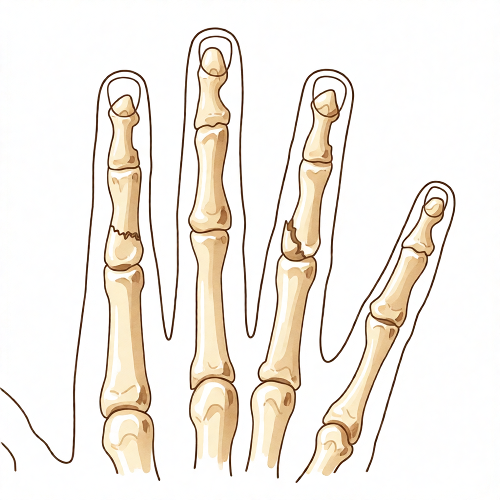
Fracture dislocations of the proximal interphalangeal joint are classified into three types based on the fracture geometry of the base of the middle phalanx: palmar lip fractures, dorsal lip fractures, and pilon fractures [50]. Fracture-dislocations of the proximal interphalangeal joint encompass a spectrum of injury severity, ranging from injuries that require little intervention to those that require advanced reconstructive surgery for optimal outcome [105].
Acromioclavicular Joint Dislocations
Rockwood type VI acromioclavicular joint dislocations are exceedingly rare, with only 24 cases reported to date [30].
Sternoclavicular Dislocations
The Allman clinical classification for sternoclavicular dislocation includes Stage 1 (contusion or minor distortion with a stable joint and no radiological signs of lesions), Stage 2 (subluxation due to partial rupture of the sternoclavicular ligaments with an intact costoclavicular ligament), and Stage 3 (dislocation with complete rupture of all ligaments and the articular disk) [53].
Tarsometatarsal Injuries
The Quénu and Küss classification for Lisfranc fracture–dislocations describes three types of injury patterns: homolateral, isolated, and divergent [162]. The Hardcastle classification for Lisfranc fracture–dislocations distinguishes three main groups: complete (Type A), partial (Type B), and divergent (Type C) dislocation patterns [162]. Type A (complete) Lisfranc fracture–dislocations are characterized by the involvement of all parts of the Lisfranc joint complex with dislocation within one plane [162]. Type B (partial) Lisfranc fracture–dislocations are identified by partial incongruity of the joint complex [162]. Type C (divergent) Lisfranc fracture–dislocations exhibit complete and partial injury patterns, with medialization of the first metatarsal in conjunction with lateral translation of a variable number of the lateral four metatarsal bones [162].
Clinical Presentation
Glenohumeral instability is defined as the inability of the humeral head to remain centered in the glenoid fossa, a condition distinct from laxity, which is a physical finding often unassociated with clinical instability [9]. Instability is classified as acute if seen within the first days after its onset, and otherwise it is chronic [9]. If a glenohumeral joint has been dislocated on many occasions, the instability is classified as recurrent [9]. Recurrent traumatic instability typically produces symptoms when the arm is placed in a position near that of the original injury [9]. Patients with a traumatic etiology for recurrent instability usually have unidirectional instability and often have obvious pathology such as a Bankart lesion [9]. In most situations, pain and limited shoulder range of motion lead patients with chronic glenohumeral dislocations to seek medical evaluation [59]. The injury of chronic glenohumeral dislocation can be overlooked by the treating physician unless a careful and systematic history, physical examination, and radiographic evaluation are performed [59].
Diagnosis of posterior dislocation of the shoulder is often overlooked due to infrequent occurrence and the failure of standard antero-posterior roentgenograms to demonstrate the posterior displacement [54]. The recurrent type of posterior shoulder dislocation can be diagnosed far more easily provided the dislocation can be witnessed by a medical observer either accidentally or through voluntary reproduction by the patient [54]. Posterior shoulder dislocations are rare in the pediatric population and require a high level of suspicion for diagnosis [48]. Bilateral posterior shoulder dislocations with reverse Hill-Sachs lesions are uncommon and prone to misdiagnosis [93]. Clinicians and radiologists agreed on the presence or absence of fracture or dislocation in 897 of 1011 cases (89 per cent) in an accident department setting [5]. Early intervention and low pain levels at presentation were the two most important factors that significantly favor successful reduction of a shoulder dislocation [27]. Females and children were significantly more likely to present with a basketball-related shoulder dislocation by sustaining player contact [89].
When clinical findings suggest a neurologic injury, electrodiagnostic studies and nerve conduction velocity studies should be obtained, with these studies being positive within 3 to 4 weeks following the injury [33]. It is important to document a neurologic deficit before surgical intervention so that the deficit cannot be construed as a result of the procedure performed [33]. Vascular compromise associated with shoulder dislocation is very uncommon, but when it is suspected, evaluation by a vascular specialist is important to determine whether additional angiographic imaging studies are indicated [33].
Traumatic dislocation of the metacarpophalangeal joint is a relatively uncommon injury that may be simple (easily reducible) or complex (requiring surgical intervention) [14]. Carpometacarpal joint dislocations and fracture dislocations are uncommon injuries that can be very disabling if not recognized early [26]. Clinical results for PIP joint dislocations and fracture-dislocations vary and are often difficult to predict due to the complexity of fracture patterns and potential for sub-acute or chronic presentation [24]. Fracture dislocations of the PIP joint may rapidly develop fixed deformity, leaving an athlete with a poor outlook for complete correction if there is persistent incongruity of the joint [95]. Accurate diagnosis and management of hand and carpal fractures and dislocations are predicated on a thorough physical examination and appropriate imaging to limit joint stiffness while preserving mobility and function [32].
Foot fractures and dislocations are common traumatic injuries associated with high rates of morbidity and dysfunction [52]. The most common complex fracture-dislocation was of the knee, followed by the wrist and the ankle [58]. Complete anterior dislocation of the sacro-iliac joint is a very unusual injury that can occur in skeletally mature individuals following severe trauma [28]. Rockwood type VI acromioclavicular joint dislocations remain exceedingly rare, with only 24 cases reported to date [30]. Superior dislocation of the sternoclavicular joint can be treated nonoperatively [21].
Investigations
Physical Examination: A careful physical examination is essential to direct care and future testing if indicated, as diagnostic tests such as imaging can be expensive, time consuming, and often nonspecific [44].
MRI: An increased glenoid index on MRI may help identify patients at risk for primary or recurrent anterior glenohumeral instability events and may therefore help with guiding treatment and prevention [148].
Other Considerations: Machine learning–aided diagnosis may help to decrease misdiagnosis of perilunate dislocations, particularly when subspecialist evaluation is delayed [167]. Although diagnosing the cause of ulnar collateral ligament locking may be complicated by the lack of evidence in imaging studies, open surgical treatment has traditionally been the most often used with a high success rate [165]. Missed posterior dislocation of the shoulder after intramedullary fixation of proximal humeral fractures can be missed due to inadequate initial and postoperative x-ray images and incorrect interpretation [168]. Posterior shoulder dislocations require a high level of suspicion for diagnosis, and serial radiographs may be beneficial to monitor for sequelae [48].
Treatment
Non-Operative
Conservative management remains a viable option for specific dislocation presentations. In pediatric patients, the absence of Hill-Sachs and Bankart lesions suggests that conservative treatment is a viable option for managing shoulder dislocation [129]. For acute, initial anterior shoulder dislocations, patients opting for nonoperative treatment undergo a supervised rehabilitation program involving a sling for 3 weeks, followed by a restrengthening program emphasizing internal rotation and adduction muscles [102]. Regarding immobilization position, the current best evidence does not support a relative effectiveness of immobilization in external rotation compared with internal rotation to avoid recurrent shoulder dislocations in patients with traumatic anterior shoulder dislocations [104]. Specifically, the position of external rotation immobilization of the arm in the nonoperative treatment of a younger patient with first-time shoulder dislocation does not offer any obvious clinical advantage over an arm placed in internal rotation in a traditional sling and, at this time, cannot be recommended [107].
For hand and wrist injuries, nonoperative strategies vary by joint and timing. A patient's outcome after a short period of splinting, followed by hand therapy, resulted in a fully functioning hand and long finger for dorsal dislocation of the distal interphalangeal joint and volar dislocation of the metacarpophalangeal joint [4]. When a trans-scaphoid perilunate dislocation is diagnosed late, an enduring functional result may be achieved by nonoperative treatment [15]. If the diagnosis of an ulnar CMC joint dislocation or fracture–dislocation is early accomplished and a concentric and stable reduction is initially achieved, nonoperative treatment may be a successful option, requiring close follow-up for the first week [35]. Similarly, although operative treatment is recommended in the literature, conservative treatment through immediate reduction and splint immobilization can be sufficient for acute, uncomplicated ulnar CMC dislocations [135]. Many acute injuries of the metacarpophalangeal joint can be managed nonsurgically with extension splints, while optimal management of subacute or chronic injuries remains undefined [116]. Management of habitual dislocation of the thumb metacarpophalangeal joint in children should be conservative, resisting surgical attempts to tighten the joint, and encouraging children to unlearn the habit [70].
For knee and elbow dislocations, nonoperative care is standard for many cases. Most simple elbow dislocations can be treated nonoperatively with closed manipulative reduction and early rehabilitation, resulting in good functional outcomes, though some patients may report residual stiffness, pain, or instability [122]. Nonoperative care for patellar dislocations begins initially with reduction of the joint and observation for vascular injury, nerve injury, or developing compartment syndrome [163]. Assuming that there are no concomitant limb-threatening injuries or open wounds, and the joint remains concentrically reduced, the knee is immobilized in full extension in a long leg cylindrical cast (or splint) for at least 3 weeks [163]. The period of immobilization for patellar dislocations may be extended as long as 6 weeks, depending on the extent of the original injury and the treating physician's impression of the degree of ligamentous laxity persisting at 3 weeks [163]. Immobilization for longer than 6 weeks is not recommended for patellar dislocations [163]. The goal of nonoperative care for patellar dislocations is the “titration” of a sufficient amount of stiffness so that the joint will remain reduced but will maintain a functional arc of motion [163].
Operative
Indications: Surgical intervention is indicated based on specific anatomical and clinical criteria. For acute irreducible shoulder dislocations, open or arthroscopic procedures are utilized, with recurrence being low and none reporting recurrent dislocation in limited follow-up [3]. Treatment selection for locked posterior dislocation of the shoulder depends on the duration of dislocation, size of the impression defect, and glenoid condition [63]. The modified McLaughlin procedure is a reliable option for the treatment of acute locked posterior shoulder dislocation with a head defect of 25–45% [40]. It is also a safe and effective treatment option for patients with locked posterior dislocation of the shoulder and a reverse Hill-Sachs lesion between 20% and 50% of the humeral head articular surface, demonstrating favorable clinical and radiographic outcomes with low complication and recurrent instability rates [114]. For knee dislocations, surgery is indicated in the acute setting for physically active patients and in the chronic setting for instability without significant arthrosis [103]. In hand and wrist injuries, timely treatment of complex fracture-dislocations ensures optimal outcome in range of motion and overall hand function [31]. In patients with delayed presentation of CMC fracture dislocations, ORIF is recommended [36]. While closed management and immobilization can be considered if the injury is recognized early, surgical reduction and stabilization are more commonly warranted for lesser digit carpometacarpal joint fracture-dislocations [127]. An algorithm is recommended to guide the acute management of anterior shoulder fracture dislocations [62].
Surgical Approach / Technique: Operative techniques vary by joint and pathology. For chronic glenohumeral dislocations, a wide variety of management options are available, including non-operative management, closed reduction, open reduction with tendon and bone transfers, allograft procedures, and prosthetic arthroplasty [59]. Suture-button stabilisation offers a less-invasive alternative to traditional stabilisation methods with favourable outcomes in both chronic instability and acute traumatic dislocation of the thumb carpometacarpal joint [8]. The surgical treatment using a suture button implant for delayed ulnar carpometacarpal fracture–dislocations allowed early motion from 2 weeks after surgery, which resulted in good motion of the ulnar CMC joints and increased hand grip [66]. In a patient with dorsal dislocation of the distal interphalangeal joint with fracture of the volar base, closed reduction had failed to reduce the dislocation and open reduction was necessary [61]. Results of dynamic distraction external fixation for unstable fracture-dislocations of the proximal interphalangeal joint are comparable with other techniques used in the management of unstable PIP joint fracture-dislocations [41]. Delayed treatment of unstable proximal interphalangeal joint fracture-dislocations with a dynamic external fixator is effective in restoring function to the PIPJ [101]. Occasionally, the application of an external fixator may be required to maintain reduction of a highly unstable knee joint [163].
Implant Selection: Current evidence suggests that operative rather than non-operative treatment of Rockwood grade III acromioclavicular dislocations results in better cosmetic and radiological results, similar functional outcomes and longer time off work [112].
Other Considerations: Surgeons and patients should try to avoid opioids if possible after nonoperatively treated fractures and dislocations [117]. In specific situations where clinical findings suggest a neurologic injury in shoulder dislocation, electrodiagnostic studies and nerve conduction velocity studies should be obtained [33]. Although the presence of a neurologic deficit likely will not alter the approach to management, it is important to document the deficit before surgical intervention so that the deficit cannot be construed as a result of the procedure performed [33]. When vascular compromise associated with shoulder dislocation is suspected, evaluation by a vascular specialist is important to determine whether additional angiographic imaging studies are indicated [33]. Despite relatively high surgical complication rates, operative management of non-thumb carpometacarpal joint fracture dislocations results in good-to-excellent functional outcomes [12]. Outcomes following surgical treatment of the volar fracture subluxation/dislocation of the proximal interphalangeal joint are poor, with most patients developing arthritis and some requiring salvage procedures [39]. A case of ulnar collateral ligament rupture of the metacarpophalangeal joint was initially treated conservatively, but instability was still present after 3 weeks [128]. Supervised self-reduction and physician-assisted techniques could be used as an effective first-line treatment for anterior shoulder dislocation, potentially reducing the need for analgesics and emergency room visits [111].
Complications
Shoulder Dislocation Complications
Instability and Recurrence: The risk of recurrent dislocation and instability is highest in patients aged 15 to 19 years [1]. A previous history of shoulder dislocation is a significant factor for recurrent instability requiring capsular reconstruction [7]. In the context of reverse shoulder arthroplasty, the strongest patient-related factors associated with dislocation are a history of postoperative subluxations and a primary diagnosis of fracture nonunion [29].
Arthropathy: Age at primary dislocation, recurrence, high-energy sports participation, and alcohol abuse are factors associated with the development of arthropathy after primary anterior shoulder dislocation [151]. Specifically, two-thirds of patients with first-time dislocations at the age of <25 years develop different stages of arthropathy within 25 years [20].
Surgical Complications: The choice of open reduction and stabilization technique for chronic anterior dislocation leads to a wide variation in reported outcomes, with a high number of complications such as resubluxation and early arthrosis [11].
Vascular Compromise: Vascular compromise associated with shoulder dislocation is very uncommon [33].
Hand and Wrist Dislocation Complications
Arthritis and Salvage: Outcomes following surgical treatment of volar fracture subluxation/dislocation of the proximal interphalangeal joint are poor, with most patients developing arthritis and some requiring salvage procedures [39].
Functional Outcomes: Operative management of carpometacarpal fracture dislocations results in good-to-excellent functional outcomes despite relatively high surgical complication rates [12]. In patients with perilunate dislocations and transscaphoid perilunate fracture–dislocations, the presence of radiological arthritis and static carpal instability did not cause reduced function at a minimum follow-up of 10 years [13]. The outcome of simultaneous dislocations of the five carpometacarpal joints remains uncertain, with hand and wrist functions maintained but often reduced grip strength [57].
Other Joint Dislocation Complications
Acromioclavicular Joint: Type I and II acromioclavicular joint disruptions impair long-term shoulder function in about half of patients 10 years after injury [68].
Pelvic Ring: Long-term outcomes for pelvic ring injuries are complicated by posterior pelvic pain and are largely multifactorial [65].
Recovery
Other Considerations: The evidence base for this section does not provide specific timelines for light activity, full activity, or complete recovery phases; therefore, these fields are omitted. Recovery outcomes and functional trajectories vary significantly by joint and injury mechanism.
Shoulder Dislocations
Recurrent instability is a primary concern in young patients. The group aged 15 to 19 years is at the highest risk of recurrent dislocation and instability following glenohumeral dislocation [1]. Almost half of all first-time dislocations at the age of <25 years will have stabilising surgery, and two-thirds will develop different stages of arthropathy within 25 years [20]. Recurrent instability requiring capsular reconstruction seems to be more prevalent in patients with a previous history of shoulder dislocation who present with luxatio erecta [7].
Surgical outcomes and complications vary by technique and pathology. The choice of open reduction and stabilization technique for chronic anterior shoulder dislocation was highly variable, leading to a wide variation in reported outcomes with a high number of complications such as resubluxation and early arthrosis [11]. In contrast, acute arthroscopic Bankart repair for first-time traumatic anterior glenohumeral dislocations resulted in excellent subjective function and return to athletics in young, active patients with an acceptable rate of recurrence and reoperation at long-term follow-up [141]. For acute locked posterior shoulder dislocation with a head defect of 25–45%, the modified McLaughlin procedure is a reliable option for functional outcome [40].
Non-operative management and specific pathologies present distinct recovery profiles. Non-operated patients with primary anterior shoulder dislocation and self-reported shoulder trouble three-six weeks after initial injury do not have less shoulder impairment (self-reportedly or objectively measured) than non-operated patients with recurrent anterior shoulder dislocation and self-reported shoulder trouble three-six weeks after their latest shoulder dislocation event [67]. In children with Erb’s obstetric brachial plexus palsy treated with low rotation humeral osteotomy, there is a significant long-term decrease in shoulder abduction despite maintenance of external rotation and improvement in elbow extension [142]. The strongest patient-related factors associated with dislocation after reverse shoulder arthroplasty were a history of postoperative subluxations and having a primary diagnosis of fracture nonunion [29].
Hand and Wrist Dislocations
Treatment strategies for hand and wrist dislocations prioritize functional restoration and stability. Closed reduction is the treatment of choice for simultaneous dislocations of the small finger without associated lesions [2]. For dorsal dislocation of the distal interphalangeal joint and volar dislocation of the metacarpophalangeal joint, a short period of splinting followed by hand therapy resulted in a fully functioning hand and long finger [4]. The suture-button stabilisation technique offers a less-invasive alternative to traditional stabilisation methods with favourable outcomes in both chronic instability and acute traumatic dislocation of the thumb carpometacarpal joint [8].
Fracture-dislocations of the proximal interphalangeal (PIP) joint are managed with specific pinning techniques. Extension-block pinning for unstable dorsal PIP joint fracture-dislocations is technically straightforward and cheap, producing excellent functional outcomes with minimal long-term disability [143]. The long-term outcomes of extension block pinning for PIP joint fracture-dislocations were satisfactory and similar to the mid-term follow-up results of the same patient cohort reported 11 years earlier [69]. The prognosis is far better after irreducible than after reducible volar dislocations of the PIP joint because the extensor mechanism is displaced, not disrupted [177]. Open dorsal metacarpophalangeal joint dislocations of the four long fingers are unusual and should be addressed intraoperatively with minimal delay [176].
Wrist and complex hand injuries require tailored interventions. A patient achieved excellent functional results with no major complications at 1 year follow-up following combined reverse perilunate and axial–ulnar dislocation of the wrist [71]. There was no objective or subjective evidence of instability at either of the two joints following simultaneous dislocations of the carpometacarpal and metacarpophalangeal joints of the thumb [74]. The approach for floating index metacarpal associated with multiple carpometacarpal fracture-dislocations was associated with an excellent short-term functional outcome [145]. In mallet fracture, fragment size, fragment displacement, and interval between injury and treatment were independently associated with both initial and late subluxation [175].
Other
Diagnostic accuracy and rare syndromic presentations influence recovery trajectories. Clinicians and radiologists agreed on the presence or absence of fracture or dislocation in 897 of 1011 cases (89 per cent) in an accident department [5]. In a patient with bilateral elbow dislocation and Rubinstein-Taybi syndrome, there has been no re-dislocation on the right elbow, five years postsurgery [16]. Long-term outcomes for pelvic fractures are dependent on the pelvic ring injury as well as the associated injuries, and are complicated by posterior pelvic pain but also largely multifactorial [65].
Key Evidence
- [L3] The group aged 15 to 19 years was at the highest risk of recurrent dislocation and instability. [1] (10.1016/j.jse.2017.09.006)
- [L4] Closed reduction is the treatment of choice for simultaneous dislocations without associated lesions. [2] (10.1016/j.jhsa.2012.10.043)
- [L4] When patients were treated with an open or arthroscopic procedure, recurrence was low, with none reporting recurrent dislocation in limited follow-up. [3] (10.1177/23259671221121633)
- [Case_report] The patient's outcome after a short period of splinting, followed by hand therapy, resulted in a fully functioning hand and long finger. [4] (10.1007/s11552-009-9218-3)
- [L4] Clinicians and radiologists agreed on the presence or absence of fracture or dislocation in 897 of 1011 cases (89 per cent). [5] (10.1016/s0020-1383(80)80048-9)
- [L1] The systematic review found that the available literature supports surgical intervention in patients under the age of 25 to reduce re-dislocation. [6] (10.1177/17585732241254693)
- [L4] Recurrent instability requiring capsular reconstruction seems to be more prevalent in patients with a previous history of shoulder dislocation. [7] (10.1016/j.jse.2009.07.062)
- [L4] The technique offers a less-invasive alternative to traditional stabilisation methods with favourable outcomes in both chronic instability and acute traumatic dislocation. [8] (10.1177/15589447261469607)
- [L4] The choice of open reduction and stabilization technique for chronic anterior dislocation was highly variable and led to a wide variation in reported outcomes with a high number of complications such as resubluxation and early arthrosis. [11] (10.1016/j.jse.2020.10.010)
- [L4] Despite relatively high surgical complication rates, operative management of CMC fracture dislocations results in good-to-excellent functional outcomes. [12] (10.1016/j.jhsg.2024.11.003)
- [L4] The presence of radiological arthritis and static carpal instability did not cause reduced function at our minimum follow-up of 10 years. [13] (10.1016/j.jhsa.2009.09.003)
- [L5] Traumatic dislocation of the metacarpophalangeal joint is a relatively uncommon injury that may be simple (easily reducible) or complex (requiring surgical intervention). [14] (10.5435/00124635-200905000-00006)
- [L4] The authors conclude that when this injury is diagnosed late, an enduring functional result may be achieved by nonoperative treatment. [15] (10.1016/j.jhsa.2007.05.003)
- [Case_report] There has been no re-dislocation on the right elbow, five years postsurgery, and we hope that our experience will help clinicians in the future when treating patients with similar conditions. [16] (10.1016/j.jseint.2023.03.021)
- [L4] In patients with locked posterior shoulder dislocation, the primary factor influencing clinical outcomes is the duration of the dislocation, rather than humeral head defect size or patient age. [17] (10.1186/s12891-025-08886-4)
- [L3] The most common treatment for a shoulder dislocation in paediatric patients is a closed shoulder reduction. [18] (10.3390/ijerph17082834)
- [L1] Surgical treatments are more effective than conservative options in preventing recurrent instability in adolescents and young adults under 40 years of age with first-time anterior shoulder dislocation. [19] (10.1016/j.arthro.2025.07.044)
- [L2] Almost half of all first-time dislocations at the age of <25 years will have stabilising surgery and two-thirds will develop different stages of arthropathy within 25 years. [20] (10.1007/s00167-015-3980-2)
- [L4] Superior dislocation of the SCJ can be treated nonoperatively. [21] (10.1016/j.jse.2007.02.126)
- [L5] Recent studies suggest an operative approach to the first-time anterior dislocation of the shoulder, especially if the patient is young, active, and involved in highly demanding sports. [22] (10.1007/s00167-009-0950-6)
- [L5] Clinical results for PIP joint dislocations and fracture-dislocations vary and are often difficult to predict due to the complexity of fracture patterns and potential for sub-acute or chronic presentation. [24] (10.1177/17531934231183259)
- [L5] Carpometacarpal joint dislocations and fracture dislocations are uncommon injuries that can be very disabling if not recognized early. [26] (10.5435/jaaos-d-25-00583)
- [L1] Early intervention and low pain levels at presentation were the two most important factors that significantly favor successful reduction of a shoulder dislocation. [27] (10.1016/j.jse.2012.01.004)
- [Case_report] Complete anterior dislocation of the sacro-iliac joint is a very unusual injury that can occur in skeletally mature individuals following severe trauma. [28] (10.2106/00004623-197658010-00028)
- [L3] The strongest patient-related factors associated with dislocation were a history of postoperative subluxations and having a primary diagnosis of fracture nonunion. [29] (10.1016/j.jse.2023.05.028)
- [L5] RW type VI AC dislocations remain exceedingly rare, with only 24 cases reported to date. [30] (10.1016/j.jisako.2025.101042)
- [L5] Timely treatment of complex fracture-dislocations ensures optimal outcome in range of motion and overall hand function. [31] (10.1016/j.csm.2019.10.006)
- [L5] [33] (10.5435/00124635-200807000-00004)
- [L4] If the diagnosis of an ulnar CMC joint dislocation or fracture–dislocation is early accomplished and a concentric and stable reduction is initially achieved, the nonoperative treatment may be a successful option to take into account but requiring a close follow-up for the first week. [35] (10.1055/s-0039-1688468)
- [L4] In patients with delayed presentation of CMC fracture dislocations, we recommend ORIF. [36] (10.1016/j.jhsa.2015.07.017)
- [L4] Surgery improves the clinical and radiological outcome in Rockwood type IV dislocations, whereas Rockwood type III dislocations benefit from conservative treatment. [38] (10.1007/s00167-020-06423-5)
- [L4] Outcomes following surgical treatment of the volar fracture subluxation/dislocation are poor, with most patients developing arthritis and some requiring salvage procedures. [39] (10.1016/j.jhsa.2017.03.030)
- [L4] With respect to functional outcome, modified McLaughlin procedure is a reliable option for the treatment of acute locked posterior shoulder dislocation with a head defect of 25–45%. [40] (10.1007/s00167-012-2217-x)
- [L4] Our results are comparable with other techniques used in the management of unstable PIP joint fracture-dislocations. [41] (10.1016/j.jhsa.2007.07.018)
- [Case_report] Posterior shoulder dislocations are rare in the pediatric population and require a high level of suspicion for diagnosis; positioning the shoulder in abduction and external rotation helps maintain reduction, and serial radiographs may be beneficial to monitor for sequelae. [48] (10.1016/j.xrrt.2020.12.003)
- [L5] [50] (10.1053/jssh.2002.33317)
- [Case_report] [53] (10.1007/s00167-004-0504-x)
- [L4] [54] (10.2106/00004623-195840020-00022)
- [L5] The outcome of these injuries remains uncertain, with hand and wrist functions maintained but often reduced grip strength. [57] (10.1016/s0020-1383(02)00098-0)
- [L3] The most common complex fracture-dislocation was of the knee, followed by the wrist and the ankle. [58] (10.1186/1471-2474-12-253)
- [L5] In this patient closed reduction had failed to reduce the dislocation and open reduction was necessary. [61] (10.1016/s0020-1383(97)00121-6)
- [L3] We recommend an algorithm to guide the acute management of anterior shoulder fracture dislocations. [62] (10.1016/j.jseint.2020.09.014)
- [L4] Treatment selection depends on the duration of dislocation, size of the impression defect, and glenoid condition. [63] (10.2106/jbjs.l.01588)
- [L4] This retrospective study suggests that there is no significant difference between the side of shoulder dislocation and hand dominance of patients who sustain shoulder dislocations requiring shoulder stabilization surgery. [64] (10.1016/j.jses.2018.04.001)
- [L4] The surgical treatment using a suture button implant allowed early motion from 2 weeks after surgery, which resulted in good motion of the ulnar CMC joints and increased hand grip. [66] (10.1055/s-0039-1693051)
- [L4] Non-operated patients with primary anterior shoulder dislocation and self-reported shoulder trouble three-six weeks after initial injury do not have less shoulder impairment (self-reportedly or objectively measured) than non-operated patients with recurrent anterior shoulder dislocation and self-reported shoulder trouble three-six weeks after their latest shoulder dislocation event. [67] (10.1186/s12891-019-2444-0)
- [L4] Type I and II acromioclavicular joint disruptions impair long-term shoulder function in about half of patients 10 years after injury. [68] (10.1177/0363546508319047)
- [L4] The long-term outcomes were satisfactory and similar to the mid-term follow-up results of the same patient cohort reported 11 years earlier. [69] (10.1177/17531934221102251)
- [L4] Management should be conservative, resisting surgical attempts to tighten the joint, and encouraging children to unlearn the habit. [70] (10.1177/1753193416687915)
- [Case_report] The patient achieved excellent functional results with no major complications at 1 year follow-up. [71] (10.1177/1753193408091348)
- [L5] There was no objective or subjective evidence of instability at either of the two joints. [74] (10.1016/0020-1383(93)90062-b)
- [L4] Females and children were significantly more likely to present with a dislocation by sustaining player contact. [89] (10.1016/j.jse.2024.07.040)
- [L4] Bilateral posterior shoulder dislocations with reverse Hill-Sachs lesions are uncommon and prone to misdiagnosis; early recognition and tailored treatment strategies are essential for satisfactory functional outcomes. [93] (10.1186/s12891-026-09537-y)
- [L5] Timely diagnosis is imperative, especially if there is any persistent incongruity of the joint, as fracture dislocations of the PIP joint may rapidly develop fixed deformity, leaving an athlete with a poor outlook for complete correction. [95] (10.1016/j.hcl.2012.05.036)
- [L4] Palmar dislocation of the thumb metacarpophalangeal joint is classified into three types: Type A (stable joint), Type B (tendon block), and Type C (joint instability). [97] (10.1177/1753193413499291)
- [Paper] Delayed treatment of unstable PIPJ fracture-dislocations with a DDEF is effective in restoring function to the PIPJ. [101] (10.1016/j.injury.2015.06.027)
- [L3] [102] (10.1177/03635465010290051101)
- [L2] The current best evidence does not support a relative effectiveness of immobilization in external rotation compared with internal rotation to avoid recurrent shoulder dislocations in patients with traumatic anterior shoulder dislocations. [104] (10.1016/j.jse.2013.07.037)
- [L5] Fracture-dislocations of the proximal interphalangeal joint encompass a spectrum of injury severity, ranging from injuries that require little intervention to those that require advanced reconstructive surgery for optimal outcome. [105] (10.5435/jaaos-21-02-88)
- [L1] [107] (10.2106/jbjs.9616.ebo209)
- [L1] These techniques could be used as an effective first-line treatment for anterior shoulder dislocation, potentially reducing the need for analgesics and emergency room visits. [111] (10.1186/s12891-024-07379-0)
- [L2] Current evidence suggests that operative rather than non-operative treatment of Rockwood grade III dislocations results in better cosmetic and radiological results, similar functional outcomes and longer time off work. [112] (10.1302/0301-620x.95b12.31802)
- [L4] The modified McLaughlin procedure is a safe and effective treatment option for patients with locked posterior dislocation of the shoulder and a reverse Hill-Sachs lesion between 20% and 50% of the humeral head articular surface, demonstrating favorable clinical and radiographic outcomes with low complication and recurrent instability rates. [114] (10.1016/j.xrrt.2023.08.007)
- [L5] Many acute injuries can be managed nonsurgically with extension splints, while optimal management of subacute or chronic injuries remains undefined. [116] (10.5435/jaaos-d-13-00203)
- [L4] Surgeons and patients should try to avoid opioids if possible after nonoperatively treated fractures and dislocations. [117] (10.2106/jbjs.17.01475)
- [L5] While closed management and immobilization can be considered if the injury is recognized early, surgical reduction and stabilization are more commonly warranted. [127] (10.1016/j.jhsa.2026.02.016)
- [L4] The case reported here was initially treated conservatively, but instability was still present after 3 weeks. [128] (10.1054/jhsb.1999.0334)
- [Case_report] The absence of Hill-Sachs and Bankart lesions in this pediatric patient suggests that conservative treatment is a viable option for managing shoulder dislocation in children. [129] (10.1016/j.xrrt.2024.01.012)
- [L4] Although operative treatment is recommended in the literature, these cases show that conservative treatment through immediate reduction and splint immobilization can be sufficient for acute, uncomplicated ulnar CMC dislocations. [135] (10.1007/s11552-011-9347-3)
- [L4] At long-term follow-up, acute arthroscopic Bankart repair for first-time traumatic anterior glenohumeral dislocations resulted in excellent subjective function and return to athletics in young, active patients with an acceptable rate of recurrence and reoperation. [141] (10.1177/0363546508328416)
- [L4] The study reports a significant long-term decrease in shoulder abduction despite maintenance of external rotation and improvement in elbow extension. [142] (10.1177/1753193409104552)
- [L4] It is technically straightforward and cheap, and produces excellent functional outcomes with minimal long-term disability. [143] (10.1177/15589447211066352)
- [Case_report] This approach was associated with an excellent short-term functional outcome. [145] (10.1007/s12593-015-0191-5)
- [L3] This useful MRI measurement may help identify patients at risk for primary or recurrent anterior glenohumeral instability events and may therefore help with guiding treatment and prevention. [148] (10.1177/2325967120986139)
- [L2] Age at primary dislocation, recurrence, high-energy sports, and alcohol abuse were factors associated with the development of arthropathy. [151] (10.1016/j.jse.2008.11.004)
- [L4] Although diagnosing the cause of UCL locking may be complicated by the lack of evidence in imaging studies, open surgical treatment has traditionally been the most often used with a high success rate. [165] (10.1016/j.jhsg.2022.08.003)
- [Paper] This may help to decrease misdiagnosis of perilunate dislocations, particularly when subspecialist evaluation is delayed. [167] (10.1177/15589447241308603)
- [L4] Missed posterior dislocation of the shoulder after intramedullary fixation of proximal humeral fractures is an extremely rare injury that can be missed due to inadequate initial and postoperative x-ray images and incorrect interpretation. [168] (10.1016/j.jse.2008.10.020)
- [L4] Fragment size, fragment displacement, and interval between injury and treatment were independently associated with both initial and late subluxation. [175] (10.1177/1753193416669929)
- [Case_report] Open dorsal metacarpophalangeal joint dislocations of the four long fingers are unusual and should be addressed intraoperatively with minimal delay. [176] (10.1007/s11552-014-9646-6)
- [L4] The prognosis is far better after irreducible than after reducible volar dislocations because the extensor mechanism is displaced, not disrupted. [177] (10.2106/00004623-197860010-00023)
See Also
References
[1] Epidemiology of glenohumeral dislocation and subsequent instability in an urban population. Journal of Shoulder and Elbow Surgery. 2018. DOI: 10.1016/j.jse.2017.09.006
[2] Simultaneou Triple Dislocation of the Small Finger. The Journal of Hand Surgery. 2013. DOI: 10.1016/j.jhsa.2012.10.043
[3] A Systematic Review of Acute Irreducible Shoulder Dislocations in the 21st Century. Orthopaedic Journal of Sports Medicine. 2022. DOI: 10.1177/23259671221121633
[4] Dorsal Dislocation of the Distal Interphalangeal Joint and Volar Dislocation of the Metacarpophalangeal Joint in the Same Finger: A Case Report. HAND. 2009. DOI: 10.1007/s11552-009-9218-3
[5] Correlation between clinical and radiological diagnosis for fractures and dislocations in an accident department. Injury. 1980. DOI: 10.1016/s0020-1383(80)80048-9
[6] A systematic review and meta-analysis of operative versus non-operative management for first time traumatic anterior shoulder dislocation in young adults. Shoulder & Elbow. 2024. DOI: 10.1177/17585732241254693
[7] Results of treatment of luxatio erecta (inferior shoulder dislocation). Journal of Shoulder and Elbow Surgery. 2010. DOI: 10.1016/j.jse.2009.07.062
[8] Suture-Button Stabilisation for Thumb Carpometacarpal Joint Instability and Dislocation: A Retrospective Case Series. HAND. 2026. DOI: 10.1177/15589447261469607
[9] Fractures, Dislocations, and Acquired Problems of the Shoulder in Children. 2016.
[11] Results and complications of head-preserving techniques in chronic neglected shoulder dislocation: a systematic review. Journal of Shoulder and Elbow Surgery. 2021. DOI: 10.1016/j.jse.2020.10.010
[12] Operative Treatment of Non-Thumb Carpometacarpal Joint Fracture Dislocations. Journal of Hand Surgery Global Online. 2025. DOI: 10.1016/j.jhsg.2024.11.003
[13] Perilunate Dislocations and Transscaphoid Perilunate Fracture–Dislocations: A Retrospective Study With Minimum Ten-Year Follow-Up. The Journal of Hand Surgery. 2010. DOI: 10.1016/j.jhsa.2009.09.003
[14] Metacarpophalangeal Joint Dislocation. Journal of the American Academy of Orthopaedic Surgeons. 2009. DOI: 10.5435/00124635-200905000-00006
[15] Long-Term Follow-Up of an Undiagnosed Trans-Scaphoid Perilunate Dislocation Demonstrating Articular Remodeling and Functional Adaptation. The Journal of Hand Surgery. 2007. DOI: 10.1016/j.jhsa.2007.05.003
[16] A case of bilateral elbow dislocation in a patient with Rubinstein-Taybi syndrome. JSES International. 2023. DOI: 10.1016/j.jseint.2023.03.021
[17] The duration of dislocation is the most important prognostic factor in chronic locked posterior shoulder dislocations treated with the modified McLaughlin surgical procedure: a multicentre study. BMC Musculoskeletal Disorders. 2025. DOI: 10.1186/s12891-025-08886-4
[18] Epidemiology of Paediatric Shoulder Dislocation: A Nationwide Study in Italy from 2001 to 2014. International Journal of Environmental Research and Public Health. 2020. DOI: 10.3390/ijerph17082834
[19] Surgical Treatment Is Superior to Conservative Options in Preventing Recurrence of First-Time Anterior Shoulder Dislocation in Adolescents and Adults Under 40 Years of Age: A Systematic Review and Network Meta-analysis. Arthroscopy: The Journal of Arthroscopic & Related Surgery. 2025. DOI: 10.1016/j.arthro.2025.07.044
[20] Primary anterior dislocation of the shoulder: long-term prognosis at the age of 40 years or younger. Knee Surgery, Sports Traumatology, Arthroscopy. 2016. DOI: 10.1007/s00167-015-3980-2
[21] Superior dislocation of the sternoclavicular joint. Journal of Shoulder and Elbow Surgery. 2008. DOI: 10.1016/j.jse.2007.02.126
[22] Primary anterior shoulder dislocation in young athletes: fix them!. Knee Surgery, Sports Traumatology, Arthroscopy. 2009. DOI: 10.1007/s00167-009-0950-6
[24] Proximal interphalangeal joint dislocations and fracture-dislocations. Journal of Hand Surgery (European Volume). 2023. DOI: 10.1177/17531934231183259
[26] Carpometacarpal Joint Dislocations and Fracture Dislocations of the Index Through Small Digits. Journal of the American Academy of Orthopaedic Surgeons. 2025. DOI: 10.5435/jaaos-d-25-00583
[27] Milch versus Stimson technique for nonsedated reduction of anterior shoulder dislocation: a prospective randomized trial and analysis of factors affecting success. Journal of Shoulder and Elbow Surgery. 2012. DOI: 10.1016/j.jse.2012.01.004
[28] Complete anterior dislocation of the sacro-iliac joint. A case report. The Journal of Bone & Joint Surgery. 1976. DOI: 10.2106/00004623-197658010-00028
[29] Predictors of dislocations after reverse shoulder arthroplasty: a study by the ASES complications of RSA multicenter research group. Journal of Shoulder and Elbow Surgery. 2024. DOI: 10.1016/j.jse.2023.05.028
[30] Rockwood type VI acromioclavicular joint dislocations: A systematic review. Journal of ISAKOS. 2026. DOI: 10.1016/j.jisako.2025.101042
[31] Management of Finger Joint Dislocation and Fracture-Dislocations in Athletes. Clinics in Sports Medicine. 2020. DOI: 10.1016/j.csm.2019.10.006
[32] Chapter 29 Hand/Carpal Fractures and Dislocations. 2021.
[33] Chronic Glenohumeral Dislocation. Journal of the American Academy of Orthopaedic Surgeons. 2008. DOI: 10.5435/00124635-200807000-00004
[35] Nonoperative Treatment of Ulnar Carpometacarpal Fracture–Dislocations. Journal of Wrist Surgery. 2019. DOI: 10.1055/s-0039-1688468
[36] The Effect of Timing on the Treatment and Outcome of Combined Fourth and Fifth Carpometacarpal Fracture Dislocations. The Journal of Hand Surgery. 2015. DOI: 10.1016/j.jhsa.2015.07.017
[37] Elbow Dislocations and Complex Instability. 2021.
[38] Surgery improves the clinical and radiological outcome in Rockwood type IV dislocations, whereas Rockwood type III dislocations benefit from conservative treatment. Knee Surgery, Sports Traumatology, Arthroscopy. 2021. DOI: 10.1007/s00167-020-06423-5
[39] The Central Slip Fracture: Results of Operative Treatment of Volar Fracture Subluxations/Dislocations of the Proximal Interphalangeal Joint. The Journal of Hand Surgery. 2017. DOI: 10.1016/j.jhsa.2017.03.030
[40] Excellent results of lesser tuberosity transfer in acute locked posterior shoulder dislocation. Knee Surgery, Sports Traumatology, Arthroscopy. 2012. DOI: 10.1007/s00167-012-2217-x
[41] Use of Dynamic Distraction External Fixation for Unstable Fracture-Dislocations of the Proximal Interphalangeal Joint. The Journal of Hand Surgery. 2008. DOI: 10.1016/j.jhsa.2007.07.018
[42] Hip Dislocations and Femoral Head Fractures. 2019.
[43] Exam Of The Hand Wrist 2Ed. INTRODUCTION.
[44] Orthopaedic Knowledge Update 13 Ebook Without Multimedia. Anatomy, Evaluation, Clinical Examination, and Imaging > Evaluation and Clinical Examination: Current Concepts.
[46] A Lange Medical Book Current Diagnosis Treatment In Orthopedics Fifth Edition. 9Hand Surgery > Image DISORDERS OF THE MUSCULATURE OF THE HAND.
[47] Exam Of The Hand Wrist 2Ed. 1.1 SKELETON OF THE HAND > The osseous skeleton.
[48] Case report: pediatric posterior shoulder dislocation. JSES Reviews, Reports, and Techniques. 2021. DOI: 10.1016/j.xrrt.2020.12.003
[50] Fracture dislocations of the proximal interphalangeal joint. Journal of the American Society for Surgery of the Hand. 2002. DOI: 10.1053/jssh.2002.33317
[52] Chapter 46 Foot Fractures and Dislocations. 2021.
[53] Anterior subluxation after reduction of a posterior traumatic sterno‐clavicular dislocation: a case report and a review of the literature. Knee Surgery, Sports Traumatology, Arthroscopy. 2004. DOI: 10.1007/s00167-004-0504-x
[54] Bilateral Recurrent Posterior Dislocation of the Shoulder. The Journal of Bone & Joint Surgery. 1958. DOI: 10.2106/00004623-195840020-00022
[57] Simultaneous dislocations of the five carpometacarpal joints. Injury. 2002. DOI: 10.1016/s0020-1383(02)00098-0
[58] Epidemiological survey of orthopedic joint dislocations based on nationwide insurance data in Taiwan, 2000-2005. BMC Musculoskeletal Disorders. 2011. DOI: 10.1186/1471-2474-12-253
[59] 5. Chronic Glenohumeral Dislocations and Impression Fractures: Evaluation, Imaging, Treatment, and Outcomes. 2003.
[61] Dorsal dislocation of the distal interphalangeal joint with fracture of the volar base: an unusual type. Injury. 1997. DOI: 10.1016/s0020-1383(97)00121-6
[62] Should closed reduction be attempted in the emergency department for an acute anterior shoulder fracture dislocation?. JSES International. 2021. DOI: 10.1016/j.jseint.2020.09.014
[63] Anatomic Shoulder Arthroplasty as Treatment for Locked Posterior Dislocation of the Shoulder. Journal of Bone and Joint Surgery. 2014. DOI: 10.2106/jbjs.l.01588
[64] Hand dominance in traumatic shoulder dislocations. JSES Open Access. 2018. DOI: 10.1016/j.jses.2018.04.001
[65] Chapter 32 Pelvic Fractures: Definitive Treatment and Outcomes. 2021.
[66] Surgical Treatment for Delayed Ulnar Carpometacarpal Fracture–Dislocations. Journal of Wrist Surgery. 2019. DOI: 10.1055/s-0039-1693051
[67] Patients with non-operated traumatic primary or recurrent anterior shoulder dislocation have equally poor self-reported and measured shoulder function: a cross-sectional study. BMC Musculoskeletal Disorders. 2019. DOI: 10.1186/s12891-019-2444-0
[68] Long-Term Shoulder Function after Type I and II Acromioclavicular Joint Disruption. The American Journal of Sports Medicine. 2008. DOI: 10.1177/0363546508319047
[69] Long-term outcomes after extension block pinning for fracture-dislocations of the proximal interphalangeal joint. Journal of Hand Surgery (European Volume). 2022. DOI: 10.1177/17531934221102251
[70] Habitual dislocation of the thumb metacarpophalangeal joint in children. Journal of Hand Surgery (European Volume). 2017. DOI: 10.1177/1753193416687915
[71] Combined Reverse Perilunate and Axial–Ulnar Dislocation of the Wrist: A Case Report. Journal of Hand Surgery (European Volume). 2008. DOI: 10.1177/1753193408091348
[72] Chapter 98 Hip Dislocations and Femoral Head Fractures. 2019.
[74] Simultaneous dislocations of the carpometacarpal and metacarpophalangeal joints of the thumb. Injury. 1993. DOI: 10.1016/0020-1383(93)90062-b
[78] Exam Of The Hand Wrist 2Ed. The arches of the hand > The metacarpal arch.
[82] A Lange Medical Book Current Diagnosis Treatment In Orthopedics Fifth Edition. 9Hand Surgery > FLEXOR TENDON INJURY.
[89] Sex and age-specific analysis of basketball-related shoulder dislocations in the United States: a national injury data review. Journal of Shoulder and Elbow Surgery. 2025. DOI: 10.1016/j.jse.2024.07.040
[93] Bilateral posterior shoulder dislocations with reverse Hill-Sachs lesions: a report of two cases and a literature review. BMC Musculoskeletal Disorders. 2026. DOI: 10.1186/s12891-026-09537-y
[95] Proximal Interphalangeal Joint Fracture Dislocations. Hand Clinics. 2012. DOI: 10.1016/j.hcl.2012.05.036
[97] Palmar dislocation of the thumb metacarpophalangeal joint: report of four cases and a review of the literature. Journal of Hand Surgery (European Volume). 2013. DOI: 10.1177/1753193413499291
[101] Delayed treatment of unstable proximal interphalangeal joint fracture-dislocations with a dynamic external fixator. Injury. 2015. DOI: 10.1016/j.injury.2015.06.027
[102] Prospective Evaluation of Arthroscopic Stabilization of Acute, Initial Anterior Shoulder Dislocations in Young Athletes. The American Journal of Sports Medicine. 2001. DOI: 10.1177/03635465010290051101
[103] Chapter 101 Knee Dislocations and Patellar Fractures. 2019.
[104] Immobilization in internal or external rotation does not change recurrence rates after traumatic anterior shoulder dislocation. Journal of Shoulder and Elbow Surgery. 2014. DOI: 10.1016/j.jse.2013.07.037
[105] Fracture-dislocations of the Proximal Interphalangeal Joint. Journal of the American Academy of Orthopaedic Surgeons. 2013. DOI: 10.5435/jaaos-21-02-88
[107] Immobilization with an External Rotation Brace Was Similar to an Internal Rotation Sling for Shoulder Dislocation. Journal of Bone and Joint Surgery. 2014. DOI: 10.2106/jbjs.9616.ebo209
[111] Efficacy of supervised self-reduction vs. physician-assisted techniques for anterior shoulder dislocations: a systematic review and meta-analysis. BMC Musculoskeletal Disorders. 2024. DOI: 10.1186/s12891-024-07379-0
[112] Controversies relating to the management of acromioclavicular joint dislocations. The Bone & Joint Journal. 2013. DOI: 10.1302/0301-620x.95b12.31802
[114] Clinical and radiographic outcomes of the modified McLaughlin procedure for locked posterior dislocation of the shoulder: a systematic review. JSES Reviews, Reports, and Techniques. 2024. DOI: 10.1016/j.xrrt.2023.08.007
[116] Closed Sagittal Band Injury of the Metacarpophalangeal Joint. Journal of the American Academy of Orthopaedic Surgeons. 2015. DOI: 10.5435/jaaos-d-13-00203
[117] A Prospective Evaluation of Patient-Reported Opioid Utilization After Nonoperative Treatment of Fractures and Dislocations. Journal of Bone and Joint Surgery. 2018. DOI: 10.2106/jbjs.17.01475
[122] Elbow Dislocations and Terrible Triad Injuries. 2019.
[127] Current Concepts: Lesser Digit Carpometacarpal Joint Fracture-Dislocations. The Journal of Hand Surgery. 2026. DOI: 10.1016/j.jhsa.2026.02.016
[128] Rupture of the Ulnar Collateral Ligament of the Metacarpophalangeal Joint of the Index Finger. Journal of Hand Surgery. 2000. DOI: 10.1054/jhsb.1999.0334
[129] Anterior shoulder dislocation and clavicle fracture in a 6-year-old boy: a case report and literature review. JSES Reviews, Reports, and Techniques. 2024. DOI: 10.1016/j.xrrt.2024.01.012
[135] Acute Ulnar Carpometacarpal Dislocations. Can it be Treated Conservatively? A Review of Four Cases. HAND. 2011. DOI: 10.1007/s11552-011-9347-3
[141] Long-term Follow-up of Acute Arthroscopic Bankart Repair for Initial Anterior Shoulder Dislocations in Young Athletes. The American Journal of Sports Medicine. 2009. DOI: 10.1177/0363546508328416
[142] Long-Term Results of Low Rotation Humeral Osteotomy in Children with Erb’s Obstetric Brachial Plexus Palsy. Journal of Hand Surgery (European Volume). 2009. DOI: 10.1177/1753193409104552
[143] Extension-Block Pinning for Unstable Dorsal Proximal Interphalangeal Joint Fracture-Dislocations: A Simple, Percutaneous Technique With Reproducible Outcomes. HAND. 2022. DOI: 10.1177/15589447211066352
[145] Floating Index Metacarpal Associated with Multiple Carpometacarpal Fracture-Dislocations: a Case Report. Journal of Hand and Microsurgery. 2015. DOI: 10.1007/s12593-015-0191-5
[148] Increased Glenoid Index as a Risk Factor for Pediatric and Adolescent Anterior Glenohumeral Dislocation: An MRI-Based, Case-Control Study. Orthopaedic Journal of Sports Medicine. 2021. DOI: 10.1177/2325967120986139
[151] Neer Award 2008: Arthropathy after primary anterior shoulder dislocation–223 shoulders prospectively followed up for twenty-five years. Journal of Shoulder and Elbow Surgery. 2009. DOI: 10.1016/j.jse.2008.11.004
[162] Fractures and Dislocations of the Midfoot and Forefoot. 2019.
[163] Patellar Fractures and Dislocations and Extensor Mechanism Injuries. 2019.
[165] Middle Finger Metacarpophalangeal Joint Locked in Flexion Caused by Entrapped Ulnar Collateral Ligament. Journal of Hand Surgery Global Online. 2022. DOI: 10.1016/j.jhsg.2022.08.003
[167] Machine Learning–Aided Diagnosis Enhances Human Detection of Perilunate Dislocations. HAND. 2025. DOI: 10.1177/15589447241308603
[168] Missed posterior dislocation of the shoulder after intramedullary fixation of humeral fractures: A report of three cases. Journal of Shoulder and Elbow Surgery. 2009. DOI: 10.1016/j.jse.2008.10.020
[175] Factors associated with subluxation in mallet fracture. Journal of Hand Surgery (European Volume). 2016. DOI: 10.1177/1753193416669929
[176] Open Metacarpophalangeal Dislocations: Literature Review and Case Report. HAND. 2014. DOI: 10.1007/s11552-014-9646-6
[177] Irreducible volar dislocation of the proximal interphalangeal joint of a finger caused by interposition of the intact central slip. The Journal of Bone & Joint Surgery. 1978. DOI: 10.2106/00004623-197860010-00023