Clinicians › Shoulder
Diagnosis, Outcomes & Rehabilitation
Clinical assessment of shoulder pathology, patient-reported outcome measures (PROs), and evidence-based rehabilitation protocols for optimizing functional recovery.
Overview
Rehabilitation following traumatic anterior shoulder dislocation lacks consensus regarding the definition of accelerated protocols or the selection of outcome measures [1]. A comprehensive evaluation approach prior to and throughout treatment assists clinicians in selecting appropriate interventions based on patient need [7]. Combining pathoanatomic causes with movement impairments provides a more focused rehabilitation strategy for the painful shoulder [7]. For the overhead throwing athlete, effective treatment relies on a thorough clinical examination and appropriate differential diagnosis to develop a program focusing on restoring adaptations, controlling inflammation, and improving neuromuscular control [14]. An objective criteria-based shoulder diagnosis and rehabilitation model may minimize the time required to regain functional capacity and recover from symptoms among patients with work-related shoulder injuries [12].
Prognostic factors for response to physiotherapy in musculoskeletal shoulder pain include baseline pain severity, disability, and psychological factors, though evidence quality is generally low [3]. Short-term repeat assessment of pain predicts long-term disability improvement better than short-term change or baseline scores across all cohorts [4]. No appropriately designed prognostic studies exist to identify predictors of success with physiotherapy in patients with massive irreparable rotator cuff tears [53].
Rehabilitation outcomes vary by condition. Improvement in outcomes for rotator cuff tears was observed up to 16 sessions of physical therapy, after which outcomes plateaued [2]. Delayed passive rehabilitation does not bring about superior outcomes compared to early rehabilitation after arthroscopic rotator cuff repair [5]. The majority of functional and symptomatic improvements following rotator cuff repair occur within the first year, with minimal clinically meaningful gains observed between 1 and 2 years [6]. Patients undergoing rehabilitation using a home-based protocol showed largely similar functional scores and healing to those with supervised physical therapy after arthroscopic rotator cuff repair of massive rotator cuff tears at the latest follow-up [8]. Functional outcomes after arthroscopic rotator cuff repair improved during midterm follow-up, regardless of retear [15]. Aspects of treatment that maximize the functional outcome are important in achieving patient satisfaction after rotator cuff repair [18]. Comprehensive rehabilitation, compared to conventional physiotherapy, has shown a statistically and clinically significant difference in improving pain, range of motion, functional disability, quality of life, and treatment effectiveness in patients after arthroscopic rotator cuff tendon repair [25]. The data available in the referenced systematic review are insufficient to make reliable clinical implications or establish a gold standard rehabilitation protocol for rotator cuff impingement [31]. Compared with surgery, physical therapy is associated with less improvement in perceived functional outcomes and a higher clinical failure rate for irreparable massive rotator cuff tears [10]. Only one study showed moderate evidence of early physical therapy promoting a more rapid return of short-term improvement in function and pain following total shoulder arthroplasty [13]. The lack of strong evidence warrants the need for future controlled studies regarding accelerated rehabilitation following reverse shoulder arthroplasty, and postoperative rehabilitation should be individualized [43]. Rehabilitation guidelines developed to manage patients who have undergone reverse total shoulder replacement aim to achieve optimal pain relief and maximize functional outcomes while mitigating risks associated with the surgery [45]. Use of the patient-centred ULTRA guideline can yield statistically significant improvements in outcome and range of motion for patients undergoing primary reverse total shoulder arthroplasty, which are maintained at the two-year post-surgery time point [107]. At more than 2 years after surgery, earlier repairs (less than 6 months) are associated with better functional outcomes for type II SLAP repair [9]. Rehabilitation is a valuable intervention for patients with atraumatic posterior shoulder subluxation [11]. Although worse functional results were found in the group with surgical indication for extra-articular scapula fractures treated nonoperatively, this difference disappears when adjusting for age as a confounding factor [41].
Anatomy & Pathophysiology
Bony Anatomy
The proximal humerus comprises four main parts: the humeral head, greater tuberosity, lesser tuberosity, and humeral shaft [72]. The articular head is spherical with a diameter of 37 to 57 mm [72] and an arc of approximately 160 degrees covered by articular cartilage [86]. The radius of curvature of the humeral head is approximately 25 mm, slightly larger in men than in women [86], with a range of 23 to 28 mm [86]. The most superior portion of the articular surface averages 8 mm above the greater tuberosity [72], or 8 to 10 mm superior to the top of the greater tuberosity [86]. Head height is approximately 5.6 cm above the superior border of the pectoralis major tendon [89].
Humeral version averages 29.8 degrees, with a range of 10 to 55 degrees [72]. Proximal humeral retroversion is highly variable, ranging from 0 to 55 degrees depending on the measurement method [86]. Mean humeral retroversion is around 26 degrees in healthy adults [80], while the humeral head is retroverted 30 degrees relative to the transepicondylar axis [89] and averages 19 degrees of retroversion [75]. The humeral head is inclined approximately 130 degrees with respect to the humeral shaft [72], with an inclination range of 30 to 55 degrees [86] and an average of 41 degrees [75]. The average neck-shaft angle is 45 degrees, with a range of 30 to 50 degrees [86], and measures an average of 135 degrees [73]. Arthritic shoulders exhibit a flatter neck-shaft angle close to 50 degrees [86].
The anatomic neck is located at the junction of the articular surface and the tuberosities [72], directly below the humeral head, and serves as an attachment for the shoulder capsule [89]. Fractures involving the anatomic neck are prognostically worse than those involving other regions regarding potential disruption of vascular supply and avascular necrosis [72]. The surgical neck represents an indistinct region below the tuberosities but above the humeral shaft [72]. It is more distal than the anatomic neck and is more often involved in fractures [89]. The bicipital groove lies between the greater and lesser tuberosities and serves as a pathway for the long head of the biceps [72]. The transverse humeral ligament is an important stabilizer of the biceps tendon [89].
The greater tuberosity serves as the attachment site for the supraspinatus, infraspinatus, and teres minor tendons [72, 80]. The lesser tuberosity serves as the attachment site for the subscapularis tendon [72, 80]. The humeral shaft extends from the level of the insertion of the pectoralis major muscle proximally to the supracondylar ridge distally [73]. The upper portion of the humeral shaft is cylindrical and becomes more flattened in an anteroposterior direction as it proceeds distally [73]. Medial and lateral intermuscular septae divide the arm into anterior and posterior compartments [73]. The anterior compartment contains the biceps brachii, coracobrachialis, and brachialis muscles, along with the neurovascular bundle [73]. The posterior compartment contains the triceps brachii muscle and the radial nerve [73].
The glenoid is a convex structure of shallow depth shaped like an inverted pear [72]. It is a shallow socket, approximately one third the size of the humeral head [73]. The subchondral bone of the glenoid is relatively flat, with articular concavity augmented by cartilage and a circumferential labrum [75]. The glenoid articular surface radius of curvature is 2 to 3 mm larger than that of the humeral head [86], with a range of 22 to 28 mm [86]. The glenoid diameter ranges from 18 to 30 mm superior anteroposteriorly and 21 to 35 mm inferior anteroposteriorly [86]. The superoinferior height of the glenoid ranges from 30 to 48 mm [86]. The glenoid inclination averages 4.2 degrees, with a range of -7 to 20 degrees [86]. The glenoid version averages 1.5 degrees of retroversion, with a range of 10.5 degrees anteversion to 9.5 degrees retroversion [86]. The glenoid averages 5 degrees of retroversion in relation to the axis of the scapular body [75, 89]. The normal position of the glenoid surface in relation to the axis of the scapular body ranges from 2 degrees of anteversion to 7 degrees of retroversion [86]. The glenoid surface area ranges from 4 to 6 mm [86], and the glenoid cartilage thickness is 2.16 mm [86]. The humeral head surface area ranges from 11 to 19 mm [86], and the humeral head cartilage thickness is 1.44 mm [86].
The scapula is attached to the axial skeleton by the acromioclavicular and sternoclavicular joints [74]. It is separated from the chest wall by thin gliding fibro-fatty tissue, allowing smooth excursion over the chest wall [74]. The scapula spans the second through seventh ribs and serves as an attachment for 17 muscles [89]. It is anteverted on the chest wall approximately 30 degrees relative to the body [89]. The basic part of the scapula is the body, which is triangular when viewed anteroposteriorly with its base situated superiorly and its apex inferiorly [74]. The glenoid is connected with the flat body of the scapula by the scapular neck [74]. The coracoid process curves forwards from the superior surface of the scapular neck [74]. The scapular spine ends in a flattened bony process, the acromion, which curves forwards [74].
The highest concentration of bony mass in the scapula is found in the glenoid, the scapular neck, and the lateral border of the scapular body [74]. Two bony pillars transmit compressive forces from the glenoid fossa: the lateral pillar and the spinal pillar [74]. The lateral pillar connects the inferior border of the glenoid with the inferior angle [74]. The spinal pillar arises from the central part of the glenoid and continues medially to become part of the base of the scapular spine [74]. The weakest bone in the scapula is located primarily in the central part of the biomechanical body, specifically in the infraspinous fossa [74]. The weakest area of the circumference of the biomechanical body of the scapula is the spinomedial angle [74].
The clavicle is the first bone to ossify at the fifth week of gestation and is the only long bone to ossify by intramembranous ossification [75, 89]. The medial epiphysis of the clavicle is the last ossification center to fuse, occurring at age 20 to 25 years [75, 89]. The primary blood supply to the clavicle is periosteal, with no nutrient artery present [75]. Fracture of the clavicle is the most common musculoskeletal birth injury [89].
Ossification of the scapular body begins at the eighth week of gestation [75]. The acromion has three ossification centers: the metacromion, mesoacromion, and preacromion [75]. Failure of fusion of the acromial ossification centers results in os acromiale [75]. Os acromiale is incomplete fusion of secondary ossification centers, most commonly between the mesoacromion and meta-acromion [89]. The coracobrachialis muscle and the short head of the biceps tendon originate from the coracoid process [75]. The pectoralis minor muscle inserts onto the medial coracoid process [75].
The proximal humerus has three centers of ossification: the humeral head, greater tuberosity, and lesser tuberosity [75]. The humeral head ossification center is usually present at birth [80]. The greater tuberosity ossification center appears by 1 to 3 years of age [80]. The lesser tuberosity ossification center appears by 5 years of age [80]. Proximal humeral ossification centers fuse by 5 to 7 years of age to form the humeral head [80]. The proximal humeral physis closes by 14 to 17 years of age in girls and by 16 to 18 years in boys [80]. Humeral retroversion averages 65 degrees in infants and young children and gradually decreases, approaching adult values by 11 years of age [80]. Eighty percent of subsequent humeral growth comes from the proximal humeral physis [80]. The proximal humeral physis accounts for approximately 40% of the growth of the entire upper extremity [80]. Less than 75% of humeral growth occurs before 2 years of age [80]. More than 85% of humeral growth occurs by 8 years of age [80].
The capsule of the glenohumeral joint extends from the glenoid rim, progressing laterally toward the surgical neck of the humerus and blending with the tendons of the rotator cuff musculature [80]. The posteromedial metaphysis, a portion of the physis, and the epiphysis are intracapsular [80]. A large part of the proximal humeral physis is extracapsular, making it susceptible to traumatic injury [80]. The proximal humeral physis is irregularly shaped, with its apex located on the posteromedial portion of the proximal humerus [80]. The periosteum is thicker and stronger in the posteromedial portion of the proximal humerus as opposed to the anterolateral portion [80].
The distance from the lateral base of the coracoid process to the lateral margin of the greater tuberosity is called the lateral humeral offset [86]. A significant decrease in lateral humeral offset reduces the lever arms for the deltoid and supraspinatus muscles, weakening abduction and impairing function [86]. A significant increase in lateral humeral offset causes excessive tension on soft tissues, resulting in loss of motion and likely accelerating polyethylene wear [86]. Humeral articular malposition of more than 4 mm led to increased subacromial contact [86]. Offset of 8 mm in any direction significantly decreased passive range of motion [86]. The medial humeral offset in the coronal plane ranges from 4 to 14 mm [86]. The posterior humeral offset in the transverse plane ranges from -2 to 10 mm [86].
Joints and Ligaments
The superior shoulder suspensory complex provides a stable connection between the scapula and the axial skeleton [75]. The superior shoulder suspensory complex is composed of the glenoid, coracoid process [75]. The acromion, coracoacromial ligament, and coracoid process form the coracoacromial arch, a rigid bony-ligamentous structure that imparts stability to the shoulder girdle [72]. The coracoacromial ligament contributes to anterosuperior stability in rotator cuff deficiency and should be preserved with irreparable cuff tears to prevent anterosuperior escape [89]. The acromial branch of the thoracoacromial artery runs on the medial aspect of the coracoacromial ligament [89]. The coracoacromial ligament is the arthroscopic landmark for a complete release of the rotator interval for adhesive capsulitis [89].
The scapula has only one true diarthrodial articulation, the acromioclavicular joint [75]. The acromioclavicular joint is a small diarthrodial joint with an interposed fibrocartilaginous disk [75]. The superior and posterior acromioclavicular ligaments are the primary stabilizers to anterior and posterior translation of the clavicle [75]. The coracoclavicular ligaments are the primary stabilizers to superior translation of the distal clavicle [75]. The sternoclavicular joint is the only true diarthrodial articulation between the upper appendicular and axial skeletons [75]. The posterior sternoclavicular joint capsule and ligaments are the primary stabilizers to anterior and posterior translation of the medial clavicle [75].
Vascular & Neural
The anterolateral ascending branch of the anterior humeral circumflex artery provides the primary blood supply to the humeral head [75]. The terminal intraosseous portion of the anterior humeral circumflex artery enters at the proximal aspect of the intertubercular groove as the arcuate artery [75]. 64% of the humeral head blood supply arises from the posterior humeral circumflex artery [80]. The axillary nerve circles the humeral neck just inferior to the glenohumeral joint as it courses posteriorly [80].
Kinematics & Evolutionary Context
Normal shoulder motion is approximately two-thirds glenohumeral and one-third scapulothoracic [75]. The rotator cuff consists of the subscapularis, supraspinatus, infraspinatus, and teres minor muscles [73]. The teres major is not a rotator cuff muscle [73]. The cuff muscles serve as depressors of the humeral head to allow the deltoid to efficiently abduct the humerus [73]. The infraspinatus and teres minor are external rotators, while the subscapularis is an internal rotator of the humerus [73]. The deltoid forward flexes and abducts the shoulder and courses from the clavicle and acromion superiorly, coalescing into a common tendinous insertion onto the lateral upper third of the humeral shaft [80]. The pectoralis major powers adduction and internal rotation due to its tendinous insertion anteriorly onto the lateral wall of the bicipital groove [80]. The pectoralis major forms the roof of the distal continuation of the bicipital tunnel [80]. The deltoid and pectoralis major muscles, along with the rotator cuff, cause predictable displacement of fractures around the proximal humerus [73].
The scapula has shifted caudally from the cervical position in lower animals, freeing the shoulder from the head and neck [83]. The scapular index is extremely high in pronograde animals with a long, narrow scapula [83]. The scapula is broader in humans and other primates, with the most pronounced differences observed in the infraspinatus fossa [83]. Broadening of the infraspinatus fossa has resulted in a change in the vector of muscle pull from the axillary border of the scapula to the glenoid fossa [83]. This adaptation allows the infraspinatus and teres minor muscles to be more effective in their roles as depressors and external rotators of the humeral head [83]. The supraspinatus fossa and muscle have changed little in size or shape over time [83]. The acromion has enlarged over time, reflecting the increasing role of the deltoid muscle in shoulder function [83]. The broader attachment of the deltoid on the acromion and its more distal insertion on the humerus have increased its mechanical advantage in shoulder motion [83]. The coracoid process has undergone an increase in size over time [83]. With the shoulder in 90 degrees of abduction, the coracoid extension over the glenohumeral joint can mechanically limit anterior translation of the humerus relative to the glenoid [83].
Classification
Scapular Dyskinesis: Scapular dyskinesis is defined as an alteration in the normal position or motion of the scapula during coupled scapulohumeral movements [16]. It represents a nonspecific response to shoulder dysfunction, as no specific pattern of dyskinesis is associated with a specific shoulder diagnosis [16]. Classification of scapular dyskinesis patterns and positions can help to determine treatment [16].
Stanmore Classification: The Stanmore Classification classifies shoulder instability into Type 1 (traumatic with structural pathology), Type 2 (atraumatic with structural pathology), and Type 3 (atraumatic with no structural pathology but abnormal muscle pattern) [191]. Surgical treatment is generally recommended for patients closest to Type 1, while non-operative treatment is recommended for those in the Type 2 or 3 categories [191].
Diagnostic Triage System: A diagnostic triage system for shoulder pain classifies patients into red flags, specific pain, and non-specific pain [38]. Red flags refer to serious diseases such as systemic, infectious, neoplastic conditions, fractures, or dislocations that masquerade as musculoskeletal conditions [38]. Specific shoulder pain is classified as symptoms that could refer to a pathology with a clear structural, patho-anatomic, or patho-physiologic origin [38]. Non-specific shoulder pain is classified as clinical features that do not belong to the categories of red flags or specific pain [38].
Sugaya MRI Classification: The Sugaya MRI classification system is used to stratify patients by early postoperative repair status after rotator cuff repair [105].
Other Considerations: The Owen, Sugaya, and Hayashida classifications are used for the magnetic resonance imaging evaluation of subscapularis tendon retears [130]. Proximal humerus fractures require accurate clinical evaluation, imaging, and classification for informed treatment decisions [150].
Clinical Presentation
History Taking and Diagnostic Approach
A comprehensive history is the first and arguably the most important aspect of evaluating a patient with a suspected rotator cuff tear [35]. Rotator cuff tears, even those of substantial size, can be asymptomatic, and the amount of shoulder discomfort experienced is not related to the size of the tear [35]. Pain may not be the primary symptom; rotator cuff failure may also produce weakness, stiffness, crepitus, or instability [35]. The patient’s physiologic and chronologic age should be considered throughout history and examination [35]. Degenerative tearing typically occurs in older patients, while a greater injury is required to tear the cuff in younger persons [35]. Traumatic glenohumeral dislocations in persons older than 40 years have a strong association with rotator cuff tears [35]. Age is a strong predictor of rotator cuff healing if operative intervention is considered, with older patients being less likely to achieve a durable repair [35]. Chronicity is an important tear characteristic impacting healing potential [35]. Acute rotator cuff tears from a distinct injury causing weakness often do well and have good healing potential with early surgery [35]. In patients with acute tears, the acute component of shoulder pain must be accentuated by history and imaging from more chronic symptoms that may have preceded the injury [35]. Patients who present with a painful shoulder problem have often endured their symptoms for months or years as a result of cuff degeneration rather than injury [35].
Activity level and expectations of treatment are essential to the overall decision-making process [35]. An inactive patient without high functional demands will likely perform activities of daily living without difficulty even with a full-thickness rotator cuff tear treated nonoperatively [35]. Conversely, a small full-thickness tear could present difficulties to a young laborer with high functional demands who requires overhead strength [35]. Hand dominance, smoking, genetic predisposition, medical comorbidities, and social factors affecting rehabilitation are variables that should be taken into account when deciding between nonoperative and operative care [35]. Prior treatment including physical therapy, injections, and previous surgery should be noted in all patients [35]. Neck pain, numbness and tingling in the arm, symptoms radiating below the elbow, or medial scapular pain may indicate cervical radiculopathy [35]. In patients with signs of cervical radiculopathy, a comprehensive cervical spine examination should follow [35].
Subjective mechanical symptoms in the affected shoulder are a common complaint in patients with suspected rotator cuff pathology [48]. Identification of whether a patient presents with pain or stiffness as their predominant symptom further guides treatment selection [24]. Combining pathoanatomic causes with movement impairments provides a more focused rehabilitation approach [7]. The key to effective treatment is a thorough clinical examination and appropriate differential diagnosis to develop a program focusing on restoring adaptations, controlling inflammation, and improving neuromuscular control [14]. A comprehensive review highlights the importance of a thorough understanding of shoulder physical examination and specific diagnostic tests, emphasizing history-taking and examination to ensure diagnostic accuracy and optimize patient outcomes [51].
Nontraumatic pathologic conditions of the upper extremity, such as neuropathy, vascular disease, and degenerative arthritis, can be difficult to diagnose [37]. It is crucial to recognize common clinical presentations of each condition with appropriate history and physical examination, followed by well-chosen diagnostic studies as needed to confirm the suspected diagnosis [37]. Compression neuropathies occur at known anatomic locations in the upper extremity and are diagnosed by clinical history and examination, which may be augmented by electrodiagnostic studies of sensory and motor function [37]. Compression neuropathies with intermittent symptoms are generally treated first with physical therapy and splinting prior to proceeding to surgical decompression [37]. Constant symptoms or motor involvement are cues to proceed with surgical intervention to preserve remaining nerve function [37].
Idiopathic brachial plexopathy, often preceded by shoulder pain, can progress to severe weakness in the distribution of affected nerves [37]. Idiopathic brachial plexitis has an insidious onset and can involve weakness in multiple but well-defined nerve distributions [37]. There is no good evidence for steroid administration or surgical intervention for idiopathic brachial plexitis [37]. Most patients with idiopathic brachial plexitis will have recovery with a variable functional end point over the course of multiple months to more than one year [37]. Patients with idiopathic brachial plexopathy generally have a slow and variable course of recovery despite interventions [37].
Vascular conditions of the upper extremity can be occlusive or vasospastic in nature [37]. Nontraumatic vascular conditions can cause a range of symptoms from intermittent discomfort to critical limb ischemia [37]. It is of utmost importance to elucidate whether the presenting condition is occlusive versus vasospastic, as the management for each is vastly different and early intervention is often critical for tissue preservation [37]. A thorough workup is imperative to determine the cause and formulate an appropriate treatment plan for vascular conditions, ranging from lifestyle modification to pharmaceutical to surgical intervention [37].
Degenerative osteoarthritis of the digits can have a profound effect on daily functions and lifestyle of patients [37]. Degenerative arthritis in the finger joints can cause severe functional deficit [37]. Treatment for pain in degenerative osteoarthritis of the digits is tailored to the individual’s age and occupation to optimize outcome [37]. The treatment algorithm for degenerative arthritis in the finger joints is largely based on relief of pain symptoms which improves function [37]. The choice of nonsurgical management or surgical intervention (arthrodesis versus arthroplasty) for degenerative arthritis in the finger joints is based on patient lifestyle and usual level of physical activities [37].
History and physical examination are important with respect to diagnosis, treatment, and surgical planning for neuromuscular disorders [118]. Upon presentation of a new patient with a neuromuscular finding, defining the onset of symptoms is critical [118]. Acute weakness, facial droop, or loss of bowel or bladder function is highly concerning and warrants emergent diagnostic workup and potentially urgent or emergent intervention [118]. Findings or a time course that is more subacute may be worked up on a less urgent, elective outpatient schedule and time course [118]. For concerning lesions which are slowly progressive, a neurology consult is often warranted to provide specific diagnostic and prognostic information which could be clinically useful to the orthopaedic surgeon [118]. The pattern of “positive” (findings in addition to normal) and “negative” (missing or deficient factors from neurotypical function) symptoms along with pathologic reflexes is often helpful in localizing the lesion geographically as an upper motor neuron injury (brain and spinal cord), versus lower motor neuron (peripheral nervous system) [118]. Localizing signs can additionally be used to estimate the site of the lesion and direct imaging and neurodiagnostic studies [118].
Upper motor neuron injuries are characterized by mild-moderate weakness, spastic tone, minimal disuse atrophy, hyperreflexia, pathological Babinski sign, clonus may be present, and no fasciculations [118]. Lower motor neuron injuries are characterized by severe weakness, flaccid tone, marked muscular atrophy, reduced or absent reflexes, no Babinski sign, no clonus, and fasciculations [118].
Scapulothoracic dissociation is a rare, traumatic disruption of the scapulothoracic articulation caused by a severe direct force applied over the shoulder accompanied by traction applied to the upper extremity [106]. Although the skin remains intact, the scapula is torn away from the posterior chest wall in scapulothoracic dissociation, prompting some to call this injury a closed traumatic forequarter amputation [106]. Because of the violent forces involved, any of the three bones in the shoulder complex (the clavicle, the scapula, and the proximal end of the humerus) may be fractured [106]. Any of the remaining three articulations (the glenohumeral, acromioclavicular, and sternoclavicular joints) may be disrupted in scapulothoracic dissociation [106]. Neurovascular injury is common in scapulothoracic dissociation [106]. Disruption of the subclavian or axillary artery (more commonly the former) and complete or partial disruptions of the brachial plexus are well described in scapulothoracic dissociation [106]. The soft tissue supportive structures can suffer severe damage, especially those that run from the chest wall to the scapula or from the chest wall to the humerus [106]. Complete or partial tears of the trapezius, levator scapulae, rhomboids, pectoralis minor, and latissimus dorsi have all been reported in scapulothoracic dissociation [106].
A presumptive diagnosis of scapulothoracic dissociation is based on a history of violent trauma and the presence of massive soft tissue swelling over the shoulder girdle [106]. A pulseless upper extremity, indicating a complete vascular disruption, and a complete or partial neurologic deficit, indicating an injury to the brachial plexus, are quite suggestive of scapulothoracic dissociation [106]. Significant lateral displacement of the scapula seen on a nonrotated chest radiograph confirms the diagnosis of scapulothoracic dissociation [106]. As with all rare injuries, awareness of the clinical entity is critical to making the diagnosis of scapulothoracic dissociation [106]. Injury to the sternal-clavicular-acromial linkage (a disruption of the sternoclavicular or acromioclavicular joint or a fracture of the clavicle) is usually, if not invariably, present for posterolateral displacement of the scapula to occur [106]. This component of scapulothoracic dissociations has been largely ignored in terms of diagnosis and treatment [106]. Of the three possible disruptions, a fracture of the clavicle seems to be the most common in scapulothoracic dissociation [106]. The anatomic situation of a clavicle fracture with scapulothoracic dissociation is very unstable—the clavicular injury allows maximal displacement of the scapula, and the unstable scapulothoracic articulation often leads to significant displacement at the clavicular fracture site [106]. ORIF of the clavicular disruption (screw-and-plate or tension band fixation) is considered in order to avoid delayed union or nonunion, to restore as much stability as possible to the shoulder complex, to avoid adverse long-term functional consequences, and to protect the brachial plexus as well as the subclavian and axillary vessels from further injury caused by tensile forces [106]. Uhl and Hospeder described a lesser injury characterized by progressive subluxation of the scapulothoracic articulation and a clavicle fracture (no neurovascular involvement) requiring ORIF of the clavicle [106]. Similar therapeutic reasoning would apply to the lesser injury described by Uhl and Hospeder [106].
Physical Examination
Scapular dyskinesis is an alteration in the normal position or motion of the scapula during coupled scapulohumeral movements [16]. Scapular dyskinesis occurs in a large number of injuries involving the shoulder joint [16]. Scapular dyskinesis is often caused by injuries that result in the inhibition or disorganization of activation patterns in scapular stabilizing muscles [16]. Scapular dyskinesis may increase the functional deficit associated with shoulder injury by altering the normal scapular role during coupled scapulohumeral motions [16]. Scapular dyskinesis appears to be a nonspecific response to shoulder dysfunction because no specific pattern of dyskinesis is associated with a specific shoulder diagnosis [16]. Scapular dyskinesis should be suspected in patients with shoulder injury [16]. Scapular dyskinesis can be identified and classified by specific physical examination [16]. Treatment of scapular dyskinesis is directed at managing underlying causes and restoring normal scapular muscle activation patterns by kinetic chain–based rehabilitation protocols [16]. The term scapular dyskinesis describes the loss of control of scapular motion and position seen clinically [16]. The term scapular dyskinesis does not suggest etiology or define patterns that correlate with specific shoulder injuries [16]. The understanding of shoulder function in throwing or work activities and of shoulder dysfunction after injuries has focused on coupled arm motion [16]. Alterations in coupled arm motion are commonly associated with injuries that create clinical dysfunction of the shoulder [16]. These alterations may be the result of injury or may exacerbate an existing injury (and thus may increase symptoms) [16].
The Multiple Angle Testing Method can assess the strength of various muscle groups following disease or surgery [52]. The Multiple Angle Testing Method can determine the effectiveness of therapy [52]. The Multiple Angle Testing Method can detect old muscle injuries that have escaped detection by existing methods of diagnosis [52]. There is insufficient evidence to recommend the Shoulder Symptom Modification Procedure as a reliable or validated evidence tool for physical examination of patients with shoulder problems [101]. Approximately 50% of patients with shoulder pain report persistent problems 6 months after onset [101]. Approximately 40% of patients with shoulder pain report incomplete recovery at 1 year [101]. There is considerable diagnostic uncertainty in shoulder pain management [101]. The structures within the subacromial space such as the bursa and rotator cuff tendons are thought to be the most common causes of shoulder pain [101]. Despite such commonality and global burden, these disorders are poorly understood and poorly managed [101]. The main reason for poor understanding and management could be the considerable diagnostic uncertainty, which is related to the limitations of the examination tests, in terms of reliability and sensitivity/specificity, used to diagnose pathology and inform treatment selection [101]. Musculoskeletal conditions are now ranked as the second highest cause of number of years lived with disability [101]. After low back and neck pain, shoulder pain is the third most common cause for musculoskeletal consultations [101]. Up to 20% of people report a shoulder-related issue at any one time [101]. A large percentage of these shoulder complaints do not recover spontaneously [101]. Some of the limitations associated with current approaches to the physical examination of the shoulder were highlighted by May et al in their systematic review [101]. May et al concluded there is no consistent evidence that any physical examination test used in the assessment of the shoulder demonstrates acceptable levels of reliability [101].
The evaluation begins with a good history for patients with continued pain and dysfunction after rotator cuff repair [36]. The patient will often present with ongoing shoulder pain and weakness after rotator cuff repair [36]. Details should be sought regarding the location of the pain, duration before and after the index surgery, and the current intensity and quality [36]. Radiation of pain past the elbow into the distal extremity, numbness, tingling, and burning symptoms may indicate an extrinsic etiology, such as cervical radiculopathy [36]. Whether there was a period of time after the initial surgery and rehabilitation when the patient was pain-free and regained shoulder function should be determined [36]. History of a new traumatic event versus insidious onset of shoulder pain and dysfunction is also important in determining possible failure mechanisms [36]. The length of physical therapy and rehabilitation after the initial cuff surgery as well as compliance with sling immobilization, the duration of immobilization, and compliance with physical therapy should be assessed [36]. Additionally, the specifics of the index surgery should be sought from the patient as well as reviewing the reports of the previous surgeries [36]. Original imaging prior to surgery may also help in determining the size of the original tear, the degree of muscle atrophy, and fatty infiltration [36]. The social history and review of systems are important factors to assess as well [36]. The use of tobacco products, history of diabetes, and obesity may have bearing on the likelihood of cuff healing [36]. The patient should be asked about fevers, chills, or malaise, and whether there were any problems with wound healing after the initial surgery as possible indications of postoperative infection [36]. In addition, the patient’s work and recreational activity status are important considerations when discussing therapeutic options [36]. Following history, a thorough physical examination of the shoulder should be performed as detailed in the physical examination section [36]. The neck should also be examined as cervical spine radiculopathy is a common extrinsic etiology for shoulder pain [36]. Proper evaluation of the cervical spine should include range-of-motion and specific tests for nerve root compression such as Spurling’s test and upper motor neuron signs such as Hoffman’s sign and clonus [36]. A thorough neurovascular examination of both upper extremities will help identify possible brachial plexus lesions, and rare conditions such as thoracic outlet syndrome may be detected [36]. The shoulder exam should progress in the usual systematic fashion, starting with observation of both shoulders and
Investigations
Plain radiography: Standardized plain films are almost always sufficient for shoulder care and provide information that CT scans cannot obtain [34]. The purpose of imaging is to establish diagnosis, determine pathoanatomy severity, assist surgical planning, and illustrate the condition to the patient [34]. Proper radiographic technique is as critical as surgical technique for achieving desired outcomes [34]. Unless a specific research protocol exists, the temptation to “overimage” should be resisted, obtaining only necessary scans [34]. The standard shoulder series includes orthogonal views: a true AP view in the scapular plane, an AP view, an axillary view, and a scapular Y view [121].
The true AP view in the scapular plane is taken with the x-ray beam perpendicular to the scapular plane and the arm in neutral rotation with slight abduction to dynamically load the cuff and deltoid [121]. This view visualizes the anterior greater tuberosity in profile and can reveal proximal humeral migration [121]. The standard AP view is taken with the arm in internal rotation, beam perpendicular to the coronal plane, visualizing the posterior aspect of the greater tuberosity and lesser tuberosity in profile [121]. The axillary view is taken with the arm in functional elevation in the scapular plane, oriented to show the spinoglenoid notch and scapular neck [34]. It is referred to as the “truth view” because it demonstrates glenohumeral relationships in the functional position of elevation [34]. This view shows a different perspective of humeral anatomy, glenoid bone amount, shape, version, and the relationship of the humeral head to the glenoid fossa [34]. It is necessary for evaluating glenohumeral joint instability and determining humeral head position [121]. The axillary view may detect occult, locked posterior shoulder dislocation in patients lacking passive external rotation [121]. It is also helpful for evaluating glenoid morphology in osteoarthritis and visualizing the coracoid process, acromion, and distal clavicle [121].
The scapular Y view provides visualization of the coracoacromial arch, revealing coracoacromial spurs associated with rotator cuff pathology [121]. It is a reliable alternative for evaluating glenohumeral subluxation and dislocation, particularly when the patient cannot abduct the arm [124]. The Y view can show scapular body abnormalities such as osteochondroma or fracture, as well as acromial shape [121]. In a systematic review of posterior shoulder dislocations, a missed initial diagnosis was reported in 73% of patients (150) due to the lack of an axillary view, Y view, or CT imaging [124]. Of these 150 patients, 98% (147/150) had only AP or lateral views [124]. When axillary or Y-view radiographs were made subsequently, the diagnosis was confirmed in 100% of patients [124]. Silfverskiold et al. compared the axillary and scapular “Y” view in 75 consecutive patients with suspected dislocations and found that in 69 patients (92%), both views resulted in the same diagnosis [124]. 81% of patients preferred the scapular “Y” view due to less pain, and radiology technicians preferred it for ease of acquisition [124].
For patients who are guarding and unable to abduct, a Velpeau view can be obtained with the patient in a sling, plate positioned posteriorly under the shoulder, patient leaning back, and beam directed down to the plate [124]. A modified axillary view is proposed by positioning the patient sitting on the table with the affected hand on the table and arm abducted 60 degrees [124]. The x-ray beam is pointed down to the glenohumeral joint, perpendicular to the table, in a superior-to-inferior direction [124]. The radiographic plate is positioned on the table under the shoulder shadow with the anterior border behind the greater tuberosity [124]. The body should lean slightly (approximately 10 degrees) toward the plate and tilted slightly backwards [124]. Another modified axillary view is obtained with the patient leaning slightly forward, plate positioned behind the patient, and beam aiming down about 45 degrees toward the plate [124]. This forward-leaning position provides greater comfort, especially in acute traumatic dislocation [124].
Special radiographic views assist in identifying pathology related to shoulder instability. The Stryker Notch view evaluates Hill-Sachs lesions after dislocation and is taken with the patient supine, affected arm on top of the head with fingers toward the back of the head, beam centered over the coracoid process with 10° cephalic tilt [121]. The West Point view is indicated for anterior glenoid bone loss and is taken with the patient prone, involved shoulder raised above table level, centered on axilla with beam directed 25° downward and 25° medial [121]. The Apical oblique view evaluates glenoid rim fracture in instability and is taken with the patient seated, cassette placed posterior and parallel to the scapular spine, beam directed 45° to the plane of the thorax and 45° caudally [121]. The Serendipity view is indicated for the sternoclavicular joint and is taken with the patient supine, 40° cephalic tilt view centered on sternum [121]. The Zanca view is indicated for the AC joint and is taken as an AP with 10° cephalic tilt centered over the AC joint, patient supine, using one-half the voltage of a routine shoulder AP view [121].
Plain radiographs are often the only required studies for assessing acute shoulder trauma, including fractures or dislocations [117]. Initially, all patients are usually asked to have AP and lateral plain radiographs related to their chief report [117]. Arthritis, calcific tendinitis, and osteolysis of the distal clavicle can be observed on plain radiograph [117]. Radiographs provide an overview of bony anatomy, orientation of the humeral head relative to the glenoid, and initial assessment for bony Bankart and Hill–Sachs lesions [124]. For shoulder instability and dislocations, AP, Grashey (true AP view), Y, and axillary views are typically obtained [124]. The AP view is aligned with the body, while the Grashey view is oriented to the scapula with the beam centered on the glenohumeral joint line [124]. If the patient can abduct, an axillary view must be obtained to evaluate for anterior or posterior subluxation or dislocation [124]. The axillary view is centered on the epicenter of the humeral head and glenoid, providing an unambiguous view of anteroposterior glenohumeral alignment [124]. Clinical concerns of anterior or posterior subluxation/dislocation and osseous Bankart lesions are best evaluated with the axillary view [124]. If the patient cannot abduct due to injury acuity, a scapular “Y” view must be obtained to evaluate the relationship of the humeral head to the glenoid [124].
When taken properly, standardized anteroposterior and axillary views indicate cartilage space thickness, relative positions of the humeral head and glenoid, presence of osteophytes, degree of osteopenia, and extent of bony deformity and erosion [34]. The AP view in the scapular plane shows the superoinferior position of the humeral head relative to the glenoid, osteophytes on the humeral head and glenoid, joint space narrowing, medial displacement of the humerus relative to the lateral acromial line, bone quality, loose bodies, and humeral head collapse or deformity [34]. Joint space narrowing is most evident on the axillary truth view compared to images made with the arm at the side [34]. The axillary truth view can show posterior subluxation or “functional decentering” not evident in images taken with the arm at the side [34]. The degree of posterior subluxation can be measured as (1) the position of the center of the humeral head in relation to the plane of the scapula, (2) the position of the center of the humeral head in relation to the glenoid face, or (3) the point of contact of the humeral articular surface on the glenoid articular surface [34]. The point of contact reflects the degree of centering of the net humeral joint reaction force on the glenoid [34]. Malcentering of the joint reaction force leads to posterior instability, posterior glenoid wear, and “rocking horse” loosening of prosthetic glenoid components [34]. Many “axillary views” sent for consultation are taken without standardization, making it impossible to determine important features of the glenohumeral joint [34].
Normal radiographic measurements include an acromiohumeral distance of 7 to 14 mm and a symmetric glenohumeral joint space width superiorly and inferiorly [121]. The coracoclavicular distance is normally 1.1 to 1.3 cm [121]. Neer classified acromial morphology as type I (flat), type II (curved), and type III (hooked) [121]. Type III acromial morphology correlates with the presence of rotator cuff disease, although no direct causal relationship has been demonstrated [121]. The Neer classification has shown relatively poor interobserver reliability [121].
MRI: Magnetic resonance imaging (MRI) is a diagnostic tool to complement physical examination and standard radiographs in managing patients with anterior shoulder instability [111]. MRI is utilized for evaluating soft tissues with high contrast and spatial resolution [111]. The acquired multi-planar imaging allows detailed evaluation of the glenoid, labrum, joint capsule, and rotator cuff in different planes [111]. MRI is the modality of choice for evaluating the rotator cuff, biceps, and subacromial/subdeltoid bursa [117]. T2-weighted MRI provides better visualization of full-thickness rotator cuff tears [117]. T1-weighted MRI can reveal Hill-Sachs lesions and is often used with magnetic resonance (MR) arthrograms to provide a more detailed picture of joint surfaces [117]. MRI is useful to identify osteonecrosis of the humeral head or a bone tumour [100]. MRI can identify labral tears and rotator cuff tears, although accuracy for these is enhanced by combining the scan with arthrography [100]. Magnetic resonance (MR) accuracy in identifying labral and rotator cuff tears in the literature ranges from 70% to 100% [111].
Magnetic resonance arthrography (MRA) refers to MRI of a joint injected with an intra-articular contrast agent such as diluted gadolinium or saline solution [111]. The contrast material is injected prior to MRI by fluoroscopic or ultrasound guidance under strict aseptic technique [111]. By distending the joint capsule, the cartilage, ligaments, and labrum are outlined with contrast, increasing sensitivity for detecting tears and other lesions [111]. In the acute dislocation setting, a joint effusion with distension of the joint may outline these structures similarly, making the arthrogram unnecessary [111]. MRA has proven utility by increasing both sensitivity and specificity in detecting injuries to the capsulolabral–ligamentous complex compared to traditional MRI [111]. In a meta-analysis of diagnostic test accuracy for MRA compared to MRI for glenoid labral injuries, Smith et al. evaluated 6 studies including 4,667 shoulders [111]. Smith et al. found greater diagnostic test accuracy for MRA over MRI in the detection of glenoid labral lesions (MRA sensitivity 88% and specificity 93% vs. MRI sensitivity 76% [111]. MR arthrography is considered the benchmark for evaluation of labral tears and is rarely indicated for evaluation of rotator cuff pathology [117]. Arthrography involves injection of contrast agent in conjunction with either an MRI or CT scan, enhancing imaging of the joint to enable better identification of normal structures and pathology involving the joint surfaces [117]. When MRI or MR arthrography is contraindicated (eg, pacemaker, vascular clips), CT arthrography is indicated [117].
CT: Computed tomography (CT) is helpful for planning fracture surgery and shoulder joint replacement [100]. CT imaging is frequently used to evaluate fractures of the shoulder, assess for bony lesions in recurrent instability cases, or for preoperative templating for shoulder arthritis [117]. CT with three-dimensional reconstructions is the advanced imaging study of choice for determining the extent of glenoid bone loss in the setting of shoulder instability [121]. CT scans have the disadvantage of being taken with the arm in the adducted position [34]. CT scans may offer a few degrees of increased precision in the measurement of glenoid version, but this precision does not improve the quality of the surgery or the clinical outcome [34]. Three-dimensional reconstructions can reveal fine details of shoulder anatomy, but this additional information rarely changes the planning or conduct of the arthroplasty [34].
Ultrasonography: Ultrasonography is a simple and accurate test for identifying rotator cuff tears and calcific tendinitis [100]. It is a low-cost alternative to MRI and arthrography for evaluating both skeletal and soft-tissue structures of the shoulder [117]. Ultrasonography can provide immediate, real-time visualization of the rotator cuff, biceps tendon, and calcific deposits [117]. It can be used to measure the subacromial space and detect atrophy of rotator cuff muscles [117]. As a result of providing images in real-time, ultrasonography can evaluate impingement in various positions and motions [117]. Ultrasonography can be useful in guiding injections or barbotage (aspirating calcific deposits in the rotator cuff) [100]. However, ultrasonography is highly operator dependent and is not as useful for evaluating labral tears or rotator cuff tears that are very small or larger than 3 cm [117].
Arthroscopy: Arthroscopy is useful for diagnosing and treating subacromial impingement, intra-articular lesions, detachment of the glenoid labrum and rotator cuff tears [100].
Treatment
General Principles & Prognosis
A comprehensive evaluation approach prior to and throughout treatment assists clinicians in selecting appropriate treatments based on patient need [7]. For work-related shoulder injuries, a proposed objective criteria-based diagnosis and rehabilitation model could effectively minimize the time required to regain functional capacity and recover from symptoms [12]. The SANE is valid for assessing patient outcomes across operative and nonoperative treatment for a range of common shoulder diagnoses [20]. In total shoulder arthroplasty, PROMIS PF scores were responsive to the functional improvements observed clinically [21]. Following rotator cuff repair, outcome models showed consistent relationships between patient baseline covariates and 12-month ASES scores and Recovery Period shoulder-related encounters [50]. In patients with full-thickness rotator cuff tears, mental health has a stronger association with patient-reported shoulder pain and function than tear size [96]. Further studies are needed to determine the effect of mental health on the outcome of the treatment of rotator cuff disease [96].
Non-Operative
Conservative management remains the initial line of treatment for many shoulder pathologies. For rotator cuff tears, improvement in outcomes was observed up to 16 sessions of physical therapy, after which outcomes plateaued [2]. Prognostic factors for response to physiotherapy for musculoskeletal shoulder pain include baseline pain severity, disability, and psychological factors, though the evidence quality was generally low [3]. Successful nonoperative treatment of superior labral tears results in improved pain relief and functional outcomes compared with pretreatment assessments [39]. Non-operative treatment with an appropriate regimen provided satisfactory clinical outcomes in middle-aged patients with symptomatic SLAP lesions and should be considered before recommending operative treatment [136]. A multimodal nonoperative treatment program is effective for most patients with adhesive capsulitis [165]. However, surgery (after failure of conservative treatment) ranked highest across all ROM domains for adhesive capsulitis [63]. Manipulative therapy in addition to usual medical care accelerates recovery and is more effective than usual medical care alone on the long term for shoulder complaints, but is associated with higher costs [29]. The authors argue that the data available in referenced systematic reviews are insufficient to make reliable clinical implications or establish a gold standard rehabilitation protocol for rotator cuff impingement, emphasizing the need for further research on dose-response relationships and supervised exercise [31]. Stable, nondisplaced ligament injuries without significant MRI findings can be treated nonoperatively, with most athletes safely returning to sport within 6–10 weeks and minimal complications [61]. The key to effective treatment of the overhead throwing athlete is a thorough clinical examination and appropriate differential diagnosis to develop a program focusing on restoring adaptations, controlling inflammation, and improving neuromuscular control [14]. Clinical case examples show techniques that may help guide the clinician in establishing effective nonoperative or postoperative treatments for thoracic outlet syndrome in overhead athletes [184].
Operative
Indications: Surgical intervention for rotator cuff tears was reserved for patients who did not respond to conservative management [202]. For patients aged ≤20 years, arthroscopic Bankart repair is associated with poor outcomes, with more than half of the patients having recurrence; alternative stabilization techniques should probably be considered for these patients [171]. For the minority of patients with multidirectional instability for whom nonoperative management fails, surgical reconstruction with an open inferior capsular shift can be reasonably recommended [67]. Constant symptoms or motor involvement in compression neuropathies are cues to proceed with surgical intervention to preserve remaining nerve function [37].
Surgical Approach / Technique: Operative treatment resulted in greater improvement in Constant scores and significantly decreased pain scores compared to nonoperative management for full-thickness rotator cuff tears [138]. At > 2 years after surgery, earlier repairs (< 6 months) are associated with better functional outcomes for type II SLAP repair [9]. Although worse functional results were found in the group with surgical indication for extra-articular scapula fractures, this difference disappears when adjusting for age as a confounding factor [41].
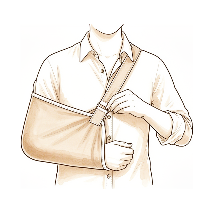
Postoperative Immobilization & Rehabilitation: The most common immobilization device prescribed postoperatively for rotator cuff repair was an abduction pillow sling (arm in neutral or slight internal rotation) at 70% [188]. Internal rotation was employed by 45% of surgeons for the position of immobilization following rotator cuff repair [188]. Following arthroscopic rotator cuff repair, the operated arm was immobilized in a Mayo clinic sling for 4 weeks with the elbow in 90° flexion and by the side of the body [198]. All patients undergoing the Latarjet procedure wore a sling for the first week postoperatively [157]. The postoperative protocol for revision arthroscopic type II SLAP repairs did not differ from the standard protocol for primary repairs, with patients initially placed in a sling for 7 to 10 days [179].
Formal physical therapy was most commonly initiated postoperatively within the first 2 weeks (37%) following rotator cuff repair [188]. The majority of surgeons initiated passive shoulder ROM within the first 2 weeks (69%) following rotator cuff repair [188]. Unrestricted passive shoulder ROM was started at 6 to 7 weeks by 35% of surgeons following rotator cuff repair [188]. Gentle Codman-Pendulum exercises were started at 4 weeks postoperatively following arthroscopic rotator cuff repair [198]. Active ROM was begun by most surgeons at 7 to 10 weeks (61%) following rotator cuff repair [188]. Unrestricted active ROM was started at 7 to 10 weeks with a consensus response of 53% following rotator cuff repair [188]. Active assisted range of motion exercises, except for overhead active motion, were started at 6 weeks postoperatively following arthroscopic rotator cuff repair [198]. Patients undergoing the Latarjet procedure were encouraged to start self-assisted rehabilitation for 3 weeks at the beginning of the second week [157]. Patients undergoing the Latarjet procedure were referred to a physical therapist to start active mobilization in elevation and external rotation after 1 month postoperatively [157].
Strengthening (resistance) exercises were most commonly prescribed between 6 weeks and 3 months (56%) following rotator cuff repair [188]. Resisted isotonic strengthening exercises were started at 10 weeks postoperatively following arthroscopic rotator cuff repair [198]. Unrestricted return to all activities was started at 5 to 6 months by nearly half of participants (42%) following rotator cuff repair [188]. Comprehensive rehabilitation, compared to conventional physiotherapy, has shown a statistically and clinically significant difference in improving pain, ROM, functional disability, quality of life, and treatment effectiveness in patients after arthroscopic rotator cuff repair [25]. There is strong evidence that early initiation of rehabilitation and early functional loading does not adversely affect clinical outcome after rotator cuff repair, though there is a marginally higher, statistically non-significant incidence of tendon re-tear [154].
Fracture & Arthroplasty Rehabilitation: The lack of strong evidence warrants the need for future controlled studies; subsequently, postoperative rehabilitation following reverse shoulder arthroplasty should be individualized [43]. The review outlines rehabilitation guidelines developed to manage patients who have undergone reverse total shoulder replacement, aiming to achieve optimal pain relief and maximize functional outcomes while mitigating risks associated with the surgery [45]. At early follow-up, navigated and non-navigated reverse shoulder arthroplasties yielded similar rates of improvement in range of motion and functional outcome scores [167]. Task-oriented exercises improve disability of working patients with surgically-treated proximal humeral fractures [194]. After early mobilization by the end of the first week after surgery for proximal humeral fractures, the control program incorporated segmentary exercises for humeral mobilisation, strengthening, segmentary muscle stretching, and postural control [194]. The interventional programs for proximal humeral fractures were performed individually and lasted twelve weeks, involving three individual 1-hour sessions a week of physical training (medium intensity), attending a total of 36 meetings [194]. The experimental group for proximal humeral fractures met with the occupational therapist once a week for a 60-min session [194].
Neuromuscular & Special Populations: An interdisciplinary approach with regular longitudinal follow-ups will hopefully provide patients and their family an improved day-to-day satisfaction alongside with optimal clinical outcomes for neuromuscular diseases [17]. The orthopaedic approach to patients with neuromuscular diseases can be challenging as both surgical and supportive methods of interventions are available, and the timing of each should be chosen carefully and be placed in context of the patients’ overall function and the point in time of recovery [17]. Idiopathic brachial plexopathy, often preceded by shoulder pain, can progress to severe weakness in the distribution of effected nerves and patients generally have a slow and variable course of recovery despite interventions [37]. There is no good evidence for steroid administration or surgical intervention for idiopathic brachial plexitis, and most will have recovery with a variable functional end point over the course of multiple months to more than one year [37].
Complications
Other Considerations: A thorough, well-designed postoperative or postinjury rehabilitation program may prevent most complications [159]. If complications arise, a team approach working to develop an evidenced-based treatment program designed specifically for the underlying complication can successfully treat these issues [159]. Limb salvage has higher rates of postoperative complications, including infection, aseptic loosening, and graft or prosthetic failure, compared with amputation [125]. Prehabilitation, or rehabilitation after diagnosis and prior to treatment, enhances quality of life and postoperative outcomes in cancer-related procedures [125]. The cause of failure leading to early revision varies between late and early revision cases [160].
Recovery
Prognostic Factors: Baseline pain severity, disability, and psychological factors are identified prognostic factors for response to physiotherapy for musculoskeletal shoulder pain, though the evidence quality is generally low [3]. Pain intensity, neck pain, and longer duration of complaints predict poorer outcome in patients with shoulder pain [54]. In shoulder surgery patients, worse functional outcomes were associated with worse PROMIS Depression and Anxiety scores [133], with more severe two-year PROMIS Anxiety identified as the strongest predictor of worse functional outcomes [133]. Patients with a worse prognosis may be monitored more frequently and the treatment plan modified if complaints persist [54].
Measurement Agreement: Some measures of shoulder range of motion showed a moderate to high level of agreement between patient-reported measurements and the physician's measurements [65].
Rotator Cuff Rehabilitation: Although functional status improved with time after 6 months, the structural status of repaired cuffs remained unchanged between 6 and 19 months postoperatively [113]. At 6 months postoperatively, superior functional outcomes were observed in the delayed repair group compared with the immediate repair group for partial-thickness rotator cuff tears [59].
Arthroplasty & Instability Outcomes: Six distinct early recovery trajectories were identified after total shoulder arthroplasty, with 83.7% of patients (the 'Faster group') experiencing very low pain scores after only 2 weeks [60]. Revision reverse shoulder arthroplasty leads to an improvement in patient-reported outcome measures and pain levels and demonstrates high durability at mid-term follow-up with low complication and failure rates [30]. Physical therapy plays an important role subsequent to reverse shoulder arthroplasty and has an effect in the mid-term to long-term follow-up regarding the range of motion as well as activities of daily living [135]. Although there is a significant and longitudinal improvement in shoulder function and pain relief after total shoulder replacement, long-term follow-up of fifteen to twenty years demonstrated a high revision rate in this cohort [56]. Nonmodifiable risk factors, such as age less than 20 years and duration of symptoms greater than 5 months, significantly affect risk of recurrence after arthroscopic instability repair [213].
Fractures & Other Conditions: Delayed rehabilitation by 3 weeks of shoulder immobilization produces a slower recovery, which continues for at least 2 years after the time of injury for two-part fractures of the neck of the humerus [205]. Rehabilitation is a valuable intervention when faced with atraumatic posterior shoulder subluxation [11]. Significant deficits in the literature regarding adhesive capsulitis include a paucity of randomized controlled trials, failure to report response to treatment in a stage-based fashion, and an incomplete understanding of the disease's natural course [214]. Three months follow-up is too early for outcome evaluation using five shoulder outcome measures [55].
Key Evidence
- [L4] There is no consensus on the definition of accelerated rehabilitation or outcome measure selection. [1] (10.1177/17585732221089636)
- [L3] Improvement in outcomes was observed up to 16 sessions of physical therapy, after which outcomes plateaued. [2] (10.1016/j.jse.2018.10.001)
- [L2] The review identified several prognostic factors for response to physiotherapy, including baseline pain severity, disability, and psychological factors, though the evidence quality was generally low. [3] (10.1186/1471-2474-14-203)
- [L2] Short-term repeat assessment of pain was better than short-term change or baseline score at predicting long-term disability improvement across all cohorts. [4] (10.1186/s12891-017-1502-8)
- [L2] Delayed passive rehabilitation does not bring about superior outcomes compared to early rehabilitation. [5] (10.5397/cise.2019.22.4.190)
- [L4] The majority of functional and symptomatic improvements following RCR occur within the first year, with minimal clinically meaningful gains observed between 1 and 2 years. [6] (10.1016/j.jse.2025.05.020)
- [L5] Combining pathoanatomic causes with movement impairments provides a more focused rehabilitation approach, and applying a comprehensive evaluation approach prior to and throughout treatment can assist clinicians in selecting appropriate treatments based on patient need. [7] (10.1016/j.jse.2023.07.013)
- [L3] Patients undergoing rehabilitation using a home-based protocol showed largely similar functional scores and healing to those with supervised PT after ARCR of MRCTs at the latest follow-up. [8] (10.1016/j.arthro.2024.06.037)
- [L3] However, at > 2 years after surgery, earlier repairs ( < 6 months) are associated with better functional outcomes. [9] (10.1177/17585732211015825)
- [L4] Compared with surgery, physical therapy is associated with less improvement in perceived functional outcomes and a higher clinical failure rate. [10] (10.1016/j.jse.2020.07.030)
- [L4] These results support the view that rehabilitation is a valuable intervention when faced with such a patient presentation. [11] (10.1177/1758573213517218)
- [L5] The proposed objective criteria-based shoulder diagnosis and rehabilitation model could be a new effective strategy for minimizing the time required to regain functional capacity and recover from symptoms among patients with work-related shoulder injuries. [12] (10.1186/s12891-017-1435-2)
- [L1] Only one study showed moderate evidence of early physical therapy promoting a more rapid return of short-term improvement in function and pain. [13] (10.1177/1758573218812038)
- [L5] The key to effective treatment is a thorough clinical examination and appropriate differential diagnosis to develop a program focusing on restoring adaptations, controlling inflammation, and improving neuromuscular control. [14] (10.1177/03635465020300011201)
- [L3] Functional outcomes after ARCR improved during midterm follow-up, regardless of retear. [15] (10.1177/03635465241305742)
- [L5] [16] (10.5435/00124635-200303000-00008)
- [L3] Aspects of treatment that maximize the functional outcome are important in achieving patient satisfaction. [18] (10.1016/j.jse.2007.02.136)
- [L2] The study demonstrates that the SANE is valid for a range of common shoulder diagnoses to assess patient outcomes across operative and nonoperative treatment for shoulder complaints. [20] (10.1177/0363546518807924)
- [L3] PROMIS PF scores were responsive to the functional improvements observed clinically. [21] (10.1016/j.jse.2018.08.040)
- [L5] Identification of whether a patient presents with pain or stiffness as their predominant symptom further guides treatment selection. [24] (10.1177/1758573215586152)
- [L1] Comprehensive rehabilitation, compared to conventional physiotherapy, has shown a statistically and clinically significant difference in improving pain, ROM, functional disability, quality of life, and treatment effectiveness in patients after ARCR. [25] (10.1016/j.jht.2023.09.009)
- [L1] Manipulative therapy in addition to usual medical care accelerates recovery and is more effective than usual medical care alone on the long term, but is associated with higher costs. [29] (10.1186/1471-2474-11-200)
- [L4] Revision rTSA leads to an improvement in patient-reported outcome measures and pain levels and demonstrates high durability at mid-term follow-up with low complication and failure rates. [30] (10.1016/j.jsea.2026.100004)
- [Letter] The authors argue that the data available in the referenced systematic review are insufficient to make reliable clinical implications or establish a gold standard rehabilitation protocol, emphasizing the need for further research on dose-response relationships and supervised exercise. [31] (10.1016/j.jse.2009.03.015)
- [L4] [38] (10.1177/1758573220913246)
- [L4] Successful nonoperative treatment of superior labral tears results in improved pain relief and functional outcomes compared with pretreatment assessments. [39] (10.1177/0363546510370937)
- [L3] Although worse functional results were found in the group with surgical indication, this difference disappears when adjusting for age as a confounding factor. [41] (10.1016/j.jse.2025.02.026)
- [L1] The lack of strong evidence warrants the need for future controlled studies; subsequently, postoperative rehabilitation should be individualized. [43] (10.1177/17585732221144007)
- [L5] The review outlines rehabilitation guidelines developed to manage patients who have undergone reverse total shoulder replacement, aiming to achieve optimal pain relief and maximize functional outcomes while mitigating risks associated with the surgery. [45] (10.1111/j.1758-5740.2011.00138.x)
- [L2] Subjective mechanical symptoms in the affected shoulder are a common complaint in patients with suspected rotator cuff pathology. [48] (10.1016/j.jse.2024.02.024)
- [L3] Outcome models showed consistent relationships between patient baseline covariates and the 12-month ASES scores and Recovery Period shoulder-related encounters. [50] (10.1016/j.jseint.2026.101763)
- [L5] This comprehensive review highlights the importance of a thorough understanding of shoulder physical examination and specific diagnostic tests, emphasizing history-taking and examination to ensure diagnostic accuracy and optimize patient outcomes for surgeons. [51] (10.5435/jaaos-d-25-00024)
- [L4] It can assess the strength of various muscle groups following disease or surgery, determine the effectiveness of therapy, and detect old muscle injuries that have escaped detection by existing methods of diagnosis. [52] (10.2106/00004623-196345010-00011)
- [L5] There are no appropriately designed prognostic studies to identify predictors of success with physiotherapy in patients with massive irreparable rotator cuff tears. [53] (10.3390/ijerph20075242)
- [L1] Those with a worse prognosis may be monitored more frequently and the treatment plan modified if complaints persist. [54] (10.1186/s12891-015-0738-4)
- [L2] Our results suggest that 3 months follow-up is too early for outcome evaluation. [55] (10.1186/s12891-021-04483-3)
- [L4] Although there is a significant and longitudinal improvement in shoulder function and pain relief after total shoulder replacement, long-term follow-up of fifteen to twenty years demonstrated a high revision rate in this cohort. [56] (10.2106/jbjs.m.00079)
- [L2] However, at 6 months postoperatively, superior functional outcomes were observed in the delayed repair group compared with the immediate repair group. [59] (10.1177/0363546518757425)
- [L2] Six distinct early recovery trajectories were identified after total shoulder arthroplasty, with 83.7% of patients (the 'Faster group') experiencing very low pain scores after only 2 weeks. [60] (10.1016/j.jse.2025.06.016)
- [L5] Stable, nondisplaced ligament injuries without significant MRI findings can be treated nonoperatively, with most athletes safely returning to sport within 6–10 weeks and minimal complications. [61] (10.1002/ksa.70244)
- [L1] Surgery (after failure of conservative treatment) ranked highest across all ROM domains. [63] (10.1016/j.arthro.2020.09.041)
- [L3] Some measures of shoulder ROM showed a moderate to high level of agreement between patient-reported measurements and the physician's measurements. [65] (10.1016/j.jse.2016.02.010)
- [L5] For the minority of patients for whom nonoperative management fails, surgical reconstruction with an open inferior capsular shift can be reasonably recommended. [67] (10.5435/00124635-199801000-00007)
- [L2] Further studies are needed to determine its effect on the outcome of the treatment of rotator cuff disease. [96] (10.2106/jbjs.o.00444)
- [L3] [101] (10.1136/bmjsem-2018-000342)
- [L2] No differences in clinical scores existed between patients stratified by the Sugaya MRI classification system at 16 weeks. [105] (10.1016/j.jse.2015.09.019)
- [L4] Use of the patient-centred ULTRA guideline can yield statistically significant improvements in outcome and range of motion for patients undergoing primary RTSA, which are maintained at the two-year post-surgery time point. [107] (10.1177/17585732221133532)
- [L4] Although functional status improved with time after 6 months, the structural status of repaired cuffs remained unchanged between 6 and 19 months. [113] (10.1016/j.jse.2011.05.027)
- [L4] The dichotomized and trichotomized classifications as well as the combined classifications did not lead to superior agreements. [130] (10.1016/j.arthro.2021.12.005)
- [L3] Worse functional outcomes were associated with worse PROMIS Depression and Anxiety; however, more severe two-year PROMIS Anxiety was the strongest predictor of worse functional outcomes. [133] (10.1302/0301-620x.104b4.bjj-2021-1089.r1)
- [Paper] Physical therapy plays an important role subsequent to reverse shoulder arthroplasty and has an effect in the mid-term to long-term follow-up regarding the range of motion as well as activities of daily living. [135] (10.1007/s00402-018-2977-y)
- [L4] Non-operative treatment with an appropriate regimen provided satisfactory clinical outcomes in middle-aged patients with symptomatic SLAP lesions and should be considered before recommending operative treatment. [136] (10.1007/s00167-016-4226-7)
- [L1] Operative treatment resulted in greater improvement in Constant scores and significantly decreased pain scores compared to nonoperative management. [138] (10.1016/j.jse.2017.09.032)
- [L1] There is strong evidence that early initiation of rehabilitation and early functional loading does not adversely affect clinical outcome, though there is a marginally higher, statistically non-significant incidence of tendon re-tear. [154] (10.1177/1758573214567702)
- [L3] [157] (10.1177/0363546519879929)
- [Paper] A thorough well-designed postoperative or postinjury rehabilitation program may prevent most complications, and if complications do arise, a team approach working to develop an evidenced-based treatment program designed specifically for the underlying complication can successfully treat these issues. [159] (10.1016/j.csm.2017.12.010)
- [L3] The cause of failure leading to early revision varies between late and early revision cases. [160] (10.1016/j.jse.2015.05.035)
- [L3] A multimodal nonoperative treatment program is effective for most patients with adhesive capsulitis. [165] (10.1177/0363546510385403)
- [L3] At early follow-up, navigated and non-navigated RSAs yielded similar rates of improvement in range of motion and functional outcome scores. [167] (10.1016/j.jse.2022.07.007)
- [L4] Patients aged ≤20 years did poorly with more than half of the patients having recurrence; alternative stabilization techniques should probably be considered for these patients. [171] (10.1007/s00167-017-4504-z)
- [L4] [179] (10.1177/0363546511398648)
- [L5] The clinical case example shows techniques that may help guide the clinician in establishing effective nonoperative or postoperative treatments for TOS. [184] (10.1016/j.asmr.2021.11.007)
- [L4] [188] (10.1177/2325967116684775)
- [L4] [191] (10.1177/1758573215592266)
- [L1] [194] (10.1186/s12891-021-04140-9)
- [L4] [198] (10.1016/j.jse.2011.02.009)
- [L1] Delayed rehabilitation by 3 weeks of shoulder immobilization produces a slower recovery, which continues for at least 2 years after the time of injury. [205] (10.1016/j.jse.2006.06.003)
- [L2] Nonmodifiable risk factors, such as age less than 20 years and duration of symptoms greater than 5 months, significantly affect risk of recurrence. [213] (10.1177/0363546520949840)
- [L5] Significant deficits in the literature include a paucity of randomized controlled trials, failure to report response to treatment in a stage-based fashion, and an incomplete understanding of the disease's natural course. [214] (10.1177/0363546509348048)
See Also
- Rotator Cuff
- Rotator cuff repair
- Total shoulder arthroplasty
- Reverse Shoulder Arthroplasty
- Fractures
- Os Acromiale
- Shoulder Instability
- Clavicle Fracture
- Thoracic Outlet Syndrome
- Calcific Tendinitis
- Shoulder Arthritis
- Latarjet Procedure
References
[1] Post-operative rehabilitation following traumatic anterior shoulder dislocation: A systematic scoping review. Shoulder & Elbow. 2022. DOI: 10.1177/17585732221089636
[2] Physical therapy versus natural history in outcomes of rotator cuff tears: the Rotator Cuff Outcomes Workgroup (ROW) cohort study. Journal of Shoulder and Elbow Surgery. 2019. DOI: 10.1016/j.jse.2018.10.001
[3] Predicting response to physiotherapy treatment for musculoskeletal shoulder pain: a systematic review. BMC Musculoskeletal Disorders. 2013. DOI: 10.1186/1471-2474-14-203
[4] Brief pain re-assessment provided more accurate prognosis than baseline information for low-back or shoulder pain. BMC Musculoskeletal Disorders. 2017. DOI: 10.1186/s12891-017-1502-8
[5] Early and Delayed Postoperative Rehabilitation after Arthroscopic Rotator Cuff Repair: A Comparative Study of Clinical Outcomes. Clinics in Shoulder and Elbow. 2019. DOI: 10.5397/cise.2019.22.4.190
[6] Patient-reported and clinical outcomes up to 2 years after primary rotator cuff repair: do we need to collect patient-reported outcomes at 2-year follow-up visit?. Journal of Shoulder and Elbow Surgery. 2026. DOI: 10.1016/j.jse.2025.05.020
[7] Rehabilitation of the painful shoulder. Journal of Shoulder and Elbow Surgery. 2024. DOI: 10.1016/j.jse.2023.07.013
[8] Home‐Based or Supervised Physical Therapy Shows Similar Functional Outcomes and Healing After Massive Rotator Cuff Repair. Arthroscopy. 2024. DOI: 10.1016/j.arthro.2024.06.037
[9] Is timing of superior labrum anterior to posterior (SLAP) repair important? A cohort study evaluating the effect of the duration of symptoms prior to surgery on the outcomes of patients who underwent type II SLAP repair. Shoulder & Elbow. 2021. DOI: 10.1177/17585732211015825
[10] Management of irreparable massive rotator cuff tears: a systematic review and meta-analysis of patient-reported outcomes, reoperation rates, and treatment response. Journal of Shoulder and Elbow Surgery. 2020. DOI: 10.1016/j.jse.2020.07.030
[11] Patient-reported outcomes following a physiotherapy rehabilitation programme for atraumatic posterior shoulder subluxation. Shoulder & Elbow. 2014. DOI: 10.1177/1758573213517218
[12] A protocol for a new methodological model for work-related shoulder complex injuries: From diagnosis to rehabilitation. BMC Musculoskeletal Disorders. 2017. DOI: 10.1186/s12891-017-1435-2
[13] Effectiveness of formal physical therapy following total shoulder arthroplasty: A systematic review. Shoulder & Elbow. 2018. DOI: 10.1177/1758573218812038
[14] Current Concepts in the Rehabilitation of the Overhead Throwing Athlete. The American Journal of Sports Medicine. 2002. DOI: 10.1177/03635465020300011201
[15] Midterm Functional Outcomes After Retear of an Arthroscopic Rotator Cuff Repair: A Propensity Score–Matched Comparative Study. The American Journal of Sports Medicine. 2025. DOI: 10.1177/03635465241305742
[16] Scapular Dyskinesis and Its Relation to Shoulder Pain. Journal of the American Academy of Orthopaedic Surgeons. 2003. DOI: 10.5435/00124635-200303000-00008
[17] Orthopaedic Basic Science Fifth Edition Print Ebook. Rare Genetic Bone Diseases of Orthopaedic Significance > Summary.
[18] Factors influencing patient satisfaction after rotator cuff repair. Journal of Shoulder and Elbow Surgery. 2007. DOI: 10.1016/j.jse.2007.02.136
[20] Validity and Responsiveness of the Single Alpha-numeric Evaluation for Shoulder Patients. The American Journal of Sports Medicine. 2018. DOI: 10.1177/0363546518807924
[21] Preoperative Patient-Reported Outcomes Measurement Information System (PROMIS) scores predict postoperative outcome in total shoulder arthroplasty patients. Journal of Shoulder and Elbow Surgery. 2019. DOI: 10.1016/j.jse.2018.08.040
[24] Physiotherapy assessment of shoulder stiffness and how it influences management. Shoulder & Elbow. 2015. DOI: 10.1177/1758573215586152
[25] Comparison of the physiotherapy with and without focus on the scapulothoracic joint on pain, range of motion, functional disability, quality of life, and treatment effectiveness of patients after arthroscopic shoulder rotator cuff tendon repair: A randomized controlled trial with short-term follow-up. Journal of Hand Therapy. 2024. DOI: 10.1016/j.jht.2023.09.009
[29] Manipulative therapy in addition to usual medical care accelerates recovery of shoulder complaints at higher costs: economic outcomes of a randomized trial. BMC Musculoskeletal Disorders. 2010. DOI: 10.1186/1471-2474-11-200
[30] Pain relief and durable outcomes after revision reverse shoulder arthroplasty for failed primary arthroplasty. Journal of Shoulder and Elbow Arthroplasty. 2026. DOI: 10.1016/j.jsea.2026.100004
[31] Regarding “Exercise in the treatment of rotator cuff impingement: A systematic review and a synthesized evidence-based rehabilitation protocol”. Journal of Shoulder and Elbow Surgery. 2009. DOI: 10.1016/j.jse.2009.03.015
[34] Rockwood And Matsen S The Shoulder. Arthroscopic Management of Prearthritic and Arthritic Conditions of the Shoulder and the Postarthroplasty Shoulder > Radiographic Evaluation.
[35] Rockwood And Matsen S The Shoulder. Fractures, Dislocations, and Acquired Problems of the Shoulder in Children > CLINICAL EVALUATION AND PHYSICAL EXAMINATION.
[36] Rockwood And Matsen S The Shoulder. Fractures, Dislocations, and Acquired Problems of the Shoulder in Children > APPROACH TO FAILED CUFF SURGERY.
[37] Orthopaedic Knowledge Update 13 Ebook Without Multimedia. Neuropathies, Vascular Conditions: Buerger’s, Raynaud’s; Degenerative Conditions > Summary.
[38] Shoulder Pain and Disability Index: Italian cross-cultural validation in patients with non-specific shoulder pain. Shoulder & Elbow. 2020. DOI: 10.1177/1758573220913246
[39] Nonoperative Treatment of Superior Labrum Anterior Posterior Tears. The American Journal of Sports Medicine. 2010. DOI: 10.1177/0363546510370937
[41] Functional outcomes and healing rates in patients with extra-articular scapula fractures treated nonoperatively: a comparison of fractures meeting and not meeting surgical indications. Journal of Shoulder and Elbow Surgery. 2025. DOI: 10.1016/j.jse.2025.02.026
[43] Can we accelerate rehabilitation following reverse shoulder arthroplasty? A systematic review. Shoulder & Elbow. 2022. DOI: 10.1177/17585732221144007
[45] Rehabilitation following Reverse Total Shoulder Replacement. Shoulder & Elbow. 2011. DOI: 10.1111/j.1758-5740.2011.00138.x
[48] The significance of subjective mechanical symptoms in rotator cuff pathology. Journal of Shoulder and Elbow Surgery. 2024. DOI: 10.1016/j.jse.2024.02.024
[50] Assessing the properties of an encounter-based “process of recovery” measure of patient benefit following rotator cuff repair. JSES International. 2026. DOI: 10.1016/j.jseint.2026.101763
[51] Upper Extremity Surgeon's Guide to the Evaluation of the Shoulder Girdle and Diagnosis of Associated Pathology. Journal of the American Academy of Orthopaedic Surgeons. 2025. DOI: 10.5435/jaaos-d-25-00024
[52] The Multiple Angle Testing Method for the Evaluation of Muscle Strength. The Journal of Bone & Joint Surgery. 1963. DOI: 10.2106/00004623-196345010-00011
[53] Massive Irreparable Rotator Cuff Tears: Which Patients Will Benefit from Physiotherapy Exercise Programs? A Narrative Review. International Journal of Environmental Research and Public Health. 2023. DOI: 10.3390/ijerph20075242
[54] Pain intensity, neck pain and longer duration of complaints predict poorer outcome in patients with shoulder pain – a systematic review. BMC Musculoskeletal Disorders. 2015. DOI: 10.1186/s12891-015-0738-4
[55] Responsiveness of five shoulder outcome measures at follow-ups from 3 to 24 months. BMC Musculoskeletal Disorders. 2021. DOI: 10.1186/s12891-021-04483-3
[56] Longitudinal Observational Study of Total Shoulder Replacements with Cement. Journal of Bone and Joint Surgery. 2014. DOI: 10.2106/jbjs.m.00079
[59] When Should We Repair Partial-Thickness Rotator Cuff Tears? Outcome Comparison Between Immediate Surgical Repair Versus Delayed Repair After 6-Month Period of Nonsurgical Treatment. The American Journal of Sports Medicine. 2018. DOI: 10.1177/0363546518757425
[60] Trajectories of pain recovery during the first 8 weeks after shoulder arthroplasty: results from the shoulder diary study using latent growth curve modeling. Journal of Shoulder and Elbow Surgery. 2026. DOI: 10.1016/j.jse.2025.06.016
[61] Diagnostic evaluation and nonoperative management of Lisfranc injuries in athletes. Knee Surgery, Sports Traumatology, Arthroscopy. 2025. DOI: 10.1002/ksa.70244
[63] Efficacy of Arthroscopic Surgery in the Management of Adhesive Capsulitis: A Systematic Review and Network Meta‐analysis of Randomized Controlled Trials. Arthroscopy. 2020. DOI: 10.1016/j.arthro.2020.09.041
[65] Agreement between patient self-assessment and physician assessment of shoulder range of motion. Journal of Shoulder and Elbow Surgery. 2016. DOI: 10.1016/j.jse.2016.02.010
[67] Multidirectional Instability of the Shoulder: Pathophysiology, Diagnosis, and Management. Journal of the American Academy of Orthopaedic Surgeons. 1998. DOI: 10.5435/00124635-199801000-00007
[72] Rockwood And Matsen S The Shoulder. Shoulder and Elbow Specialty Clinic Workers’ Survey > ANATOMY.
[73] A Lange Medical Book Current Diagnosis Treatment In Orthopedics Fifth Edition. 2Musculoskeletal Trauma Surgery > SHOULDER AND ARM INJURIES.
[74] Rockwood And Green S Fractures In Adults. 29: Principles of Nonunion and Bone Defect Treatment > Applied Anatomy Related to Scapular Fractures.
[75] Aaos Comprehensive Orthopaedic Review 3. Anatomy of the Shoulder, Arm, and Elbow > I. Shoulder.
[80] Rockwood And Matsen S The Shoulder. Fractures, Dislocations, and Acquired Problems of the Shoulder in Children > FRACTURES OF THE PROXIMAL HUMERUS.
[83] Rockwood And Matsen S The Shoulder. Developmental Anatomy of the Shoulder and Anatomy of the Glenohumeral Joint > Development of Individual Regions.
[86] Campbell S Operative Orthopaedics 4 Volume Set. RECONSTRUCTIVE PROCEDURES OF THE SHOULDER AND ELBOW IN ADULTS > ANATOMY AND BIOMECHANICS.
[89] Miller S Review Of Orthopaedics. Genetics of musculoskeletal conditions and abnormalities are summarized in Table 1.27 > UPPER EXTREMITY > SHOULDER.
[96] Mental Health Has a Stronger Association with Patient-Reported Shoulder Pain and Function Than Tear Size in Patients with Full-Thickness Rotator Cuff Tears. Journal of Bone and Joint Surgery. 2016. DOI: 10.2106/jbjs.o.00444
[100] Apley And Solomon S Concise System Of Orthopaedics And Trauma. INVESTIGATION.
[101] Reliability of the Shoulder Symptom Modification Procedure and association of within-session and between-session changes with functional outcomes. BMJ Open Sport & Exercise Medicine. 2018. DOI: 10.1136/bmjsem-2018-000342
[105] Early postoperative repair status after rotator cuff repair cannot be accurately classified using questionnaires of patient function and isokinetic strength evaluation. Journal of Shoulder and Elbow Surgery. 2016. DOI: 10.1016/j.jse.2015.09.019
[106] Rockwood And Matsen S The Shoulder. Shoulder and Elbow Specialty Clinic Workers’ Survey > OTHER DISORDERS > Lateral Dislocation of the Scapula (Scapulothoracic Dissociation).
[107] Physiotherapy treatment for reverse total shoulder arthroplasty: Results of a patient-centred rehabilitation guideline using functional and patient-reported outcome measures. Shoulder & Elbow. 2022. DOI: 10.1177/17585732221133532
[111] Rockwood And Green S Fractures In Adults. 29: Principles of Nonunion and Bone Defect Treatment > Magnetic Resonance Imaging and Arthrography.
[113] Serial structural and functional assessments of rotator cuff repairs: do they differ at 6 and 19 months postoperatively?. Journal of Shoulder and Elbow Surgery. 2012. DOI: 10.1016/j.jse.2011.05.027
[117] Orthopaedic Knowledge Update 13 Ebook Without Multimedia. Shoulder Anatomy and Biomechanics, Clinical Evaluation, Imaging > Clinical Evaluation > Imaging.
[118] Orthopaedic Basic Science Fifth Edition Print Ebook. Rare Genetic Bone Diseases of Orthopaedic Significance > History and Physical Examination.
[121] Aaos Comprehensive Orthopaedic Review 3. Imaging of the Shoulder and Elbow > I. Shoulder.
[124] Rockwood And Green S Fractures In Adults. 29: Principles of Nonunion and Bone Defect Treatment > Radiography.
[125] Miller S Review Of Orthopaedics. SECTION 16 PATELLAR TRACKING IN TOTAL KNEE ARTHROPLASTY > MUSCULOSKELETAL TUMORS.
[130] Owen, Sugaya, and Hayashida Classifications Give Poor Intra‐ and Inter‐Rater Agreement on a Magnetic Resonance Imaging Evaluation of Subscapularis Tendon Retears. Arthroscopy. 2021. DOI: 10.1016/j.arthro.2021.12.005
[133] PROMIS Depression and Anxiety in shoulder surgery patients. The Bone & Joint Journal. 2022. DOI: 10.1302/0301-620x.104b4.bjj-2021-1089.r1
[135] Reverse shoulder arthroplasty: the role of physical therapy on the clinical outcome in the mid-term to long-term follow-up. Archives of Orthopaedic and Trauma Surgery. 2018. DOI: 10.1007/s00402-018-2977-y
[136] Clinical outcomes of non-operative treatment for patients presenting SLAP lesions in diagnostic provocative tests and MR arthrography. Knee Surgery, Sports Traumatology, Arthroscopy. 2016. DOI: 10.1007/s00167-016-4226-7
[138] Operative versus nonoperative treatment for the management of full-thickness rotator cuff tears: a systematic review and meta-analysis. Journal of Shoulder and Elbow Surgery. 2018. DOI: 10.1016/j.jse.2017.09.032
[150] 1. Clinical Evaluation, Imaging, and Classification of Proximal Humerus Fractures. 2011.
[154] Rehabilitation following rotator cuff repair: a systematic review. Shoulder & Elbow. 2015. DOI: 10.1177/1758573214567702
[157] Outcomes After Latarjet Procedure: Patients With First-Time Versus Recurrent Dislocations. The American Journal of Sports Medicine. 2019. DOI: 10.1177/0363546519879929
[159] Rehabilitation. Clinics in Sports Medicine. 2018. DOI: 10.1016/j.csm.2017.12.010
[160] Early revision within 1 year after shoulder arthroplasty: patient factors and etiology. Journal of Shoulder and Elbow Surgery. 2015. DOI: 10.1016/j.jse.2015.05.035
[165] Predictors of Outcome After Nonoperative and Operative Treatment of Adhesive Capsulitis. The American Journal of Sports Medicine. 2010. DOI: 10.1177/0363546510385403
[167] Early clinical outcomes following navigation-assisted baseplate fixation in reverse total shoulder arthroplasty: a matched cohort study. Journal of Shoulder and Elbow Surgery. 2023. DOI: 10.1016/j.jse.2022.07.007
[171] Long-term results of arthroscopic Bankart repair: Minimum 10 years of follow-up. Knee Surgery, Sports Traumatology, Arthroscopy. 2017. DOI: 10.1007/s00167-017-4504-z
[179] Outcomes of Revision Arthroscopic Type II Superior Labral Anterior Posterior Repairs. The American Journal of Sports Medicine. 2011. DOI: 10.1177/0363546511398648
[184] Rehabilitation in Overhead Athletes With Thoracic Outlet Syndrome. Arthroscopy, Sports Medicine, and Rehabilitation. 2022. DOI: 10.1016/j.asmr.2021.11.007
[188] Postoperative Rehabilitation After Rotator Cuff Repair. Orthopaedic Journal of Sports Medicine. 2017. DOI: 10.1177/2325967116684775
[191] Physiotherapy treatment for atraumatic recurrent shoulder instability: early results of a specific exercise protocol using pathology-specific outcome measures. Shoulder & Elbow. 2015. DOI: 10.1177/1758573215592266
[194] Task-oriented exercises improve disability of working patients with surgically-treated proximal humeral fractures. A randomized controlled trial with one-year follow-up. BMC Musculoskeletal Disorders. 2021. DOI: 10.1186/s12891-021-04140-9
[198] Does arthroscopic rotator cuff repair actually heal? Anatomic evaluation with magnetic resonance arthrography at minimum 2 years follow-up. Journal of Shoulder and Elbow Surgery. 2012. DOI: 10.1016/j.jse.2011.02.009
[202] Surgical Repair of Tears of the Rotator Cuff of the Shoulder: FACTORS INFLUENCING THE RESULT.. The Journal of Bone and Joint Surgery. American Volume. 1974.
[205] Rehabilitation of two-part fractures of the neck of the humerus (two-year follow-up). Journal of Shoulder and Elbow Surgery. 2007. DOI: 10.1016/j.jse.2006.06.003
[213] Risk Factors for Recurrence After Arthroscopic Instability Repair—The Importance of Glenoid Bone Loss >15%, Patient Age, and Duration of Symptoms: A Matched Cohort Analysis. The American Journal of Sports Medicine. 2020. DOI: 10.1177/0363546520949840
[214] Adhesive Capsulitis. The American Journal of Sports Medicine. 2010. DOI: 10.1177/0363546509348048