Clinicians › Hand
Flexor Sheath Ganglion
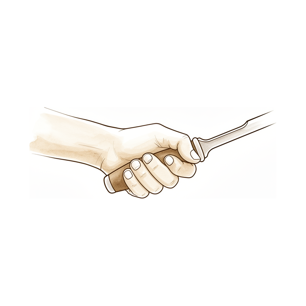
Flexor tendon sheath ganglia (palmar A1-pulley seed cysts).
Overview
A flexor sheath ganglion is a painful cystic lesion of the digital flexor tendon sheath that is effectively managed by surgical excision, a procedure characterized as simple, safe, and effective [1]. Accurate preoperative diagnosis relies on a detailed physical examination of the metacarpophalangeal region to distinguish the ganglion from trigger digit or Dupuytren's contracture [2]. While decision tree and Markov modeling approaches have accurately recreated the management of these lesions, mirroring observed clinical experience [3], the condition is distinct from other flexor tendon pathologies. For instance, pseudo-Volkmann contracture most commonly involves the ring finger flexor digitorum superficialis but can occur in all fingers [4], and tendon-associated ganglion cysts are not usual, although flexor hallucis longus tendinopathy is common in athletes [9].
Clinical presentation often involves significant thickening of the flexor tendon before patients experience triggering, except in the thumb [5]. Anatomical variations, such as the absence of the palmaris longus tendon in one or both hands, were found in 13% of patients [6]. Operative findings support the hypothesis that intraosseous ganglia arise from penetration of bone by synovial tissue or fluid [7]. In cases where a midpalmar ganglion displaces carpal tunnel contents, this displacement strongly suggests it is a major factor in developing carpal tunnel syndrome [10]. Furthermore, motor recovery following excision of a ganglion compressing the deep palmar branch of the ulnar nerve was complete [12].
Conservative and minimally invasive options exist alongside open surgery. Percutaneous puncture is a practical option for managing flexor tendon sheath ganglions given its low cost, lack of downtime, and low recurrence rate, with no recurrences observed after a second puncture in this cohort [8]. The mid-axial route is a dependable site for infiltration of drug into the flexor sheath and is devoid of complications if meticulous care is given during the procedure [13]. Additionally, triggering pathology at the wrist has been treated with interventional radiological measures rather than open surgery, demonstrating the efficacy of the technique [11].
Anatomy & Pathophysiology
Flexor Tendon Anatomy
The extrinsic flexor tendons of the hand serve as the terminal functional units of the forearm motors to the digits, named according to the location of their originating forearm muscles [25]. The flexor digitorum profundus (FDP) arises from the deeper layer of flexor muscles, while the flexor digitorum superficialis (FDS) continues from the more superficial muscle layer [25]. The flexor pollicis longus (FPL) originates from the deeper muscle layer and is the only thumb flexor with a tendon occupying a sheath [25]. Within the carpal tunnel, the profundus tendons are arranged deepest, with the index and small finger superficialis tendons positioned above them, followed by the superficialis tendons of the middle and ring fingers [25].
Muscle Origins: The flexor digitorum profundus originates from the proximal ulna and the interosseous membrane [58]. The flexor digitorum superficialis possesses two heads: a radial head originating from the proximal shaft of the radius, and a humeral ulnar head originating from the medial humeral epicondyle and coronoid process of the ulna [58]. The flexor pollicis longus originates from two heads: a radial head from the proximal radius and interosseous membrane, and an accessory head from the coronoid process of the ulna and medial epicondyle of the humerus [58].
Functional Roles: The flexor digitorum profundus provides digital flexion at both the proximal and distal interphalangeal joints [58]. The primary function of the flexor digitorum superficialis is digital flexion at the proximal interphalangeal joint [58]. The flexor pollicis longus flexes both the interphalangeal and metacarpophalangeal joints of the thumb [58].
Flexor Sheath and Pulley System
As flexor tendons pass distal to the metacarpal neck, they enter the fibroosseous tunnel, or digital flexor sheath, which extends distally to the proximal aspect of the distal phalanx [58]. The tendinous sheath consists of annular pulleys, which provide mechanical stability, and cruciate pulleys, which provide flexibility [58]. The first, third, and fifth annular pulleys (A1, A3, and A5) are located over the metacarpophalangeal, proximal interphalangeal, and distal interphalangeal joints, respectively [58]. The second and fourth pulleys (A2 and A4) are situated over the middle portion of the proximal and middle phalanges [58]. The A2 and A4 pulleys are the most essential in maintaining the mechanical advantage of the flexor tendons [58].
In zone II, the synovial sheath is organized into thickenings or segments of transverse or oblique fibers comprising annular or cruciate pulleys [25]. The entirety of the sheath, pulleys and all, serves to retain the flexor tendons close to the phalanges throughout a complete range of motion [25]. Bowstringing of tendons occurs when the pulley system fails to retain the flexor tendons close to the phalanges [25]. The tenosynovium that lines the fibroosseous tunnel supplies both nutrition and lubrication to the poorly vascularized flexor tendons [58]. Within the sheath, tendon vascularity is supplied via the vincula system: the vinculum longus and brevis [58]. The fibrous sheath assumes the role of a pulley when the tendon changes direction [59]. The synovial sheaths at specific sites are surrounded by fibrous sheaths that keep the tendon close to the skeleton, in particular when the pulling tendons cross the sinus of an articular angle [59].
Pathophysiology and Differential Diagnosis
Giant-cell tumors of the tendon sheath typically occur in the hand where they represent the second most common type of soft tissue tumors after synovial ganglions [23]. Giant-cell tumors of the tendon sheath usually present as a solitary and firm slow-growing nodular lesion, which affects the volar aspect of the hand [23]. Multifocal giant-cell tumors of the tendon sheath are rarely described in the literature and commonly involve the same finger or the volar aspect of different fingers [23].
Functional Deficits and Deformities: Loss of profundus function prevents subterminal and terminal pinch, unless the DIP joint is fused [29]. If the profundus tendon becomes adherent to the remaining sublimis tendon or fracture callus it may tether the profundus tendons of adjacent uninjured fingers, preventing full digitopalmar grip [29]. Loss of the superficialis with preservation of the profundus tendon may result in hyperextension of the PIP joint in supple individuals, a phenomenon called recurvatum [29]. With loss of both profundus and superficialis tendons, flexion of the MP joint to 45 degrees may be possible if intrinsic function is intact [29]. Retraction of the profundus tendon following more proximal amputations may result in shortening and contracture of the corresponding lumbrical [29]. During flexion, contraction of the profundus muscle belly places stretch on the shortened lumbrical, which results in paradoxical extension of the PIP joint, termed the lumbrical plus deformity [29].
Imaging and Evaluation
Ultrasonography facilitates dynamic, real-time evaluation of bones, joints, tendons, nerves, and vessels, making it an ideal imaging modality for hand and wrist conditions [18]. A careful physical examination is essential to direct care and future testing if indicated [38].
Classification
Other Considerations: An ideal classification system for congenital hand differences should incorporate etiology, diagnosis, treatment, and prognosis [30]. The system must be detailed enough to differentiate among various differences but not too cumbersome to be useful [30]. It must also be widely accepted to facilitate standardized communication [30]. The congenital upper limb anomaly (CULA) classification scheme needs to be an ongoing process open to revision as we continue to discover and refine our understanding of underlying mechanisms [86].
Clinical Presentation
Flexor sheath ganglions present with variable symptoms depending on their anatomical origin and the structures they compress. A midpalmar ganglion can cause concomitant compressive neuropathy of the ulnar and median nerves [10]. The displacement of carpal tunnel contents by a midpalmar ganglion strongly suggests it was a major factor in the development of carpal tunnel syndrome [10]. Similarly, a ganglion originating from the third carpometacarpal joint can compress the deep palmar branch of the ulnar nerve [12].
A volar wrist ganglion can present as trigger finger [11]. A ganglion cyst can be contiguous with the flexor hallucis longus tendon [9]. A synovial cyst of the pulp of the little finger can originate from the wrist joint [16]. Distant as well as local sources of the contents of synovial cysts should be considered when local anatomy permits communication between a degenerate joint and an adjacent tendon sheath [16].
A persistent median artery can occur simultaneously with a reversed palmaris longus muscle and a volar ganglion [31].
Investigations
Clinical Examination
A detailed physical examination of the metacarpophalangeal (MCP) region of the affected digit distinguishes between a flexor sheath ganglion, trigger digit, and Dupuytren's disease [2]. Careful physical examination remains essential to direct care and future testing for hand and wrist conditions [38].
Imaging
In the presence of diagnostic uncertainty in hand infection, imaging by sonography or MRI may help to identify and localise collections [82]. Wrist arthrography demonstrated the origin of a little finger synovial cyst at the volar aspect of the distal radioulnar joint [24]. MRI-confirmed origin from the third carpometacarpal joint was used to diagnose ulnar distal motor branch compression by a ganglion [12].
Other Considerations
Distant as well as local sources of the contents of synovial cysts should be considered when the local anatomy permits communication between a degenerate joint and an adjacent tendon sheath [16]. Operative findings support the hypothesis that intraosseous ganglia arise from penetration of bone by synovial tissue or fluid [7].
Treatment
Non-Operative
Percutaneous puncture serves as a practical management option for flexor tendon sheath ganglions due to its low cost, lack of downtime, and low recurrence rate [8]. In the cohort assessed for this technique, no recurrences were observed following a second puncture [8]. Additionally, interventional radiological measures have demonstrated efficacy in treating volar wrist ganglions presenting as trigger finger, offering an alternative to open surgery [11].
Operative
Indications: Surgical intervention is indicated when nonsurgical treatment fails, as demonstrated by the successful surgical closure of a little finger synovial cyst at the volar aspect of the distal radioulnar joint [24].
Surgical Approach / Technique: Excision of the ganglion and its origin is the definitive operative strategy. In cases of ulnar distal motor branch compression by a ganglion with an MRI-confirmed origin from the third carpometacarpal joint, excision resulted in complete motor recovery [12]. For synovial cysts, surgical closure of the origin at the volar aspect of the distal radioulnar joint cures the cyst [24].
Complications
Nerve palsy: A midpalmar ganglion can cause concomitant compressive neuropathy of the ulnar and median nerves in the hand [10]. The displacement of carpal tunnel contents by a midpalmar ganglion is a major factor in the development of carpal tunnel syndrome [10]. Motor recovery following excision of a ganglion compressing the deep palmar branch of the ulnar nerve can be complete [12].
Other Considerations: A volar wrist ganglion can present as trigger finger pathology at the wrist [11]. Triggering pathology at the wrist caused by a ganglion can be treated with interventional radiological measures rather than open surgery [11]. A persistent median artery and reversed palmaris longus muscle may be identified incidentally during surgical exploration for a volar ganglion [31].
Recovery
Other Considerations: The median active range of motion of the proximal interphalangeal joint was 0° in extension and 95° in flexion at final follow-up for patients undergoing a stepwise surgical approach for triggering distally to the A2 pulley after A1 pulley release [14].
Key Evidence
- [L4] Surgical excision is a simple, safe, and effective method for treating a painful ganglion of the digital flexor tendon sheath. [1] (10.1007/s11552-007-9028-4)
- [L4] Distinguishing between a flexor sheath ganglion, trigger digit, and Dupuytren's may be accomplished with a detailed physical examination of the MCP region of the affected digit. [2] (10.1177/15589447221109644)
- [L5] Both decision tree and Markov modeling approaches accurately recreated the management of flexor sheath ganglia and mirrored the observed clinical experience reported in the literature. [3] (10.1007/s11552-007-9060-4)
- [L4] It most commonly involves the ring finger flexor digitorum superficialis but can occur in all fingers. [4] (10.5435/jaaosglobal-d-18-00031)
- [L4] The flexor tendon thickened significantly before patients experienced triggering except in the thumb. [5] (10.1016/j.jhsa.2012.06.027)
- [L3] Palmaris longus tendon absence in one or both hands was found in 13% of patients. [6] (10.1177/17531934261474769)
- [L4] The operative findings support the hypothesis that intraosseous ganglia arise from penetration of bone by synovial tissue or fluid. [7] (10.1054/jhsb.1999.0159)
- [L4] Percutaneous puncture is a practical option for managing flexor tendon sheath ganglions given its low cost, lack of downtime, and low recurrence rate, with no recurrences observed after a second puncture in this cohort. [8] (10.1177/17531934221115983)
- [L4] Tendon associated ganglion cyst is not usual although flexor hallucis longus tendinopathy is common in athletes. [9] (10.1177/2325967114s00211)
- [L4] The ganglion's displacement of carpal tunnel contents strongly suggests it was a major factor in the patient's developing carpal tunnel syndrome. [10] (10.1007/s11552-012-9416-2)
- [L4] This is the first reported case of triggering pathology at the wrist to be treated with interventional radiological measures rather than open surgery and demonstrates the efficacy of the technique. [11] (10.1177/1753193412453699)
- [L4] In this first reported case of ulnar distal motor branch compression by a ganglion with MRI-confirmed origin from the third carpometacarpal joint, motor recovery following excision was complete. [12] (10.1007/s11552-006-9008-0)
- [Paper] The mid-axial route is a dependable site for infiltration of the drug into the flexor sheath and is devoid of complications if meticulous care is given during the procedure. [13] (10.1007/s12593-014-0120-z)
- [L4] The median active range of motion of the proximal interphalangeal joint was 0° in extension and 95° in flexion at final follow-up. [14] (10.1177/17531934261460239)
- [L4] Distant as well as local sources of the contents of synovial cysts should be considered when the local anatomy permits communication between a degenerate joint and an adjacent tendon sheath. [16] (10.1054/jhsb.2002.0844)
- [L5] Ultrasonography facilitates dynamic, real-time evaluation of bones, joints, tendons, nerves, and vessels, making it an ideal imaging modality for hand and wrist conditions. [18] (10.5435/jaaos-d-15-00170)
- [L5] [23] (10.1007/s12593-015-0185-3)
- [Case_report] Wrist arthrography demonstrated the origin of the cyst at the volar aspect of the distal radioulnar joint, and after nonsurgical treatment failed, surgical closure of its origin cured the little finger synovial cyst. [24] (10.1016/j.jhsa.2009.02.015)
- [L5] [25] (10.1016/j.hcl.2004.11.003)
- [L5] [29] (10.1016/s0749-0712(02)00130-0)
- [L5] An ideal classification system for congenital hand differences should incorporate etiology, diagnosis, treatment, and prognosis, be detailed enough to differentiate among various differences but not too cumbersome to be useful, and be widely accepted to facilitate standardized communication. [30] (10.1016/j.hcl.2009.02.002)
- [L4] This case report describes a rare simultaneous occurrence of a reversed palmaris longus muscle and a persistent median artery, which were identified incidentally during surgical exploration for wrist masses. [31] (10.1016/j.jhsg.2022.04.005)
- [L4] In the presence of diagnostic uncertainty in hand infection, imaging by sonography or MRI may help to identify and localise collections, then guide drainage and debridement. [82] (10.1177/1753193408087113)
- [L5] The congenital upper limb anomaly (CULA) classification scheme needs to be an ongoing process open to revision as we continue to discover and refine our understanding of underlying mechanisms. [86] (10.1177/1753193419889806)
See Also
- Tumors
- Neuropathy
- Trigger Finger
- Dupuytren's Disease
References
[1] Flexor Tendon Sheath Ganglions: Results of Surgical Excision. HAND. 2007. DOI: 10.1007/s11552-007-9028-4
[2] A Simple Physical Exam Maneuver to Distinguish Trigger Digit, Dupuytren’s Nodule, and Flexor Sheath Ganglion. HAND. 2022. DOI: 10.1177/15589447221109644
[3] Modeling the Management of a Flexor Sheath Ganglion. HAND. 2007. DOI: 10.1007/s11552-007-9060-4
[4] Pseudo-Volkmann Contracture: A Case Report and Review of the Current Literature. JAAOS: Global Research and Reviews. 2018. DOI: 10.5435/jaaosglobal-d-18-00031
[5] Sonographic Appearance of the Flexor Tendon, Volar Plate, and A1 Pulley With Respect to the Severity of Trigger Finger. The Journal of Hand Surgery. 2012. DOI: 10.1016/j.jhsa.2012.06.027
[6] A prospective pilot study of two new clinical tests for the presence of the palmaris longus tendon. Journal of Hand Surgery (European Volume). 2026. DOI: 10.1177/17531934261474769
[7] Intraosseous Ganglion of the Trapezium in Communication with the Flexor Carpi Radialis Tendon Sheath. Journal of Hand Surgery. 1999. DOI: 10.1054/jhsb.1999.0159
[8] Percutaneous puncture of flexor sheath ganglions: an assessment of recurrence. Journal of Hand Surgery (European Volume). 2022. DOI: 10.1177/17531934221115983
[9] Ganglion Cyst Contiguity of the Flexor Hallusis Longus Tendon in a National Swimmer. Orthopaedic Journal of Sports Medicine. 2014. DOI: 10.1177/2325967114s00211
[10] Concomitant compressive neuropathy of the ulnar and median nerves in the hand by midpalmar ganglion. HAND. 2012. DOI: 10.1007/s11552-012-9416-2
[11] Volar wrist ganglion presenting as trigger finger. Journal of Hand Surgery (European Volume). 2012. DOI: 10.1177/1753193412453699
[12] Compression of the Deep Palmar Branch of the Ulnar Nerve by a Ganglion. HAND. 2006. DOI: 10.1007/s11552-006-9008-0
[13] Mid-Axial Injection of Steroid into the Flexor Sheath for Trigger Fingers. Journal of Hand and Microsurgery. 2014. DOI: 10.1007/s12593-014-0120-z
[14] Stepwise surgical approach for triggering distally to the A2 pulley after A1 pulley release. Journal of Hand Surgery (European Volume). 2026. DOI: 10.1177/17531934261460239
[16] Synovial Cyst of the Pulp of the Little Finger – Origin from the Wrist Joint. Journal of Hand Surgery. 2002. DOI: 10.1054/jhsb.2002.0844
[18] Ultrasonography for Hand and Wrist Conditions. Journal of the American Academy of Orthopaedic Surgeons. 2016. DOI: 10.5435/jaaos-d-15-00170
[23] Multiple Giant Cell Tumors of the Tendon Sheath : Separate Volar and Dorsal Lesions Involving Three Digits of the Same Hand Following Repetitive Trauma. Journal of Hand and Microsurgery. 2015. DOI: 10.1007/s12593-015-0185-3
[24] Treatment of a Little Finger Synovial Cyst by Repair of an Opening in the Wrist Capsule: Case Report. The Journal of Hand Surgery. 2009. DOI: 10.1016/j.jhsa.2009.02.015
[25] Flexor Tendons: Anatomy and Surgical Approaches. Hand Clinics. 2005. DOI: 10.1016/j.hcl.2004.11.003
[29] Biomechanics and hand trauma: what you need. Hand Clinics. 2003. DOI: 10.1016/s0749-0712(02)00130-0
[30] Congenital Hand Differences: Embryology and Classification. Hand Clinics. 2009. DOI: 10.1016/j.hcl.2009.02.002
[31] Persistent Median Artery With a Reversed Palmaris Longus and Volar Ganglion. Journal of Hand Surgery Global Online. 2022. DOI: 10.1016/j.jhsg.2022.04.005
[38] Orthopaedic Knowledge Update 13 Ebook Without Multimedia. Anatomy, Evaluation, Clinical Examination, and Imaging > Evaluation and Clinical Examination: Current Concepts.
[58] A Lange Medical Book Current Diagnosis Treatment In Orthopedics Fifth Edition. 9Hand Surgery > FLEXOR TENDON INJURY.
[59] Exam Of The Hand Wrist 2Ed. 1.3 MOVEMENTS OF THE HAND AND WRIST > Gliding mechanisms.
[82] Re: Safe, tension-free stabilisation of retrieved flexor tendon ends. Journal of Hand Surgery (European Volume). 2008. DOI: 10.1177/1753193408087113
[86] Re: Boeckstyns MEH, Merser S, Cool P. Reporting implant survival. J Hand Surg Eur. 2019, 44: 761–3. Journal of Hand Surgery (European Volume). 2019. DOI: 10.1177/1753193419889806