Clinicians › Shoulder
Ligaments & Soft Tissue
Glenohumeral and coracoclavicular ligamentous injuries, focusing on HAGL lesions and their role in anterior instability and preoperative diagnostic challenges.
Overview
Recurrent shoulder instability, whether anterior or posterior, necessitates a comprehensive preoperative assessment to identify all contributing bony and soft-tissue pathologies [2]. Surgical intervention is indicated when conservative management fails, requiring accurate definition of the instability pattern and addressing all associated injuries [3]. In recurrent anterior instability, simply repeating the index procedure typically yields poor outcomes; therefore, a detailed assessment of soft tissue injury is mandatory as it directly influences surgical results [5, 14]. Proper evaluation of bone loss is critical in determining surgical indications and predicting outcomes [215]. The indications for an isolated soft-tissue procedure in anterior instability are narrow, with the ideal candidate presenting with minimal glenoid bone loss of 13.5% [15]. Ultimately, repair and reconstruction indications must be properly assessed to ensure functions are returned to the pre-injury state, a factor particularly important for athletes' return to sports [61].
Treatment principles for anterior shoulder instability include anatomic dissection, identification and repair of responsible lesions, returning tissues to their anatomic locations, and early postoperative range of motion [17]. There is no single procedure that fits all patients; surgeons must individualize their approach based on bone changes, soft tissue quality, activity requirements, and surgical experience [18]. The failure rate with a soft tissue procedure alone is high in contact athletes with recurrent anterior instability [34]. Arthroscopic ligamentoplasty offers advantages in preserving native anatomy, maintaining joint integrity, and allowing for alternative interventions if failure occurs [62]. For posterior instability, the "Pinch-and-Tuck" arthroscopic technique provides efficient individualized plication, greater confidence in reducing capsular laxity, and reduced risk to neurovascular structures due to controlled soft tissue penetration depth [214].
Natural anatomical reconstruction of the coracoclavicular ligaments has excellent potential to replicate normal ligament function, achieving maximum therapeutic efficacy and patient satisfaction [4]. Biologic soft-tissue reconstruction techniques offer robust time zero strength and a diminished complication profile [19]. Arthroscopic reconstruction of acromioclavicular separation using a soft tissue graft provides adequate fixation and stability while promoting sound biologic healing [32]. Coracoclavicular ligament reconstruction is a relatively common and safe procedure in physically active populations [36]. A mini-open technique for acute acromioclavicular joint dislocation provides adequate exposure of the coracoid base with minimal damage to surrounding soft tissues [213]. An arthroscopy-assisted all-suture adjustable system for acromioclavicular separation shows favorable early results, including stability, accelerated rehabilitation, and an absence of common hardware complications [1]. Long-term clinical studies demonstrate good outcomes for Mason–Allen labral repair [20]. Achieving the best result depends on a procedure that allows observation of joint surfaces, provides anatomical repair, maintains range of motion, and carries low rates of complications and recurrence [33]. Unanimous consensus exists on relative indications for nonoperative management and labral repair, as well as steps to minimize complications in posterior shoulder instability [49].
Anatomy & Pathophysiology
Bony Anatomy
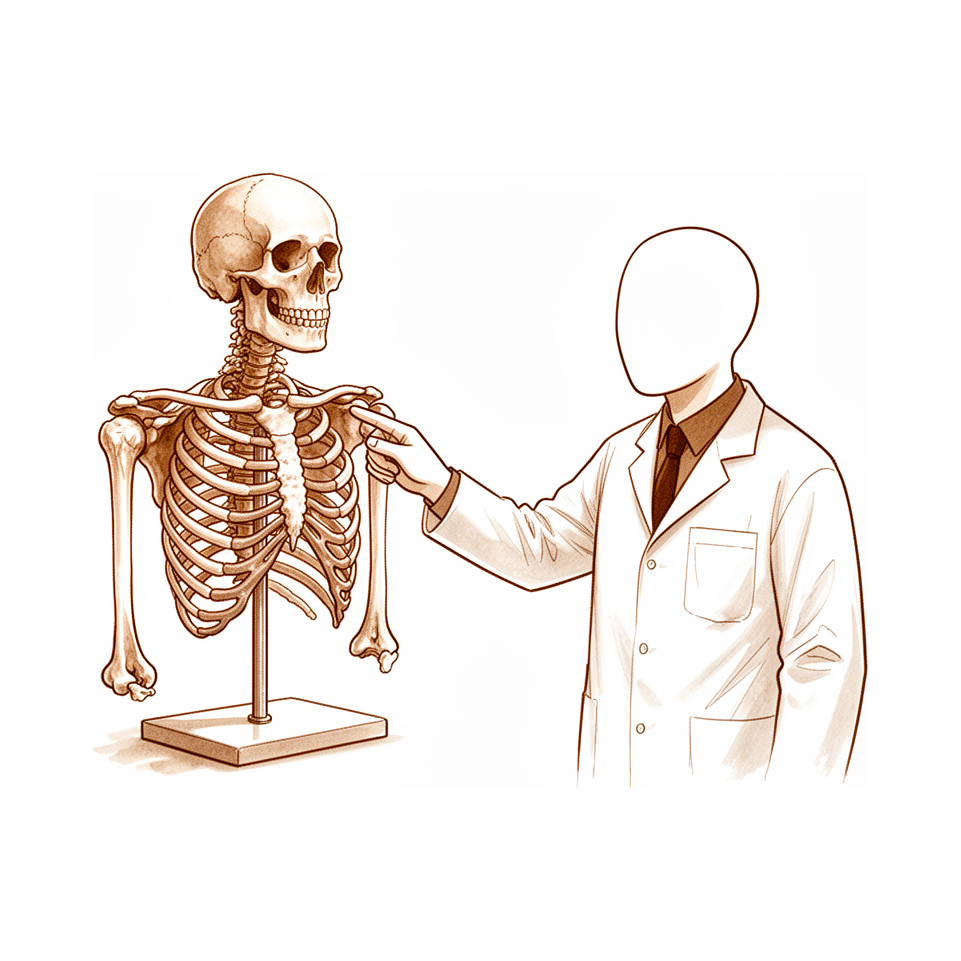
The scapula is attached to the axial skeleton by the acromioclavicular (AC) and sternoclavicular (SC) joints [71]. The basic part of the scapula is the body, which is triangular when viewed anteroposteriorly, with its base situated superiorly and its apex inferiorly [71]. The scapula spans second through seventh ribs and serves as an attachment for 17 muscles [84]. It is anteverted on the chest wall approximately 30 degrees relative to the body [84]. The glenoid is connected with the flat body of the scapula by the scapular neck [71]. The hook-shaped coracoid process curves forwards from the superior surface of the scapular neck [71]. The scapular spine ends in a flattened bony process, the acromion, which curves forwards [71]. The highest concentration of bony mass in the scapula is found in the glenoid, the scapular neck (including the base of the coracoid process), and the lateral border of the scapular body [71]. Two bony pillars transmit compressive forces from the glenoid fossa: the lateral pillar and the spinal pillar [71]. The lateral pillar connects the inferior border of the glenoid with the inferior angle [71]. The spinal pillar arises from the central part of the glenoid and continues medially to become part of the base of the scapular spine [71]. The weakest bone in the scapula is located primarily in the central part of the biomechanical body, i.e., in the infraspinous fossa [71]. The weakest area of the circumference of the biomechanical body of the scapula is the spinomedial angle, which is the connection of the scapular spine and the medial border of the scapula [71]. In most scapular body fractures, one of the main fracture lines passes through the spinomedial angle [71].
The glenoid is a convex structure of shallow depth shaped like an inverted pear [69]. The glenoid cavity is a shallow socket, approximately one third the size of the humeral head [70]. The subchondral bone of the glenoid is relatively flat; the articular concavity is augmented by cartilage and a circumferential labrum [72]. The glenoid averages 5° of retroversion in relation to the axis of the scapular body [72]. The glenoid is retroverted approximately 5 degrees relative to the scapular body [84]. The normal position of the glenoid surface in relation to the axis of the scapular body ranged from 2 degrees of anteversion to 7 degrees of retroversion [81]. The glenoid version is 1.5 degrees retroversion (10.5-9.5 degrees anteversion) [81]. The glenoid inclination averages 4.2 degrees (–7 to 20 degrees) [81]. The glenoid diameter superior anteroposterior is 18-30 mm [81]. The glenoid diameter inferior anteroposterior is 21-35 mm [81]. The glenoid superoinferior height is 30-48 mm [81]. The glenoid surface area is 4-6 mm [81]. The glenoid cartilage thickness is 2.16 mm [81]. The glenoid radius of curvature is 22-28 mm [81]. The glenoid articular surface radius of curvature is 2 to 3 mm larger than that of the humeral head [81].
The articular head of the humerus is spherical and has a diameter of 37 to 57 mm [69]. The articular surface of the humeral head is essentially spherical, with an arc of approximately 160 degrees covered by articular cartilage [81]. The radius of curvature of the humeral head is approximately 25 mm and is slightly larger in men than in women [81]. The humeral head radius of curvature is 23-28 mm (smaller in women than men) [81]. The humeral head surface area is 11-19 mm [81]. The humeral head cartilage thickness is 1.44 mm [81]. The most superior portion of the articular surface of the humeral head averages 8 mm above the greater tuberosity [69]. The superior margin of the humeral head articular surface normally is superior to the top of the greater tuberosity by 8 to 10 mm [81]. The head height is approximately 5.6 cm above the superior border of the pectoralis major tendon [84]. Humeral version averages 29.8 degrees, with a range of 10 to 55 degrees [69]. The humeral head is retroverted an average of 30 degrees [70]. The humeral head is retroverted 30 degrees relative to the transepicondylar axis of the humerus [84]. Mean humeral retroversion values are around 26 degrees in healthy adults [76]. Proximal humeral retroversion is highly variable, ranging from 0 to 55 degrees, depending on the method used for measurement [81]. The humeral head version is 0-55 degrees retroversion (dependent on measurement method; highly variable among individuals) [81]. The humeral head is inclined approximately 130 degrees with respect to the humeral shaft [69]. The neck-shaft angle measures an average of 135 degrees [70]. The humeral head averages 19° of retroversion and 41° of inclination (neck-shaft angle) [72]. The average neck-shaft angle is 45 degrees (±5 degrees), with a range of 30 to 50 degrees [81]. Arthritic shoulders have a flatter neck-shaft angle close to 50 degrees [81]. The humeral head inclination is 30-55 degrees [81]. The head-shaft angle is 30-55 degrees [81]. The medial (coronal) humeral offset is 4-14 mm [81]. The posterior (transverse) humeral offset is –2 to 10 mm [81].
The anatomic neck is located directly below the humeral head and serves as an attachment for the shoulder capsule [84]. The surgical neck is more distal than the anatomic neck and is more often involved in fractures [84]. The distance from the lateral base of the coracoid process to the lateral margin of the greater tuberosity is called the lateral humeral offset [81]. A significant decrease in lateral humeral offset reduces the lever arms for the deltoid and supraspinatus muscles, which weakens abduction and impairs function [81]. A significant increase in lateral humeral offset causes excessive tension on the soft tissues ("overstuffing" of the joint), which results in loss of motion and likely accelerates polyethylene wear [81]. Humeral articular malposition of more than 4 mm led to increased subacromial contact [81]. Offset of 8 mm in any direction significantly decreased passive range of motion [81]. Anatomic reconstruction of the humeral head/humeral shaft offset should be within 4 mm of normal to minimize subacromial contact and maximize glenohumeral motion [81].
The proximal humerus has three ossification centers: the humeral head (4 to 6 months), the greater tuberosity (1 to 3 years), and the lesser tuberosity (3 to 5 years) [72]. The proximal humeral ossification centers fuse to the shaft at age 17 to 20 years [72]. The formation of the humerus begins with the appearance of the cartilage anlage, which is present by the fifth week of gestation [76]. The primary ossification center for the humerus appears at about the sixth week [76]. By the time of birth, the entire humeral diaphysis is completely ossified [76]. The proximal humerus is primarily cartilaginous at birth [76]. Ossification centers for the proximal humerus can be detected with ultrasonography as early as the 38th week of gestation and are generally present between the 38th and 42nd week of gestation [76]. The ossification center for the humeral head is usually present at birth [76]. The greater tuberosity ossification center appears by 1 to 3 years of age [76]. The lesser tuberosity ossification center appears by 5 years of age [76]. The proximal humeral ossification centers fuse by 5 to 7 years of age to form the humeral head [76]. The proximal humeral physis closes by 14 to 17 years of age in girls and by 16 to 18 years in boys [76]. In infants and young children, humeral retroversion averages 65 degrees and gradually decreases, approaching adult values by 11 years of age [76]. Eighty percent of subsequent growth of the humerus comes from the proximal humeral physis [76]. The proximal humeral physis accounts for approximately 40% of the growth of the entire upper extremity [76]. Less than 75% of growth from the proximal humerus occurs before 2 years of age [76]. More than 85% of growth from the proximal humerus occurs by 8 years of age [76].
The clavicle is the first bone to ossify (fifth week of gestation) and is the only long bone to ossify by intramembranous ossification [72]. The medial (sternal) epiphysis of the clavicle is the last ossification center to fuse, at age 20 to 25 years [72]. The primary blood supply to the clavicle is periosteal; no nutrient artery is present [72]. The acromion has three ossification centers: the metacromion (base), the mesoacromion (middle), and the preacromion (tip) [72]. Failure of fusion of the acromial ossification centers results in os acromiale [72].
Vascular Anatomy
The proximal humerus receives its blood supply from the anterior and posterior humeral circumflex branches from the third division of the axillary artery [69]. The anterior humeral circumflex artery (AHCA) arises from the axillary artery at the inferior border of the subscapularis [69]. The AHCA provides vascular inflow to the humeral head by way of its terminal anterolateral branch known as the artery of Laing (also known as the arcuate artery) [69]. The ascending branch of the AHCA courses parallel to the lateral aspect of the long head biceps tendon and enters the humeral head at the interface of the bicipital groove and greater tuberosity [69]. The anterolateral ascending branch of the anterior humeral circumflex artery provides the primary blood supply to the humeral head [72]. The terminal intraosseous portion of the anterior humeral circumflex artery enters at the proximal aspect of the intertubercular groove as the arcuate artery [72]. The major blood supply to the humeral head is through the ascending branch of the anterior humeral circumflex artery, which penetrates the head at the bicipital groove and becomes the arcuate artery [70]. Injury to the arcuate artery may result in osteonecrosis of the humeral head [69]. Additional extraosseous collateral branches can permit humeral head perfusion despite complete ligation of the arcuate artery [69]. The posterior humeral circumflex artery travels with the axillary nerve, enters the quadrilateral space posteriorly, and anastomoses with a branch of the anterior circumflex to supply the posterior cuff [69]. Fractures of the anatomic neck have a poor prognosis because of complete disruption of the blood supply to the head [70]. Surgical neck fractures are common, and with these, the blood supply to the head is preserved [70].
Joints and Ligaments
The shoulder joint is composed of four articulations: the sternoclavicular, acromioclavicular, glenohumeral, and scapulothoracic [82]. The sternoclavicular (SC) joint is the only true diarthrodial articulation between the upper appendicular and axial skeletons [72]. The SC joint has double gliding with an articular disc [84]. The SC joint rotates 30 degrees with shoulder motion [84]. The posterior SC joint capsule and ligaments are the primary stabilizers to anterior and posterior translation of the medial clavicle [72]. The posterior sternoclavicular ligament is the strongest and primary restraint to anteroposterior instability [84].
The acromioclavicular (AC) joint is a small diarthrodial joint with an interposed fibrocartilaginous disk [72]. The AC joint is a plane/gliding joint with a fibrocartilaginous disc [84]. The AC joint is a diarthrodial joint that serves as a primary link between the axial skeleton and the upper extremity [63]. The AC joint has dynamic and static stabilizers and is movable in all planes so it is not a rigid structure [63]. The complex ligamentous structure of the AC joint is critical to the normal function of the shoulder girdle [63]. The main ligaments of the AC joint are superior, inferior, anterior, and posterior [63]. The main function of the AC joint ligaments is reinforcement of the capsule surrounding the joint [63]. The AC and CC ligaments are the static stabilizers of the AC joint [63]. The deltoid and trapezoid muscles are the dynamic stabilizers of the AC joint [63]. The superior and posterior AC ligaments are the primary stabilizers to anterior and posterior (horizontal) translation of the clavicle [72]. The AC ligaments prevent anteroposterior displacement [84]. The posterior and superior AC ligaments are considered the strongest [84]. The normal AC joint is capable of translating 4 to 6mm in the anterior, posterior, and superior planes under 70-N loads [63]. The AC joint accommodates rotary motion of 5° to 8° during scapulothoracic motion [63]. The AC joint accommodates rotary motion of 40° to 45° with shoulder abduction and elevation [63].
The coracoclavicular (CC) ligaments are the primary stabilizers to superior (vertical) translation of the distal clavicle [72]. The CC ligaments prevent superior displacement of the distal clavicle [84]. The conoid ligament is medial and the trapezoid ligament is lateral [72]. The trapezoid ligament is anterolateral and located approximately 25 mm from the AC joint [84]. The conoid ligament is posteromedial, stronger, and located approximately 45 mm from the AC joint [84].
The glenohumeral joint depends on static and dynamic stabilizers for movement and stability [81]. The rotator cuff stabilizes the glenohumeral joint via joint compression [72]. Positioning of the scapulothoracic joint contributes to dynamic stability of the glenohumeral joint [72]. Static stabilizers of the glenohumeral joint include articular congruity, the glenoid labrum, concavity-compression, negative intra-articular pressure, and the glenohumeral capsule and ligaments [72]. The glenoid labrum provides concavity and up to 50% of marginal glenoid socket depth [72]. The fibrocartilaginous glenoid labrum deepens the socket by 50% around the humeral head and increases stability [82]. The glenoid articular surface and the labrum combine to create a socket that is approximately 9 mm deep in the superoinferior direction and 5 mm deep in the anteroposterior direction [82]. Adding the glenoid labrum increases the glenoid surface to 75% of the humeral head vertically and 57% horizontally [82].
The rotator interval is defined medially by the base of the coracoid, superiorly by the supraspinatus tendon, and inferiorly by the subscapularis tendon [72]. The rotator interval contains the coracohumeral (CH) ligament, the superior glenohumeral ligament (SGHL), and the intra-articular portion of the long head of the biceps tendon [72]. Laxity of the rotator interval results in inferior laxity (the sulcus sign) [72]. Contracture of the rotator interval is seen with adhesive capsulitis [72]. The transverse humeral ligament is an important stabilizer of the biceps tendon [84].
Classification
General Principles
Shoulder instability presents with a variety of clinical presentations and a complex nature that has until recently been poorly understood [21]. A new classification system categorizes instability based on frequency, aetiology, direction, and severity [146]. The 2024 ESSKA–ESA Formal Consensus provides a framework for individualized decision-making in traumatic anterior shoulder instability by integrating recurrence status, bone loss, soft tissue injury, and sport type [57]. Recurrent shoulder instability occurs more frequently following soft-tissue stabilization procedures compared to bony stabilization procedures [10]. In a substantial number of patients with a long-standing history of instability, classification of lesions was impossible at arthroscopy due to scar tissue formation and advanced degenerative changes in the labroligamentous complex [8].
Acromioclavicular Joint
Rockwood: This system is based on the degree and direction of disrupted anatomy [44]. Type I is established when 0% to <10% superior displacement of the distal clavicle is found [24]. Type II is present when the affected side differs by 10% to ≤25% superior displacement of the distal clavicle [24]. Type III dislocation is defined as a coracoclavicular distance (CCD) of >25% to ≤100% compared with the contralateral side [24]. A CCD of >100% compared with the contralateral side indicates a type V separation [24]. The modified Rockwood classification system includes deltotrapezial fascial injury in the grading of ACJ injuries [192].
New AC Joint Instability Classification: This system defines two groups based on a cutoff of a 30% difference in the CCD compared with the contralateral side [24]. Group 1 includes patients with a CCD ≤30%, encompassing all Rockwood type I, type II, and borderline low-grade type III patients [24]. Group 2 is defined as having a CCD >30%, representing high-grade AC joint dislocations including all Rockwood type V patients and the majority of Rockwood type III patients [24]. High-grade acromioclavicular injuries are always based on additional structural damage to the coracoclavicular ligaments [157].
ISAKOS: This classification system, including type IIIA and IIIB, requires a comprehensive clinical evaluation to establish the true pathoanatomy and pathomechanics [11]. The ‘low grade/high grade’ system for AC joint injuries also requires a comprehensive clinical evaluation to establish the true pathoanatomy and pathomechanics [11]. For acute AC joint injuries, the clinical exam should be performed at the initial visit and again 3–6 weeks after the injury to allow for reduction in pain and better evaluation of motions [11].
Imaging Discrepancies: Ultrasound, MRI, and radiography results were in agreement in 24 of 47 cases when assessing the contribution of ultrasound to the management of acromioclavicular joint injuries [27]. In 17 cases, ultrasound showed a grade 3 injury suggestive of ruptured coracoclavicular ligaments, which was confirmed on MRI, while radiography had given grade 2 [27]. Ultrasound was wrong in six cases and radiography in 24 cases when comparing imaging grades for acromioclavicular joint injuries [27].
Glenohumeral Joint
A detailed assessment of soft tissue injury is mandatory as it influences surgical outcomes in anterior shoulder instability [14]. Nonsurgical treatment is successful in most cases of recurrent posterior shoulder instability, but surgical intervention is indicated when conservative treatment fails [3]. Surgical intervention for recurrent posterior shoulder instability requires accurate definition of the instability pattern and addressing all soft-tissue and bony injuries [3]. It is important to identify all contributing pathoanatomic soft-tissue and bone factors for the selection of the adequate revision repair procedure in recurrent anterior shoulder instability [2]. All bony and soft tissue pathology should be identified and assessed in recurrent anterior shoulder instability after surgical stabilization in children and adolescents [5]. Simply repeating the steps of the index procedure typically results in poor outcomes in recurrent anterior shoulder instability, often requiring an escalation of surgical complexity at the time of revision [5].
Clinical Presentation
General Principles & Assessment
Shoulder instability presents with diverse clinical manifestations and a complex nature that was poorly understood until recently [21]. A detailed assessment of soft tissue injury is mandatory because it influences surgical outcomes, with various non-operative and operative options available based on patient characteristics [14]. Surgeons must identify all contributing pathoanatomic soft-tissue and bone factors while accounting for the patient's functional demands and activity level to select the adequate revision repair procedure [2]. In recurrent anterior shoulder instability, all bony and soft tissue pathology should be identified and assessed, as simply repeating the steps of the index procedure typically results in poor outcomes [5]. For recurrent posterior shoulder instability, nonsurgical treatment is successful in most cases, but surgical intervention is indicated when conservative treatment fails, requiring accurate definition of the instability pattern and addressing all soft-tissue and bony injuries [3]. Surgeons should individualize their approach based on patients' bone changes, soft tissue quality, activity requirements, and surgical experience to maximize success and reduce complications [18]. Anatomical variants should not be confounded with pathological findings when evaluating shoulder instability [26]. Different anatomical lesions can be found depending on the type of instability and the functional requirements of the patient [26]. Non-traumatic shoulder instability's aetiologies and clinical manifestations are multifactorial [50]. Rotator cuff disease, shoulder instability, and associated lesions are common pathologic conditions of the shoulder involving soft tissues [132].
Acromioclavicular (AC) Joint
The clinical evaluation for AC joint injuries requires a comprehensive assessment to establish the true pathoanatomy and pathomechanics to better identify treatment options [11]. The AC joint exam comprises visualization/inspection/palpation, provocative maneuvers, observation of motion, and corrective maneuvers that may alter the clinical symptoms [11]. For acute AC joint injuries, the exam should be performed both at the initial visit and 3–6 weeks after the injury to allow for reduction in pain and better ability to evaluate motions [11]. Clinical evaluation of acute AC injury is often deferred for 7 to 14 days after injury to minimize acute pain and swelling and to allow recovery of arm motion [23].
Inspection: Visualization and inspection of the AC joint should be accomplished by direct evaluation of the symptomatic joint and comparison to the asymptomatic contralateral joint [11]. Common visualized alterations in AC separations include varying amounts of prominence of the distal clavicle due to an apparent superior position relative to the inferiorly and medially displaced acromion [11]. Altered posture of the scapula and arm into protraction due to muscle weakness or imbalance associated with AC ligament sprains may be observed during inspection [11].
Palpation: Palpation of the clavicle along its entire length from the sternoclavicular joint to the AC joint can reveal point tenderness suggesting bony involvement [11]. Palpation of the acromion, scapular spine, coracoid, and CC ligament area allows pain in these areas to be differentiated from other anatomic areas around the joint [11]. The AC joint is palpated for point tenderness and crepitus on motion during clinical evaluation [23].
Stability & Special Tests: Ligament instability at the AC joint is evaluated by manual manipulation of the clavicle and acromion in the inferior-superior and anterior-posterior directions [23]. Increased translation of the AC joint compared with the uninvolved side is considered a positive finding for ligament instability [23]. Visual observation of bone and joint motion with arm motion in flexion and abduction is used to evaluate dynamic AC joint stability [23]. A positive finding for dynamic AC joint instability is noted if the acromion can be observed to displace around the clavicle with arm motion [23]. The clinical examination for AC joint instability includes evaluation of the labrum, which has been shown to be involved in AC joint injuries [23]. The modified dynamic labral shear test is used to identify labral injury in the context of AC joint evaluation [23]. The clinical examination for AC joint instability consists of three parts: the AC joint examination, the scapular evaluation, and the glenohumeral examination [23]. Scapular dyskinesis is graded as present or not present when the arms move through and return from forward elevation during AC joint evaluation [23]. If the AC joint can be manually reduced, the effect of the reduction on scapular dyskinesis is determined during clinical evaluation [23].
Imaging & Classification: Post-traumatic radiography for AC joint dislocations includes bilateral anteroposterior stress views with a 10-kg axial load and bilateral stress views according to Alexander [24]. The coracoclavicular (CC) distance is measured as the interspace between the inferior cortex of the clavicle and the highest part of the coracoid, measured parallel to the spine [24]. * Rockwood Type I: Established when 0% to <10% superior displacement of the distal clavicle is found [24]. * Rockwood Type II: Present when the affected side differs by 10% to ≤25% in CC distance [24]. * Rockwood Type III: Defined as a CC distance difference of >25% to ≤100% compared with the contralateral side [24]. * Rockwood Type V: Indicated by a CC distance difference of >100% compared with the contralateral side [24].
A new classification of AC joint instability defines Group 1 as having a CC distance difference ≤30%, including all RW type I, type II, and borderline low-grade type III patients [24]. A new classification of AC joint instability defines Group 2 as having a CC distance difference >30%, representing high-grade AC joint dislocations including all RW type V patients and the majority of RW type III patients [24]. Horizontal translation at the AC joint is graded as none, partial translation, or complete translation on bilateral Alexander views [24]. In cases of AC joint ligament disruption, the distal clavicle is displaced posterosuperiorly and overlapping with the acromion is less or nullified [24]. The Rockwood classification system is currently almost universally used for AC joint injuries and is based on the degree and direction of disrupted anatomy [44]. Postoperative ossifications of the AC joint ligaments are common but seem to have no influence on the clinical results [51].
Glenohumeral Instability (Anterior/Posterior)
In a substantial number of patients with a long-standing history of instability, classification of lesions was impossible at arthroscopy because of scar tissue formation and advanced degenerative changes in the labroligamentous complex [8]. Age less than 22 years old, ligamentous laxity, the presence of an off-track lesion, and a concomitant SLAP tear were significantly associated with revision failure in anterior shoulder instability [22]. Age less than 22 years old, ligamentous laxity, the presence of an off-track lesion, and a concomitant SLAP tear were significantly associated with revision failure in recurrent anterior shoulder instability [25]. If patients demonstrate symptomatic instability after arthroscopic soft tissue stabilization without evidence of labral retears on imaging scans, an anterior capsular tear should be considered as a possible factor for recurrence [47]. It is essential to show subtle capsulolabral and ligamentous findings that reduce joint stability in posterior shoulder instability [41]. Surgeons should have a heightened suspicion for posterior and combined anteroposterior labral pathology when performing arthroscopic stabilization procedures in young, active individuals [140]. Extensive labral lesions can occur even in patients with few previous dislocations, and clinical outcomes can be excellent with appropriate treatment [134]. Athletes with an initial instability injury and primary soft-tissue pathology may return to sport within 3 weeks, while those with bone loss, recurrent instability, or an inability to perform sport-specific drills are candidates for surgical stabilization [135]. By treating the intra-articular pathology, the extra-articular symptoms can be relieved in the vast majority of patients with minor or occult shoulder instability presenting with subacromial impingement symptoms [139].
Imaging & Assessment: Advanced imaging modalities are essential for identifying associated lesions, and bony reconstruction procedures should be considered for patients with significant glenoid bone loss or recurrent instability after soft tissue reconstruction [52]. MRI or MR-Arthrogram are the best methods for evaluating soft tissue quality, specifically assessing capsular volume, labral ring integrity and labral tissue degeneration (size and signal) [131]. Evaluation of soft tissue quality should also consider medical records (including the time from dislocation and the number of dislocation episodes), the mechanism of injury and the clinical examination [131]. In older adults with traumatic anterior shoulder instability, the most critical soft tissue lesion is the rotator cuff tear [131]. The most effective method for evaluating rotator cuff quality in older adults is MRI, though ultrasound can also be used depending on user experience [131]. Patients who present with glenohumeral instability undergo a standardized workup including a detailed history and physical as well as advanced imaging to critically assess soft tissues and glenoid bone loss [141]. The exact degree of glenoid bone loss is calculated in each patient and, in conjunction with history and physical examination, used to determine eligibility for specific procedures [141]. Bone loss of 20% is a relative indication for a bone augmentation procedure in anterior shoulder instability [141]. Radiographs are crucial to confirming the diagnosis after dislocation, traumatic or otherwise, and at least 2 views are required to prove a dislocation [43]. After reduction, MRA is used to assess for soft tissue injury in glenohumeral instability [43]. Imaging would show disruption of the capsular fibers with possible extravasation of contrast through the deficiency in glenohumeral ligament avulsion [43]. MRA can be limited in its clarity and, in the case of a GAGL lesion, may not clearly delineate the separation of the glenohumeral ligaments from the intact labrum, leading to a misdiagnosis of an anterior capsulolabral injury [43]. Diagnostic arthroscopy with proper portal placement becomes integral to identifying glenohumeral ligament avulsion pathology that may be missed on MRA [43].
Imaging & Diagnostic Limitations
The diagnostic value of US, MR, and MR arthrography in shoulder instability is supported by references to pathophysiology and stabilizing structures [12]. Ultrasound of the coracoclavicular ligaments in the acute phase of an acromioclavicular dislocation has been compared with radiographic and MRI findings [27]. The role of ultrasound in the evaluation of sports medicine injuries of the upper extremity includes assessment of glenohumeral ligaments and shoulder capsular mechanisms [30]. Patients searching the internet for information regarding shoulder labral pathology often look for facts regarding the diagnosis and management of their conditions [138]. Ultrasound, MRI, and radiography results were in agreement in 24 of 47 cases when assessing AC joint injuries [27]. In 23 of 47 cases, one of the three imaging techniques (ultrasound, MRI, radiography) had a different grade than the others for AC joint injuries [27]. Ultrasound was wrong in six cases and radiography in 24 cases when compared to MRI for grading AC joint injuries [27]. MRI should not be the imaging modality of first choice for AC joint injuries but could be useful to clinically assess low-grade injuries that have not settled to exclude higher-grade injury or if there are associated glenohumeral soft tissue injuries [44].
Investigations
Plain radiography: Standard radiographs provide an overview of bony anatomy, humeral head orientation relative to the glenoid, and initial assessment for bony Bankart and Hill–Sachs lesions [104]. The axillary view is necessary to evaluate for anterior or posterior humeral head subluxation or dislocation [99]. This view enables determination of the humeral head position in the glenoid fossa and may detect occult, locked posterior shoulder dislocation in patients with a lack of passive external rotation [99]. In a systematic review of posterior shoulder dislocations, 73% of patients had a missed initial diagnosis due to the lack of an axillary view, Y view, or CT imaging [104]. When axillary or Y-view radiographs were subsequently obtained in patients with initially missed posterior dislocations, the diagnosis was confirmed in 100% of patients [104]. Specific views are indicated for targeted pathology: the Stryker notch view evaluates Hill–Sachs lesions after dislocation [99]; the West Point view evaluates anterior glenoid bone loss [99]; the Zanca view evaluates the acromioclavicular joint and involves an AP projection with a 10° cephalic tilt centered over the AC joint [99]; and the apical oblique view evaluates glenoid rim fractures in instability [99].
CT: CT with three-dimensional reconstructions is the advanced imaging study of choice for determining the extent of glenoid bone loss in the setting of shoulder instability [99]. CT imaging proved to be more important than MRI for evaluating glenoid defects in recurrent anterior shoulder instability [172]. CT is the best technique for depicting and quantifying skeletal changes in shoulder instability [222]. Accurate imaging and assessment of the size and relations of bone defects are crucial for further treatment planning in bony-mediated shoulder instability [202]. Appropriate preoperative imaging is essential for detection and quantification of osseous abnormalities in recurrent shoulder instability [190].
MRI: MRI is the modality of choice for evaluating the rotator cuff, biceps, and subacromial/subdeltoid bursa [97]. T1-weighted MRI can reveal Hill–Sachs lesions and is often used with MR arthrograms to provide a more detailed picture of joint surfaces [97]. T2-weighted MRI provides better visualization of full-thickness rotator cuff tears [97]. MRI is a valid imaging tool to diagnose and measure osseous lesions of the shoulder in adolescents [204]. MRI is the best imaging study for the diagnosis of Humeral Avulsion of Glenohumeral Ligaments (HAGL) lesions [219]. ArthroMRI with contrast is necessary to make the diagnosis of isolated reverse HAGL lesions [223]. Careful interpretation of MR arthrograms and thorough diagnostic arthroscopy are essential to identify posterior humeral avulsion of the glenohumeral ligaments [178]. The sensitivity, specificity, and reliability of magnetic resonance arthrogram for detecting Kim's lesions in posterior shoulder instability are substantial [196]. Identification of critical radiographic variables on magnetic resonance arthrography assists in the accurate diagnosis and management of clinically significant posterior shoulder instability [217]. There is evidence to support the use of both CT and MRI imaging modalities in assessing the glenoid track in recurrent shoulder instability [221]. MRA can be limited in its clarity for Glenoid Avulsion of the Glenohumeral Ligaments (GAGL) lesions and may not clearly delineate the separation of the glenohumeral ligaments from the intact labrum, leading to a misdiagnosis of an anterior capsulolabral injury [43].
MR Arthrography: MR arthrography is considered the benchmark for evaluation of labral tears and is rarely indicated for evaluation of rotator cuff pathology [97]. MR arthrography increases the sensitivity and specificity for detecting injuries to the capsulolabral–ligamentous complex compared to traditional MRI [94]. A meta-analysis found greater diagnostic test accuracy for MRA over MRI in the detection of glenoid labral lesions, with MRA sensitivity of 88% and specificity of 93% versus MRI sensitivity of 76% and specificity of 87% [94]. Abduction and external rotation (ABER) positioning during MRA is utilized to increase the sensitivity and specificity for detecting anteroinferior labroligamentous injury [94]. MDCT arthrography showed better accuracy than MR arthrography in the detection of osseous, cartilage, and labroligamentous injuries related to anterior shoulder instability [220].
Ultrasonography: Ultrasonography is a low-cost alternative to MRI and arthrography for evaluating both skeletal and soft-tissue structures of the shoulder [97]. Ultrasonography can provide immediate, real-time visualization of the rotator cuff, biceps tendon, and calcific deposits [97]. Ultrasound is a simple and accurate test for identifying rotator cuff tears and calcific tendinitis [88]. The sensitivity of ultrasonography for the detection of full-thickness rotator cuff tears is 98% and the specificity is 80% [99]. In contrast, the sensitivity of MRI for the detection of full-thickness rotator cuff tears is 100% and the specificity is 68% [99]. Ultrasonography is highly operator dependent and is not as useful for evaluating labral tears or rotator cuff tears that are very small or larger than 3 cm [97]. In the acute phase of acromioclavicular dislocation, ultrasound was wrong in six cases and radiography in 24 cases when compared to MRI [27].
Other Considerations: The purpose of imaging the shoulder is to help establish the diagnosis, determine the severity of the pathoanatomy, assist in surgical planning, and enable the surgeon to illustrate the condition of the shoulder to the patient [39]. Standardized plain films are almost always sufficient to garner the information needed for shoulder care, and there is information that can be gathered from properly taken plain films that cannot be obtained from CT scans [39]. The shoulder is a three-dimensional structure that cannot be represented by a single planar view, and critical relationships such as the degree of centering of the humeral head change with the position of the arm [92]. Advanced imaging modalities are essential for identifying associated lesions in shoulder instability [52]. Radiography remains an essential tool to provide a general overview and rule out serious conditions in shoulder instability [222]. The indications for an isolated soft-tissue procedure in anterior shoulder instability are narrower, with the ideal candidate presenting with minimal glenoid bone loss of 13.5% [15]. In acute acromioclavicular dislocations, the coracoclavicular distance (CCD) is measured as the interspace between the inferior cortex of the clavicle and the highest part of the coracoid, measured parallel to the spine [24]. The normal coracoclavicular distance is 1.1 to 1.3 cm [99]. A new classification of AC joint instability defines Group 1 as a CCD ≤30% and Group 2 as a CCD >30% compared with the contralateral side [24].
Treatment
Non-Operative
Nonsurgical treatment remains successful in most cases of recurrent posterior shoulder instability [3], and recent studies continue to demonstrate a role for nonoperative treatment in the successful long-term management of anterior glenohumeral instability [155]. However, primary non-operative management is a prominent risk factor for recurrence of shoulder instability [54]. Treatment algorithms have traditionally included a period of non-operative management in all patients, although young athletic patients may often benefit from early operative treatment [137]. In the context of acromioclavicular (AC) injuries, nonsurgical management of type V injuries allows most patients to return to work, though functional outcome scores are limited [151]. From the standpoint of objective strength, nonsurgical treatment of Grade III AC injury is as effective as surgical treatment [171]. For athletes, conservative management is highly effective for those whose goal is to return to the same level of sport in the next season, calling into question routine fixation for first-time dislocators [166]. Most athletes are able to complete the season with non-operative treatment, however over 1/3rd suffered recurrent instability events during the year [170]. Of the 32 NHL team physicians surveyed, 28 preferred nonoperative management for the treatment of initial anterior shoulder dislocations with isolated soft tissue injuries [165]. While nonoperative treatment is associated with faster return to play, operative management is associated with fewer recurrent instability events, greater time between recurrent instability events, and greater career longevity [158]. Patients with a chief complaint of pain were much more likely to succeed with nonoperative treatment while those with instability were more likely to undergo surgical management [56]. Nonoperative treatment is commonly unsuccessful in active patients with posterior shoulder instability, and surgical stabilization can be considered in patients who do not respond [129]. Conservative treatment can be used as the first-line therapy for secondary frozen shoulder after traumatic anterior shoulder instability, followed by effective arthroscopic mobilization when conservative treatment fails [149]. Specific non-operative protocols exist for the in-season management of AC injuries to quickly and safely return athletes to play [143]. Clinicians are urged to err on the conservative side when possible until better evidence defines when surgery is necessary for unstable acromioclavicular injuries [154]. At a minimum 5-year follow-up, patients with successful non-operative treatment for type III-V ACJ injuries achieved similar clinical outcomes compared to those who were converted to anatomic coracoclavicular ligament reconstruction [163].
Operative
Indications: It is important to identify all contributing pathoanatomic soft-tissue and bone factors and to take into account the patients' functional demands and activity level for selection of the adequate revision repair procedure for recurrent anterior shoulder instability [2]. There is no one procedure that fits all patients with anterior instability; surgeons should individualize their approach based on patients' bone changes, soft tissue quality, activity requirements, and surgical experience to maximize success and reduce complications [18]. By integrating recurrence status, bone loss, soft tissue injury and sport type, the 2024 ESSKA–ESA Formal Consensus provides a clinically valuable framework for individualized decision-making for traumatic anterior shoulder instability [57]. Overall, 77% of statements reached unanimous or strong consensus, covering patient history evaluation, prognostic factors for nonoperative management, and Bankart repair steps for anterior shoulder instability [31]. The indications for an isolated soft-tissue procedure in anterior shoulder instability are now narrower; the ideal candidate presents with minimal glenoid bone loss (13.5%) [15]. The failure rate in contact athletes with recurrent anterior shoulder instability is high when treated with a soft tissue procedure alone [34].
Surgical Approach / Technique: Regardless of which procedure is chosen by a surgeon for anterior shoulder instability, the treatment should follow the guidelines taught by Rowe: anatomic dissection, identification and repair of the lesions responsible for the instability, returning tissues to their anatomic locations, and early postoperative range of motion [17]. Achieving the best result for any particular patient depends on the procedure that allows observation of the joint surfaces, provides the anatomical repair, maintains range of motion, and also can be applied with low rates of complications and recurrence [33]. Long-term clinical studies demonstrate good clinical outcomes of patients operated with a Mason–Allen labral repair for recurrent anterior shoulder instability [20]. The treatment strategy of arthroscopic soft tissue stabilization combined with selected augmentations provided good clinical outcomes for competitive collision athletes, characterized by low rates of recurrence and complication, a high rate of return to sports, and good shoulder function [148]. Soft tissue stabilization combined with selective augmentation procedures for shoulder instability in collision athletes demonstrated satisfactory outcomes with low recurrence rate [156]. Soft tissue stabilization combined with selective augmentation procedures for traumatic shoulder instability in collision athletes demonstrated satisfactory outcomes with extremely low recurrence rate [167]. The procedure of arthroscopic transfer of the long head of the biceps brachii is indicated for patients younger than 45 years with specific activity requirements or capsule-ligament insufficiency, and for patients 45 years or older with combined SLAP lesions [55]. In the case series with 1 year follow-up, 20 patients who underwent arthroscopic labrum repair with the addition of a dermal allograft had a significant improvement in outcome scores, and excellent incorporation of the graft as seen on MRI [37]. This procedure has the advantages of being an anatomic reconstruction that addresses bony and soft tissue instability for recurrent anterior shoulder instability [6]. Capsular reconstruction has emerged as option for the management of instability with poor quality or absent capsular tissue [164]. This case demonstrates a clear indication for the usefulness of diagnostic and therapeutic arthroscopy in the situation of soft tissue instability complicating a previously successful total shoulder arthroplasty [59].
Acromioclavicular Joint Reconstruction: Its emphasis on flexible, anatomic ligament reconstruction, combined with favorable early results showing stability, accelerated rehabilitation, and an absence of common hardware complications, positions the N-way technique as a viable alternative to overcome the drawbacks of established methods for AC joint separation [1]. Natural anatomical reconstruction of the CC ligaments has excellent potential to replicate the function of normal ligaments, achieving both maximum therapeutic efficacy and patient satisfaction [4]. The authors do not recommend surgery for grade-III ACD using Ligastic-type reconstruction due to the risks inherent to surgery and evolutive clavicular osteolysis [16]. Biologic soft-tissue reconstruction techniques offer robust time zero strength and a diminished complication profile for the management of an ipsilateral acromioclavicular and sternoclavicular joint dislocation [19]. The arthroscopic reconstruction of the AC separation is a low-morbidity, safe, and reproducible operation that provides adequate fixation and stability combined with the use of a soft tissue graft to promote sound biologic healing [32]. Combined anatomic reconstruction of both AC and CC ligaments using nylon tape by the described technique provides overall 88.2% satisfaction, 94% radiologic reduction, and a low complication rate [174]. Comparable subjective outcomes after surgical treatment of AC joint instability were reported for all modalities, with relatively low unplanned reoperation rates [175]. Future surgical approaches should systematically evaluate and address both CC ligament-mediated vertical stability and AC ligament-mediated horizontal stability to optimize clinical outcomes [176]. An arthroscopically assisted reconstruction offers the advantage of less soft tissue dissection and superior visualization to the base of the coracoid compared to traditional open approaches for high-grade AC separation [45]. All patients improved subjectively with regard to pain and function at a minimum followup of 3 months (mean, 5 months; range, 3-18 months) following the arthroscopically assisted coracoclavicular ligament reconstruction technique [45]. Protection of the repair with a sling for 6 weeks significantly improved functional and anatomical results in the surgical treatment of chronic acromioclavicular dislocations [65]. Medialized clavicular bone tunnel position predicts failure after anatomic coracoclavicular ligament reconstruction in young, active male patients [160]. A total of 16 studies reporting on 327 CC ligament reconstruction procedures were included in a systematic review, with 11 studies (253 cases) reporting a minimum follow-up of 5 years and 5 studies (74 cases) reporting a minimum follow-up of 10 years [9]. According to the MINORS criteria, noncomparative studies had a mean score of 12/16, and comparative studies had a mean score of 16/24, indicating moderate quality of evidence for both study types in CC ligament reconstruction [9].
Posterior Shoulder Instability: Operative treatment for posterior shoulder instability can be divided into two broad groups: soft-tissue repair and osseous stabilization [145].
Postoperative Rehabilitation: The arm was placed in a sling for comfort for a few days following radiofrequency capsular shrinkage for voluntary shoulder instability, with motion allowed avoiding extremes in all directions for the first 6 weeks [133]. Proprioceptive physiotherapy was initiated within the first week after surgery for radiofrequency capsular shrinkage [133]. Patients were placed in an abduction sling and instructed to wear the sling with the arm supported and without tension on the reconstructed ligaments for 6 weeks following anatomic coracoclavicular ligament reconstruction [160]. Range of motion exercises were initiated in physical therapy at 6 weeks postoperatively following anatomic coracoclavicular ligament reconstruction [160]. Strengthening exercises were begun at approximately 3 months following anatomic coracoclavicular ligament reconstruction [160]. The patient is to remain non-weightbearing on the left upper extremity with no active range of motion of the left shoulder for 2 weeks following revision shoulder instability surgery using distal tibial allograft [161]. Patients are non-weight bearing in a shoulder immobilizer for the first 6 weeks following arthroscopic anatomic knotless coracoclavicular ligament repair [168]. Passive range of motion is allowed during the first 6 weeks up to 90 of forward flexion and abduction following arthroscopic anatomic knotless coracoclavicular ligament repair [168]. The patient is restricted from carrying any objects greater than 5 pounds until 12 weeks postoperation following arthroscopic anatomic knotless coracoclavicular ligament repair [168]. The patient begins gentle progressive strengthening exercises at 12 weeks following arthroscopic anatomic knotless coracoclavicular ligament repair [168]. The patient starts advanced strengthening and sport-specific exercises at 4 months following arthroscopic anatomic knotless coracoclavicular ligament repair [168]. Return to sport occurs between 6 and 9 months postoperatively following arthroscopic anatomic knotless coracoclavicular ligament repair [168]. The patients' shoulders were immobilized for 4 weeks with the elbow against the body in a light removable sling following endoscopically assisted reconstruction of acute acromioclavicular joint dislocation using a synthetic ligament [152]. Passive hanging movements were authorized beginning the 1st postoperative day following endoscopically assisted reconstruction of acute acromioclavicular joint dislocation using a synthetic ligament [152]. The extremity is protected in a broad arm sling for 6 weeks following surgical treatment of acute type-V acromioclavicular injuries in athletes [153]. Isometric and pendulum exercises are started a few days after surgery for acute type-V acromioclavicular injuries [153]. Active mobilization is allowed after 6 weeks following surgical treatment of acute type-V acromioclavicular injuries in athletes [153]. The patient is initially placed in an immobilization sling for the first 6 postoperative weeks following arthroscopic labral repair in the setting of recurrent posterior shoulder instability [162]. Passive range-of-motion exercises may be initiated in the sagittal and frontal plane as well as shoulder external rotation to 40° in neutral following arthroscopic labral repair for posterior shoulder instability [162]. It is important to restrict internal rotation past neutral until 6 weeks after surgery and internal rotation with abduction until 8 weeks after surgery to protect repaired tissues in posterior shoulder instability [162].
Complications
Recurrence and Failure
Recurrent instability occurs more frequently following soft-tissue compared to bony stabilization procedures [10]. Long-term results suggest higher than optimal recurrence and revision rates, especially in patients younger than 23 years old, despite using modern day suture anchor and capsular tightening techniques [195]. Instability-related complications occurred only in the capsulabral group, and the incidence increased with time [169]. A history of multiple instability episodes prior to presentation was the greatest predictor of recurrent instability and failure of nonoperative treatment and progression to surgery [66]. Age less than 22 years old, ligamentous laxity, the presence of an off-track lesion, and a concomitant SLAP tear were significantly associated with revision failure [22]. Characteristics related to a history of instability (age <20 years at first instability episode, larger number of dislocations, ≥2 years between first dislocation and surgery) were found to be risk factors for the development of an off-track HS lesion [67]. Patients with hyperlaxity and instability are more likely to experience episodes of recurrent subluxation than they are to have recurrent dislocation [181]. Poor quality of the soft tissues surrounding the glenohumeral joint, whether caused by multiple previous operations, multiple subluxation or dislocation episodes, or an underlying connective tissue disorder, has the potential to threaten the success of any shoulder stabilization procedure [181].
Hardware and Graft Complications
The most disabling failures occurred when internal fixation was performed too soon after soft-tissue healing [179]. In a series of 31 patients undergoing primary anatomic coracoclavicular ligament reconstruction, 7 patients (22.6%) experienced a complication requiring a subsequent surgical procedure, including graft rupture/attenuation (2), clavicle fractures (2), distal clavicle hypertrophy (2), and adhesive capsulitis (1) [209]. A case of tunnel widening after CC ligament reconstruction with a semitendinous autograft tendon was reported, noting no definite detrimental effect on the functional outcome at more than 2 years' follow-up [58]. AC joint reconstruction with the LARS achieves good-to-excellent clinical and functional outcomes at long-term follow-up with a surgical revision rate of 8.5% [60]. Anatomic AC reconstruction with LARS1 artificial ligament resulted in both satisfactory functional outcome and low complication rate [177]. CC ligament augmentation is highly recommended to improve short-term outcomes and decrease complications for Rockwood type V AC separation treated by hook plate [180].
Degenerative and Long-Term Outcomes
Although uncommon, an age of at least 30 years at the time of diagnosis and surgical intervention were identified as risk factors for developing symptomatic arthritis at long-term follow-up after posterior shoulder instability [68]. Patients with recurrent shoulder instability who required more invasive stabilization procedures were more likely to have severe bone loss [185].
Recovery
Light activity (weeks): Clinical evaluation of acute acromioclavicular joint injury is deferred for 7 to 14 days after injury to minimize acute pain and swelling and to allow recovery of arm motion [23].
Other Considerations: Early repairs of acromioclavicular dislocations within 3 weeks showed a trend for better results and return to sports or heavy labor compared to late reconstructions, though this was not statistically significant in the overall comparison [227]. The acromioclavicular ligament exhibits early and dynamic healing responses following acute traumatic rupture [229]. No long-term disability results from the loss of the coraco-acromial ligament from its normal site [53]. A case report of tunnel widening after coracoclavicular ligament reconstruction with a semitendinous autograft tendon noted no definite detrimental effect on the functional outcome at more than 2 years' follow-up [58]. In a case series with 1 year follow-up, 20 patients who underwent arthroscopic labrum repair with the addition of a dermal allograft had a significant improvement in outcome scores, and excellent incorporation of the graft as seen on MRI [37]. The combination of arthroscopic remplissage in addition to the classic Bankart repair for recurrent anterior shoulder instability with engaging Hill–Sachs lesions has long-term outcomes in terms of the recurrence rate and does not significantly influence the range of motion of the shoulder [183]. Early to midterm follow-up of the arthroscopic J-bone-graft technique has revealed excellent clinical results, and the traditional open technique has been in use successfully for over 3 decades [228]. Surgical stabilization appears to restore the rate and timing of instability to that of players with no prior history of instability in professional football players [226]. At long-term follow-up of 17 years, a high rate of poor outcomes was observed following nonoperative management of anterior shoulder instability [187]. Long-term follow-up demonstrates that nearly 40% of patients treated non-operatively for posterior shoulder instability eventually require surgery [194]. Patients who underwent surgery at any time point for posterior shoulder instability were at an increased risk of radiographic progression of arthritis at a minimum of 5 years of follow-up [225].
Key Evidence
- [L4] Its emphasis on flexible, anatomic ligament reconstruction, combined with favorable early results showing stability, accelerated rehabilitation, and an absence of common hardware complications, positions it as a viable alternative to overcome the drawbacks of established methods. [1] (10.1016/j.eats.2025.103906)
- [L5] It is important to identify all contributing pathoanatomic soft-tissue and bone factors and to take into account the patients' functional demands and activity level for selection of the adequate revision repair procedure. [2] (10.2106/jbjs.st.k.00023)
- [L5] Nonsurgical treatment is successful in most cases, but surgical intervention is indicated when conservative treatment fails, requiring accurate definition of the instability pattern and addressing all soft-tissue and bony injuries. [3] (10.5435/00124635-200608000-00004)
- [L4] Natural anatomical reconstruction of the CC ligaments has excellent potential to replicate the function of normal ligaments, achieving both maximum therapeutic efficacy and patient satisfaction. [4] (10.1007/s00264-010-1124-3)
- [L4] Specifically, all bony and soft tissue pathology should be identified and assessed, with an understanding that simply repeating the steps of the index procedure typically results in poor outcomes, and often an ' escalation ' of surgical complexity is required at the time of revision. [5] (10.1007/s12178-020-09612-4)
- [L4] This procedure has the advantages of being an anatomic reconstruction that addresses bony and soft tissue instability. [6] (10.1177/0363546520960119)
- [L2] In a substantial number of patients with a long-standing history of instability, classification of lesions was impossible at arthroscopy because of scar tissue formation and advanced degenerative changes in the labroligamentous complex. [8] (10.1148/radiol.2372041429)
- [L4] [9] (10.1177/03635465261457302)
- [L2] Recurrent instability does not vary by region but occurs more frequently following soft-tissue compared to bony stabilization procedures. [10] (10.1016/j.xrrt.2023.08.005)
- [L5] [11] (10.1177/17585732221122335)
- [L3] [12] (10.1016/s0020-1383(13)70194-3)
- [L5] A detailed assessment of soft tissue injury is mandatory as it influences surgical outcomes, with various non-operative and operative options available based on patient characteristics. [14] (10.1177/2325967121s00835)
- [Paper] The indications for an isolated soft-tissue procedure in anterior shoulder instability are now narrower; the ideal candidate presents with minimal glenoid bone loss (13.5%). [15] (10.2106/jbjs.rvw.26.00033)
- [L4] The authors do not recommend surgery for grade-III ACD using Ligastic-type reconstruction due to the risks inherent to surgery and evolutive clavicular osteolysis. [16] (10.1016/j.otsr.2010.06.004)
- [L5] Regardless of which procedure is chosen by a surgeon, the treatment should follow the guidelines taught by Rowe: anatomic dissection, identification and repair of the lesions responsible for the instability, returning tissues to their anatomic locations, and early postoperative range of motion. [17] (10.1177/03635465030310011001)
- [L5] There is no one procedure that fits all patients with anterior instability; surgeons should individualize their approach based on patients' bone changes, soft tissue quality, activity requirements, and surgical experience to maximize success and reduce complications. [18] (10.1016/j.arthro.2017.09.028)
- [L5] Biologic soft-tissue reconstruction techniques, such as those described in the present chronicle, offer robust time zero strength and a diminished complication profile. [19] (10.1016/j.eats.2023.01.006)
- [Paper] Long-term clinical studies demonstrate good clinical outcomes of patients operated with a Mason–Allen labral repair. [20] (10.1016/j.eats.2021.04.018)
- [L5] Shoulder instability is a phenomenon with a variety of clinical presentations, and its complex nature has until recently been poorly understood. [21] (10.1016/j.cuor.2004.04.002)
- [L3] Age less than 22 years old, ligamentous laxity, the presence of an offtrack lesion, and a concomitant SLAP tear were significantly associated with revision failure. [22] (10.1016/j.arthro.2018.10.084)
- [L4] [23] (10.1016/j.arthro.2016.08.023)
- [L1] [24] (10.1016/j.jse.2020.10.026)
- [L3] Age less than 22 years old, ligamentous laxity, the presence of an off-track lesion, and a concomitant SLAP tear were significantly associated with revision failure. [25] (10.1016/j.arthro.2018.10.083)
- [L2] [27] (10.1007/s00330-016-4413-4)
- [L5] [30] (10.1016/j.csm.2006.03.004)
- [L5] Overall, 77% of statements reached unanimous or strong consensus, covering patient history evaluation, prognostic factors for nonoperative management, and Bankart repair steps. [31] (10.1016/j.arthro.2021.07.022)
- [L4] The arthroscopic reconstruction of the AC separation is a low-morbidity, safe, and reproducible operation that provides adequate fixation and stability combined with the use of a soft tissue graft to promote sound biologic healing. [32] (10.1016/j.jse.2009.12.014)
- [L4] Achieving the best result for any particular patient depends on the procedure that allows observation of the joint surfaces, provides the anatomical repair, maintains range of motion, and also can be applied with low rates of complications and recurrence. [33] (10.12998/wjcc.v2.i11.676)
- [L5] The failure rate in these situations with a soft tissue procedure alone is high. [34] (10.1016/j.ocl.2014.09.005)
- [L3] Coracoclavicular ligament reconstruction is a relatively common and safe procedure in a physically active population. [36] (10.1177/2325967121s00708)
- [L4] In the case series with 1 year follow-up, 20 patients who underwent arthroscopic labrum repair with the addition of a dermal allograft had a significant improvement in outcome scores, and excellent incorporation of the graft as seen on MRI. [37] (10.1016/j.arthro.2024.11.027)
- [L5] It is essential to show subtle capsulolabral and ligamentous findings that reduce joint stability. [41] (10.1016/j.mric.2019.12.005)
- [Paper] [43] (10.1016/j.eats.2019.06.001)
- [L1] [44] (10.1016/j.injury.2015.10.060)
- [L4] [45] (10.1007/s11999-007-0085-3)
- [L3] If patients demonstrate symptomatic instability after arthroscopic soft tissue stabilization without evidence of labral retears on imaging scans, an anterior capsular tear should be considered as a possible factor for recurrence. [47] (10.1177/2325967121995891)
- [L5] Unanimous consensus was reached on relative indications for nonoperative management and labral repair, as well as steps to minimize complications for labral repair. [49] (10.1016/j.arthro.2024.04.035)
- [L5] Non-traumatic shoulder instability's aetiologies and clinical manifestations are multifactorial. [50] (10.1177/17585732251320070)
- [L4] Postoperative ossifications of the AC joint ligaments are common, but seem to have no influence on the clinical results. [51] (10.1055/s-2008-1038577)
- [Paper] Advanced imaging modalities are essential for identifying associated lesions, and bony reconstruction procedures should be considered for patients with significant glenoid bone loss or recurrent instability after soft tissue reconstruction. [52] (10.1016/j.csm.2014.06.006)
- [L4] No long term disability results from the loss of the coraco-acromial ligament from its normal site. [53] (10.1016/s0020-1383(80)80045-3)
- [L2] Primary non-operative management is a prominent risk factor for recurrence of shoulder instability. [54] (10.1136/bjsports-2016-096895)
- [Paper] The procedure is indicated for patients younger than 45 years with specific activity requirements or capsule-ligament insufficiency, and for patients 45 years or older with combined SLAP lesions. [55] (10.1016/j.eats.2017.07.009)
- [L3] Patients with a chief complaint of pain were much more likely to succeed with nonoperative treatment while those with instability were more likely to undergo surgical management. [56] (10.1093/milmed/usaa122)
- [L2] By integrating recurrence status, bone loss, soft tissue injury and sport type, the consensus provides a clinically valuable framework for individualized decision-making. [57] (10.1002/ksa.70497)
- [L5] The authors present a case of tunnel widening after CC ligament reconstruction with a semitendinous autograft tendon, noting no definite detrimental effect on the functional outcome at more than 2 years' follow-up. [58] (10.1016/j.jse.2005.02.019)
- [L5] This case demonstrates a clear indication for the usefulness of diagnostic and therapeutic arthroscopy in the situation of soft tissue instability complicating a previously successful total shoulder arthroplasty. [59] (10.1007/s11420-013-9373-5)
- [L4] AC joint reconstruction with the LARS achieves good-to-excellent clinical and functional outcomes at long-term follow-up with a surgical revision rate of 8.5%. [60] (10.1016/j.otsr.2018.02.010)
- [L5] Repair and reconstruction indications should be properly assessed on patients to ensure functions returned to their pre-injury state which is important for athletes' return to sports. [61] (10.1177/2325967121s00842)
- [L2] Arthroscopic ligamentoplasty is advantageous in preserving the patient's native anatomy, maintaining joint integrity, and allowing for alternative interventions in case of failure. [62] (10.1186/s13018-025-06422-7)
- [L4] [63] (10.1302/2058-5241.3.170027)
- [L3] Protection of the repair with a sling for 6 weeks significantly improved functional and anatomical results. [65] (10.1016/j.otsr.2015.09.002)
- [L3] A history of multiple instability episodes prior to presentation was the greatest predictor of recurrent instability and failure of nonoperative treatment and progression to surgery. [66] (10.1016/j.asmr.2023.03.014)
- [L3] Characteristics related to a history of instability (age <20 years at first instability episode, larger number of dislocations, ≥2 years between first dislocation and surgery) were found to be risk factors for the development of an off-track HS lesion. [67] (10.1177/23259671231213858)
- [L3] Although uncommon, an age of at least 30 years at the time of diagnosis and surgical intervention were identified as risk factors for developing symptomatic arthritis at long-term follow-up. [68] (10.1177/23259671221112973)
- [L4] Nonoperative treatment is commonly unsuccessful in active patients, and surgical stabilization can be considered in patients who do not respond. [129] (10.1177/1941738116672446)
- [L2] [131] (10.1002/ksa.70336)
- [L4] [133] (10.1016/j.jse.2005.11.011)
- [L3] Extensive labral lesions can occur even in patients with few previous dislocations, and clinical outcomes can be excellent with appropriate treatment. [134] (10.1007/s00167-012-2045-z)
- [L5] Athletes with an initial instability injury and primary soft-tissue pathology may return to sport within 3 weeks, while those with bone loss, recurrent instability, or an inability to perform sport-specific drills are candidates for surgical stabilization. [135] (10.5435/jaaos-20-08-518)
- [L4] Treatment algorithms have traditionally included a period of non-operative management in all patients, however young athletic patients may often benefit from early operative treatment. [137] (10.1007/s12178-011-9092-9)
- [L4] Patients searching the internet for information regarding shoulder labral pathology often look for facts regarding the diagnosis and management of their conditions. [138] (10.1016/j.asmr.2024.100983)
- [L3] By treating the intra-articular pathology, the extra-articular symptoms can be relieved in the vast majority of patients. [139] (10.1007/s00167-011-1552-7)
- [L3] Surgeons should have a heightened suspicion for posterior and combined anteroposterior labral pathology when performing arthroscopic stabilization procedures in young, active individuals. [140] (10.1097/corr.0000000000001530)
- [Paper] [141] (10.1016/j.eats.2019.09.016)
- [L4] This study represents a specific non-operative protocol for the in-season management of AC injuries to quickly and safely return athletes to play. [143] (10.1177/2325967117s00381)
- [L4] [145] (10.2106/jbjs.rvw.n.00090)
- [L5] The system categorizes instability based on frequency, aetiology, direction, and severity. [146] (10.1136/bjsm.2009.071183)
- [L4] The treatment strategy of arthroscopic soft tissue stabilization combined with selected augmentations provided good clinical outcomes for competitive collision athletes, characterized by low rates of recurrence and complication, a high rate of return to sports, and good shoulder function. [148] (10.1177/03635465211003091)
- [L4] Conservative treatment can be used as the first-line therapy, followed by effective arthroscopic mobilization when conservative treatment fails. [149] (10.1016/j.jses.2019.10.100)
- [L4] Following nonsurgical management of type V AC injuries, most patients are able to return to work but have limited functional outcome scores. [151] (10.5435/jaaos-d-16-00176)
- [L4] [152] (10.1016/j.otsr.2010.10.004)
- [L4] [153] (10.1007/bf00662287)
- [L5] The author urges clinicians to err on the conservative side when possible until better evidence defines when surgery is necessary. [154] (10.1016/j.arthro.2018.03.022)
- [L4] Recent studies continue to demonstrate a role for nonoperative treatment in the successful long-term management of anterior glenohumeral instability. [155] (10.1007/s12178-017-9432-5)
- [L4] Soft tissue stabilization combined with selective augmentation procedures for shoulder instability in collision athletes demonstrated satisfactory outcomes with low recurrence rate. [156] (10.1177/2325967119s00270)
- [L5] High-grade injuries were always based on additional structural damage to the coracoclavicular ligaments. [157] (10.1007/s00402-014-2045-1)
- [L3] Whereas nonoperative treatment is associated with faster return to play, operative management is associated with fewer recurrent instability events, greater time between recurrent instability events, and greater career longevity. [158] (10.1016/j.arthro.2020.12.225)
- [L4] [160] (10.1177/0363546516651613)
- [L4] [161] (10.1016/j.eats.2021.12.013)
- [Paper] [162] (10.1016/j.eats.2017.06.055)
- [L4] At a minimum 5-year follow-up, patients with successful non-operative treatment for type III-V ACJ injuries achieved similar clinical outcomes compared to those who were converted to ACCR. [163] (10.1007/s00167-020-06159-2)
- [L5] Capsular reconstruction has emerged as option for the management of instability with poor quality or absent capsular tissue. [164] (10.1007/s12178-019-09569-z)
- [L4] Of the 32 NHL team physicians surveyed, 28 preferred nonoperative management for the treatment of initial anterior shoulder dislocations with isolated soft tissue injuries, while initial operative management was preferred by the same number of team physicians for any injuries with bony involvement. [165] (10.1177/23259671241271704)
- [L3] The data suggests conservative management is highly effective for patients whose goal is to return to the same level of sport in the next season, calling into question routine fixation for first-time dislocators. [166] (10.1177/2325967117s00284)
- [L4] Soft tissue stabilization combined with selective augmentation procedures for traumatic shoulder instability in collision athletes demonstrated satisfactory outcomes with extremely low recurrence rate. [167] (10.1177/2325967119s00192)
- [Paper] [168] (10.1016/j.eats.2023.08.006)
- [L3] Instability-related complications occurred only in the capsulabral group, and the incidence increased with time. [169] (10.1177/03635465211029022)
- [L5] Most athletes are able to complete the season with non-operative treatment, however over 1/3rd suffered recurrent instability events during the year. [170] (10.1016/j.csm.2013.07.005)
- [L3] From the standpoint of objective strength, nonsurgical treatment of Grade III AC injury is as effective as surgical treatment. [171] (10.1177/036354658501300302)
- [L3] Despite the advantages of MRI in the detection of soft tissue damages in recurrent anterior shoulder instability CT imaging proved to be more important for glenoid defects. [172] (10.1007/s00402-012-1656-7)
- [L4] Combined anatomic reconstruction of both AC and CC ligaments using nylon tape by the described technique provides overall 88.2% satisfaction, 94% radiologic reduction, and a low complication rate. [174] (10.1016/j.arthro.2012.02.001)
- [L1] Comparable subjective outcomes after surgical treatment of AC joint instability were reported for all modalities, with relatively low unplanned reoperation rates. [175] (10.1016/j.arthro.2018.01.016)
- [L5] Future surgical approaches should systematically evaluate and address both CC ligament-mediated vertical stability and AC ligament-mediated horizontal stability to optimize clinical outcomes. [176] (10.5397/cise.2025.00843)
- [L4] Anatomic AC reconstruction with LARS1 artificial ligament resulted in both satisfactory functional outcome and low complication rate. [177] (10.1016/j.injury.2010.09.023)
- [Case_report] Careful interpretation of MR arthrograms and thorough diagnostic arthroscopy are essential to identify this rare lesion. [178] (10.1016/j.jse.2006.09.009)
- [L4] The most disabling failures occurred when internal fixation was performed too soon after soft-tissue healing, highlighting the importance of surgical judgment regarding timing. [179] (10.2106/jbjs.m.01524)
- [L3] Therefore, CC ligament augmentation is highly recommended to improve short-term outcomes and decrease complications for Rockwood type V AC separation treated by hook plate. [180] (10.1186/s12891-020-03726-z)
- [Paper] [181] (10.1016/j.csm.2017.12.001)
- [L4] This combination has long-term outcomes in terms of the recurrence rate and does not significantly influence the range of motion of the shoulder. [183] (10.1007/s00167-018-5261-3)
- [L3] Correspondingly, they reported a more extensive history of instability, were more likely to have severe bone loss, and required more invasive stabilization procedures. [185] (10.1177/2325967119841079)
- [L4] At long-term follow-up of 17 years, a high rate of poor outcomes was observed following nonoperative management of anterior shoulder instability. [187] (10.1016/j.jse.2021.07.016)
- [L5] Appropriate preoperative imaging is essential for detection and quantification of osseous abnormalities, and treatment decisions should be guided by the extent of osseous deficiency and patient-specific factors. [190] (10.2106/jbjs.j.00906)
- [L5] These findings validate the inclusion of deltotrapezial fascial injury being considered in the grading of ACJ injuries as outlined by the modified Rockwood classification system. [192] (10.1177/23259671221118943)
- [L3] Long-term follow-up demonstrates that nearly 40% of patients treated non-operatively for posterior shoulder instability eventually require surgery. [194] (10.1177/2325967118s00098)
- [L4] Long-term results suggest higher than optimal recurrence and revision rates, especially in patients younger than 23 years old, despite using modern day suture anchor and capsular tightening techniques. [195] (10.1016/j.jisako.2023.03.343)
- [L4] The sensitivity, specificity, and reliability of magnetic resonance arthrogram for detecting these lesions are substantial, while the interobserver reliability of Kim's classification is fair. [196] (10.1016/j.arthro.2014.02.038)
- [L4] Accurate imaging and assessment of the size and relations of bone defects are crucial for further treatment planning. [202] (10.1302/2058-5241.5.200049)
- [L4] Additionally, MRI is a valid imaging tool to diagnose and measure osseous lesions of the shoulder. [204] (10.1007/s00247-018-4318-2)
- [L4] [209] (10.1016/j.arthro.2015.03.034)
- [L4] The proposed mini-open technique provides adequate exposure of the base of the coracoid with minimal damage to the soft tissues surrounding the CC ligaments while ensures an excellent cosmetic result. [213] (10.1016/j.injury.2013.01.002)
- [L5] The authors believe the technique has advantages including efficient individualized plication, greater confidence in reduction of capsular laxity, and reduced risk to neurovascular structures due to control of soft tissue penetration depth. [214] (10.1016/j.eats.2025.103794)
- [L5] Proper evaluation of bone loss best determines shoulder instability surgical indications and outcomes. [215] (10.1016/j.arthro.2021.01.004)
- [L3] Identification of these critical radiographic variables on magnetic resonance arthrography assists in the accurate diagnosis and management of clinically significant posterior shoulder instability. [217] (10.1177/0363546516660076)
- [L5] Careful history and physical examination are critical in the diagnosis of HAGL lesions, with MRI being the best imaging study. [219] (10.5435/00124635-201103000-00001)
- [L2] MDCT arthrography showed better accuracy than did MR arthrography in the detection of osseous, cartilage, and labroligamentous injuries related to anterior shoulder instability. [220] (10.2214/ajr.11.7251)
- [L4] There is evidence to support the use of both CT and MRI imaging modalities in assessing the GT. [221] (10.1177/23259671211006750)
- [L5] Radiography remains an essential tool to provide a general overview and rule out serious conditions, while CT is the best technique for depicting and quantifying skeletal changes. [222] (10.21037/qims.2017.08.05)
- [L5] ArthroMRI with contrast is necessary to make the diagnosis. [223] (10.1016/j.tcr.2020.100312)
- [L3] Patients who underwent surgery at any time point were at an increased risk of radiographic progression of arthritis at a minimum of 5 years of follow-up. [225] (10.1016/j.arthro.2019.01.056)
- [L3] Surgical stabilization appears to restore the rate and timing of instability to that of players with no prior history of instability. [226] (10.1177/1941738112472156)
- [L4] Early repairs (within 3 weeks) showed a trend for better results and return to sports or heavy labor compared to late reconstructions, though this was not statistically significant in the overall comparison. [227] (10.1177/036354659502300313)
- [L5] Early to midterm follow-up of the arthroscopic J-bone-graft technique has revealed excellent clinical results, and the traditional open technique has been in use successfully for over 3 decades. [228] (10.2106/jbjs.st.18.00109)
- [L4] The acromioclavicular ligament exhibits early and dynamic healing responses following acute traumatic rupture. [229] (10.1186/s12891-020-03614-6)
See Also
- Shoulder Instability
- Fractures
- Os Acromiale
- Rotator Cuff
- Calcific Tendinitis
- Frozen Shoulder
- Total shoulder arthroplasty
- Internal Fixation
References
[1] An Arthroscopy‐Assisted All‐Suture Adjustable System for Anatomic Reduction of Acromioclavicular Joint Separation: N‐way Technique. Arthroscopy Techniques. 2025. DOI: 10.1016/j.eats.2025.103906
[2] Arthroscopic Capsulolabral Revision Repair for Recurrent Anterior Shoulder Instability. JBJS Essential Surgical Techniques. 2012. DOI: 10.2106/jbjs.st.k.00023
[3] Recurrent Posterior Shoulder Instability. Journal of the American Academy of Orthopaedic Surgeons. 2006. DOI: 10.5435/00124635-200608000-00004
[4] Arthroscopically assisted anatomical coracoclavicular ligament reconstruction using tendon graft. International Orthopaedics. 2010. DOI: 10.1007/s00264-010-1124-3
[5] Management of Recurrent Anterior Shoulder Instability After Surgical Stabilization in Children and Adolescents. Current Reviews in Musculoskeletal Medicine. 2020. DOI: 10.1007/s12178-020-09612-4
[6] Arthroscopic Anatomic Glenoid Reconstruction Using Distal Tibial Allograft for Recurrent Anterior Shoulder Instability: Clinical and Radiographic Outcomes. The American Journal of Sports Medicine. 2020. DOI: 10.1177/0363546520960119
[8] Anterior Shoulder Instability: Accuracy of MR Arthrography in the Classification of Anteroinferior Labroligamentous Injuries. Radiology. 2005. DOI: 10.1148/radiol.2372041429
[9] Mid- to Long-term Outcomes of Coracoclavicular Ligament Reconstruction for Acromioclavicular Joint Dislocations: A Systematic Review of Studies With Minimum 5-Year Follow-up. The American Journal of Sports Medicine. 2026. DOI: 10.1177/03635465261457302
[10] Global variations in treatment and outcomes reported for anterior shoulder instability: a systematic review of the literature. JSES Reviews, Reports, and Techniques. 2023. DOI: 10.1016/j.xrrt.2023.08.005
[11] Acromioclavicular joint injuries revisited: Pathoanatomy, pathomechanics, and clinical presentation. Shoulder & Elbow. 2022. DOI: 10.1177/17585732221122335
[12] Diagnostic value of US, MR and MR arthrography in shoulder instability. Injury. 2013. DOI: 10.1016/s0020-1383(13)70194-3
[14] Decision-Making in Anterior Shoulder Instability. Orthopaedic Journal of Sports Medicine. 2023. DOI: 10.1177/2325967121s00835
[15] Current Approaches to Arthroscopic Management of Anterior Shoulder Instability. JBJS Reviews. 2026. DOI: 10.2106/jbjs.rvw.26.00033
[16] Acute grade III and IV acromioclavicular dislocations: Outcomes and pitfalls of reconstruction procedures using a synthetic ligament. Orthopaedics & Traumatology: Surgery & Research. 2010. DOI: 10.1016/j.otsr.2010.06.004
[17] Open Repairs for the Treatment of Anterior Shoulder Instability. The American Journal of Sports Medicine. 2003. DOI: 10.1177/03635465030310011001
[18] Editorial Commentary: We Need to Customize Surgical Treatment When Treating Patients With Recurrent Anterior Shoulder Instability. Arthroscopy. 2018. DOI: 10.1016/j.arthro.2017.09.028
[19] The Floating Clavicle: Surgical Technique for Management of an Ipsilateral Acromioclavicular and Sternoclavicular Joint Dislocation. Arthroscopy Techniques. 2023. DOI: 10.1016/j.eats.2023.01.006
[20] Modified Mason–Allen Knot for Arthroscopic Complex Bankart Lesion Repair in Recurrent Anterior Shoulder Instability. Arthroscopy Techniques. 2021. DOI: 10.1016/j.eats.2021.04.018
[21] (ii) The classification of shoulder instability: new light through old windows!. Current Orthopaedics. 2004. DOI: 10.1016/j.cuor.2004.04.002
[22] Radiographic Analysis of Glenoid Morphology After Arthroscopic Latarjet Versus Distal Tibial Allograft in the Treatment of Anterior Shoulder Instability. Arthroscopy. 2018. DOI: 10.1016/j.arthro.2018.10.084
[23] Treatment of Symptomatic Acromioclavicular Joint Instability by a Docking Technique: Clinical Indications, Surgical Technique, and Outcomes. Arthroscopy. 2016. DOI: 10.1016/j.arthro.2016.08.023
[24] The ligamentous injury pattern in acute acromioclavicular dislocations and its impact on clinical and radiographic parameters. Journal of Shoulder and Elbow Surgery. 2021. DOI: 10.1016/j.jse.2020.10.026
[25] Risk Factors for Recurrent Shoulder Instability after Arthroscopic Revision Anterior Stabilization. Arthroscopy. 2018. DOI: 10.1016/j.arthro.2018.10.083
[26] 360° Around Shoulder Instability. 2020.
[27] Ultrasound of the coracoclavicular ligaments in the acute phase of an acromioclavicular disjonction: Comparison of radiographic, ultrasound and MRI findings. European Radiology. 2016. DOI: 10.1007/s00330-016-4413-4
[30] The Role of Ultrasound in the Evaluation of Sports Medicine Injuries of the Upper Extremity. Clinics in Sports Medicine. 2006. DOI: 10.1016/j.csm.2006.03.004
[31] Anterior Shoulder Instability Part I—Diagnosis, Nonoperative Management, and Bankart Repair—An International Consensus Statement. Arthroscopy. 2021. DOI: 10.1016/j.arthro.2021.07.022
[32] Arthroscopic stabilization of acromioclavicular joint dislocation using the AC graftrope system. Journal of Shoulder and Elbow Surgery. 2010. DOI: 10.1016/j.jse.2009.12.014
[33] Recurrent anterior shoulder instability: Review of the literature and current concepts. World Journal of Clinical Cases. 2014. DOI: 10.12998/wjcc.v2.i11.676
[34] The Latarjet-Patte Procedure for Recurrent Anterior Shoulder Instability in Contact Athletes. Orthopedic Clinics of North America. 2015. DOI: 10.1016/j.ocl.2014.09.005
[36] Poster 147: Operative Treatment of Acromioclavicular Injuries in the Military: Trends and Complications. Orthopaedic Journal of Sports Medicine. 2022. DOI: 10.1177/2325967121s00708
[37] Podium Presentation Title: Clinical Outcomes for Dermal Allograft Glenoid Augmentation for Anterior Shoulder Instability. Arthroscopy. 2025. DOI: 10.1016/j.arthro.2024.11.027
[39] Rockwood And Matsen S The Shoulder. Arthroscopic Management of Prearthritic and Arthritic Conditions of the Shoulder and the Postarthroplasty Shoulder > Radiographic Evaluation.
[41] Posterior Shoulder Instability. Magnetic Resonance Imaging Clinics of North America. 2020. DOI: 10.1016/j.mric.2019.12.005
[43] Understanding the Glenoid Avulsion of the Glenohumeral Ligaments as a Cause of Shoulder Instability: Surgical and Postsurgical Management. Arthroscopy Techniques. 2019. DOI: 10.1016/j.eats.2019.06.001
[44] Is AC TightRope fixation better than Bosworth screw fixation for minimally invasive operative treatment of Rockwood III AC joint injury?. Injury. 2015. DOI: 10.1016/j.injury.2015.10.060
[45] A Modified Technique of Arthroscopically Assisted AC Joint Reconstruction and Preliminary Results. Clinical Orthopaedics & Related Research. 2008. DOI: 10.1007/s11999-007-0085-3
[47] Failure Rates After Revision Arthroscopic Stabilization for Recurrent Anterior Shoulder Instability Based on Anterior Capsulolabral Complex Conditions. Orthopaedic Journal of Sports Medicine. 2021. DOI: 10.1177/2325967121995891
[49] Posterior Shoulder Instability, Part I—Diagnosis, Nonoperative Management, and Labral Repair for Posterior Shoulder Instability—An International Expert Delphi Consensus Statement. Arthroscopy. 2024. DOI: 10.1016/j.arthro.2024.04.035
[50] Assessment and diagnosis of non-traumatic shoulder instability: A scoping review. Shoulder & Elbow. 2025. DOI: 10.1177/17585732251320070
[51] Versorgung der Schultereckgelenksprengung mittels akromioklavikulärer und korakoklavikulärer FiberWire®-Cerclage. Zeitschrift für Orthopädie und Unfallchirurgie. 2008. DOI: 10.1055/s-2008-1038577
[52] Shoulder Instability in the Military. Clinics in Sports Medicine. 2014. DOI: 10.1016/j.csm.2014.06.006
[53] Disruption of the acromioclavicular joint: surgical anatomy and biological reconstruction. Injury. 1980. DOI: 10.1016/s0020-1383(80)80045-3
[54] Recurrence and return to play after shoulder instability events in young and adolescent athletes: a systematic review and meta-analysis. British Journal of Sports Medicine. 2016. DOI: 10.1136/bjsports-2016-096895
[55] Arthroscopic Transfer of the Long Head of the Biceps Brachii for Anterior Shoulder Instability. Arthroscopy Techniques. 2017. DOI: 10.1016/j.eats.2017.07.009
[56] Risk Factors for Failure of Nonoperative Treatment of Posterior Shoulder Labral Tears on Magnetic Resonance Imaging. Military Medicine. 2020. DOI: 10.1093/milmed/usaa122
[57] Age‐ and time‐specific management of traumatic anterior shoulder instability: The 2024 ESSKA–ESA Formal Consensus. Part 2: Treatment and return to sports. Knee Surgery, Sports Traumatology, Arthroscopy. 2026. DOI: 10.1002/ksa.70497
[58] Distal clavicle tunnel widening after coracoclavicular ligament reconstruction with semitendinous tendon: A case report. Journal of Shoulder and Elbow Surgery. 2006. DOI: 10.1016/j.jse.2005.02.019
[59] Shoulder Instability after Total Shoulder Arthroplasty: A Case of Arthroscopic Repair. HSS Journal®: The Musculoskeletal Journal of Hospital for Special Surgery. 2014. DOI: 10.1007/s11420-013-9373-5
[60] The use of the LARS system in the treatment of AC joint instability – Long-term results after a mean of 7.4 years. Orthopaedics & Traumatology: Surgery & Research. 2018. DOI: 10.1016/j.otsr.2018.02.010
[61] Acromioclavicular Joint Problem in Athletes. Orthopaedic Journal of Sports Medicine. 2023. DOI: 10.1177/2325967121s00842
[62] Shoulder ligamentoplasty, arthroscopic Latarjet, dynamic anterior stabilization, and arthroscopic Trillat for the treatment of shoulder instability: a systematic review of original studies on surgical techniques. Journal of Orthopaedic Surgery and Research. 2025. DOI: 10.1186/s13018-025-06422-7
[63] Acromioclavicular joint injuries: diagnosis, classification and ligamentoplasty procedures. EFORT Open Reviews. 2018. DOI: 10.1302/2058-5241.3.170027
[65] Prognostic factors to succeed in surgical treatment of chronic acromioclavicular dislocations. Orthopaedics & Traumatology: Surgery & Research. 2015. DOI: 10.1016/j.otsr.2015.09.002
[66] Patients Aged >50 Years With Anterior Shoulder Instability Have a Decreased Risk of Recurrent Dislocation After Operative Treatment Compared With Non‐Operative Treatment. Arthroscopy, Sports Medicine, and Rehabilitation. 2023. DOI: 10.1016/j.asmr.2023.03.014
[67] Association of Instability History and Off-Track Hill-Sachs Lesions in Anterior Shoulder Instability. Orthopaedic Journal of Sports Medicine. 2023. DOI: 10.1177/23259671231213858
[68] Patient Age and Surgical Intervention as Risk Factors for the Development of Osteoarthritis After Posterior Shoulder Instability: A Population-Based Study. Orthopaedic Journal of Sports Medicine. 2022. DOI: 10.1177/23259671221112973
[69] Rockwood And Matsen S The Shoulder. Shoulder and Elbow Specialty Clinic Workers’ Survey > ANATOMY.
[70] A Lange Medical Book Current Diagnosis Treatment In Orthopedics Fifth Edition. 2Musculoskeletal Trauma Surgery > SHOULDER AND ARM INJURIES.
[71] Rockwood And Green S Fractures In Adults. 29: Principles of Nonunion and Bone Defect Treatment > Applied Anatomy Related to Scapular Fractures.
[72] Aaos Comprehensive Orthopaedic Review 3. Anatomy of the Shoulder, Arm, and Elbow > I. Shoulder.
[76] Rockwood And Matsen S The Shoulder. Fractures, Dislocations, and Acquired Problems of the Shoulder in Children > FRACTURES OF THE PROXIMAL HUMERUS.
[81] Campbell S Operative Orthopaedics 4 Volume Set. RECONSTRUCTIVE PROCEDURES OF THE SHOULDER AND ELBOW IN ADULTS > ANATOMY AND BIOMECHANICS.
[82] Campbell S Operative Orthopaedics 4 Volume Set. ANTERIOR CRUCIATE LIGAMENT RECONSTRUCTION WITH BONE-PATELLAR TENDON-BONE GRAFT > SHOULDER INJURIES > ANATOMY AND BIOMECHANICS.
[84] Miller S Review Of Orthopaedics. Genetics of musculoskeletal conditions and abnormalities are summarized in Table 1.27 > UPPER EXTREMITY > SHOULDER.
[88] Apley And Solomon S Concise System Of Orthopaedics And Trauma. INVESTIGATION.
[92] Rockwood And Matsen S The Shoulder. Developmental Anatomy of the Shoulder and Anatomy of the Glenohumeral Joint > SENIOR EDITOR COMMENTARY.
[94] Rockwood And Green S Fractures In Adults. 29: Principles of Nonunion and Bone Defect Treatment > Magnetic Resonance Imaging and Arthrography.
[97] Orthopaedic Knowledge Update 13 Ebook Without Multimedia. Shoulder Anatomy and Biomechanics, Clinical Evaluation, Imaging > Clinical Evaluation > Imaging.
[99] Aaos Comprehensive Orthopaedic Review 3. Imaging of the Shoulder and Elbow > I. Shoulder.
[104] Rockwood And Green S Fractures In Adults. 29: Principles of Nonunion and Bone Defect Treatment > Radiography.
[129] Posterior Shoulder Instability. Sports Health: A Multidisciplinary Approach. 2016. DOI: 10.1177/1941738116672446
[131] Age‐ and time‐specific management of traumatic anterior shoulder instability: The 2024 ESSKA‐ESA formal consensus. Part 1: History taking, physical exam and imaging studies. Knee Surgery, Sports Traumatology, Arthroscopy. 2026. DOI: 10.1002/ksa.70336
[132] Chapter 24 Shoulder Instability, Rotator Cuff Disorders, Muscular Ruptures, Adhesive Capsulitis, Calcific Tendinitis. 2020.
[133] Radiofrequency capsular shrinkage for voluntary shoulder instability. Journal of Shoulder and Elbow Surgery. 2007. DOI: 10.1016/j.jse.2005.11.011
[134] Relationship between the extent of labral lesions and the frequency of glenohumeral dislocation in shoulder instability. Knee Surgery, Sports Traumatology, Arthroscopy. 2012. DOI: 10.1007/s00167-012-2045-z
[135] Management of Mid-season Traumatic Anterior Shoulder Instability in Athletes. Journal of the American Academy of Orthopaedic Surgeons. 2012. DOI: 10.5435/jaaos-20-08-518
[137] Anterior shoulder instability: a review of pathoanatomy, diagnosis and treatment. Current Reviews in Musculoskeletal Medicine. 2011. DOI: 10.1007/s12178-011-9092-9
[138] Patients With Shoulder Labral Tears Search the Internet to Understand Their Diagnoses and Treatment Options. Arthroscopy, Sports Medicine, and Rehabilitation. 2024. DOI: 10.1016/j.asmr.2024.100983
[139] Minor or occult shoulder instability: an intra‐articular pathology presenting with extra‐articular subacromial impingement symptoms. Knee Surgery, Sports Traumatology, Arthroscopy. 2011. DOI: 10.1007/s00167-011-1552-7
[140] The Incidence of Posterior and Combined AP Shoulder Instability Treatment with Surgical Stabilization Is Higher in an Active Military Population than in the General Population: Findings from the US Naval Academy. Clinical Orthopaedics & Related Research. 2020. DOI: 10.1097/corr.0000000000001530
[141] Arthroscopic Technique for Bone Augmentation With Suture Button Fixation for Anterior Shoulder Instability. Arthroscopy Techniques. 2019. DOI: 10.1016/j.eats.2019.09.016
[143] A Novel Non-operative Protocol for the Acute Management of In-season Acromioclavicular Separations. Orthopaedic Journal of Sports Medicine. 2017. DOI: 10.1177/2325967117s00381
[145] Posterior Shoulder Instability in Athletes. JBJS Reviews. 2015. DOI: 10.2106/jbjs.rvw.n.00090
[146] A new classification system for shoulder instability. British Journal of Sports Medicine. 2010. DOI: 10.1136/bjsm.2009.071183
[148] Arthroscopic Soft Tissue Stabilization With Selective Augmentations for Traumatic Anterior Shoulder Instability in Competitive Collision Athletes. The American Journal of Sports Medicine. 2021. DOI: 10.1177/03635465211003091
[149] Secondary frozen shoulder after traumatic anterior shoulder instability. JSES International. 2020. DOI: 10.1016/j.jses.2019.10.100
[151] Functional Outcomes of Type V Acromioclavicular Injuries With Nonsurgical Treatment. Journal of the American Academy of Orthopaedic Surgeons. 2016. DOI: 10.5435/jaaos-d-16-00176
[152] Endoscopically assisted reconstruction of acute acromioclavicular joint dislocation using a synthetic ligament. Outcomes at 12 months. Orthopaedics & Traumatology: Surgery & Research. 2011. DOI: 10.1016/j.otsr.2010.10.004
[153] Surgical treatment of acute type-V acromioclavicular injuries in athletes. Archives of Orthopaedic and Trauma Surgery. 1993. DOI: 10.1007/bf00662287
[154] Editorial Commentary: Similar Outcomes Among Surgical Techniques for Unstable Acromioclavicular Injuries: Is There More to the Story?. Arthroscopy. 2018. DOI: 10.1016/j.arthro.2018.03.022
[155] The Epidemiology and Natural History of Anterior Shoulder Instability. Current Reviews in Musculoskeletal Medicine. 2017. DOI: 10.1007/s12178-017-9432-5
[156] Arthroscopic Soft Tissue Stabilization for Traumatic Anterior Shoulder Instability in Elite Collision Athletes: Is it Sufficient?. Orthopaedic Journal of Sports Medicine. 2019. DOI: 10.1177/2325967119s00270
[157] Acromioclavicular joint dislocations: radiological correlation between Rockwood classification system and injury patterns in human cadaver species. Archives of Orthopaedic and Trauma Surgery. 2014. DOI: 10.1007/s00402-014-2045-1
[158] Career Longevity and Performance After Shoulder Instability in National Football League Athletes. Arthroscopy. 2021. DOI: 10.1016/j.arthro.2020.12.225
[160] Medialized Clavicular Bone Tunnel Position Predicts Failure After Anatomic Coracoclavicular Ligament Reconstruction in Young, Active Male Patients. The American Journal of Sports Medicine. 2016. DOI: 10.1177/0363546516651613
[161] Revision Shoulder Instability Surgery After Failed Latarjet: Glenoid Reconstruction Using Distal Tibial Allograft and Humeral Head Reconstruction Using Osteochondral Allograft. Arthroscopy Techniques. 2022. DOI: 10.1016/j.eats.2021.12.013
[162] Arthroscopic Labral Repair in the Setting of Recurrent Posterior Shoulder Instability. Arthroscopy Techniques. 2017. DOI: 10.1016/j.eats.2017.06.055
[163] Conversion to anatomic coracoclavicular ligament reconstruction (ACCR) shows similar clinical outcomes compared to successful non‐operative treatment in chronic primary type III to V acromioclavicular joint injuries. Knee Surgery, Sports Traumatology, Arthroscopy. 2020. DOI: 10.1007/s00167-020-06159-2
[164] Innovative Approaches in the Management of Shoulder Instability: Current Concept Review. Current Reviews in Musculoskeletal Medicine. 2019. DOI: 10.1007/s12178-019-09569-z
[165] Treatment of Initial Anterior Shoulder Instability in National Hockey League Players: A Survey of NHL Team Physicians. Orthopaedic Journal of Sports Medicine. 2024. DOI: 10.1177/23259671241271704
[166] Return to Sport as Outcome Measure for Shoulder Instability: Surprising Findings in Non-Operative Management in a High School Athlete Population. Orthopaedic Journal of Sports Medicine. 2017. DOI: 10.1177/2325967117s00284
[167] Arthroscopic Soft Tissue Stabilization for Traumatic Anterior Shoulder Instability in Elite Collision Athletes. Orthopaedic Journal of Sports Medicine. 2019. DOI: 10.1177/2325967119s00192
[168] Arthroscopic Anatomic Knotless Coracoclavicular Ligament Repair Without Coracoid Drilling. Arthroscopy Techniques. 2023. DOI: 10.1016/j.eats.2023.08.006
[169] Return to Professional Australian Rules Football After Surgery for Traumatic Anterior Shoulder Instability. The American Journal of Sports Medicine. 2021. DOI: 10.1177/03635465211029022
[170] Decision Making in the In-Season Athlete with Shoulder Instability. Clinics in Sports Medicine. 2013. DOI: 10.1016/j.csm.2013.07.005
[171] Shoulder strength following acromioclavicular injury. The American Journal of Sports Medicine. 1985. DOI: 10.1177/036354658501300302
[172] The importance of CT for the pre-operative surgical planning in recurrent anterior shoulder instability. Archives of Orthopaedic and Trauma Surgery. 2012. DOI: 10.1007/s00402-012-1656-7
[174] Midterm Results of Combined Acromioclavicular and Coracoclavicular Reconstruction Using Nylon Tape. Arthroscopy. 2012. DOI: 10.1016/j.arthro.2012.02.001
[175] Acromioclavicular and Coracoclavicular Ligament Reconstruction for Acromioclavicular Joint Instability: A Systematic Review of Clinical and Radiographic Outcomes. Arthroscopy. 2018. DOI: 10.1016/j.arthro.2018.01.016
[176] Critical role of the acromioclavicular ligament in posterior stability: a paradigm shift in acromioclavicular joint reconstruction. Clinics in Shoulder and Elbow. 2025. DOI: 10.5397/cise.2025.00843
[177] Surgical treatment of chronic acromioclavicular dislocation: Comparison between two surgical procedures for anatomic reconstruction. Injury. 2010. DOI: 10.1016/j.injury.2010.09.023
[178] Open treatment of posterior humeral avulsion of the glenohumeral ligaments: A case report and review of the literature. Journal of Shoulder and Elbow Surgery. 2007. DOI: 10.1016/j.jse.2006.09.009
[179] Is Open Stabilization Superior to Arthroscopic Stabilization for the Treatment of Recurrent Traumatic Anterior Shoulder Instability?. Journal of Bone and Joint Surgery. 2014. DOI: 10.2106/jbjs.m.01524
[180] Hook plate with or without coracoclavicular ligament augmentation in the treatment of acute acromioclavicular separation. BMC Musculoskeletal Disorders. 2020. DOI: 10.1186/s12891-020-03726-z
[181] Shoulder Instability. Clinics in Sports Medicine. 2018. DOI: 10.1016/j.csm.2017.12.001
[183] Long‐term outcome of arthroscopic remplissage in addition to the classic Bankart repair for the management of recurrent anterior shoulder instability with engaging Hill–Sachs lesions. Knee Surgery, Sports Traumatology, Arthroscopy. 2018. DOI: 10.1007/s00167-018-5261-3
[185] Recurrent Shoulder Instability: Do Morbidity and Treatment Differ Based on Insurance?. Orthopaedic Journal of Sports Medicine. 2019. DOI: 10.1177/2325967119841079
[187] Nonoperative management of anterior shoulder instability can result in high rates of recurrent instability and pain at long-term follow-up. Journal of Shoulder and Elbow Surgery. 2022. DOI: 10.1016/j.jse.2021.07.016
[190] Recurrent Shoulder Instability: Current Concepts for Evaluation and Management of Glenoid Bone Loss. Journal of Bone and Joint Surgery. 2010. DOI: 10.2106/jbjs.j.00906
[192] The Role of the Trapezius in Stabilization of the Acromioclavicular Joint: A Biomechanical Evaluation. Orthopaedic Journal of Sports Medicine. 2022. DOI: 10.1177/23259671221118943
[194] Non-operative Management of Posterior Shoulder Instability: An Assessment of Survival and Predictors for Conversion to Surgery at 1 to 13 Years After Diagnosis. Orthopaedic Journal of Sports Medicine. 2018. DOI: 10.1177/2325967118s00098
[195] Arthroscopic Bankart Repair For Recurrent Anterior Shoulder Instability: Outcomes with Minimum 10-Year Follow-Up. Journal of ISAKOS. 2023. DOI: 10.1016/j.jisako.2023.03.343
[196] Arthroscopic and Magnetic Resonance Arthrogram Features of Kim's Lesion in Posterior Shoulder Instability. Arthroscopy. 2014. DOI: 10.1016/j.arthro.2014.02.038
[202] Clinical and radiological examination of bony-mediated shoulder instability. EFORT Open Reviews. 2020. DOI: 10.1302/2058-5241.5.200049
[204] Magnetic resonance imaging predictors of shoulder instability in adolescents. Pediatric Radiology. 2018. DOI: 10.1007/s00247-018-4318-2
[209] Two‐Year Outcomes After Primary Anatomic Coracoclavicular Ligament Reconstruction. Arthroscopy. 2015. DOI: 10.1016/j.arthro.2015.03.034
[213] Management of acute acromioclavicular joint dislocation with a double-button fixation system. Injury. 2013. DOI: 10.1016/j.injury.2013.01.002
[214] “Pinch‐and‐Tuck” Arthroscopic Technique for Capsular Laxity in Posterior Shoulder Instability. Arthroscopy Techniques. 2025. DOI: 10.1016/j.eats.2025.103794
[215] Proper Evaluation of Bone Loss Determines Shoulder Instability Indications and Outcomes. Arthroscopy: The Journal of Arthroscopic & Related Surgery. 2021. DOI: 10.1016/j.arthro.2021.01.004
[217] Critical Findings on Magnetic Resonance Arthrograms in Posterior Shoulder Instability Compared With an Age-Matched Controlled Cohort. The American Journal of Sports Medicine. 2016. DOI: 10.1177/0363546516660076
[219] Humeral Avulsion of Glenohumeral Ligaments. American Academy of Orthopaedic Surgeon. 2011. DOI: 10.5435/00124635-201103000-00001
[220] Preoperative Imaging of Anterior Shoulder Instability: Diagnostic Effectiveness of MDCT Arthrography and Comparison With MR Arthrography and Arthroscopy. American Journal of Roentgenology. 2012. DOI: 10.2214/ajr.11.7251
[221] Imaging Modalities for the Glenoid Track in Recurrent Shoulder Instability: A Systematic Review. Orthopaedic Journal of Sports Medicine. 2021. DOI: 10.1177/23259671211006750
[222] Imaging of shoulder instability. Quantitative Imaging in Medicine and Surgery. 2017. DOI: 10.21037/qims.2017.08.05
[223] Posterior shoulder instability due to isolated reverse HAGL lesion in a young gymnast: A rare mechanism of injury and surgical technique. Trauma Case Reports. 2020. DOI: 10.1016/j.tcr.2020.100312
[225] Nonoperative Management of Posterior Shoulder Instability: An Assessment of Survival and Predictors for Conversion to Surgery at 1 to 10 Years After Diagnosis. Arthroscopy. 2019. DOI: 10.1016/j.arthro.2019.01.056
[226] Shoulder Instability in Professional Football Players. Sports Health: A Multidisciplinary Approach. 2013. DOI: 10.1177/1941738112472156
[227] Surgical Treatment of Complete Acromioclavicular Dislocations. The American Journal of Sports Medicine. 1995. DOI: 10.1177/036354659502300313
[228] Arthroscopic, Implant-Free Bone-Grafting for Shoulder Instability with Glenoid Bone Loss. JBJS Essential Surgical Techniques. 2020. DOI: 10.2106/jbjs.st.18.00109
[229] The acromioclavicular ligament shows an early and dynamic healing response following acute traumatic rupture. BMC Musculoskeletal Disorders. 2020. DOI: 10.1186/s12891-020-03614-6