Patients › Shoulder
Total Shoulder Arthroplasty
Total shoulder replacement for severe arthritis — stemless options if rotator cuff is intact.
Why this operation has been suggested
Dr Kieran Hirpara, an upper-limb surgeon at Mater Private Hospital Rockhampton, starts with the least invasive options that suit your condition. Patients are generally referred to our clinic by their GP; if a physiotherapist has suggested you see us, you will still need a referral from your GP in order to be eligible for the Medicare rebate. A clinic assessment, including your history, an examination and imaging where needed, establishes the diagnosis. For wear-and-tear problems like arthritis (the smooth cartilage covering the joint wears away), we usually try non-operative care first: activity change, physiotherapy, and splinting. We consider surgery when that has not given enough improvement.
Total shoulder arthroplasty means shoulder joint replacement: the worn surfaces of your shoulder joint are removed and replaced with artificial parts. We suggest it when shoulder pain and stiffness limit your daily life and other treatment has not helped enough. The operation aims to relieve pain and improve how your shoulder moves and works. Pain is relieved in approximately 90% to 95% of all patients undergoing this operation. Over 80% of shoulder replacements last more than 10 years, and 75% last more than 20 years. Shoulder replacement is considered as safe as other major joint replacements. We will talk through your options with you and decide together whether this operation suits your shoulder and your goals.
Before the operation
Your surgeon will plan your operation using X-rays of your shoulder, and sometimes an MRI (a scan that shows soft tissues) or an ultrasound (a scan using sound waves). These pictures show the shape of the joint, the amount of wear, and the condition of the tendons around it. That detail helps your surgeon choose the right replacement parts for your shoulder.
In the days before surgery, you will be given clear instructions to follow. You will need to stop eating and drinking seven hours before your operation. This longer gap lets us bring you forward if the theatre list runs early. Some medications may need to be paused, and your surgeon will tell you which ones and when. Bring a written list of everything you take, including tablets, drops and natural remedies. Arrange for someone to drive you home, as you will not be able to drive yourself. Wear loose, comfortable clothing that is easy to put on and take off. If you have other medical conditions, you may need blood tests or a review with the anaesthetist (the doctor who gives the anaesthetic) before the day of surgery.
On the day
You will arrive at the hospital's surgical admissions unit, where you are checked in and prepared for theatre. You will meet the anaesthetist before the operation and talk through the plan with them. This operation is done under general anaesthetic combined with a regional nerve block. The anaesthetist will meet you before the operation and talk you through both parts. You are then taken into the operating theatre, where the operation is performed.
When you wake up, you will be in the recovery area. Nurses will monitor you while the anaesthetic wears off. Once you are stable, you will move to the ward. If you are going home, the person you arranged to drive you will take you there.
What the operation involves
Your surgeon makes a single cut over the front of your shoulder to reach the joint. Through this opening, they remove the worn-out joint surfaces: the rounded ball at the top of your arm bone and the shallow socket it moves against. These are replaced with artificial parts made of metal and plastic, fitted to match the shape of your own joint.
One tendon in front of the joint, the subscapularis (a muscle-tendon unit that helps rotate your arm inward), is gently moved aside to give access, then repaired back into place at the end. Your surgeon takes care throughout to protect the soft tissues around the joint and to position the new parts accurately.
Once the new parts are in place and the tendon is repaired, the wound is closed. A fine self-adhesive mesh is laid over the closed wound first, holding the skin edges together. A liquid skin adhesive is then painted over the mesh, where it sets to seal the whole thing. This stays on for roughly one to two weeks and then lifts and peels away by itself, so there is nothing to be taken out.
After the operation
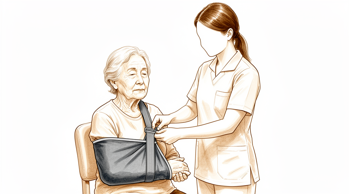
When you wake up, you will be in the recovery area, then move to the ward. Most patients stay one or two nights in hospital after this operation. Your arm will rest in a simple sling for comfort. It comes off for exercises and washing. Pain control is planned for you before you leave theatre, and your care team will keep checking how you are feeling and adjust things as needed. We leave the dressing on for about 10 days; please do not take it off before then unless we tell you to. We change or remove it when we see you. A physiotherapist will usually see you on the ward to start gentle movements. Please arrange for someone to stay with you for the first 24 hours after you go home.
Recovery
Everyone's recovery follows its own path, and your timeline may differ. Your surgeon and physiotherapist will guide you at each visit.
In the first days and weeks, expect some pain and swelling around the shoulder. This settles gradually for most people. Your care team will plan pain relief before you leave hospital and adjust it as needed. Many people manage well with a simple, non-opioid pain plan. Keeping your arm in the sling between exercises helps with comfort. Rest, gentle movement as your physiotherapist shows you, and following the plan will ease the discomfort.
A physiotherapist will usually see you on the ward to start gentle movements. At home, you will keep doing these exercises as directed. The sling comes off for exercises and washing. You will need help with some daily tasks at first, such as dressing, since the operated arm will be limited. Sleep can be disturbed early on, but most people find their sleep improves as the shoulder settles.
Movement and strength return in stages. As the swelling settles and your movement grows, everyday tasks become easier. Once your surgeon clears you to drive, typically at the six-week review, you can get back behind the wheel. See our guide on Driving after upper-limb surgery. Returning to work and sport depends on your job, your activities and how your shoulder is healing. Your surgeon and physiotherapist will talk you through what is safe at each stage.
What can go wrong
Most patients do well, but problems can occasionally happen. Your surgeon and the team monitor you closely to spot any issue early.
Infection is the most serious problem to watch for. It can show up as a deep, throbbing pain that does not ease with simple painkillers, redness that spreads out from the wound, or a fever. Tell the clinic straight away if you notice any of these signs. Some infections appear months later, so mention anything unusual at your reviews, even if it seems minor.
A clot can form in a vein after this operation. This may cause sudden swelling and tenderness in the calf. If a piece of that clot travels to the lungs, you might feel sudden breathlessness or chest discomfort. Go to the emergency department if this happens.
The nerves around the shoulder can be stretched or bruised during surgery. You might notice numbness, tingling or weakness in your arm or hand. Many of these recover fully, some only partly. Mention it at your next review so it can be tracked.
A fracture near the new joint is uncommon but can happen, either during the operation or afterwards. You would feel sharp pain and lose the ability to move the arm normally. Call the clinic if this occurs.
The artificial parts can loosen over time. This usually feels like a return of pain, sometimes with a clicking or grinding feeling in the shoulder. Bring this up at your review, as scans can show what is happening.
The shoulder can also become stiff or unstable, or the new parts can come out of place. You would notice a sudden loss of movement, or a feeling that the joint has shifted. Contact the clinic promptly.
If you have had shoulder surgery before, some of these risks are higher. Your surgeon will discuss this with you before the operation.
The complications table on this page lists typical rates if you want the specifics.
When to call us
Trust your instincts. If something feels wrong, contact us. Call the clinic if you have a fever, increasing redness or discharge from the wound, or pain that keeps getting worse. Go to emergency if you have sudden swelling in your calf, shortness of breath, or chest discomfort. These can be signs of a clot. Call us straight away if you lose feeling in your arm or hand, or you cannot move it at all. Most problems are easier to sort out early.
Where to read more about the condition
This page is about the operation itself. The condition it treats, including what the evidence shows about when surgery helps and when it does not, is covered in more detail on the Shoulder Arthritis page.
Evidence & references
This is the clinical evidence summary written for health professionals. It is technical, and it lists the research this page was built from. You do not need to read it to understand your treatment or to make a decision about it.
Anatomy & Pathophysiology
Bony Anatomy
- The proximal humerus comprises four main parts: the humeral head, greater tuberosity, lesser tuberosity, and humeral shaft [3].
- The articular head of the humerus is spherical with a diameter of 37 to 57 mm [3].
- The most superior portion of the articular surface of the humeral head averages 8 mm above the greater tuberosity [3].
- Humeral version averages 29.8 degrees, with a range of 10 to 55 degrees [3].
- The humeral head is inclined approximately 130 degrees with respect to the humeral shaft [3].
- The neck-shaft angle measures an average of 135 degrees [4].
- The humeral head is retroverted an average of 30 degrees [4].
- The humeral head averages 19° of retroversion and 41° of inclination (neck-shaft angle) [6].
- The anatomic neck of the proximal humerus is located at the junction of the articular surface and the tuberosities [3].
- The surgical neck represents an indistinct region below the tuberosities but above the humeral shaft [3].
- The glenoid is a convex structure of shallow depth shaped like an inverted pear [3].
- The glenoid cavity is a shallow socket, approximately one third the size of the humeral head [4].
- The subchondral bone of the glenoid is relatively flat, with articular concavity augmented by cartilage and a circumferential labrum [6].
- The glenoid averages 5° of retroversion in relation to the axis of the scapular body [6].
- The scapula is attached to the axial skeleton by the acromioclavicular and sternoclavicular joints [5].
- The scapula is separated from the chest wall by thin gliding fibro-fatty tissue, allowing smooth excursion over the chest wall [5].
- The basic part of the scapula is the body, which is triangular when viewed anteroposteriorly with its base situated superiorly and its apex inferiorly [5].
- The glenoid is connected with the flat body of the scapula by the scapular neck [5].
- The hook-shaped coracoid process curves forwards from the superior surface of the scapular neck [5].
- The scapular spine ends in a flattened bony process, the acromion, which curves forwards [5].
- The highest concentration of bony mass in the scapula is located in the glenoid, the scapular neck, and the lateral border of the scapular body [5].
- Two bony pillars transmit compressive forces from the glenoid fossa: the lateral pillar and the spinal pillar [5].
- The lateral pillar connects the inferior border of the glenoid with the inferior angle [5].
- The spinal pillar arises from the central part of the glenoid and continues medially to become part of the base of the scapular spine [5].
- The weakest bone in the scapula is located primarily in the central part of the biomechanical body, specifically in the infraspinous fossa [5].
- The weakest area of the circumference of the biomechanical body of the scapula is the spinomedial angle [5].
- The clavicle is the first bone to ossify, occurring in the fifth week of gestation [6].
- The clavicle is the only long bone to ossify by intramembranous ossification [6].
- The medial (sternal) epiphysis of the clavicle is the last ossification center to fuse, at age 20 to 25 years [6].
- Ossification of the scapular body begins at the eighth week of gestation [6].
- The acromion has three ossification centers: the metacromion, mesoacromion, and preacromion [6].
- Failure of fusion of the acromial ossification centers results in os acromiale [6].
- The proximal humerus has three centers of ossification: the humeral head, greater tuberosity, and lesser tuberosity [6].
- The humeral head ossification center appears at 4 to 6 months [6].
- The greater tuberosity ossification center appears at 1 to 3 years [6].
- The lesser tuberosity ossification center appears at 3 to 5 years [6].
- The proximal humeral ossification centers fuse to the shaft at age 17 to 20 years [6].
Soft Tissue Anatomy
- The greater tuberosity serves as the attachment site for the supraspinatus, infraspinatus, and teres minor tendons [3].
- The lesser tuberosity serves as the attachment site for the subscapularis tendon [3].
- The bicipital groove lies between the greater and lesser tuberosities and serves as a pathway for the long head of the biceps [3].
- The distal aspect of the bicipital groove is internally rotated with respect to the proximal portion [3].
- The rotator cuff consists of four muscles: the subscapularis, supraspinatus, infraspinatus, and teres minor [4].
- The teres major is not a rotator cuff muscle [4].
- The rotator cuff muscles serve as depressors of the humeral head to allow the deltoid to efficiently abduct the humerus [4].
- The infraspinatus and teres minor are external rotators of the humerus [4].
- The subscapularis is an internal rotator of the humerus [4].
- The acromion, coracoacromial ligament, and coracoid process form the coracoacromial arch [3].
- The rotator cuff, subacromial bursa, and subdeltoid bursa pass underneath the coracoacromial arch [3].
- The subscapular bursa lies between the subscapularis tendon and the neck of the scapula [7].
- The subscapular bursa communicates with the joint cavity between the superior and middle glenohumeral ligaments [7].
- The subscapular bursa is linked to the coracoid process by a suspensory ligament [7].
- In 28% of dissected specimens, the subscapular bursae merged with the subcoracoid bursae, forming a unique wide bursa [7].
- The rotator interval is defined medially by the base of the coracoid, superiorly by the supraspinatus tendon, and inferiorly by the subscapularis tendon [6].
- The rotator interval contains the coracohumeral ligament, the superior glenohumeral ligament, and the intra-articular portion of the long head of the biceps tendon [6].
- The average area of the rotator interval is 20.96 mm [7].
- The superior transverse scapular ligament arises from the medial base of the coracoid overlying the suprascapular notch [6].
- The suprascapular artery runs superior to the superior transverse scapular ligament, while the nerve runs deep to it [6].
- The spinoglenoid ligament overlies the suprascapular nerve at the spinoglenoid notch [6].
Vascular and Neural Anatomy
- The proximal humerus receives its blood supply from the anterior and posterior humeral circumflex branches from the third division of the axillary artery [3].
- The posterior humeral circumflex artery travels with the axillary nerve and enters the quadrilateral space posteriorly [3].
- The anterior humeral circumflex artery arises from the axillary artery at the inferior border of the subscapularis [3].
- The anterior humeral circumflex artery provides vascular inflow to the humeral head via its terminal anterolateral branch, known as the artery of Laing or arcuate artery [3].
- The ascending branch of the anterior humeral circumflex artery courses parallel to the lateral aspect of the long head biceps tendon [3].
- The ascending branch of the anterior humeral circumflex artery enters the humeral head at the interface of the bicipital groove and greater tuberosity [3].
- The anterolateral ascending branch of the anterior humeral circumflex artery provides the primary blood supply to the humeral head [6].
- The terminal intraosseous portion of the anterior humeral circumflex artery enters at the proximal aspect of the intertubercular groove as the arcuate artery [6].
- Injury to the arcuate artery may result in osteonecrosis of the humeral head [3].
- Additional extraosseous collateral branches can permit humeral head perfusion despite complete ligation of the arcuate artery [3].
- Fractures of the anatomic neck have a poor prognosis because of complete disruption of the blood supply to the head [4].
- Surgical neck fractures are common, and with these, the blood supply to the head is preserved [4].
- The brachial plexus and axillary artery are anterior to the coracoid process of the scapula and humeral head [4].
- Nerves innervating muscles around the shoulder include the axillary, suprascapular, subscapular, and musculocutaneous nerves [4].
- An axillary nerve injury from proximal humeral fracture or fracture-dislocation results in paralysis of the deltoid muscle and anesthesia over the “badge” region at the lateral proximal arm [4].
- Entrapment of the suprascapular nerve at the superior transverse scapular ligament causes denervation of both the supraspinatus and the infraspinatus [6].
- Entrapment, traction, or compression of the suprascapular nerve at the spinoglenoid notch causes denervation of the infraspinatus [6].
Joint Stability and Ligaments
- Stability and function of the glenohumeral joint are provided by the interaction of structures that promote a near global range of motion and purposeful function [3].
- External loads transferred to the shoulder girdle are initially offset by joint surface anatomy, joint volume, atmospheric pressure, and joint fluid cohesion and adhesion [3].
- Moderate and large loads are counterbalanced by the deltoid and rotator cuff and by the capsulolabral and bone structures, respectively [3].
- The glenohumeral joint is stabilized dynamically by the rotator cuff via joint compression [6].
- Static stabilizers of the glenohumeral joint include articular congruity, the glenoid labrum, concavity-compression, negative intra-articular pressure, and the glenohumeral capsule and ligaments [6].
- The glenoid labrum provides concavity and up to 50% of marginal glenoid socket depth [6].
- The coracohumeral ligament restricts external rotation in adduction [6].
- The coracohumeral ligament is a static restraint to inferior and posterior translation in adduction and external rotation [6].
- The superior glenohumeral ligament is a primary static restraint against anterior translation with the arm at the side [6].
- The superior glenohumeral ligament and coracohumeral ligament form a pulley that provides restraint against medial subluxation of the long head of the biceps tendon [6].
- The middle glenohumeral ligament is a primary static restraint against anterior translation with the arm in external rotation and 45° of abduction [6].
- The anterior band of the inferior glenohumeral ligament is a primary static restraint against anterior-inferior dislocation of the glenohumeral joint in 90° of abduction and external rotation [6].
- The posterior band of the inferior glenohumeral ligament is a primary static restraint against posterior-inferior translation in internal rotation and adduction [6].
- Laxity of the rotator interval results in inferior laxity, known as the sulcus sign [6].
- Contracture of the rotator interval is seen with adhesive capsulitis [6].
- The superior shoulder suspensory complex provides a stable connection between the scapula and the axial skeleton [6].
- The superior shoulder suspensory complex is composed of the glenoid, coracoid process, coracoclavicular ligaments, distal clavicle, acromioclavicular joint, and acromion [6].
- The superior strut of the superior shoulder suspensory complex comprises the middle clavicle [6].
- The inferior strut of the superior shoulder suspensory complex comprises the lateral scapular border and spine of the scapula [6].
- The sternoclavicular joint is the only true diarthrodial articulation between the upper appendicular and axial skeletons [6].
- The posterior sternoclavicular joint capsule and ligaments are the primary stabilizers to anterior and posterior translation of the medial clavicle [6].
- The acromioclavicular joint is a small diarthrodial joint with an interposed fibrocartilaginous disk [6].
- The superior and posterior acromioclavicular ligaments are the primary stabilizers to anterior and posterior translation of the clavicle [6].
- The coracoclavicular ligaments are the primary stabilizers to superior translation of the distal clavicle [6].
Pathophysiology
- Post-traumatic shoulder fractures alter complex interactions of the shoulder girdle, resulting in pain, decreased range of motion and stiffness, and disability [3].
- Displacement of proximal humeral fracture fragments is based on deforming forces created by the tendinous insertions of the pectoralis major, subscapularis, supraspinatus, and infraspinatus [3].
- The subscapularis inserts on the lesser tuberosity and causes medial displacement of the fragment [3].
- The supraspinatus and infraspinatus insert on the greater tuberosity and cause superior and posterior displacement of the fragment [3].
- The pectoralis major inserts on the humeral shaft and displaces it medially [3].
- A fracture involving the anatomic neck is prognostically worse than fractures involving other regions of the proximal humerus regarding potential disruption of the vascular supply to the humeral head and subsequent development of avascular necrosis [3].
- Displaced proximal humeral fractures can impede normal movement of structures passing under the coracoacromial arch, causing impingement and disruption of normal glenohumeral motion [3].
- In proximal humeral fractures, the subdeltoid and subacromial bursae can become thickened and fibrotic, forming adhesions that limit normal glenohumeral motion [3].
- The pathogenesis of shoulder stiffness is still elusive, though basic science research has provided insight into cellular and biochemical pathways [1].
- The diagnosis of a stiff shoulder depends on awareness of the problem, with history and physical examination being paramount [1].
- No treatment for a stiff shoulder has proved to be definitive [1].
- The literature supports many forms of treatment for a stiff shoulder, both operative and nonoperative [1].
- The treatment approach for a stiff shoulder should be tailored to each individual patient to ensure the best possible outcome [1].
- Arthritis usually involves the central aspect of the humeral head [2].
- Joint space narrowing in arthritis is most evident on the axillary view taken with the arm in elevation, as opposed to images made with the arm at the side [2].
- The axillary view taken with the arm in elevation can show posterior subluxation or “functional decentering” that is not evident in images taken with the arm at the side [2].
- Malcentering of the joint reaction force leads to posterior instability, posterior glenoid wear, and “rocking horse” loosening of prosthetic glenoid components [2].
- The degree of posterior subluxation can be measured by the position of the center of the humeral head in relation to the plane of the scapula [2].
- The degree of posterior subluxation can be measured by the position of the center of the humeral head in relation to the glenoid face [2].
- The degree of posterior subluxation can be measured by the point of contact of the humeral articular surface on the glenoid articular surface [2].
- The point of contact of the humeral articular surface on the glenoid articular surface reflects the degree of centering of the net humeral joint reaction force on the glenoid [2].
- Normal shoulder motion is approximately two-thirds glenohumeral and one third scapulothoracic [6].
- The relationship between acromial anatomy and rotator cuff disease remains controversial [6].
- The classification of acromial morphology (flat, curved, or hooked) is challenged by poor interobserver reliability [6].
- The relationship between coracoid morphology and subscapularis tears is controversial [6].
Investigations
Radiographic Evaluation
- The purpose of shoulder imaging is to help establish the diagnosis, determine the severity of the pathoanatomy, assist in surgical planning, and enable the surgeon to illustrate the condition of the shoulder to the patient [2].
- Unless a specific research protocol is in place, the tendency to "overimage" should be resisted by obtaining only the scans or reconstructions necessary for patient care [2].
- Standardized plain films are almost always sufficient to garner the information needed for total shoulder arthroplasty [2].
- CT scans may offer increased precision in the measurement of glenoid version, but this precision does not improve the quality of the surgery or the clinical outcome [2].
- Proper radiographic technique is as important as proper surgical technique to achieve the desired outcome [2].
- The first key radiographic view is the anteroposterior (AP) view in the plane of the scapula, taken so that the x-ray beam passes through the glenohumeral joint [2].
- The AP view in the plane of the scapula shows the superoinferior position of the humeral head relative to the glenoid, presence of osteophytes, narrowing of the joint space, degree of medial displacement of the humerus, quality of bone, presence of loose bodies, and humeral head collapse or deformity [2].
- The second key radiographic view is the axillary view taken with the arm in the functional position of elevation in the plane of the scapula [2].
- The axillary view is oriented so that both the spinoglenoid notch and the scapular neck are visible [2].
- The axillary view demonstrates the amount of glenoid bone, shape of the glenoid, its version in relation to the plane of the scapula, and the relationship of the humeral head to the glenoid fossa [2].
- The axillary view is referred to as the "truth view" because it demonstrates glenohumeral relationships in the functional position of elevation [2].
- CT scans have the disadvantage of being taken with the arm in the adducted position, unlike the axillary truth view [2].
- Many axillary views sent for consultation are taken without standardization, making it impossible to determine important features of the glenohumeral joint [2].
- Standardized anteroposterior and axillary views indicate the thickness of the cartilage space between the humerus and the glenoid, relative positions of the humeral head and glenoid, presence of osteophytes, degree of osteopenia, and extent of bony deformity and erosion [2].
- Joint space narrowing is most evident on the axillary truth view as opposed to images made with the arm at the side [2].
- The axillary truth view shows posterior subluxation or "functional decentering" that is not evident in images taken with the arm at the side [2].
- Three-dimensional reconstructions can reveal fine details of shoulder anatomy, but this additional information rarely changes the planning or conduct of the arthroplasty [2].
- At least two X-ray views should be obtained: an anteroposterior in the plane of the glenoid and an axillary projection with the arm in abduction to show the relationship of the humeral head to the glenoid [11].
- Computed tomography (CT) is helpful for planning shoulder joint replacement [11].
Magnetic Resonance Imaging
- Magnetic resonance imaging (MRI) is useful to identify osteonecrosis of the humeral head or a bone tumour [11].
- MRI can identify labral tears and rotator cuff tears, although the accuracy for these is enhanced by combining the scan with arthrography [11].
Ultrasonography
- Ultrasonography is a simple and accurate test for identifying rotator cuff tears and calcific tendinitis [11].
- Ultrasonography can be useful in guiding injections or barbotage (aspirating calcific deposits in the rotator cuff) [11].
- The most commonly performed joint examination using ultrasonography is the shoulder examination [9].
- The accuracy of rotator cuff ultrasonography depends on the skill of the scanner operator and an awareness of pitfalls encountered [9].
General Imaging Principles
- The shoulder is a three-dimensional structure that cannot be represented by a single planar view [13].
- Critical relationships, such as the degree of centering of the humeral head, change with the position of the arm [13].
- Shoulder pathology may be found in a large number of different bones and soft tissues [13].
- Overlying and superimposed structures as well as metallic implants may complicate imaging the structures of interest [13].
- Surgeons need to develop a judicious approach to imaging that yields necessary information while avoiding the tendency to "over-image" [13].
References
[1] Rockwood And Matsen S The Shoulder. Arthroscopic Management of Prearthritic and Arthritic Conditions of the Shoulder and the Postarthroplasty Shoulder > SUMMARY.
[2] Rockwood And Matsen S The Shoulder. Arthroscopic Management of Prearthritic and Arthritic Conditions of the Shoulder and the Postarthroplasty Shoulder > Radiographic Evaluation.
[3] Rockwood And Matsen S The Shoulder. Shoulder and Elbow Specialty Clinic Workers’ Survey > ANATOMY.
[4] A Lange Medical Book Current Diagnosis Treatment In Orthopedics Fifth Edition. 2Musculoskeletal Trauma Surgery > SHOULDER AND ARM INJURIES.
[5] Rockwood And Green S Fractures In Adults. 29: Principles of Nonunion and Bone Defect Treatment > Applied Anatomy Related to Scapular Fractures.
[6] Aaos Comprehensive Orthopaedic Review 3. Anatomy of the Shoulder, Arm, and Elbow > I. Shoulder.
[7] Rockwood And Matsen S The Shoulder. Developmental Anatomy of the Shoulder and Anatomy of the Glenohumeral Joint > Bursae.
[9] Orthopaedic Knowledge Update Sports Medicine 6. Diagnostic Ultrasonography and Ultrasonography-Guided Procedures > Annotated References.
[11] Apley And Solomon S Concise System Of Orthopaedics And Trauma. INVESTIGATION.
[13] Rockwood And Matsen S The Shoulder. Developmental Anatomy of the Shoulder and Anatomy of the Glenohumeral Joint > SENIOR EDITOR COMMENTARY.