Patients › Hand
Finger Fractures
Phalangeal and metacarpal fractures of the hand — non-operative care and indications for fixation.
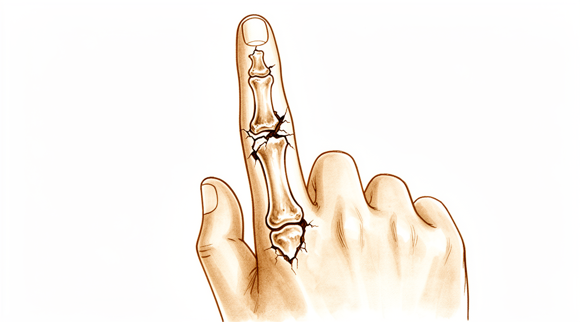
What you're feeling
A broken finger usually happens in a moment. A ball jams your fingertip, you fall onto your hand, or your finger takes a direct hit. Some people feel or hear a snap or pop at the moment of injury. A crushed fingertip, for example from a door, is another common cause.
Straight away, the finger hurts and swells. Bruising often appears. The finger may look bent or twisted, or it may sit out of line with the others. You will probably not want to move it, and everyday tasks like gripping a door handle, doing up buttons or holding a cup become painful. Sometimes a jammed finger seems minor, just swollen and sore at the middle joint, but a small piece of bone can still be broken. That is why the finger needs checking rather than waiting it out.
In the first days, the pain is often worse when you try to move the finger, and it can ache at night. Swelling and bruising are at their peak early on. Over the first couple of weeks, the pain usually settles as healing begins, though the finger may stay stiff and tender for longer. Most broken fingers heal without an operation. The aim of treatment is to line the bone up, keep the finger moving early, and protect your grip and fine control for the long term.
One thing worth knowing: a finger that heals twisted can affect how your whole hand works. If your finger crosses over or under its neighbours when you make a fist, that twist does not correct itself with time. So if your finger looks out of line, or it will not bend and straighten like normal, have it looked at promptly.
What's actually happening
Each finger is built from three small bones, stacked end to end. The end bone finishes in a rounded tip called the tuft. The bones are joined by joints that bend and straighten, held steady by strong cords of tissue on each side and a firm strap across the front of each joint. Tendons run along the back of the finger and attach to the bones, and they pull the finger straight.
When a finger is jammed or crushed, one of these bones can crack, or a tendon can pull away from the bone it anchors to. Think of a tent pole: if one section bends or snaps, the whole frame loses its shape and the canvas sags. That is what happens inside your finger. The tendons keep pulling, and they can drag the broken pieces out of line, so the finger settles bent or twisted.
Bone heals by knitting back together, the way a crack in a plate fuses when it is held still. A torn tendon heals by reattaching to the bone. But the pieces need to stay lined up while this happens, and a finger kept still for too long turns stiff.
How badly the pieces are out of line changes the picture. If the break runs into the joint surface, the smooth track the finger bends along becomes uneven. If the tendon that straightens the end joint tears or pulls off a small piece of bone, the fingertip droops and you cannot lift it yourself, though it can still be straightened by hand. A twist in the finger does not straighten itself as it heals, and it can make your finger cross over its neighbours when you grip.
Most broken fingers heal without an operation. The aim is to line the bone up, protect the joint surfaces and tendons, and get the finger moving early so it does not seize up.
What we can do about it
Dr Kieran Hirpara, an upper-limb surgeon at Mater Private Hospital Rockhampton, matches the treatment to your specific injury. Patients are generally referred to our clinic by their GP; if a physiotherapist has suggested you see us, you will still need a referral from your GP in order to be eligible for the Medicare rebate. At the clinic we examine your hand and take x-rays, and sometimes other scans, to work out exactly which bone is broken and how far the pieces have moved. Then we talk through the options with you.
Most broken fingers do not need an operation. If the break is stable, or the pieces have hardly moved, we hold the finger still with a splint or cast while it heals. The splint usually holds the finger bent at the knuckle and straight at the other joints. We keep a close eye on the finger with repeat x-rays to make sure the pieces stay lined up. Once the bone is secure, you start gentle movement, because a finger kept still too long turns stiff. Some breaks can be treated with buddy taping, where the sore finger is taped to its neighbour for support. For children, most finger fractures heal well with simple splinting or taping and early movement.
Surgery is recommended from the start when the bone pieces are badly out of line, when the break is unstable, or when it runs into a joint. It is also considered when your work, sport or hand dominance puts real demands on that finger. The aim of an operation is to hold the pieces in the right position while they knit together, so the finger can start moving sooner. We use small plates, screws or smooth wires to do this, and we will explain which suits your break. Sometimes both paths are reasonable, and the choice is genuinely shared. A finger that has had surgery is usually stiffer than one treated without it, and some people prefer to avoid an operation even if the finger ends up slightly twisted. We will talk through what matters most to you.
Whichever path you take, the first weeks are similar. We will help you keep the pain under control while the bone heals. You protect the finger from knocks and heavy use during this time. Hand therapy starts at the right stage, once the break is secure enough to move. Your therapist will guide your exercises and fit any splint you need along the way.
What to expect
Most broken fingers heal without an operation. If your break is stable, a splint or cast holds the bone still while it knits together. Most breaks of the main finger bone heal within 4 weeks when the pieces are pushed back into line and held with smooth wires. Once the bone is secure, you start gentle movement, because a finger kept still too long turns stiff.
Recovery from an injury like this is a slow build rather than a single moment. Over the first weeks, the pain settles but the finger stays stiff and tender. Over the following months, you work on bending and straightening as the swelling goes down. Hand therapy after surgery is with Ruby Doolan at Extend Rehabilitation. Ruby is a hand therapist: she directs your exercises and makes any splint you need. How well your finger recovers depends on the type of break, and on how well you stick to your exercises.
The results of surgery for finger fractures are generally good when the pieces are held firmly in the right position. Most people regain useful movement and grip. But it is honest to say things can go wrong. Stiffness is the main one: 43% of fingers treated with plates and screws for an unstable break ended up stiff after surgery. A finger that heals in a poor position can lose bending and straightening at the middle joint, especially if the bone was out of line by 25 degrees or more. Some breaks need more than one operation, most often when the finger was crushed or its blood supply was affected. About a quarter of open finger fractures, where the skin is broken, need more than one procedure.
A few things make recovery harder. If there is a long delay between the injury and treatment, or the break is an unusual type, the outcome tends to be less favourable. For children, most finger fractures heal well without surgery. A small number need prompt surgery to avoid problems, and breaks that run into a joint surface can leave lasting changes in up to 50% of cases when the pieces were displaced at first.
When to see someone
Seek urgent care if your finger is clearly bent or twisted, if the skin is broken over the injury, if you cannot bend or straighten the finger at all, or if it feels numb or tingles. A jammed finger that stays swollen and sore at the middle joint also needs checking, because a small break can hide behind a mild-looking injury. If your finger crosses over or under its neighbours when you make a fist, have it seen promptly, as that twist will not correct itself. For other injuries, see your GP first. Ask for a specialist review if the pain is not settling, or if swelling, movement or grip are not improving week on week as the bone heals.
In more depth
Advanced reading: the deeper science (optional)
This section goes further than you need for your own treatment decisions. Finger fractures are worth the extra reading for two findings that cut against standard practice: prophylactic antibiotics do not appear to help an open fingertip fracture, and for the difficult joint fracture at the middle knuckle, no fixation method has proved better than the others.
Antibiotics for an open fingertip fracture do not reduce infection
An open fracture of the distal phalanx, bone exposed through a wound, typically after a crush, is routinely given prophylactic antibiotics on the general principle that open fractures need them.
The evidence does not support it here. Across 353 patients, the results fail to show any effect of prophylactic antibiotics on the rate of superficial infections following open distal phalanx fractures, and the authors conclude the focus should be on prompt irrigation and debridement rather than administration of prophylactic antibiotics [1].
The distinction is between cleaning the wound and medicating it. Removing contamination mechanically is what reduces infection; antibiotics were not shown to add to that in this setting. Given the costs of unnecessary antibiotic use, it is worth knowing that their omission after thorough wound care reflects evidence rather than oversight.
For the middle-joint fracture-dislocation, no technique wins
Fracture-dislocations at the proximal interphalangeal joint are among the more difficult injuries in the hand, a small joint with a fractured surface that must be both reduced and kept reduced while moving.
Across 735 patients, the comparison is instructive precisely because it fails to separate the options: percutaneous fixation yielded the highest post-operative range of motion, extension-block pinning resulted in the greatest grip strength, and no treatment method or fracture type yielded consistently better outcomes than another [2].
Where more than half the joint surface is involved, one reconstructive option is to rebuild it with a graft from the hamate bone. Across 235 patients, hemi-hamate arthroplasty was found reliable and effective, affording symptomatic relief and functional restoration [3].
Why stiffness is the real enemy
The reason these injuries are difficult is not that the bone fails to heal, finger fractures unite readily. It is that the finger stiffens.
The flexor and extensor tendons run immediately against the bone with minimal soft tissue between them, so blood and swelling around a fracture organise into scar that binds the tendons to it. That process is well advanced within weeks, and once established it is difficult to reverse.
This is why finger fractures are moved early wherever the fracture is stable enough to allow it, and why fixation is chosen partly for whether it permits movement rather than solely for how rigidly it holds bone. It also explains the apparently paradoxical situation of a perfectly healed fracture on X-ray and a finger that will not bend, the bone was never the problem.
References for the advanced reading
- Metcalfe D, Aquilina AL, Hedley HM. Prophylactic antibiotics in open distal phalanx fractures: systematic review and meta-analysis. J Hand Surg Eur Vol. 2015;41(4):423-30.
- Demino C, Yates M, Fowler JR. Surgical management of proximal interphalangeal joint fracture-dislocations: a systematic review. Hand (N Y). 2019;16(4):453-60.
- Faulkner H, Graham DJ, Hile M, Lawson RD, Sivakumar BS. Hemi-hamate arthroplasty for base of middle phalanx fracture: a systematic review. Hand (N Y). 2021;18(2):300-6.
Evidence & references
This is the clinical evidence summary written for health professionals. It is technical, and it lists the research this page was built from. You do not need to read it to understand your treatment or to make a decision about it.
Overview
- The majority of hand fractures can be treated without surgery [1].
- Surgery offers distinct advantages in properly selected cases of hand fractures [1].
- Most hand fractures can be managed successfully without operation [3].
- Conservative functional techniques are the optimum treatment for the majority of patients with single metacarpal fractures [3].
- Surgeons who treat metacarpal and phalangeal fractures inevitably treat complications associated with these fractures [4].
- A quarter of open finger fractures will likely need more than one surgical procedure [5].
- The need for more than one surgical procedure in open finger fractures is especially present in more severely injured fingers due to crush or with vascular impairment [5].
- Most pediatric phalangeal fractures can be treated nonsurgically [19].
- A small subset of pediatric phalangeal fractures benefits from surgical intervention [19].
- Taping displaced extra-articular phalangeal finger fractures in children can be recommended irrespective of the degree of displacement or the need for reduction [23].
Anatomy & Pathophysiology
Bony Anatomy
- Finger phalangeal fractures account for approximately 10% of all fractures seen [22].
- The little and ring fingers are the most frequently affected digits in finger phalangeal fractures [22].
- The thumb accounts for 18.4% of finger phalangeal fractures, the index for 9.0%, the middle for 14.3%, the ring for 25.8%, and the little finger for 32.5% [22].
- All phalanges consist of a proximal base, a central diaphysis, and a distal head [32].
- In contrast to metacarpals, the bases of all phalanges develop as metaphyses rather than the heads [32].
- The distal portion of the distal phalanx is referred to as the tuft [32].
- Fingers follow a typical pattern of relative lengths where the tip of the index finger extends to the base of the nail of the middle finger, the tip of the ring finger to the mid-aspect of the middle finger nail, and the tip of the small finger to a corresponding position [32].
- The third and fourth metacarpal heads help stabilize the metacarpal arch by providing attachments for the transverse metacarpal ligament [10].
- The proximal phalanx of the middle or ring finger is functionally important because its absence creates a hole through which small objects can pass and impairs scooping maneuvers [10].
- The ring finger forms the keystone of the palmar arch and participates in power grip [51].
- The small finger plays an important role in palmar grip due to the mobility of its carpometacarpal joint and the action of the hypothenar muscles [29].
- The small finger increases the span of the hand for grasp owing to its abduction moment [29].
Joint Anatomy & Biomechanics
- The articulations of the fingers form a triarticular chain that flexes toward the thumb and the palm to allow grasp [48].
- The interphalangeal articulations of the digits function uniquely in flexion–extension with trochlear-shaped articulations that are closely congruent throughout excursion [48].
- Flexion of the metacarpophalangeal joint is approximately 85 degrees, the proximal interphalangeal joint approximately 115 degrees, and the distal interphalangeal joint 80 degrees [48].
- The index finger is capable of less flexion than the other fingers because it opposes the thumb [48].
- Intraarticular fractures that disrupt joint congruency can occur at the distal (condylar) or proximal (pilon or proximal condylar) articular surface [32].
- Most intraarticular phalangeal fractures are produced by an axial loading injury [32].
- Intrinsic and extrinsic tendon insertions act as deforming forces that create typical angulation patterns in phalangeal fractures [32].
- Proximal and middle phalangeal shaft fractures typically collapse into apex volar angulation due to the proximal flexion moment of the intrinsics and distal extensor moment of the extensor mechanism [32].
- The volar tendinous apparatus, consisting of the two flexor tendons, is considerably stronger than the dorsal extensor apparatus [48].
- The capsular structures and fibro-fatty cushions are much stronger on the flexor side than the extensor side [48].
- The finger is designed to function in flexion [48].
- The metacarpophalangeal and interphalangeal joints are ball-and-socket and hinge joints, respectively [50].
- In flexion, the distal phalanx is drawn upon the proximal phalanx, effectively shortening the palmar length of the skeleton [50].
- The soft tissue of the fingers between the fingertips and the area of the aponeurosis is mobile and flexible [50].
- The soft tissue of the fingertips is tightly anchored [50].
Soft Tissue & Skin Anatomy
- The cutaneous striations that make up fingerprints reflect the arrangement of the papillary ridges of the underlying dermis [49].
- The overall orientation of palmar skin striations is predominantly transverse, forming a typical concentric pattern at the pulps [49].
- Palmar skin striations play an important part in the retention of an object during gripping by preventing sliding [49].
- The palmar skin is anchored to the underlying fascial planes by a system of fibrous tracts [49].
- The metacarpophalangeal pad sits transversely over the base of the fingers from the ulnar to the radial border of the hand [49].
- The pulp has a lobulated palmar pad where fibrous septa join the periosteum of the distal phalanx to the deep aspect of the dermis [49].
- The nail plate is composed of keratin and originates from the germinal matrix proximal to the nail fold [71].
- The sterile matrix is directly beneath the nail plate and contributes keratin to increase plate thickness [71].
- The lunula is the proximal nail plate at the junction of the sterile and germinal matrices [71].
- The hyponychium is located between the distal nail bed and skin of the fingertip and acts as a barrier to micro-organisms [71].
- The eponychium, or cuticle, is located at the distal margin of the proximal nail fold [71].
- The paronychium forms the lateral margins of the nail [71].
- The midlateral finger incision allows the neurovascular bundle to be carried volarward with the volar flap or allows dissection superficial to the neurovascular bundle [26].
- On the radial sides of the index and middle fingers and on the ulnar side of the little finger, the dorsal branch of the digital nerve should be preserved if possible during midlateral approaches [26].
Pathophysiology & Injury Mechanisms
- Most hand fractures can be managed successfully without operation, with conservative functional techniques being the optimum treatment for the majority of patients with single metacarpal fractures [3].
- Hand and finger fractures are the second most common fracture presenting to emergency departments in the pediatric population [14].
- There is a bimodal age distribution for pediatric hand and finger fractures with peaks at 0 to 2 years of age and 12 to 16 years of age [14].
- The most commonly injured locations in pediatric hand fractures are the base of the proximal phalanx (67%) of the border rays, specifically the little finger (52.2%) and thumb (23.5%) [14].
- Salter-Harris II fractures of the digits are an extremely common hand fracture in children, with the little finger proximal phalanx being the most commonly injured [14].
- Malrotation in pediatric finger fractures does not remodel and can result in problems with grip formation [14].
- Seymour fractures are Salter-Harris I/II or juxtaphyseal fractures of the distal phalanx with interposed nail bed at the fracture site [14].
- Missed Seymour fractures have a high rate of complication including infection and nail or physeal growth disturbance [14].
- Direct blow or assault is the most common mode of injury for finger phalangeal fractures, accounting for 39.1% of cases [22].
- Falls from standing height account for 29.5% of finger phalangeal fractures [22].
- Sports injuries account for 23.8% of finger phalangeal fractures [22].
- The prevalence of fractures caused by direct blows or assaults is higher on the radial side of the hand [22].
- 34.3% of little finger and 33.9% of ring finger fractures are caused by direct blows or assaults [22].
- 47.4% of middle finger, 50.0% of index finger, and 43.4% of thumb fractures are caused by direct blows or assaults [22].
- 23.4% of finger phalangeal fractures are basal fractures of the proximal phalanges [22].
- 11.5% of finger phalangeal fractures are diaphyseal fractures of the proximal phalanges [22].
- 16.3% of all finger phalangeal fractures are basal fractures of the middle phalanges [22].
- 3.4% of phalangeal fractures are diaphyseal fractures of the middle phalanges [22].
- 1.7% of finger phalangeal fractures are distal fractures of the middle phalanges [22].
- Fractures of the base of the distal phalanges account for 21.3% of all phalangeal fractures [22].
- 8.4% of phalangeal fractures occur in the diaphyses of the distal phalanges [22].
- 9.8% of phalangeal fractures are distal fractures of the distal phalanges [22].
- Open fractures of the phalanges are relatively common, with the highest prevalence seen in 36- to 64-year-old males [22].
- The commonest site of open phalangeal fractures is the distal phalanges, where 25.3% of fractures are open [22].
- Approximately 55% of patients with multiple phalangeal fractures have other phalangeal fractures as associated injuries [22].
- The incidence of hand fracture is 3.7 per 1000 per year for men and 1.3 per 1000 per year for women [25].
- Fractures of the little finger metacarpal were common, accounting for 27% of the total in a study of hand fractures [25].
- Metacarpal stress fractures can present with significant pain and impact performance in athletes who perform sports involving repetitive movements of the hand and wrist [31].
- The mallet finger deformity is characterized by a loss of active distal interphalangeal joint extension with full passive range of motion evident [35].
- Mallet finger reflects the loss of normal extensor force transmission via the terminal tendon insertion onto the distal phalanx [35].
- The unopposed flexor digitorum profundus pulls the distal joint into flexion in mallet finger deformity [35].
- The usual mechanism of injury for mallet finger involves sudden passive flexion of the actively extended distal interphalangeal joint [35].
- Disruption of the terminal tendon in mallet finger may be confined to the tendon or may involve an avulsed fracture fragment from the dorsal lip of the distal phalanx proximal articular surface [35].
- A poorly functioning finger may represent a liability to the hand, and achievement of union or improved alignment alone may not be sufficient to justify retention of the digit [15].
- Excising the third metacarpal shaft removes the origin of the adductor pollicis and weakens pinch [10].
- Ring finger ray resection can have negative effects on hand function, including substantially decreased key and chuck pinch strengths compared to amputation through the proximal phalanx [51].
- Central ray deletion can have a negative impact on manual dexterity [51].
- Shortening and closing an injury that leads to proximal migration of the flexor digitorum profundus from its insertion at the base of the distal phalanx may result in a lumbrical-plus finger [27].
- In a lumbrical-plus finger, the flexor digitorum profundus tendon retracts and creates tension on the extensor mechanism through the lumbrical, causing paradoxical interphalangeal joint extension with active digit flexion [27].
- Fingertip injuries are the most common hand injuries seen in the emergency department [71].
- The long finger is the most commonly involved digit in fingertip injuries [71].
- Crush injuries without extensive soft tissue loss may result in nail plate avulsions, nail matrix lacerations, and distal phalanx (tuft) fractures [71].
Classification
- Treatment of fractures of the proximal phalanx and metacarpals is based on the presentation of the fracture, degree of displacement, and difficulty in maintaining fracture reduction [17].
- The Eaton classification for volar plate injuries includes Type I (avulsion of the volar plate without a fracture dislocation), Type II (dorsal dislocation of the proximal interphalangeal joint with avulsion of the volar plate and complete tear of the collateral ligament), and Type IIIa/IIIb [24].
- The Keifhaber-Stern classification for volar plate injuries categorizes injuries as "Stable," "Tenuous," or "Unstable" [24].
- Bony mallet finger is classified using the Wehbe and Schneider classification method [75].
- Dislocation in bony mallet finger is determined by the consistency of the axis of the distal phalanx and middle phalanx, with mild dislocation defined as the distal phalanx axis displaced forward but the dorsal cortical bone line not exceeding the axis of the middle phalanx, and severe dislocation defined as the dorsal cortical bone line displaced forward and exceeding the axis of the middle phalanx [75].
- Intra-articular fractures of the base of the first metacarpal include Bennett and Rolando fractures [80].
Clinical Presentation
Epidemiology and Demographics
- Finger phalangeal fractures account for approximately 10% of all fractures seen in clinical practice [22].
- Finger phalangeal fractures are the second most common fracture in males [22].
- The prevalence of little finger phalangeal fractures is 32.5% [22].
- The prevalence of ring finger phalangeal fractures is 25.8% [22].
- The prevalence of thumb phalangeal fractures is 18.4% [22].
- The prevalence of middle finger phalangeal fractures is 14.3% [22].
- The prevalence of index finger phalangeal fractures is 9.0% [22].
- Approximately 70% of all phalangeal and metacarpal fractures occur in patients between the ages of 11 and 45 years [32].
- Phalangeal fractures are more common in men than women [32].
- Pediatric hand and finger fractures exhibit a bimodal age distribution with peaks at 0 to 2 years of age and 12 to 16 years of age [14].
- The frequency, pattern, and treatment of pediatric hand fractures vary among different age groups [12].
Mechanism of Injury
- The average age of patients injured by direct blows or sports injuries is lower than those injured by standing falls [22].
- Patients injured by direct blows or sports injuries are more likely to be male [22].
- Fractures of the little and ring fingers are more frequently caused by falls and sports injuries compared to radial side digits [22].
- In pediatric populations, toddlers and preschool age children usually sustain crush injuries at home [14].
- In pediatric populations, adolescents most often get injured outside the home with sporting activities [14].
Associated Injuries
- Approximately 55.8% of patients with finger phalangeal fractures have other finger fractures [22].
- Approximately 13.9% of patients with finger phalangeal fractures have associated distal radius or ulna fractures [22].
- Approximately 9.3% of patients with finger phalangeal fractures have associated metacarpus fractures [22].
- In younger patients, 6% to 9% present with multiple fractures, a rate that rises with increasing age [22].
- The average age of patients who presented with multiple phalangeal fractures was 55.4 years [22].
- The gender ratio for patients presenting with multiple phalangeal fractures was 50/50 [22].
- 50% of patients with multiple phalangeal fractures sustained their injuries following a fall [22].
- 41.6% of patients with multiple phalangeal fractures sustained their injuries as a result of a direct blow [22].
Clinical Examination and Diagnosis
- Appropriate evaluation of hand and finger fractures includes clinical examination and radiographs [14].
- Clinical examination must assess for open injuries and angular and rotational malalignment of the injured ray [14].
- Rotational alignment can be confirmed by ensuring that all fingers point to the scaphoid tubercle when the fingers are flexed [14].
- Radiographs should include PA, lateral, and oblique views of the injured location [14].
- Only two studies were found on the diagnostic accuracy of history taking for hand and wrist fractures [7].
- A high index of suspicion should be maintained if there is discordance between the radiographic appearance of injury films and the patient’s clinical examination [56].
- The coronal plane deformity of Salter-Harris II fractures is easy to assess, but extra care must be taken to assess for rotational deformity, which is not as obvious on radiographs [14].
- The classic “jammed” finger with a swollen, painful PIP joint usually involves a volar plate injury or small nondisplaced avulsion fracture off the volar base of the middle finger epiphysis [14].
- Phalangeal neck and condyle fractures have a similar presentation to a simple “jammed” finger and are often missed [14].
- Seymour fractures are open fractures that are often missed, with the key to diagnosis being disruption of the nail plate/cuticle in addition to radiographic findings [14].
- Radiographs for Seymour fractures reveal a displaced fracture of the distal phalanx [14].
Investigations
- A radiograph should be obtained to determine whether a fracture is present in mallet finger and, if the dorsal fragment is large, whether the distal phalanx is subluxed palmarward [35].
- The key to diagnosis of Seymour fractures is disruption of the nail plate/cuticle as well as a displaced fracture of the distal phalanx on radiographs [14].
- Any patient suspected of having a carpometacarpal or proximal metacarpal injury should have a true lateral X-ray [77].
- Extensive radiographic procedures should be performed only in the limited group of cases with a clinical, radiographical, or high index of suspicion of an occult or difficult to visualise fracture [78].
Treatment
General Principles
- Timely treatment of complex fracture-dislocations ensures optimal outcome in range of motion and overall hand function [21].
- Surgical indications for fractures or fracture-dislocations include displaced articular fragments, rotational misalignment, significant digit angulation or shortening, irreducible dislocation, and significant injury to the joint supporting structures [66].
Non-Operative Management
- The majority of pediatric hand and finger fractures can be treated with closed reduction, appropriate immobilization, and early motion [14].
- Buddy taping is a non-inferior treatment modality for most paediatric finger fractures compared to splint immobilization [61].
- Traction splinting has been shown to be successful in the treatment of closed proximal phalangeal fractures [13].
- With non-operative treatment of fractures of the neck of the fifth metacarpal, similar results were achieved with dorsal angulation either above or below 30 degrees [30].
- For a closed extensor tendon rupture from its insertion into the distal phalanx, the distal interphalangeal joint is constantly held in hyperextension on a splint for 6 to 8 weeks and at night only for 2 to 4 additional weeks [44].
- Splint treatment within 2 weeks of injury has been found to be as effective as splinting more than 4 weeks after injury for mallet finger deformities [44].
- For dorsal PIP dislocations that are stable after reduction, buddy taping and range of motion are initiated [76].
- For dorsal PIP dislocations that are unstable after reduction, a dorsal blocking splint is applied [76].
- For Hastings type I and II PIP fracture-dislocations that are reducible, management involves a dorsal extension block splint with the amount of flexion decreased by 10° every week [76].
- For volar PIP dislocations, the PIP joint should be splinted in extension for 6 weeks to prevent a boutonnière deformity and allow healing of the central slip [76].
- Fingertip injuries without exposed bone involving less than 1 cm² of the tip or pulp are allowed to heal by second intention [27].
- Full-thickness skin grafts are preferred for the fingertip because they provide better durability, less contraction, and superior sensibility than composite or split-thickness skin grafts [27].
- V-Y advancement is indicated to preserve length and cover transverse or dorsal oblique fingertip injuries [27].
- The Moberg advancement flap is most useful for amputations distal to the thumb interphalangeal joint [27].
- Composite flaps for distal fingertip amputations may be attempted in patients younger than 6 years [27].
Operative Management
- Surgery offers distinct advantages in properly selected cases for hand fractures [1].
- Phalangeal neck and condyle fractures in the pediatric population usually require surgery [14].
- Displaced phalangeal neck fractures require reduction and pin fixation, which can usually be achieved through a closed fashion [14].
- Open procedures for condyle fractures increase the risk for osteonecrosis, so all attempts should be made for early diagnosis and treatment [14].
- Seymour fractures require removal of the nail plate with débridement of the fracture site, extrication of the interposed nail bed, and reduction of the fracture [14].
- If a Seymour fracture is unstable, it may require Kirschner wire placement in addition to immobilization in a splint or cast [14].
- Recommended antibiotic treatment for Seymour fractures includes a dose of IV antibiotic in the emergency department followed by a 7- to 10-day course of oral antibiotic, with a first-generation cephalosporin being preferred [14].
- Unstable PIP fracture-dislocations must be managed surgically using ORIF or hemihamate arthroplasty [76].
- Chronic PIP fracture-dislocations are managed using volar plate arthroplasty or hemihamate arthroplasty [76].
- Pilon fractures of the base of the middle phalanx are managed using longitudinal traction (pin and rubber band traction) and immediate motion [76].
- Rotatory subluxation-dislocations of the PIP often require surgical intervention for reduction because of interposed soft tissues [76].
- Indications for surgical management of metacarpal shaft fractures include unacceptable angulation, malrotation, multiple fractures, an inability to treat with cast immobilization, and open injuries [36].
- Non-locking plates are appropriate for most metacarpal and phalangeal fractures necessitating plate fixation [67].
- Retrograde intramedullary screw fixation in metacarpal fractures provides adequate stability with satisfactory clinical outcomes and minimal complications [37].
- External fixation is an alternative treatment method for combined open fractures of the thumb metacarpal and trapezium [9].
- An open transection of the central slip insertion at the distal phalanx is usually repaired with a roll stitch or a dermotenodermal suture and protected with a small transarticular Kirschner wire [44].
- For volar PIP dislocations that remain unstable after reduction, pinning for 3 weeks is required [76].
Specific Fracture Patterns and Outcomes
- A quarter of open finger fractures will likely need more than one surgical procedure, especially in more severely injured fingers due to crush or vascular impairment [5].
- Patients with combined ring and little finger carpometacarpal joint fracture-dislocations have similar functional outcomes to patients with only a little finger carpometacarpal joint fracture-dislocation [6].
- Intramedullary splinting for displaced fractures of the little finger metacarpal neck offers an aesthetic, but not a functional advantage compared to conservative treatment [79].
- The PRTS significantly increases flexion forces of the PIP joint and prevents narrowing of the joint [33].
- In a retrospective review of 105 pediatric patients treated with closed reduction pin fixation of a displaced proximal phalanx fracture, the complication rate was 4.8% [14].
- Thirty-six of 105 pediatric patients treated with closed reduction pin fixation for displaced proximal phalanx fractures had postoperative stiffness, with 31 requiring therapy [14].
- Phalangeal neck fractures had the highest rate of postoperative stiffness in pediatric patients treated with closed reduction pin fixation [14].
- Thirty-one pediatric patients available for follow-up at 1 year or greater after closed reduction pin fixation of displaced proximal phalanx fractures reported return of full motion, no pain, and happiness with function and appearance [14].
- Twenty-two percent of pediatric patients with measurable coronal plane deformity on radiograph after closed reduction pin fixation reported full motion and satisfaction at 1-year follow-up [14].
- By 6 weeks, most patients with isolated spiral fractures of the fourth metacarpal regained full range of movements and adequate grip strength [62].
Ray Amputation and Reconstruction
- The proximal phalanx of either the middle or the ring finger is important functionally, and its absence makes a hole through which small objects can pass [10].
- Transposition of the index ray ulnarward to replace the third ray may be indicated when the middle finger has been amputated proximal to the proximal interphalangeal joint in a child or woman [10].
- Transposition of the index metacarpal after partial middle finger metacarpal amputation is technically challenging and has significant complications [10].
- Index ray transposition is contraindicated if the hand is needed for heavy manual labor [10].
- Resection of the fourth metacarpal at its carpometacarpal joint and closure of the skin to create a common web permits a “folding-in” of the fifth digit to close the gap without transposing the fifth metacarpal [10].
- Disarticulation of the ring finger at the carpometacarpal joint allows the small finger metacarpal base to shift radially over the hamate facet, essentially eliminating radial deviation of the ray [10].
- Although single-ray amputation of the index, long, ring, or small finger affects the biomechanics of the hand, it does not result in substantial loss of hand function [29].
- An acute ray amputation following trauma should be performed sparingly as delayed ray resection can be performed to address functional or esthetic concerns [29].
- Ray resections without transposition avoid complications such as nonunion and minimize postoperative immobilization [29].
- Ray resections with transposition narrow the resultant open space and avoid complications such as malrotation leading to scissoring [29].
- Amputation of the index, long, and small fingers requires preservation of the metacarpal base to protect the insertion of the flexor and extensor tendons [29].
- The small finger plays an important role in palmar grip because of the mobility of its CMC joint and the action of the hypothenar muscles [29].
Complications
- A quarter of open finger fractures will likely need more than one surgical procedure, especially in more severely injured fingers, due to crush or with vascular impairment [5].
- Patients undergoing surgery for metacarpal or proximal/middle phalangeal fractures are not at greater risk for infection based on the diagnosis of open fracture alone [11].
- The outcome of simultaneous dislocations of the five carpometacarpal joints remains uncertain, with hand and wrist functions maintained but often reduced grip strength [2].
- Postoperatively, recovery of the wrist was rapid, though extension of the fingers remained poor for over 3 months in a case of unusual carpometacarpal fracture-dislocation [39].
- Union of midshaft metacarpal osteotomies is more difficult in the context of index ray transposition, and metaphyseal fixation is recommended in such instances [10].
- The absence of the proximal phalanx of either the middle or the ring finger makes a hole through which small objects can pass and impairs the hand's ability to be used as a cup or in a scooping maneuver [10].
- The absence of the proximal phalanx of either the middle or the ring finger makes the remaining fingers tend to deviate toward the midline of the hand [10].
- Differences in hamate morphology may preclude anatomical reconstruction in the setting of a dorsal PIP fracture-dislocation, thereby affecting short- and long-term outcomes [41].
Recovery
- Hand and wrist functions are maintained but often reduced grip strength following simultaneous dislocations of the five carpometacarpal joints [2].
- Early diagnosis and appropriate treatment can allow athletes to return to play quickly after they sustain fractures or dislocations of the hand or wrist [16].
- Postoperatively recovery of the wrist was rapid, though extension of the fingers remained poor for over 3 months following an unusual carpometacarpal fracture-dislocation [39].
- Mini-external fixation and Kirschner wire internal fixation have similar effects on postoperative traumatic arthritis and postoperative hand functions in Bennett fracture treatment [42].
- Each of eight patients achieved a useful, painless range of motion while in traction and afterward, and full use of the hand was obtained eight to ten weeks from the time of injury [43].
- The only variables that lessen the return-to-play time are involvement of lesser digit metacarpals and operative intervention for treatment of thumb metacarpal fractures [46].
- A patient with a subtotal thumb metacarpal defect reconstructed with a vascularized medial femoral condyle flap regained satisfactory grip and thumb function with minimal donor site morbidity [65].
Key Evidence
- [L5] The majority of hand fractures can be treated without surgery, though surgery offers distinct advantages in properly selected cases. [1] (10.1016/j.jhsa.2013.02.017)
- [L5] The outcome of these injuries remains uncertain, with hand and wrist functions maintained but often reduced grip strength. [2] (10.1016/s0020-1383(02)00098-0)
- [L5] Most hand fractures can be managed successfully without operation, and conservative functional techniques are the optimum treatment for the majority of patients with single metacarpal fractures. [3] (10.1177/1753193420928820)
- [L5] Surgeons who treat metacarpal and phalangeal fractures inevitably treat complications associated with these fractures. [4] (10.1016/j.hcl.2010.01.005)
- [L3] A quarter of open finger fractures will likely need more than one surgical procedure, especially in more severely injured fingers, due to crush or with vascular impairment. [5] (10.1177/15589447211043191)
- [L4] Patients with combined ring and little finger carpometacarpal joint fracture-dislocations have similar functional outcomes to patients with only a little finger carpometacarpal joint fracture-dislocation. [6] (10.1177/1753193414562706)
- [L1] Only two studies were found on the diagnostic accuracy of history taking for hand and wrist fractures. [7] (10.1186/s12891-019-2988-z)
- [L4] Short-term clinical and radiographic results encouraged the authors about the efficiency of external fixation as an alternative treatment method for combined open fractures of the thumb metacarpal and trapezium. [9] (10.1007/s11552-007-9026-6)
- [L2] Patients undergoing surgery for metacarpal or proximal/middle phalangeal fractures are not at greater risk for infection based on the diagnosis of open fracture alone. [11] (10.1016/j.jhsa.2018.04.032)
- [L4] The frequency, pattern, and treatment of pediatric hand fractures vary among different age groups. [12] (10.1177/1558944719900565)
- [L4] We believe we have shown its success in the treatment of closed proximal phalangeal fractures. [13] (10.1016/s0020-1383(01)00138-3)
- [L5] A poorly functioning finger may represent a liability to the hand, and achievement of union or improved alignment alone may not be sufficient to justify retention of the digit. [15] (10.2106/00004623-200506000-00028)
- [L5] Early diagnosis and appropriate treatment can allow athletes to return to play quickly after they sustain fractures or dislocations of the hand or wrist. [16] (10.1016/j.csm.2016.05.005)
- [L5] Treatment of fractures of the proximal phalanx and metacarpals is based on the presentation of the fracture, degree of displacement, and difficulty in maintaining fracture reduction. [17] (10.5435/00124635-200810000-00004)
- [Paper] Most pediatric phalangeal fractures can be treated nonsurgically, but a small subset benefits from surgical intervention. [19] (10.1016/j.jhsa.2025.08.015)
- [L5] Timely treatment of complex fracture-dislocations ensures optimal outcome in range of motion and overall hand function. [21] (10.1016/j.csm.2019.10.006)
- [L1] With the current data, we can conclude that taping these finger fractures can be recommended irrespective of the degree of displacement or the need for reduction. [23] (10.1177/17531934241293338)
- [L4] [24] (10.1177/15589447241231308)
- [L3] [25] (10.1177/1753193410381823)
- [L3] With non-operative treatment of fractures of the neck of the fifth metacarpal, similar results were achieved with dorsal angulation either above or below 30 degrees. [30] (10.1016/j.injury.2008.03.016)
- [L4] Metacarpal stress fractures can present with significant pain and impact performance in athletes who perform sports involving repetitive movements of the hand and wrist. [31] (10.1177/15589447241266965)
- [L5] [32] (10.1016/j.hcl.2012.05.032)
- [L4] The PRTS significantly increases flexion forces of the PIP joint and prevents narrowing of the joint. [33] (10.1007/s00402-007-0526-1)
- [L5] [36] (10.1016/j.hcl.2012.05.028)
- [L2] RIS use in metacarpal fractures appears to provide adequate stability with satisfactory clinical outcomes and minimal complications, although more high-quality studies are needed to fully examine this modality. [37] (10.1177/1558944720988073)
- [L5] Postoperatively recovery of the wrist was rapid, though extension of the fingers remained poor for over 3 months. [39] (10.1016/0020-1383(94)90161-9)
- [L4] The differences may still preclude anatomical reconstruction in the setting of a dorsal PIP fracture-dislocation, thereby affecting short- and long-term outcomes. [41] (10.1016/j.jhsa.2019.11.009)
- [L1] Both fixations have similar effects on postoperative traumatic arthritis and postoperative hand functions. [42] (10.1016/j.otsr.2012.07.015)
- [L4] The only variables that lessen the return-to-play time are involvement of lesser digit metacarpals and operative intervention for treatment of thumb metacarpal fractures. [46] (10.1016/j.jhsa.2022.01.011)
- [L5] [56] (10.1177/15589447241260074)
- [L1] Buddy taping is a non-inferior treatment modality for most paediatric finger fractures compared to splint immobilization. [61] (10.1177/1753193418822692)
- [Paper] [62] (10.1177/15589447251378682)
- [Case_report] The patient regained satisfactory grip and thumb function with minimal donor site morbidity. [65] (10.1016/j.jhsa.2014.06.002)
- [L5] Surgical indications for fractures or fracture-dislocations include displaced articular fragments, rotational misalignment, significant digit angulation or shortening, irreducible dislocation, and significant injury to the joint supporting structures. [66] (10.1016/j.csm.2014.09.002)
- [L5] Non-locking plates are appropriate for most metacarpal and phalangeal fractures necessitating plate fixation. [67] (10.1016/j.jhsa.2011.09.023)
- [L4] [75] (10.1186/s13018-019-1513-2)
- [L5] The author argues that any patient suspected of having a carpometacarpal or proximal metacarpal injury should have a true lateral X-ray and that the important message about these injuries should be widely publicised. [77] (10.1016/s0020-1383(98)00219-8)
- [Paper] Only in the limited group of cases with a clinical, radiographical, or a high index of suspicion of an occult or difficult to visualise fracture, extensive radiographic procedures should be performed, in order to secure the best outcome for all. [78] (10.1016/s0020-1383(98)00220-4)
- [L2] Intramedullary splinting for displaced fractures of the little finger metacarpal neck offers an aesthetic, but not a functional advantage compared to conservative treatment. [79] (10.1177/1753193410377845)
- [L4] [80] (10.1016/j.injury.2011.10.038)
References
[1] Hand Fractures: A Review of Current Treatment Strategies. The Journal of Hand Surgery. 2013. DOI: 10.1016/j.jhsa.2013.02.017
[2] Simultaneous dislocations of the five carpometacarpal joints. Injury. 2002. DOI: 10.1016/s0020-1383(02)00098-0
[3] Current methods, outcomes and challenges for the treatment of hand fractures. Journal of Hand Surgery (European Volume). 2020. DOI: 10.1177/1753193420928820
[4] Complications After the Fractures of Metacarpal and Phalanges. Hand Clinics. 2010. DOI: 10.1016/j.hcl.2010.01.005
[5] Reoperation After Operative Treatment of Open Finger Fractures. HAND. 2022. DOI: 10.1177/15589447211043191
[6] Fracture-dislocations of the carpometacarpal joints of the ring and little finger. Journal of Hand Surgery (European Volume). 2014. DOI: 10.1177/1753193414562706
[7] Diagnostic accuracy of history taking, physical examination and imaging for phalangeal, metacarpal and carpal fractures: a systematic review update. BMC Musculoskeletal Disorders. 2020. DOI: 10.1186/s12891-019-2988-z
[9] Management of Combined Open Fractures of Thumb Metacarpal and Trapezium (Surgical Tip). HAND. 2007. DOI: 10.1007/s11552-007-9026-6
[10] Campbell S Operative Orthopaedics 4 Volume Set. AMPUTATION THROUGH THE SURGICAL NECK OF THE HUMERUS > MIDDLE OR RING FINGER RAY AMPUTATIONS.
[11] Comparison of Open and Closed Hand Fractures and the Effect of Urgent Operative Intervention. The Journal of Hand Surgery. 2019. DOI: 10.1016/j.jhsa.2018.04.032
[12] Frequency, Pattern, and Treatment of Hand Fractures in Children and Adolescents: A 27-Year Review of 4356 Pediatric Hand Fractures. HAND. 2020. DOI: 10.1177/1558944719900565
[13] Old principles revisited—traction splinting for closed proximal phalangeal fractures. Injury. 2002. DOI: 10.1016/s0020-1383(01)00138-3
[14] Orthopaedic Knowledge Update 13 Ebook Without Multimedia. Pediatric Forearm, Wrist, and Hand Trauma > Hand and Finger Fractures.
[15] MALUNION AND NONUNION OF THE METACARPALS AND PHALANGES. The Journal of Bone and Joint Surgery-American Volume. 2005. DOI: 10.2106/00004623-200506000-00028
[16] Return to Play After Hand and Wrist Fractures. Clinics in Sports Medicine. 2016. DOI: 10.1016/j.csm.2016.05.005
[17] Fractures of the Proximal Phalanx and Metacarpals in the Hand: Preferred Methods of Stabilization. Journal of the American Academy of Orthopaedic Surgeons. 2008. DOI: 10.5435/00124635-200810000-00004
[19] Pediatric Finger Fractures: Preventing Big Problems After Small Fractures. The Journal of Hand Surgery. 2026. DOI: 10.1016/j.jhsa.2025.08.015
[21] Management of Finger Joint Dislocation and Fracture-Dislocations in Athletes. Clinics in Sports Medicine. 2020. DOI: 10.1016/j.csm.2019.10.006
[22] Rockwood And Green S Fractures In Adults. 3: Biologic and Biophysical Technologies for the Enhancement of Fracture Repair > Finger Phalanges.
[23] Buddy taping after reduction of displaced extra-articular phalangeal finger fractures in children: a randomized controlled trial. Journal of Hand Surgery (European Volume). 2024. DOI: 10.1177/17531934241293338
[24] Management of Pediatric Volar Plate Avulsion Fractures of the Proximal Interphalangeal Joint: A Systematic Review. HAND. 2024. DOI: 10.1177/15589447241231308
[25] The epidemiology of fractures of the hand and the influence of social deprivation. Journal of Hand Surgery (European Volume). 2010. DOI: 10.1177/1753193410381823
[26] Campbell S Operative Orthopaedics 4 Volume Set. RESULTS OF SUTURE OF THE SCIATIC NERVE > FINGER INCISIONS.
[27] Miller S Review Of Orthopaedics. SECTION 16 PATELLAR TRACKING IN TOTAL KNEE ARTHROPLASTY > 4. Fingertip injuries with tissue loss.
[29] Green S Operative Hand Surgery. SMALL FINGER RAY AMPUTATION.
[30] Effects of fusion angle on functional results following non-operative treatment for fracture of the neck of the fifth metacarpal. Injury. 2008. DOI: 10.1016/j.injury.2008.03.016
[31] Metacarpal Stress Fractures in Athletes: A Systematic Review. HAND. 2024. DOI: 10.1177/15589447241266965
[32] Phalangeal Fractures. Hand Clinics. 2012. DOI: 10.1016/j.hcl.2012.05.032
[33] Biomechanical and functional analysis of the pins and rubbers tractions system for treatment of proximal interphalangeal joint fracture dislocations. Archives of Orthopaedic and Trauma Surgery. 2007. DOI: 10.1007/s00402-007-0526-1
[35] A Lange Medical Book Current Diagnosis Treatment In Orthopedics Fifth Edition. 9Hand Surgery > 3. Mallet Finger.
[36] Fractures of the Thumb and Finger Metacarpals in Athletes. Hand Clinics. 2012. DOI: 10.1016/j.hcl.2012.05.028
[37] Retrograde Intramedullary Screw Fixation for Metacarpal Fractures: A Systematic Review. HAND. 2021. DOI: 10.1177/1558944720988073
[39] An unusual carpometacarpal fracture—dislocation. Injury. 1994. DOI: 10.1016/0020-1383(94)90161-9
[41] An Analysis of Hamate Morphology Relevant to Hemi-Hamate Arthroplasty. The Journal of Hand Surgery. 2020. DOI: 10.1016/j.jhsa.2019.11.009
[42] Three-dimensional finite element analysis of mini-external fixation and Kirschner wire internal fixation in Bennett fracture treatment. Orthopaedics & Traumatology: Surgery & Research. 2013. DOI: 10.1016/j.otsr.2012.07.015
[43] 00004623-197961020-00018. 1979.
[44] Campbell S Operative Orthopaedics 4 Volume Set. RECONSTRUCTION OF FINGER FLEXORS: SINGLE-STAGE TENDON GRAFT > EXTENSOR TENDON REPAIR.
[46] Metacarpal Fractures in the National Football League: Injury Characteristics, Management, and Return to Play. The Journal of Hand Surgery. 2023. DOI: 10.1016/j.jhsa.2022.01.011
[48] Exam Of The Hand Wrist 2Ed. Movements of the fingers > Articulations of the fingers.
[49] Exam Of The Hand Wrist 2Ed. Macroscopic appearance of palmar skin > Fingerprints.
[50] Dupuytren S Disease And Related Hyperproliferative Disorders. 2. Palmar Fibromatosis or the Loss of Flexibility of the Palmar Finger Tissue: A New Insight into the Disease Process of Dupuytren Contracture > 2.2 The Skin Anchoring Fibers in the Normal and Diseased Hand > 2.2.1 The External View of the Soft Tissue of the Fingers.
[51] Green S Operative Hand Surgery. RING FINGER RAY AMPUTATION.
[56] Thumb Metacarpal Periosteal Sleeve Avulsion: A Case Report. HAND. 2024. DOI: 10.1177/15589447241260074
[61] Buddy taping versus splint immobilization for paediatric finger fractures: a randomized controlled trial. Journal of Hand Surgery (European Volume). 2019. DOI: 10.1177/1753193418822692
[62] Isolated Spiral Fracture of the Right Fourth Metacarpal: “Power Drill Kickback Fracture”—A Distinct Fracture Pattern Caused by a Specific Mechanism of Injury. HAND. 2025. DOI: 10.1177/15589447251378682
[65] Functional Reconstruction of Subtotal Thumb Metacarpal Defect With a Vascularized Medial Femoral Condyle Flap: Case Report. The Journal of Hand Surgery. 2014. DOI: 10.1016/j.jhsa.2014.06.002
[66] Finger Joint Injuries. Clinics in Sports Medicine. 2015. DOI: 10.1016/j.csm.2014.09.002
[67] Non-Locked and Locked Plating Technology for Hand Fractures. The Journal of Hand Surgery. 2011. DOI: 10.1016/j.jhsa.2011.09.023
[71] Miller S Review Of Orthopaedics. NAIL AND FINGERTIP INJURIES.
[75] Open reduction and compression with double Kirschner wires for the treatment of old bony mallet finger. Journal of Orthopaedic Surgery and Research. 2019. DOI: 10.1186/s13018-019-1513-2
[76] Orthopaedic Knowledge Update Sports Medicine 6. Hand and Wrist Injuries > Hand Injuries > Finger Dislocations.
[77] Correspondence. Injury. 1999. DOI: 10.1016/s0020-1383(98)00219-8
[78] Correspondence. Injury. 1999. DOI: 10.1016/s0020-1383(98)00220-4
[79] Intramedullary Splinting or Conservative Treatment for Displaced Fractures of the Little Finger Metacarpal Neck? a Prospective Study. Journal of Hand Surgery (European Volume). 2010. DOI: 10.1177/1753193410377845
[80] Closed reduction intermetacarpal Kirschner wire fixation in the treatment of unstable fractures of the base of the first metacarpal. Injury. 2012. DOI: 10.1016/j.injury.2011.10.038