Clinicians › Hand
Arthroscopy
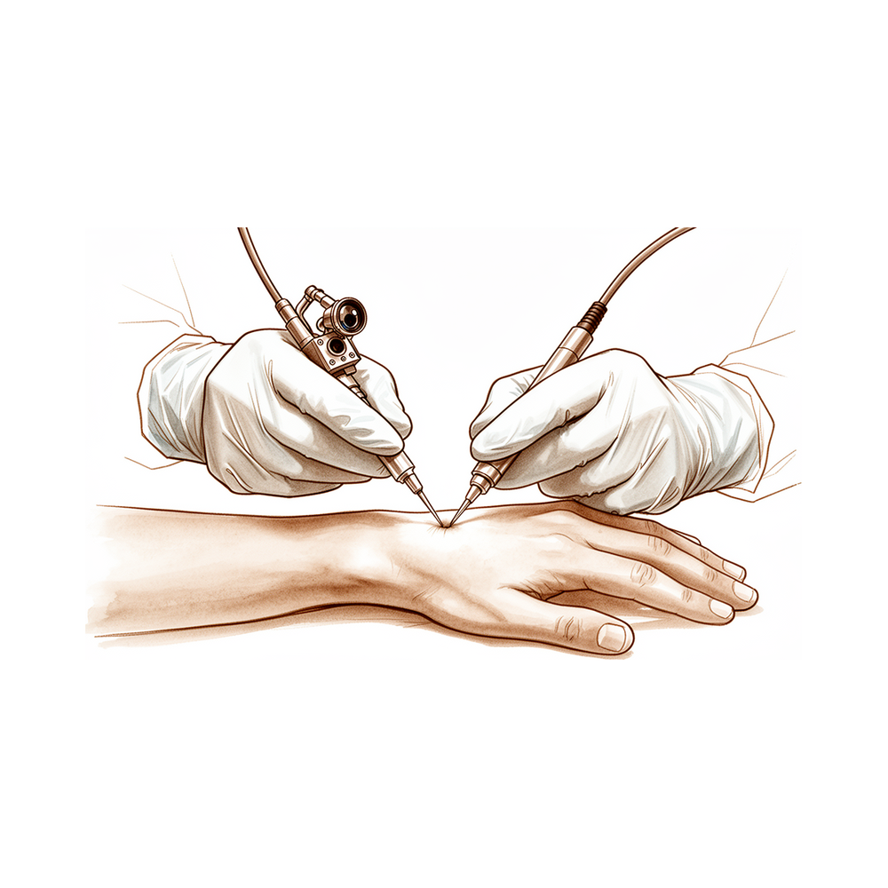
Hand & wrist arthroscopy: indications for diagnosis/treatment of TFCC tears, ligament injuries, synovitis, and dorsal wrist pain.
Overview
Arthroscopy provides adequate joint visualization with minimal damage to skin and surrounding soft tissues, thereby minimizing morbidity [3]. The procedure offers benefits including diagnostic accuracy, definitive operative treatment with minimal tissue damage, shorter rehabilitation times, fewer complications, and greater economy [16]. While a 2014 review notes the general lack of evidence-based assessment regarding the efficacy and cost-effectiveness of arthroscopic procedures [2], the Arthroscopy Association of North America (AANA) advocates for patient access and balanced interpretation of evidence-based medicine [21]. Suggested guidelines for the practice of arthroscopic surgery emphasize the need for appropriate training, privileges, practice standards, continuing education, and performance review to ensure patient safety and surgeon competence [7, 12].
The literature remains controversial in certain areas, with conclusions often unsupported due to bias and limitations; no clinical guidelines are definitive pending higher levels of evidence [4]. The rapid expansion of arthroscopy has led to abuses driven by financial motives and a lack of ethical standards [38]. In national arthroscopy registries, compliance with 2-year patient-reported outcome measures (PROMs) does not meet traditional follow-up thresholds of 60% or 80%, and reporting of missing-data methodologies is inconsistent [20]. Needle arthroscopy is considered low-risk with an extremely low complication rate when performed with appropriate technique and indications [1]. It offers potential for improved patient satisfaction, expedited care, and cost-effectiveness, but further research is necessary to quantify risks, determine cost implications, and identify best-suited patients and indications [35]. Current indications for needle arthroscopy in joints other than the shoulder are limited to rotator cuff tears, making its diagnostic value in other joints much more limited [5].
Despite being a minimally invasive procedure, elbow arthroscopy is not without complications and morbidity despite advances made in surgical technique [24]. Arthroscopic and open approaches have comparable outcome profiles regarding recurrence and complications for dorsal ganglion cysts [22]. Subtalar arthroscopy yields effective outcomes at long-term follow-up concerning patient-reported outcome measures in athletic populations, with favorable return to sport level, return to sport time, clinical outcomes, and safety outcome measures [8]. Arthroscopic treatment of talus bipartitus can be a safe and effective option with excellent short- and long-term outcomes [28]. Arthroscopic excision is the treatment of choice for localized pigmented villonodular synovitis and is currently thought to be curative [55]. The arthroscopic Hill-Sachs filling technique using an absorbable interference screw is safe, easily reproducible, does not require a long learning curve, and allows restoration of joint stability [23]. Distal radioulnar joint (DRUJ) arthroscopy should be used selectively and need not be a part of every arthroscopic wrist evaluation [18].
Anatomy & Pathophysiology
General Principles
Arthroscopy provides more precise information regarding the status of intra-articular structures than alternative imaging modalities [40]. The needle arthroscope allows for macroscopic examination of joints and the performance of biopsy under visual control, providing both microscopic and macroscopic information for diagnosis [27]. Closed synovial removal via arthroscopy aids in understanding the pathological mechanism within the joint, while arthroscopic examination helps in comparing the results of physical examination with intra-articular changes [27]. In patients with osteoarthritis, the needle arthroscope can establish the condition of joint structures but underestimates the severity of changes in cartilage and synovium compared to a standard arthroscope [27]. The use of a needle arthroscope entails less trauma for the patient compared to a standard arthroscope [27]. Needle arthroscopy can be carried out under local anesthesia as a 1-day procedure, and complications associated with needle arthroscopy are rare [27].
The needle arthroscope aids in the differential diagnosis of acute arthritis [27]. Repeated needle arthroscopy can monitor early signs of rheumatoid arthritis and predict the seriousness of the disease course [27]. Needle arthroscopy allows for monitoring the disease itself and following changes in joint structure resulting from therapy [27]. The needle arthroscope can help elucidate the role of various tissue mediators in disease [27]. Arthroscopy has historically served as a frontline treatment for painful joints in middle-aged and older people [37]. By the end of the 20th century, knee, shoulder, hip, and ankle arthroscopies were among the most frequently performed surgeries in developed countries [37]. Questions regarding the efficacy of knee arthroscopy for advanced osteoarthritis were first raised in 2002 following a landmark placebo-controlled trial by Moseley and colleagues [37]. Rigorous trials have questioned the efficacy of arthroscopic partial meniscectomy and subacromial decompression [37]. Despite high-quality evidence questioning efficacy, the number of arthroscopies for degenerative conditions continued to rise in the first decade of the 21st century [37]. Arthroscopies were increasingly billed using different procedure codes, which was the most obvious change to clinical practice [37].
Biomechanical investigations have inherent limitations, and direct clinical inference is rare when the role of biological healing is not tested [134]. A motion analysis system has been shown to be a valid means of objective assessment of arthroscopic skills in orthopaedics [72].
Hand and Wrist Anatomy
The hand is both an organ designed to obtain information and an organ of execution [46]. The hand functions efficiently only if the proximal joints of the limb are stable and yet mobile [46]. The shoulder is the most mobile joint in the body and allows orientation of the upper limb as required [46]. The combined movements of the wrist and forearm place the hand in a position for grasping [46]. The hand consists of 19 bones, 17 articulations, and 19 muscles situated entirely within the hand, along with about the same number of tendons activated by forearm muscles [46]. The skeleton of the hand and wrist consists of 27 bones, of which 19 are long bones [50]. The skeleton is divided into five rays, each making up a polyarticulated chain comprising the metacarpals and phalanges [50]. The wrist has three axes of movement, permitting the hand to be positioned in any spatial configuration [50].
The radial ray or first ray is the shortest, made up of only three bones: a metacarpal and two phalanges [50]. The trapezium is angled out in front of the carpal plane so that the first metacarpal makes an angle of about 45 degrees with the second metacarpal in the sagittal plane [50]. The thumb metacarpal is the shortest, while the index finger metacarpal is the longest [50]. The proximal and middle phalanges of the long and ring fingers are longer than those of the index finger [50]. The transverse axis of the palm is oblique, forming an acute angle of approximately 75 degrees with the longitudinal axis [50]. The epiphyseal plates are located at the proximal ends of the phalanges and the first metacarpal, whereas they are located at the distal ends of the other metacarpals [50].
The index metacarpal is the most firmly fixed, while the fifth metacarpal is semi-independent with a range of flexion–extension of approximately 20 degrees [91]. The ring metacarpal has about 10 degrees of mobility in flexion and extension [91]. The metacarpophalangeal joints are the keystones of the longitudinal arches of the hand [91]. The volar plates prevent hyperextension at the metacarpophalangeal joints [91]. The stability of the metacarpophalangeal joints is essential to the support of both the longitudinal and transverse metacarpal arches [91].
Control of digital posture requires a complex balance of extrinsic and intrinsic muscle forces [49]. Extrinsic muscles have their origin outside of the hand and their insertion on the hand or carpus, whereas intrinsic muscles have both origin and insertion within the hand [49]. The extrinsic extensors run through six different fibroosseous retinacular compartments at the wrist level [49].
Extensor Compartments: * First compartment: Contains the abductor pollicis longus and the extensor pollicis brevis [49]. * Second compartment: Contains the extensor carpi radialis longus and the extensor carpi radialis brevis [49]. * Third compartment: Contains the extensor pollicis longus, which turns abruptly radialward about Lister tubercle [49]. * Fourth compartment: Contains the extensor indicis proprius lying deep to the four tendons of the extensor digitorum communis [49]. * Fifth compartment: Contains the extensor digiti quinti [49]. * Sixth compartment: Contains the extensor carpi ulnaris tendon [49].
The digital extensor tendons are stabilized over the midline of the metacarpophalangeal joint by their attachment to sagittal band fibers [49]. With rupture or attenuation of the sagittal band fibers, the extrinsic extensor tendon can sublux to the ulnar side of the metacarpal head causing ulnar deviation of the finger [49]. The flexor digitorum profundus inserts on the proximal volar aspect of the distal phalanx [49]. The flexor digitorum superficialis acts as a flexor of the proximal interphalangeal and metacarpophalangeal joints [49].
There are seven interosseous muscles in the hand: four dorsal and three volar [89]. The dorsal interossei are abductors, while the volar interossei are adductors [89]. The middle finger has two dorsal interossei and no volar interossei because the central axis of the hand lies within it [89]. The deep head of each dorsal interosseous muscle forms a lateral tendon, or lateral band, at the level of the metacarpophalangeal joint [89]. The lateral bands are joined by the lateral slips of the extensor tendon to form the conjoined lateral band [89]. The two conjoined lateral bands to each finger unite at the distal third of the middle phalanx to form the terminal tendon [89]. The terminal tendon inserts at the base of the distal phalanx to extend it [89]. The flexor digiti quinti brevis is structurally and functionally similar to the deep head of the dorsal interossei, forming the ulnar lateral band of the little finger [89]. The opponens digiti quinti arises from the pisohamate ligament and the hook of the hamate and inserts onto the ulnar side of the diaphysis of the fifth metacarpal [89].
The extrinsic finger flexors are the flexor digitorum profundus and the flexor digitorum superficialis [95]. The flexor digitorum profundus originates from the proximal ulna and the interosseous membrane [95]. The tenosynovial sheath of the flexor pollicis longus is continuous with the radial bursa, and the tenosynovial sheath to the little finger is continuous with the ulnar digital bursa [95]. In some patients, the radial and ulnar bursae communicate, allowing a horseshoe abscess to spread between the thumb and little finger [95]. The flexor digitorum superficialis tendon bifurcates around the flexor digitorum profundus at the beginning of the A2 pulley [95]. The flexor digitorum superficialis tendon slips reunite distally at the Camper chiasm [95]. The fibroosseous tunnel, or digital flexor sheath, extends distally to the proximal aspect of the distal phalanx [95]. The A2 and A4 pulleys are the most essential in maintaining the mechanical advantage of the flexor tendons [95]. The tenosynovium that lines the fibroosseous tunnel supplies both nutrition and lubrication to the poorly vascularized flexor tendons [95]. Within the sheath, tendon vascularity is supplied via the vincula system: the vinculum longus and brevis [95].
The dorsal integument must be supple, elastic, and malleable to allow metacarpophalangeal joint flexion [96]. Interphalangeal flexion is accomplished by means of a special arrangement of skin folds on the dorsum of each articulation [96]. The gliding mechanism in narrow crowded areas is assured by the synovial sheath, which allows a considerable amplitude of movement [96]. The fibrous sheath assumes the role of a pulley when the tendon changes direction [96]. The extensor synovial tendon sheaths are present only at the level of the wrist on the dorsal aspect [96]. No single articulation in the hand is an isolated mechanical entity; they form functional groups arranged in kinetic chains [96]. The position of each articulation depends on the equilibrium of forces acting at that level and the position of the immediately proximal articulation [96]. Almost all movements in the hand are around oblique and variable axes, resulting in combined movements [96].
The dorsal skin possesses a normal pilosebaceous system, unlike the palm [97]. The dorsal skin becomes fragile in old age and has greater vulnerability to factors causing cutaneous atrophy, such as steroid therapy [97]. Flexion of the fingers produces a significant lengthening of the dorsal skin, with an average increase of 3 cm in the middle finger from extension to full flexion [97]. Flexion at the metacarpophalangeal joint alone requires an average skin lengthening of 1.25 cm [97]. The dorsal and palmar areas of skin are independent because of a system of adhesions that anchors their common boundary to the underlying plane [97].
The superficial palmar fascia lies in a coronal plane beneath the palmar subcutaneous tissue [98]. The palmaris longus tendon, when present, terminates in continuity with the fibers of the proximal corner of the superficial palmar fascia [98]. Fibers dorsal to the digital neurovascular bundle are collectively called Cleland ligament, and those palmar to the bundle are called Grayson ligament [98]. The web spaces are formed from the union of two nonsymmetrical cutaneous surfaces [88]. The dorsal slope of the web space has a gradual incline and its supple skin is not adherent to the subjacent region [88]. The palmar surface of the web space is flat and precipitously interrupted, with skin densely adherent to the commissural skeleton [88]. The commissural skeleton is formed by the interdigital palmar (natatory) ligament between the fingers and by the distal transverse ligament at the level of the thumb web [88].
The most common variations of the palmar arteries of the thumb can be schematized by dividing the thumb into three segments defined by the metacarpophalangeal and interphalangeal flexion creases [92]. In anatomical studies, only 15% of dissections of the palmar arteries of the thumb fall into the classical "typical" category [92]. The princeps pollicis artery is the terminal branch of the radial artery that crosses the first intermetacarpal space [92]. The dorsal arteries of the thumb are vascularized by two arteries which originate from the palmar arteries at the level of the first metacarpal [92].
The metacarpus is visually identifiable on the whole of the dorsum of the hand, with the third metacarpal serving as a landmark of wrist movements [99]. The metacarpophalangeal joints lie approximately on a transverse line that begins in the distal palmar crease on the ulnar side and runs into the proximal crease on the radial border [99]. The axes of flexion are arranged so that flexion of all the metacarpophalangeal and proximal interphalangeal joints causes the fingers to converge toward the scaphoid [99]. Malrotation after a metacarpal fracture is manifested by ulnar or radial deviation of the corresponding proximal phalanx when the metacarpophalangeal joints are flexed [99]. The pulley system of the thumb is composed of 4 components, as opposed to the traditional view of only 3 [115]. The trapeziometacarpal joint complex has a detailed functional anatomy that provides the basis for treatment of acquired afflictions at the base of the human thumb [135]. The position of the trapeziometacarpal prosthesis cup should be more radial or more distal in certain anatomical cases [173].
Arthroscopic Techniques and Findings
Cadaveric studies suggest horizontal placement of the hand instead of using a traction tower for arthroscopy of the proximal interphalangeal joints, as it is important to be able to flex the joint freely [54]. Recommended arthroscopic portals for the proximal interphalangeal joint are either between the central slip and the lateral bands of the extensor mechanism or between the lateral band and the collateral ligament [54]. A blunt technique of introduction is used for proximal interphalangeal joint arthroscopy to avoid iatrogenic cartilage damage and possible digital nerve injury [54]. Arthroscopy of the proximal interphalangeal joint has historically been described only in case reports prior to recent technical evaluations [54].
The novel extended application of dynamic wide-awake wrist arthroscopy has provided new insight in the assessment of carpal instability and challenged current understanding of carpal kinetics [155]. Endoscopic carpal tunnel release is a useful and safe alternative when performed by a surgeon familiar with hand anatomy and trained in endoscopic techniques, though it is more complex than standard open procedures [171]. A simple, effective, and cost-efficient solution exists to overcome oversized finger traps for wrist arthroscopy distraction [174]. The Zhang knot presents a technically feasible and mechanically promising addition to the arthroscopic armamentarium [82]. Instrumentation and experience for finger arthroscopy were noted as too small to draw firm conclusions regarding its ultimate use in 1985 [169]. Arthroscopy of the metacarpophalangeal and trapeziometacarpal joints has been described by several authors, while arthroscopy of the proximal interphalangeal joint was previously limited to case reports [54].
Classification
Palmer: The Palmer classification describes acute distal surface triangular fibrocartilage complex (TFCC) tears by location [34]. Central TFCC tears (Palmer 1A) are avascular, and débridement is recommended [34]. Ulnar or dorsal/ulnar TFCC tears (Palmer 1B) are the most amenable to arthroscopic repair due to good blood supply in this region [34]. Volar distal (Palmer 1C) and radial (Palmer 1D) TFCC tears are commonly débrided, although some are repaired [34]. Foveal TFCC tears affect the deep TFCC and bony insertion, are not included in the Palmer classification, and may be associated with distal radioulnar joint (DRUJ) instability [34].
Palmar: Palmar classified TFCC tears into two classes, with Type I denoting traumatic tears and Type II lesions denoting degenerative origin [53]. Type I TFCC injuries are further classified into four types [53]. Except for Type 1A tears, which imply a central articular disk perforation with a stable margin, the other three types of Type I TFCC tears are considered peripheral tears [53]. Peripheral TFCC tears may cause instability of the DRUJ or ulnocarpal joint, or both [53].
Nakamura: Nakamura defined the ulnar attachment as a three-dimensional complex with three components: proximal radioulnar ligaments, a distal hammock structure consisting of the centrally located fibrocartilage disk, meniscus homologue, and ulnocarpal ligaments [53]. Type 1B TFCC tear broadly includes all injuries of the TFCC at the ulnar attachment, with or without an accompanied ulnar styloid fracture [53].
Estrella and Ho: Estrella and Ho described the dorsal type of TFCC tear, located at the junction of the dorsal radioulnar ligament and the joint capsule just radial to the extensor carpi ulnaris tendon subsheath [53]. The dorsal type of TFCC tear constitutes another type of peripheral tear [53].
Copeland-Levy: The Copeland-Levy classification is suggested to be used to standardize terminology of subacromial impingement lesions [120].
Gächter: The classification according to Gächter is the most frequently used classification with clinical relevance for septic arthritis [145].
Other Considerations: Most peripheral lesions of the TFCC are traumatic in origin, though they may also occur in degenerative status due to chronic repetitive loading of the wrist [53]. Type 1B TFCC tear is among the most common peripheral tears leading to chronic ulnar wrist pain and impairment [53]. Foveal TFCC tears may be diagnosed with the arthroscopic hook test [34]. The arthroscope allows appropriate staging of the extent of cartilage degeneration in trapeziometacarpal joints to determine therapeutic options [19]. Arthroscopy allows for a true assessment of thumb carpometacarpal joint status, whereas traditional radiographic staging does not represent an accurate assessment of articular status, particularly in early stages of osteoarthritis [83]. Several classifications are available which evaluate joint infection according to pathological, anatomical, clinical, or arthroscopic aspects [145].
Clinical Presentation
Arthroscopy serves as the benchmark for identifying scapholunate and lunotriquetral ligament injuries [34]. In patients with suspected scapholunate tears, midcarpal arthroscopy frequently leads to a change in the final diagnosis and the procedure performed [57]. The diagnosis of ulnocarpal impaction may be confirmed arthroscopically by observing cartilage changes on the proximal, ulnar aspect of the lunate [34].
For trapezium fractures, arthroscopy provides direct visualization and accurate restoration of articular congruity [17]. In trapeziometacarpal arthroscopy, the arthroscope allows appropriate staging of the extent of cartilage degeneration to determine therapeutic options [19].
MCPJ arthroscopy allows clear visualization of intraarticular disorders with minimal soft tissue trauma [61]. This modality permits the diagnosis of pathology not well visualized by other means [71].
In the shoulder, diagnostic arthroscopy is a useful tool in patients with suspicion but no clear evidence of periprosthetic infection [10]. Needle arthroscopy is a promising diagnostic modality for intra-articular shoulder pathologies with comparable accuracy to MRI [51].
Regarding knee pathology, needle-based diagnostic imaging that can be used in the office setting is statistically equivalent to surgical diagnostic arthroscopy with regard to the diagnosis of intra-articular, nonligamentous knee joint pathology [30]. In a cohort of 60 knees subsequently operated on, arthroscopic findings were confirmed at operation in 48 cases, with six false negative and six false positive findings in the remaining cases [33].
The needle arthroscope allows macroscopic examination of the joints and performing biopsy under visual control [27]. It can provide micro- and macroscopic information for diagnosis [27]. With repeats, the needle arthroscope can monitor the early signs of rheumatoid arthritis and predict the seriousness of the course [27]. In patients with osteoarthritis, the condition of joint structures can be established with a needle arthroscope, but the severity of the changes in the cartilage and the synovium is underestimated compared to a standard arthroscope [27]. The operation using a needle scope entails less trauma for the patient than standard arthroscopy [27]. Complications are rare with needle arthroscopy [27].
The 70° arthroscope offers superior visualization to a 30° arthroscope in several arthroscopic procedures and circumstances [29].
Investigations
Diagnostic Accuracy and Comparison with Imaging: Needle-based diagnostic imaging performed in the office setting is statistically equivalent to surgical diagnostic arthroscopy for diagnosing intra-articular, nonligamentous knee joint pathology [30]. A VisionScope Imaging System (VSI) exam provides a more detailed and accurate diagnostic assessment of intra-articular knee pathology compared to MRI [132]. For shoulder pathologies, the combination of high-field-strength MRI plus operative arthroscopy offers the most accurate diagnostic workup [63]. Arthroscopy of finger joints provides more precise information regarding the status of intra-articular structures than alternative imaging modalities [40]. Patients with rotator cuff tendinopathy undergoing arthroscopy should be informed that the presence and severity of cartilage lesions may be underestimated on MRI [137]. Synovial chondromatosis can be invasive, and even the best preoperative imaging may not demonstrate the degree of local tissue involvement [139].
Specific Joint Applications: Arthroscopy provides direct visualization and accurate restoration of articular congruity for trapezium fractures, yielding excellent results [17]. The arthroscope allows appropriate staging of the extent of cartilage degeneration at the trapeziometacarpal joint to determine therapeutic options [19]. MCPJ arthroscopy is a simple, safe, and effective procedure that allows clear visualization of intraarticular disorders with minimal soft tissue trauma [61]. Wrist arthroscopy has firmly established its value as an essential diagnostic and powerful therapeutic tool [177].
Technique, Safety, and Limitations: The current indications for in-office needle arthroscopy in other joints are limited to rotator cuff tears in the shoulder, making its diagnostic value in other joints much more limited [5]. Practitioners should be very selective in using in-office arthroscopy [63]. There is an inherent small error of approximately 4% when arthroscopically determining anterior-inferior glenoid bone loss [45].
Image Quality and Equipment: Seven disturbances were identified that serve as measures for arthroscopic image quality [73]. The 70° arthroscope demonstrates technical advantages over the 30° arthroscope, including a wider field of view and less image distortion at the periphery [147]. Arthroscopic images are subject to distortion that increases with greater lens reflecting angles and oblique viewing angles [163].
Evidence Base and Research Context: The number of meta-analyses appearing in Arthroscopy has increased over the past 2 decades [9]. The number of publications on arthroscopy increased significantly from 1999 to 2013, with a more than 2-fold increase in volume [84].
Treatment
General Principles and Safety
Elbow arthroscopy is a reliable procedure that requires a clear understanding of anatomy to safely access the joint [104]. The procedure has become a safe and effective treatment for elbow pathology due to improved instrumentation, advanced surgical technique, and better understanding of anatomy [102]. The purpose of fluid management in arthroscopy is to review fundamental concepts and techniques to safely improve arthroscopic visualization [6].
Arthroscopy provides direct visualization and accurate restoration of articular congruity, yielding excellent results in the treatment of trapezium fractures [17]. Arthroscopic treatment can be a safe and effective option with excellent short- and long-term outcomes for talus bipartitus [28]. Arthroscopic excision is the treatment of choice and is currently thought to be curative for localized pigmented villonodular synovitis arising from the quadriceps tendon sheath [55]. The arthroscopic approach is a valuable tool if the surgeon aims to reduce the invasiveness of the procedure for capitellum osteochondritis dissecans, despite being more demanding [52]. Arthroscopic antero-superior ancillary portals help avoid bony osteotomy and limit further articular problems when addressing surgical repair perpendicularly on the talar dome [172].
In an in vitro setup, a new percutaneous procedure for lateral ulnar collateral ligament reconstruction provides isometry over the range of motion and sufficiently restores posterolateral rotatory stability comparable to the intact situation and open reconstruction [175]. The all-inside suture technique for flexor digitorum profundus attachment to bone has the advantages of avoiding an external button and the cost of anchors [170]. Arthroscopic Hill-Sachs filling technique using an absorbable interference screw is considered safe, easily reproducible, does not require a long learning curve, and allows restoration of joint stability [23].
Debridement of complete scapholunate tears is less beneficial, with complete resolution or mild symptoms in 67% of patients followed for short-term [77]. Patients treated with additional arthroscopy to remove hematoma and debris for intra-articular distal radius fractures do not have better functional outcomes compared with the nonarthroscopically treated group [69]. Arthroscopic abrasion arthroplasty is not superior to ligament reconstruction and tendon interposition for thumb carpometacarpal arthritis, with poor results in the arthroscopy group potentially secondary to inherent instability and lack of interposition material [41]. Because arthroscopic interposition of material contributes to health care costs without clear benefit to the patient, routine use of expensive interposition products for thumb basal joint arthritis should be abandoned or carefully evaluated with a prospective randomized controlled trial [152]. Osteoarthritis will likely remain the most common indication for basal joint arthroscopy while chronic pain and inflammation are useful indications for metacarpophalangeal arthroscopy [113]. Arthroscopy allows for a true assessment of the joint status in thumb carpometacarpal arthritis, which is more accurate than simple radiographic means, particularly in early stages of osteoarthritis [83]. Arthroscopic treatment of pigmented villonodular synovitis of the proximal tibiofibular joint forms the basis of further development of arthroscopic procedures of the joint and reconstruction of adjacent capsulo-ligamentous structure [74]. There was no increase in complications when total knee arthroplasty was performed more than six months after knee arthroscopy [85].
Diagnostic Arthroscopy
In-office diagnostic needle arthroscopy using the NanoScope™ system has current indications for use in other joints limited to rotator cuff tears in the shoulder, making its diagnostic value in other joints much more limited [5]. The operation using a needle scope entails less trauma for the patient, can be carried out under local anesthesia as a 1-day procedure, and complications are rare [27]. The needle arthroscope aids in the differential diagnosis of acute arthritis and can monitor the early signs of rheumatoid arthritis, predict the seriousness of the course, and monitor the disease itself [27]. With repeats, the needle arthroscope can help in elucidating the role of various tissue mediators in the disease [27].
In-office needle arthroscopy uses a 0 scope, requiring the surgeon to become familiar with this technical difference compared with a standard scope [161]. Previous surgery may increase difficulty of using small instruments through previous scars during in-office needle arthroscopy [161]. In-office needle arthroscopy may identify certain pathologies requiring the operating room setting, meaning the patient may undergo a second intervention [161]. As with the standard arthroscope, the needle arthroscope allows macroscopic examination of the joints and performing biopsy under visual control [27]. Comparing results in patients with osteoarthritis, the needle arthroscope can establish the condition of joint structures but underestimates the severity of changes in the cartilage and synovium compared to a standard arthroscope [27].
Pain Management and Perioperative Care
ERAS pathways resulted in a notable reduction in prescribing of opioids postoperatively after both arthroplasty and arthroscopic surgeries [66]. A range of nonopioid strategies exist that can reduce postarthroscopic procedural opioid consumption with equivalent pain outcomes [136]. A comprehensive solution for opioid use after arthroscopy requires identifying the problem, considering multimodal anesthesia, avoiding overprescribing, acknowledging that minimizing opioid use is not equivalent to undertreating pain, minimizing preoperative opioid use, managing patient expectations, and continuing to investigate outcomes while limiting or forgoing opioids [164]. Dexamethasone may be a valuable adjuvant to a multimodal systemic pain regimen after hip arthroscopy [176]. Magnesium sulphate added to bupivacaine may significantly improve the analgesic efficacy for arthroscopy [180]. A large gap between published recommendations and current practices regarding thromboprophylaxis after arthroscopy surgery means that the relevance of current recommendations and the cost-benefit ratio of prescribing drug-based thromboprophylaxis should be reconsidered and re-evaluated [67].
Evidence Quality and Practice Standards
There is a general lack of evidence-based assessment of the efficacy and cost-effectiveness of arthroscopic procedures [2]. Shoulder arthroscopy literature remains controversial, conclusions are often unsupported due to bias and limitations, and no clinical guidelines are definitive pending higher levels of evidence [4]. The majority of sports medicine and arthroscopy-related randomized controlled trials reporting nonsignificant results are statistically fragile [68]. The Arthroscopy Association of North America advocates for patient access and balanced interpretation of evidence-based medicine [21]. While arthroscopy has grown rapidly and offers significant benefits through minimally invasive techniques, its expansion has led to abuses driven by financial motives and lack of ethical standards [38]. As shown in the intact state, there is an inherent small error of approximately 4% when arthroscopically determining anterior-inferior glenoid bone loss [45].
Complications
General and Systemic
Elbow arthroscopy is associated with complications and morbidity despite being a minimally invasive procedure [24]. The minimal soft tissue disruption inherent in endoscopic surgery theoretically decreases the risk of infection, wound complications, peri-incisional numbness, and deep vein thrombosis or pulmonary embolism [86]. Enhanced recovery after surgery (ERAS) pathways resulted in a notable reduction in prescribing of opioids postoperatively after arthroscopic surgeries [66]. A large gap exists between published recommendations and current practices regarding drug-based thromboprophylaxis after arthroscopy surgery [67].
Infection
Post-arthroscopy septic arthritis is a recognized complication for which current data and practical recommendations have been reviewed [59]. Avoidance of magnetic resonance arthrogram within 2 weeks of shoulder arthroscopy may mitigate postoperative infection risk [217]. Patients with rheumatic diseases undergoing arthroscopy may remain on immunosuppression perioperatively without significant risk for infection [220].
Wrist and Hand
In a series of 211 wrist arthroscopies, the reported complication rate was 5.2% [162]. Major complications reported in wrist arthroscopy include permanent stiffness after arthroscopic synovectomy and development of a symptomatic dorsal wrist ganglion over the 3-4 portal site requiring surgery [162]. Minor complications reported in wrist arthroscopy include transient superficial dorsal ulnar sensory neurapraxia, transient wrist and finger joint stiffness, ulnar neurapraxia, superficial portal site infection, first-degree burns to the forearm after contact with a hot arthroscopic tower, and extensor carpi ulnaris tendinitis [162]. All minor complications in the reported wrist arthroscopy series resolved at latest follow-up evaluation with conservative care [162]. A 29% complication rate was reported in patients undergoing arthroscopic TFCC repair, requiring secondary procedures to address extensor digiti minimi tendon capture by the suture and sensory disturbances [162].
Shoulder
The overall complication rate for revision rotator cuff repair was 12% [190]. The most frequent complications in revision rotator cuff repair were repair failure (88%), shoulder stiffness (5%), and infection (4%) [190].
Knee and Lower Extremity
Arthroscopy for the treatment of septic arthritis of the knee in adults has demonstrated a lower reinfection rate compared to arthrotomy [219]. Arthroscopy for the treatment of septic arthritis of the knee in adults has demonstrated less frequent clinical signs of initial inflammatory reaction compared to arthrotomy [219].
Needle Arthroscopy
Only minor complications are reported after needle arthroscopy according to a systematic review [64].
Other Considerations
Complications after open surgery for trigger finger are more severe than those associated with nonoperative treatments [209]. Surgeons should inform patients of the likelihood of having minor complications following open trigger finger release [195]. Satisfaction and complication rates were similar across four types of skin incision for trigger thumb release, implying no clear benefit of one type over another [227]. Complications associated with minimally invasive thumb carpometacarpal joint arthrodesis include slight loss of length of the first ray, nonunion, malunion, fixation failure, implant breakage, infection, persistent pain, and pain owing to screw irritation [191]. The revision rate for the LPM ceramic coated proximal interphalangeal joint arthroplasty was higher than in published series for other PIP joint implants [225].
Recovery
Light activity (weeks): The evidence base does not specify a typical week range for desk work, driving, or light activities of daily living.
Full activity (months): The evidence base does not specify a typical month range for manual work, sport, or full range of motion and strength return.
Complete recovery / outcome plateau (months): The evidence base does not specify a month range for when pain, strength, and final functional outcomes stabilise.
Rehabilitation protocol: Adhesions following hip arthroscopy were reduced with changes to the rehabilitation protocol including hip circumduction [129]. Success of zone II combined flexor digitorum superficialis and flexor digitorum profundus repair relies on controlled early rehabilitation to minimize rupture and stiffness [133].
Functional milestones: Arthroscopic interventions for arthrofibrosis after ACL reconstruction effectively restored motion and improved patient-reported outcome scores, with gains maintained at 2-year follow-up [128]. A statistically significant clinical improvement was shown at 24 months for arthroscopic second generation autologous chondrocyte implantation [218]. Second-look arthroscopy for arthroscopic second generation autologous chondrocyte implantation demonstrated complete coverage of the grafted area with hyaline cartilage-like tissue in 12 of 15 analyzed patients [218]. In patients that do not require revision surgery following pyrocarbon proximal interphalangeal arthroplasty, pain relief was improved and motion maintained [43]. The procedure for osteochondral grafting of the metacarpophalangeal joint in rheumatoid arthritis resulted in stable, painless joints with acceptable outcomes despite some loss of motion [44]. Functional recovery following osteochondral autograft from the hamate for treating partial defect of the proximal interphalangeal joint is generally acceptable, with a well-restored joint architecture [78]. Arthroscopic DWG resection showed an improvement in functional measurements in addition to relief of pain in a significant proportion of patients [221]. Overall, results following arthroscopic management of greater trochanter pain syndrome and abductor tears have been promising, including significantly improved pain scores and functional outcomes at final post-operative assessment [214].
Other Considerations: Arthroscopic surgery offers benefits including shorter rehabilitation times and fewer complications [16]. Subtalar arthroscopy yields favorable return to sport time and return to sport level in athletic populations [8]. The endoscopic procedure for carpal tunnel release has the benefit of faster rehabilitation and return to work compared to open techniques [149]. Wrist arthroscopy is a reasonable early treatment option for athletes to give them the opportunity to return to their pre-injury level of participation and function sooner and more often than was possible in the past or after a period of immobilization [131]. Outcomes of zone 2 flexor tendon repairs were worse if there was an accompanying volar plate injury [222]. Patients with Hoffa's disease without any other concomitant pathology can expect resolution or long-term improvement in their symptoms and function after arthroscopic resection [223]. Improvement in motion or clinical score for arthroscopic treatment of elbow motion limitation was found to be closely related to preoperative range of motion [224]. The patient had full range of motion and no complaints 9 months after surgery for rocky form giant synovial chondromatosis [130]. In national arthroscopy registries, compliance with 2-year patient-reported outcome measures (PROMs) does not meet traditional follow-up thresholds of 60% or 80% [20]. Reporting of missing-data methodologies in national arthroscopy registries is inconsistent [20].
Key Evidence
- [L5] Needle arthroscopy is considered low-risk, with an extremely low complication rate when performed with appropriate technique and indications. [1] (10.1016/j.eats.2022.07.006)
- [L4] This Review reflects on both the success of arthroscopy and the general lack of evidence-based assessment of the efficacy and cost-effectiveness of arthroscopic procedures. [2] (10.1038/nrrheum.2014.174)
- [L5] Arthroscopy allows adequate visualization of joints with the smallest amount of damage to the skin and surrounding soft tissues, minimizing morbidity. [3] (10.1016/0020-1383(90)90041-r)
- [L5] The editorial states that shoulder arthroscopy literature remains controversial, conclusions are often unsupported due to bias and limitations, and no clinical guidelines are definitive pending higher levels of evidence. [4] (10.1016/j.arthro.2012.07.001)
- [L1] However, its current indications for use in other joints are limited to rotator cuff tears in the shoulder, making its diagnostic value in other joints much more limited. [5] (10.1016/j.arthro.2019.03.045)
- [L5] The purpose of this article is to review the fundamental concepts of fluid management in arthroscopy and the techniques to safely improve arthroscopic visualization. [6] (10.5435/jaaos-d-20-01057)
- [L5] This statement outlines suggested guidelines for the practice of arthroscopic surgery, emphasizing the need for appropriate training, privileges, and performance review by the Arthroscopy Association of North America. [7] (10.1016/s0749-8063(14)00453-8)
- [L4] Subtalar arthroscopy yields effective outcomes at long-term follow-up concerning patient-reported outcome measures in athletic population, with favorable return to sport level, return to sport time, clinical outcomes and safety outcome measures. [8] (10.1007/s00167-020-06385-8)
- [L4] The number of meta-analyses appearing in Arthroscopy has increased over the past 2 decades. [9] (10.1016/j.arthro.2015.08.002)
- [L3] Diagnostic arthroscopy is a useful diagnostic tool in patients with suspicion but no clear evidence of PPSI. [10] (10.1016/j.arthro.2019.03.058)
- [L5] This statement outlines suggested guidelines for the practice of arthroscopic surgery, covering privileges, training, practice standards, continuing education, and performance review to ensure patient safety and surgeon competence. [12] (10.1016/s0749-8063(14)00850-0)
- [L5] Arthroscopic surgery has had a significant impact on the whole spectrum of surgical treatment, offering benefits such as accuracy of diagnosis, definitive operative treatment with minimal tissue damage, shorter rehabilitation times, fewer complications, and greater economy. [16] (10.1016/j.arthro.2009.10.005)
- [L4] Arthroscopy provides direct visualization and accurate restoration of articular congruity, yielding excellent results. [17] (10.1016/j.arthro.2006.07.051)
- [L5] DRUJ arthroscopy should be used selectively and need not be a part of every arthroscopic wrist evaluation. [18] (10.1016/s0749-0712(21)00182-7)
- [L5] The arthroscope allows appropriate staging of the extent of cartilage degeneration to determine therapeutic options. [19] (10.1016/j.hcl.2006.02.006)
- [L4] In national arthroscopy registries, compliance with 2-year PROMs does not meet traditional follow-up thresholds of 60% or 80% and reporting of missing-data methodologies is inconsistent. [20] (10.1016/j.arthro.2020.08.007)
- [Paper] It highlights the Arthroscopy Association of North America's (AANA) advocacy for patient access and balanced interpretation of evidence-based medicine. [21] (10.1016/j.arthro.2014.06.019)
- [L2] Arthroscopic and open approaches have comparable outcome profiles regarding recurrence and complications. [22] (10.1177/1753193417734428)
- [L5] The authors believe the described arthroscopic procedure is safe, is easily reproducible, does not require a long learning curve, and allows restoration of joint stability. [23] (10.1016/j.eats.2022.08.046)
- [L3] Elbow arthroscopy is not without complications and morbidity despite being a minimally invasive procedure and advances made in surgical technique. [24] (10.1177/17585732241249393)
- [L4] [27] (10.1007/s00167-002-0329-4)
- [L4] Arthroscopic treatment can be a safe and effective option with excellent short- and long-term outcomes. [28] (10.1007/s00167-017-4613-8)
- [L1] [29] (10.1016/j.arthro.2011.02.001)
- [L2] Needle-based diagnostic imaging that can be used in the office setting is statistically equivalent to surgical diagnostic arthroscopy with regard to the diagnosis of intra-articular, nonligamentous knee joint pathology. [30] (10.1016/j.arthro.2018.03.010)
- [L4] In 48 of 60 knees subsequently operated on, arthroscopic findings were confirmed at operation, with six false negative and six false positive findings in the remaining cases. [33] (10.2106/jbjs.l.00072)
- [Paper] Needle arthroscopy offers potential for improved patient satisfaction, expedited care, and cost-effectiveness, but further research is necessary to quantify risks, determine cost implications, and identify best-suited patients and indications. [35] (10.1097/corr.0000000000003105)
- [L5] [37] (10.1136/bjsports-2020-102981)
- [L5] The paper argues that while arthroscopy has grown rapidly and offers significant benefits through minimally invasive techniques, its expansion has led to abuses driven by financial motives and lack of ethical standards. [38] (10.2106/00004623-199274100-00019)
- [L5] Arthroscopy of finger joints provides more precise information regarding the status of intra-articular structures than alternative imaging modalities. [40] (10.1016/j.hcl.2017.07.018)
- [L3] The poor results in the arthroscopy group may be secondary to the inherent instability of the CMC joint and lack of use of any biological or artificial interposition material. [41] (10.1177/1558944718778405)
- [L4] Overall, in patients that do not require revision surgery, pain relief was improved and motion maintained. [43] (10.1016/j.jhsa.2018.06.020)
- [L4] The procedure resulted in stable, painless joints with acceptable outcomes despite some loss of motion. [44] (10.1054/jhsb.2002.0839)
- [L5] As shown in the intact state, there is an inherent small error of approximately 4% when arthroscopically determining bone loss. [45] (10.1016/j.arthro.2009.05.019)
- [L2] Needle arthroscopy is a promising diagnostic modality for intra-articular shoulder pathologies with comparable accuracy to MRI. [51] (10.1016/j.arthro.2021.03.006)
- [L4] Despite being more demanding, the arthroscopic approach is a valuable tool if the surgeon aims to reduce the invasiveness of the procedure. [52] (10.1007/s00167-019-05568-2)
- [L4] [54] (10.1054/jhsb.2001.0725)
- [L4] Arthroscopic excision is the treatment of choice and is currently thought to be curative. [55] (10.1016/j.arthro.2005.12.035)
- [L3] In patients with suspected scapholunate tears, midcarpal arthroscopy often leads to a change in diagnosis and procedure performed. [57] (10.1016/j.jhsa.2013.08.040)
- [Paper] [59] (10.1016/j.otsr.2015.09.004)
- [L4] MCPJ arthroscopy is a simple, safe, and effective procedure that allows clear visualization of intraarticular disorders with minimal soft tissue trauma, providing satisfactory pain relief and good long-term functional outcomes. [61] (10.1016/j.hcl.2011.05.007)
- [L5] The author concludes that practitioners should be very selective in using in-office arthroscopy and that the combination of high-field-strength MRI plus operative arthroscopy offers the most accurate diagnostic workup. [63] (10.1016/j.arthro.2018.06.002)
- [L4] [64] (10.1016/j.asmr.2025.101158)
- [L4] ERAS pathways resulted in a notable reduction in prescribing of opioids postoperatively after both arthroplasty and arthroscopic surgeries. [66] (10.5435/jaaos-d-24-01232)
- [L4] This large gap between published recommendations and current practices means that we should reconsider the relevance of current recommendations and re-evaluate the cost-benefit ratio of prescribing a drug-based thromboprophylaxis after arthroscopy surgery. [67] (10.1016/j.otsr.2020.08.004)
- [L2] Using this methodology, we found that the majority of sports medicine and arthroscopy-related RCTs reporting nonsignificant results are fragile. [68] (10.1016/j.arthro.2023.02.022)
- [L3] Patients treated with additional arthroscopy to remove hematoma and debris do not have better functional outcomes compared with the nonarthroscopically treated group. [69] (10.1016/j.jhsa.2018.06.079)
- [L5] Arthroscopy of the MCP joint permits the diagnosis of pathology not well visualized by other means, and the treatment of these lesions with minimal soft tissue disruption. [71] (10.1016/s0749-0712(21)00186-4)
- [L4] This study shows the validity of a motion analysis system as a means of objective assessment of arthroscopic skills in orthopaedics. [72] (10.1016/j.arthro.2007.08.033)
- [L4] Seven disturbances were identified that serve as measures for arthroscopic image quality. [73] (10.1007/s00167-007-0299-7)
- [L5] This forms the basis of further development of arthroscopic procedures of the joint and reconstruction of adjacent capsulo-ligamentous structure. [74] (10.1007/s00167-014-3031-4)
- [L4] Debridement of complete tears is less beneficial, with complete resolution or mild symptoms in 67% of patients followed for short-term. [77] (10.1016/s0749-0712(21)00178-5)
- [L4] The functional recovery is generally acceptable, with a well-restored joint architecture. [78] (10.1016/j.jhsa.2021.11.007)
- [L5] The Zhang knot presents a technically feasible and mechanically promising addition to the arthroscopic armamentarium. [82] (10.1002/atn2.70108)
- [L4] [83] (10.1016/j.hcl.2011.06.004)
- [L4] The number of publications on arthroscopy increased significantly from 1999 to 2013, with a more than 2-fold increase in volume. [84] (10.1016/j.arthro.2015.03.009)
- [L3] There was no increase in complications when total knee arthroplasty was performed more than six months after knee arthroscopy. [85] (10.1016/j.arth.2015.02.023)
- [Paper] [86] (10.1016/j.eats.2019.11.022)
- [L5] Elbow arthroscopy is a reliable procedure that requires a clear understanding of the anatomy to be able to safely access the joint. [104] (10.1016/j.arthro.2019.05.014)
- [L5] Osteoarthritis will likely remain the most common indication for basal joint arthroscopy while chronic pain and inflammation are useful indications for metacarpophalangeal arthroscopy. [113] (10.1016/j.jhsa.2007.02.020)
- [L4] The pulley system of the thumb is composed of 4 components, as opposed to the traditional view of only 3. [115] (10.1016/j.jhsa.2012.08.005)
- [L4] Hence, we suggest the Copeland-Levy classification be used to standardize terminology of the subacromial impingement lesion. [120] (10.1016/j.jse.2017.07.018)
- [L4] Arthroscopic interventions for arthrofibrosis after ACL reconstruction effectively restored motion and improved patient-reported outcomes scores, with gains maintained at 2-year follow-up. [128] (10.1097/corr.0000000000003139)
- [L3] Adhesions following hip arthroscopy were reduced with changes to the rehabilitation protocol including hip circumduction. [129] (10.1016/j.arthro.2011.03.043)
- [Case_report] The patient had full range of motion and no complaints 9 months after surgery. [130] (10.1007/s00167-005-0674-1)
- [L5] Wrist arthroscopy is a reasonable early treatment option for athletes to give them the opportunity to return to their pre-injury level of participation and function sooner and more often than was possible in the past or after a period of immobilization. [131] (10.1016/s0749-0712(21)00587-4)
- [L2] When compared to MRI, a VSI exam can provide a more detailed and accurate diagnostic assessment of intra-articular knee pathology. [132] (10.1177/2325967114s00100)
- [L5] Success relies on atraumatic technique, preservation of pulleys, multistrand repair, and controlled early rehabilitation to minimize rupture and stiffness. [133] (10.1016/j.jhsa.2010.06.024)
- [L5] Biomechanical investigations, when well performed, are among a few of our favorite things, but time-zero studies have inherent limitations and direct clinical inference is rare when the role of biological healing is not tested. [134] (10.1016/j.arthro.2019.08.028)
- [L5] Detailed understanding of the functional anatomy and related pathologic features of the trapeziometacarpal joint complex provides the basis for treatment of acquired afflictions at the base of the human thumb and a model for the more general study of idiopathic osteoarthritis. [135] (10.1097/01.blo.0000176968.28247.5c)
- [L1] A range of nonopioid strategies exist that can reduce postarthroscopic procedural opioid consumption with equivalent pain outcomes. [136] (10.1177/23259671231214700)
- [L1] Patients with rotator cuff tendinopathy undergoing arthroscopy should be informed that the presence and severity of cartilage lesions may be underestimated on MRI. [137] (10.1016/j.jse.2014.01.048)
- [L4] Synovial chondromatosis can be invasive, and even the best preoperative imaging may not demonstrate the degree of local tissue involvement. [139] (10.1054/jhsb.2001.0677)
- [L4] [145] (10.1007/s00167-021-06525-8)
- [L5] The 70° arthroscope demonstrates technical advantages over the 30° arthroscope, including a wider field of view and less image distortion at the periphery. [147] (10.1007/s00167-014-3452-0)
- [L1] The endoscopic procedure is safe and has the benefit of faster rehabilitation and return to work compared to open techniques. [149] (10.1177/1753193412475247)
- [L4] Because arthroscopic interposition of material contributes to health care costs without clear benefit to the patient, routine use of expensive interposition products should be abandoned or carefully evaluated with a prospective randomized controlled trial. [152] (10.1016/j.jhsa.2015.05.010)
- [Paper] The novel extended application of dynamic wide-awake wrist arthroscopy has provided new insight in the assessment of carpal instability and challenged current understanding of carpal kinetics. [155] (10.1016/j.hcl.2018.08.010)
- [L5] [161] (10.5435/jaaosglobal-d-23-00124)
- [L5] [162] (10.5435/jaaos-20-11-725)
- [L5] Arthroscopic images are subject to distortion that increases with greater lens reflecting angles and oblique viewing angles. [163] (10.1007/s00167-014-3336-3)
- [L5] A comprehensive solution requires identifying the problem, considering multimodal anesthesia, avoiding overprescribing, acknowledging that minimizing opioid use is not equivalent to undertreating pain, minimizing preoperative opioid use, managing patient expectations, and continuing to investigate the outcomes of pain management while limiting opioid prescriptions or forgoing opioids altogether. [164] (10.1016/j.arthro.2019.04.001)
- [L4] The authors note that instrumentation and experience are too small to draw firm conclusions regarding the ultimate use for finger arthroscopy in the future. [169] (10.1177/036354658501300212)
- [L5] The all-inside suture technique also has the advantages of avoiding an external button and the cost of anchors. [170] (10.1016/j.jhsa.2013.02.015)
- [L4] Endoscopic carpal tunnel release is a useful and safe alternative when performed by a surgeon familiar with hand anatomy and trained in endoscopic techniques, though it is more complex than standard open procedures. [171] (10.1007/s001670050097)
- [L4] They help avoiding bony osteotomy and limiting articular further problems. [172] (10.1016/j.arthro.2016.03.079)
- [L4] In these cases, the position of the cup should be more radial or more distal. [173] (10.1177/17531934241274117)
- [L5] We present a simple, effective, and cost-efficient solution to overcome oversized finger traps for wrist arthroscopy distraction. [174] (10.1016/j.eats.2025.103662)
- [L5] In an in vitro setup, the new percutaneous procedure provides isometry over the range of motion and sufficiently restores posterolateral rotatory stability comparable to the intact situation and open reconstruction. [175] (10.1007/s00167-012-2019-1)
- [L1] Dexamethasone may be a valuable adjuvant to a multimodal systemic pain regimen after hip arthroscopy. [176] (10.1177/03635465241232157)
- [L5] Wrist arthroscopy has firmly established its value as an essential diagnostic and powerful therapeutic tool, with new procedures evolving to address osseous, soft tissue, and cartilage problems precisely under arthroscopic means. [177] (10.1016/j.hcl.2017.08.001)
- [L1] Magnesium sulphate added to bupivacaine may significantly improve the analgesic efficacy for arthroscopy. [180] (10.1186/s13018-021-02609-w)
- [L5] [190] (10.2106/jbjs.20.00014)
- [L4] [191] (10.1016/j.jhsa.2014.10.020)
- [L4] Surgeons should inform patients of the likelihood of having these minor complications. [195] (10.1016/j.jhsa.2009.12.040)
- [L1] Complications after open surgery are more severe; this must be taken into account when advising patients with regard to treatment. [209] (10.1016/j.jhsa.2017.02.011)
- [L4] Overall, results following arthroscopic management of GTPS and abductor tears have been promising, including significantly improved pain scores and functional outcomes at final post-operative assessment. [214] (10.1007/s00167-020-06322-9)
- [L3] Avoidance of MRA within 2 weeks of shoulder arthroscopy may mitigate postoperative infection risk. [217] (10.1016/j.jse.2022.11.018)
- [L4] A statistically significant clinical improvement was shown at 24 months, with second-look arthroscopy demonstrating complete coverage of the grafted area with hyaline cartilage-like tissue in 12 of 15 analyzed patients. [218] (10.1007/s00167-006-0265-9)
- [L1] Arthroscopy is as effective as arthrotomy in the treatment of septic arthritis of the knee in adults, but arthroscopy has demonstrated a lower reinfection rate and less frequent clinical signs of initial inflammatory reaction. [219] (10.1007/s00167-015-3918-8)
- [L3] This study is the first to suggest that patients with rheumatic diseases undergoing arthroscopy may remain on immunosuppression perioperatively without significant risk for infection. [220] (10.1177/2325967121s00664)
- [L4] Arthroscopic DWG resection showed an improvement in functional measurements in addition to relief of pain in a significant proportion of patients. [221] (10.1016/j.arthro.2010.05.008)
- [L4] The study aims to evaluate the contribution of palmar plate injury to postoperative recovery in zone 2 flexor tendon repairs, noting that outcomes were worse if there was an accompanying volar plate injury. [222] (10.1177/17531934241300510)
- [L4] Patients with Hoffa's disease without any other concomitant pathology can expect resolution or long-term improvement in their symptoms and function after arthroscopic resection. [223] (10.1016/j.arthro.2007.05.013)
- [L4] Improvement in motion or clinical score was not a satisfactory surrogate for learning curve and found to be closely related to preoperative range of motion. [224] (10.1007/s00167-010-1334-7)
- [L4] The revision rate for the LPM prosthesis was higher than in published series for other PIP joint implants, with close surveillance of all patients with this prosthesis currently in situ recommended. [225] (10.1177/1753193407087864)
- [L3] Satisfaction and complication rates were similar, implying no clear benefit of one type of incision over another. [227] (10.1177/17531934241232341)
See Also
- Soft Tissue Trauma
- Trigger Finger
- Trigger finger release
References
[1] In‐Office Diagnostic Needle Arthroscopy Using the NanoScope™ Arthroscopy System. Arthroscopy Techniques. 2022. DOI: 10.1016/j.eats.2022.07.006
[2] Advances in arthroscopy—indications and therapeutic applications. Nature Reviews Rheumatology. 2014. DOI: 10.1038/nrrheum.2014.174
[3] Arthroscopy. Injury. 1990. DOI: 10.1016/0020-1383(90)90041-r
[4] Shoulder Arthroscopy Literature Remains Controversial. Arthroscopy. 2012. DOI: 10.1016/j.arthro.2012.07.001
[5] In‐Office Needle Arthroscopy: A Systematic Review of Indications and Clinical Utility. Arthroscopy. 2019. DOI: 10.1016/j.arthro.2019.03.045
[6] Fundamentals of Arthroscopy Fluid Management and Strategies to Safely Improve Visualization. Journal of the American Academy of Orthopaedic Surgeons. 2021. DOI: 10.5435/jaaos-d-20-01057
[7] Suggested Guidelines for the Practice of Arthroscopic Surgery. Arthroscopy. 2014. DOI: 10.1016/s0749-8063(14)00453-8
[8] Six out of ten patients with sinus tarsi syndrome returned to pre‐injury type of sport after subtalar arthroscopy. Knee Surgery, Sports Traumatology, Arthroscopy. 2020. DOI: 10.1007/s00167-020-06385-8
[9] A Systematic Review of Meta‐analyses Published in Arthroscopy: The Journal of Arthroscopic and Related Surgery. Arthroscopy. 2015. DOI: 10.1016/j.arthro.2015.08.002
[10] Diagnostic Arthroscopy for Detection of Periprosthetic Infection in Painful Shoulder Arthroplasty. Arthroscopy. 2019. DOI: 10.1016/j.arthro.2019.03.058
[12] Suggested Guidelines for the Practice of Arthroscopic Surgery. Arthroscopy. 2014. DOI: 10.1016/s0749-8063(14)00850-0
[16] A History of Arthroscopy. Arthroscopy. 2010. DOI: 10.1016/j.arthro.2009.10.005
[17] Arthroscopy in the Treatment of Fracture of the Trapezium. Arthroscopy. 2007. DOI: 10.1016/j.arthro.2006.07.051
[18] ARTHROSCOPY OF THE DISTAL RADIOULNAR JOINT. Hand Clinics. 1999. DOI: 10.1016/s0749-0712(21)00182-7
[19] Trapeziometacarpal Arthroscopy: A Classification and Treatment Algorithm. Hand Clinics. 2006. DOI: 10.1016/j.hcl.2006.02.006
[20] Substantial Loss to Follow-Up and Missing Data in National Arthroscopy Registries: A Systematic Review. Arthroscopy: The Journal of Arthroscopic & Related Surgery. 2021. DOI: 10.1016/j.arthro.2020.08.007
[21] Legacies. Arthroscopy. 2014. DOI: 10.1016/j.arthro.2014.06.019
[22] Arthroscopic versus open excision of dorsal ganglion cysts: a systematic review. Journal of Hand Surgery (European Volume). 2017. DOI: 10.1177/1753193417734428
[23] Arthroscopic Hill‐Sachs Filling Technique Using an Absorbable Interference Screw. Arthroscopy Techniques. 2022. DOI: 10.1016/j.eats.2022.08.046
[24] 30-Day complications, operative time, and overnight admission following elective elbow arthroscopy. Shoulder & Elbow. 2024. DOI: 10.1177/17585732241249393
[27] Technique of synovial biopsy of metacarpophalangeal joints using the needle arthroscope. Knee Surgery, Sports Traumatology, Arthroscopy. 2002. DOI: 10.1007/s00167-002-0329-4
[28] Talus bipartitus: a systematic review and report of two cases with arthroscopic treatment. Knee Surgery, Sports Traumatology, Arthroscopy. 2017. DOI: 10.1007/s00167-017-4613-8
[29] A Practical Guide to Research: Design, Execution, and Publication. Arthroscopy. 2011. DOI: 10.1016/j.arthro.2011.02.001
[30] A Prospective, Blinded, Multicenter Clinical Trial to Compare the Efficacy, Accuracy, and Safety of In‐Office Diagnostic Arthroscopy With Magnetic Resonance Imaging and Surgical Diagnostic Arthroscopy. Arthroscopy. 2018. DOI: 10.1016/j.arthro.2018.03.010
[33] Shoulder Arthroscopy Simulator Performance Correlates with Resident and Shoulder Arthroscopy Experience. Journal of Bone and Joint Surgery. 2012. DOI: 10.2106/jbjs.l.00072
[34] Chapter 39 Wrist Arthroscopy. 2019.
[35] From Bench to Bedside: Size Matters—Needle Arthroscopy for Office-based Diagnosis. Clinical Orthopaedics & Related Research. 2024. DOI: 10.1097/corr.0000000000003105
[37] When taking a step back is a veritable leap forward. Reversing decades of arthroscopy for managing joint pain: five reasons that could explain declining rates of common arthroscopic surgeries. British Journal of Sports Medicine. 2020. DOI: 10.1136/bjsports-2020-102981
[38] Uses and abuses of arthroscopy. The Journal of Bone & Joint Surgery. 1992. DOI: 10.2106/00004623-199274100-00019
[40] Progress and Role of Finger Joint Arthroscopy. Hand Clinics. 2017. DOI: 10.1016/j.hcl.2017.07.018
[41] Arthroscopic Abrasion Arthroplasty Is Not Superior to Ligament Reconstruction and Tendon Interposition for Thumb Carpometacarpal Arthritis. HAND. 2018. DOI: 10.1177/1558944718778405
[43] Medium-Term Outcomes With Pyrocarbon Proximal Interphalangeal Arthroplasty: A Study of 170 Consecutive Arthroplasties. The Journal of Hand Surgery. 2018. DOI: 10.1016/j.jhsa.2018.06.020
[44] Osteochondral Grafting of the Metacarpophalangeal Joint in Rheumatoid Arthritis. Journal of Hand Surgery. 2003. DOI: 10.1054/jhsb.2002.0839
[45] A New Arthroscopic Technique to Determine Anterior‐Inferior Glenoid Bone Loss: Validation of the Secant Chord Theory in a Cadaveric Model. Arthroscopy. 2009. DOI: 10.1016/j.arthro.2009.05.019
[46] Exam Of The Hand Wrist 2Ed. INTRODUCTION.
[49] A Lange Medical Book Current Diagnosis Treatment In Orthopedics Fifth Edition. 9Hand Surgery > Image DISORDERS OF THE MUSCULATURE OF THE HAND.
[50] Exam Of The Hand Wrist 2Ed. 1.1 SKELETON OF THE HAND > The osseous skeleton.
[51] Needle Diagnostic Arthroscopy and Magnetic Resonance Imaging of the Shoulder Have Comparable Accuracy With Surgical Arthroscopy: A Prospective Clinical Trial. Arthroscopy. 2021. DOI: 10.1016/j.arthro.2021.03.006
[52] Encouraging mid‐term outcomes for arthroscopic autologous osteochondral transplant (OAT) in capitellum osteochondritis dissecans (OCD). Knee Surgery, Sports Traumatology, Arthroscopy. 2019. DOI: 10.1007/s00167-019-05568-2
[53] Wrist Arthroscopy. 2021.
[54] Arthroscopy of the Proximal Interphalangeal Joints of the Finger. Journal of Hand Surgery. 2002. DOI: 10.1054/jhsb.2001.0725
[55] Localized Pigmented Villonodular Synovitis: Arthroscopic Treatment of a Lesion Arising From the Quadriceps Tendon Sheath. Arthroscopy. 2006. DOI: 10.1016/j.arthro.2005.12.035
[57] Does Midcarpal Arthroscopy Alter Diagnosis and Treatment of Scapholunate or Triangular Fibrocartilage Injuries?. The Journal of Hand Surgery. 2013. DOI: 10.1016/j.jhsa.2013.08.040
[59] Post-arthroscopy septic arthritis: Current data and practical recommendations. Orthopaedics & Traumatology: Surgery & Research. 2015. DOI: 10.1016/j.otsr.2015.09.004
[61] Metacarpophalangeal Joint Arthroscopy: Indications Revisited. Hand Clinics. 2011. DOI: 10.1016/j.hcl.2011.05.007
[63] Editorial Commentary: The Desire to Take a Look: Surgeons and Patients Must Weigh the Benefits and Costs of In‐Office Needle Arthroscopy Versus Magnetic Resonance Imaging. Arthroscopy. 2018. DOI: 10.1016/j.arthro.2018.06.002
[64] Only Minor Complications Are Reported After Needle Arthroscopy: A Systematic Review. Arthroscopy, Sports Medicine, and Rehabilitation. 2025. DOI: 10.1016/j.asmr.2025.101158
[66] Application and Analysis of the Enhanced Recovery After Surgery Opioid Prescription Protocol in Arthroscopy and Arthroplasty Patients. Journal of the American Academy of Orthopaedic Surgeons. 2025. DOI: 10.5435/jaaos-d-24-01232
[67] Thromboprophylaxis in arthroscopy: Survey of current practices in France and comparison with recommendations. Orthopaedics & Traumatology: Surgery & Research. 2020. DOI: 10.1016/j.otsr.2020.08.004
[68] The Majority of Sports Medicine and Arthroscopy‐Related Randomized Controlled Trials Reporting Nonsignificant Results Are Statistically Fragile. Arthroscopy. 2023. DOI: 10.1016/j.arthro.2023.02.022
[69] Operative Treatment of Intra-Articular Distal Radius Fractures With Versus Without Arthroscopy: A Randomized Controlled Trial RADAR-trial. The Journal of Hand Surgery. 2018. DOI: 10.1016/j.jhsa.2018.06.079
[71] ARTHROSCOPY OF THE METACARPOPHALANGEAL JOINT. Hand Clinics. 1999. DOI: 10.1016/s0749-0712(21)00186-4
[72] Motion Analysis: A Validated Method for Showing Skill Levels in Arthroscopy. Arthroscopy. 2008. DOI: 10.1016/j.arthro.2007.08.033
[73] Disturbances in the arthroscopic view defined with video analysis. Knee Surgery, Sports Traumatology, Arthroscopy. 2007. DOI: 10.1007/s00167-007-0299-7
[74] Arthroscopic treatment of pigmented villonodular synovitis of the proximal tibiofibular joint. Knee Surgery, Sports Traumatology, Arthroscopy. 2014. DOI: 10.1007/s00167-014-3031-4
[77] THE ROLE OF ARTHROSCOPY IN SCAPHOLUNATE INSTABILITY. Hand Clinics. 1999. DOI: 10.1016/s0749-0712(21)00178-5
[78] Osteochondral Autograft From the Hamate for Treating Partial Defect of the Proximal Interphalangeal Joint. The Journal of Hand Surgery. 2023. DOI: 10.1016/j.jhsa.2021.11.007
[82] The Zhang Knot: A Locking Slip‐Knot for Arthroscopy. Arthroscopy Techniques. 2026. DOI: 10.1002/atn2.70108
[83] New Frontiers in Hand Arthroscopy. Hand Clinics. 2011. DOI: 10.1016/j.hcl.2011.06.004
[84] Worldwide Research Productivity in the Field of Arthroscopy: A Bibliometric Analysis. Arthroscopy. 2015. DOI: 10.1016/j.arthro.2015.03.009
[85] Total_Knee_Arthroplasty_Within_Six_Months_After_Knee_Arthroscopy_Is_Associated_W_S0883540315001345. The Journal of Arthroplasty. 2015. DOI: 10.1016/j.arth.2015.02.023
[86] Endoscopic Proximal Hamstring Tendon Repair for Nonretracted Tears: An Anatomic Approach and Repair Technique. Arthroscopy Techniques. 2020. DOI: 10.1016/j.eats.2019.11.022
[88] Exam Of The Hand Wrist 2Ed. Functional cutaneous units.
[89] Green S Operative Hand Surgery. Interosseous and Hypothenar Muscles.
[91] Exam Of The Hand Wrist 2Ed. The arches of the hand > The metacarpal arch.
[92] Exam Of The Hand Wrist 2Ed. Techniques of investigation of the arterial supply by J P Melki > Vascularization of the thumb > Palmar aspect.
[95] A Lange Medical Book Current Diagnosis Treatment In Orthopedics Fifth Edition. 9Hand Surgery > FLEXOR TENDON INJURY.
[96] Exam Of The Hand Wrist 2Ed. 1.3 MOVEMENTS OF THE HAND AND WRIST > Gliding mechanisms.
[97] Exam Of The Hand Wrist 2Ed. The dorsal skin.
[98] Green S Operative Hand Surgery. PERTINENT ANATOMY.
[99] Exam Of The Hand Wrist 2Ed. The digital rays.
[102] Chapter 6 Elbow Arthroscopy and the Thrower’s Elbow. 2019.
[104] Elbow Arthroscopy Made Simple: Indications and Techniques. Arthroscopy. 2019. DOI: 10.1016/j.arthro.2019.05.014
[113] Arthroscopy of the Trapeziometacarpal and Metacarpophalangeal Joints. The Journal of Hand Surgery. 2007. DOI: 10.1016/j.jhsa.2007.02.020
[115] Varied Anatomy of the Thumb Pulley System: Implications for Successful Trigger Thumb Release. The Journal of Hand Surgery. 2012. DOI: 10.1016/j.jhsa.2012.08.005
[120] Intraobserver and interobserver reliability of the Copeland-Levy classification for arthroscopic evaluation of subacromial impingement. Journal of Shoulder and Elbow Surgery. 2017. DOI: 10.1016/j.jse.2017.07.018
[128] CORRelations Update: Readers’ Choice in Arthroscopy/Sports. Clinical Orthopaedics & Related Research. 2024. DOI: 10.1097/corr.0000000000003139
[129] Risk_Factors_for_Adhesions_Following_Hip_Arthroscopy_(SS-39)_S074980631100260X. Arthroscopy. 2011. DOI: 10.1016/j.arthro.2011.03.043
[130] Rocky form giant synovial chondromatosis: a case report. Knee Surgery, Sports Traumatology, Arthroscopy. 2005. DOI: 10.1007/s00167-005-0674-1
[131] THE ROLE OF ARTHROSCOPY IN THE EVALUATION AND TREATMENT OF TRIANGULAR FIBROCARTILAGE COMPLEX INJURIES IN ATHLETES. Hand Clinics. 2000. DOI: 10.1016/s0749-0712(21)00587-4
[132] A Prospective Multi-Center Clinical Trial to Compare Efficiency, Accuracy and Safety Of the VisionScope Imaging System Compared to MRI and Diagnostic Arthroscopy. Orthopaedic Journal of Sports Medicine. 2014. DOI: 10.1177/2325967114s00100
[133] Zone II Combined Flexor Digitorum Superficialis and Flexor Digitorum Profundus Repair Distal to the A2 Pulley. The Journal of Hand Surgery. 2010. DOI: 10.1016/j.jhsa.2010.06.024
[134] Methods to Improve Arthroscopic and Orthopaedic Biomechanical Investigations: A Few of Our Favorite Things. Arthroscopy: The Journal of Arthroscopic & Related Surgery. 2019. DOI: 10.1016/j.arthro.2019.08.028
[135] THE ABJS 2005 NICOLAS ANDRY AWARD: Osteoarthritis and Injury at the Base of the Human Thumb. Clinical Orthopaedics and Related Research. 2005. DOI: 10.1097/01.blo.0000176968.28247.5c
[136] Arthroscopy Association of Canada Position Statement on Opioid Prescription After Arthroscopic Surgery. Orthopaedic Journal of Sports Medicine. 2023. DOI: 10.1177/23259671231214700
[137] Diagnostic accuracy of noncontrast MRI for detection of glenohumeral cartilage lesions: a prospective comparison to arthroscopy. Journal of Shoulder and Elbow Surgery. 2014. DOI: 10.1016/j.jse.2014.01.048
[139] Synovial Chondromatosis of the Metacarpophalangeal Joint. Journal of Hand Surgery. 2002. DOI: 10.1054/jhsb.2001.0677
[145] Post‐operative septic arthritis after arthroscopy: modern diagnostic and therapeutic concepts. Knee Surgery, Sports Traumatology, Arthroscopy. 2021. DOI: 10.1007/s00167-021-06525-8
[147] Comparative analysis of visual field and image distortion in 30° and 70° arthroscopes. Knee Surgery, Sports Traumatology, Arthroscopy. 2014. DOI: 10.1007/s00167-014-3452-0
[149] Carpal tunnel release: a randomized comparison of three surgical methods. Journal of Hand Surgery (European Volume). 2013. DOI: 10.1177/1753193412475247
[152] Long-Term Outcome of Arthroscopic Resection Arthroplasty With or Without Interposition for Thumb Basal Joint Arthritis. The Journal of Hand Surgery. 2015. DOI: 10.1016/j.jhsa.2015.05.010
[155] Wide-Awake Wrist and Small Joints Arthroscopy of the Hand. Hand Clinics. 2019. DOI: 10.1016/j.hcl.2018.08.010
[161] In-Office Needle Arthroscopy: Indications, Surgical Techniques, Tips, and Tricks. JAAOS: Global Research and Reviews. 2024. DOI: 10.5435/jaaosglobal-d-23-00124
[162] Advances in Wrist Arthroscopy. Journal of the American Academy of Orthopaedic Surgeons. 2012. DOI: 10.5435/jaaos-20-11-725
[163] Arthroscopic image distortion—part I: the effect of lens and viewing angles in a 2‐dimensional in vitro model. Knee Surgery, Sports Traumatology, Arthroscopy. 2014. DOI: 10.1007/s00167-014-3336-3
[164] Opioids After Arthroscopy: We're Only Halfway Through the Crisis. Arthroscopy. 2019. DOI: 10.1016/j.arthro.2019.04.001
[169] Diagnostic and operative arthroscopy of the thumb metacarpophalangeal joint. The American Journal of Sports Medicine. 1985. DOI: 10.1177/036354658501300212
[170] Comparison of an All-Inside Suture Technique With Traditional Pull-Out Suture and Suture Anchor Repair Techniques for Flexor Digitorum Profundus Attachment to Bone. The Journal of Hand Surgery. 2013. DOI: 10.1016/j.jhsa.2013.02.015
[171] Difficulties and early results of the endoscopic carpal tunnel release using the modified Chow technique. Knee Surgery, Sports Traumatology, Arthroscopy. 1998. DOI: 10.1007/s001670050097
[172] Arthroscopic Antero‐superior Ancillary Portals for Addressing Surgical Repair perpendicularly on Talar Dome: Sixteen Years’ Experience. Arthroscopy. 2016. DOI: 10.1016/j.arthro.2016.03.079
[173] Analysis of trapeziometacarpal prosthesis cup position with respect to the trapezial anatomy. Journal of Hand Surgery (European Volume). 2024. DOI: 10.1177/17531934241274117
[174] Tip to Overcome Oversized Finger Traps in Wrist Arthroscopy Distraction. Arthroscopy Techniques. 2025. DOI: 10.1016/j.eats.2025.103662
[175] Percutaneous lateral ulnar collateral ligament reconstruction. Knee Surgery, Sports Traumatology, Arthroscopy. 2012. DOI: 10.1007/s00167-012-2019-1
[176] Perioperative Intravenous Dexamethasone Significantly Reduces Postoperative Opioid Requirement and Nausea After Unilateral Elective Hip Arthroscopy: A Randomized Double-blinded Placebo-controlled Trial. The American Journal of Sports Medicine. 2024. DOI: 10.1177/03635465241232157
[177] Evolution and Inspiration from Hand and Wrist Arthroscopy. Hand Clinics. 2017. DOI: 10.1016/j.hcl.2017.08.001
[180] The effect of magnesium added to bupivacaine for arthroscopy: a meta-analysis of randomized controlled trials. Journal of Orthopaedic Surgery and Research. 2021. DOI: 10.1186/s13018-021-02609-w
[190] What’s New in Sports Medicine. Journal of Bone and Joint Surgery. 2020. DOI: 10.2106/jbjs.20.00014
[191] Minimally Invasive Thumb Carpometacarpal Joint Arthrodesis With Headless Screws and Arthroscopic Assistance. The Journal of Hand Surgery. 2015. DOI: 10.1016/j.jhsa.2014.10.020
[195] Complications of Open Trigger Finger Release. The Journal of Hand Surgery. 2010. DOI: 10.1016/j.jhsa.2009.12.040
[209] Open Surgery Versus Ultrasound-Guided Corticosteroid Injection for Trigger Finger: A Randomized Controlled Trial With 1-Year Follow-up. The Journal of Hand Surgery. 2017. DOI: 10.1016/j.jhsa.2017.02.011
[214] Arthroscopic management of greater trochanter pain syndrome and abductor tears has demonstrated promising results in terms of improvement in pain scores and functional outcomes: a scoping review. Knee Surgery, Sports Traumatology, Arthroscopy. 2020. DOI: 10.1007/s00167-020-06322-9
[217] Does needle penetration of the shoulder joint prior to arthroscopy increase infection risk? The effect of preoperative magnetic resonance arthrogram or corticosteroid injection. Journal of Shoulder and Elbow Surgery. 2023. DOI: 10.1016/j.jse.2022.11.018
[218] Arthroscopic second generation autologous chondrocyte implantation. Knee Surgery, Sports Traumatology, Arthroscopy. 2007. DOI: 10.1007/s00167-006-0265-9
[219] Arthrotomy versus arthroscopy in the treatment of septic arthritis of the knee in adults: a randomized clinical trial. Knee Surgery, Sports Traumatology, Arthroscopy. 2015. DOI: 10.1007/s00167-015-3918-8
[220] Poster 103: Incidence of postoperative infections in rheumatic disease patients undergoing arthroscopy was not affected by use of immunosuppressive medications. Orthopaedic Journal of Sports Medicine. 2022. DOI: 10.1177/2325967121s00664
[221] Arthroscopic Resection of Dorsal Wrist Ganglia: 114 Cases With Minimum Follow‐Up of 2 Years. Arthroscopy. 2010. DOI: 10.1016/j.arthro.2010.05.008
[222] Influence of palmar plate injury on outcomes of zone 2 flexor tendon repair. Journal of Hand Surgery (European Volume). 2024. DOI: 10.1177/17531934241300510
[223] Impingement of Infrapatellar Fat Pad (Hoffa's Disease): Results of High‐Portal Arthroscopic Resection. Arthroscopy. 2007. DOI: 10.1016/j.arthro.2007.05.013
[224] Arthroscopic treatment for limitation of motion of the elbow: the learning curve. Knee Surgery, Sports Traumatology, Arthroscopy. 2010. DOI: 10.1007/s00167-010-1334-7
[225] TWO TO FIVE YEAR FOLLOW-UP OF THE LPM CERAMIC COATED PROXIMAL INTERPHALANGEAL JOINT ARTHROPLASTY. Journal of Hand Surgery (European Volume). 2008. DOI: 10.1177/1753193407087864
[227] Incision techniques for trigger thumb release: a comparison of outcomes of four types of skin incision. Journal of Hand Surgery (European Volume). 2024. DOI: 10.1177/17531934241232341