Clinicians › Shoulder
Skeletal Anatomy
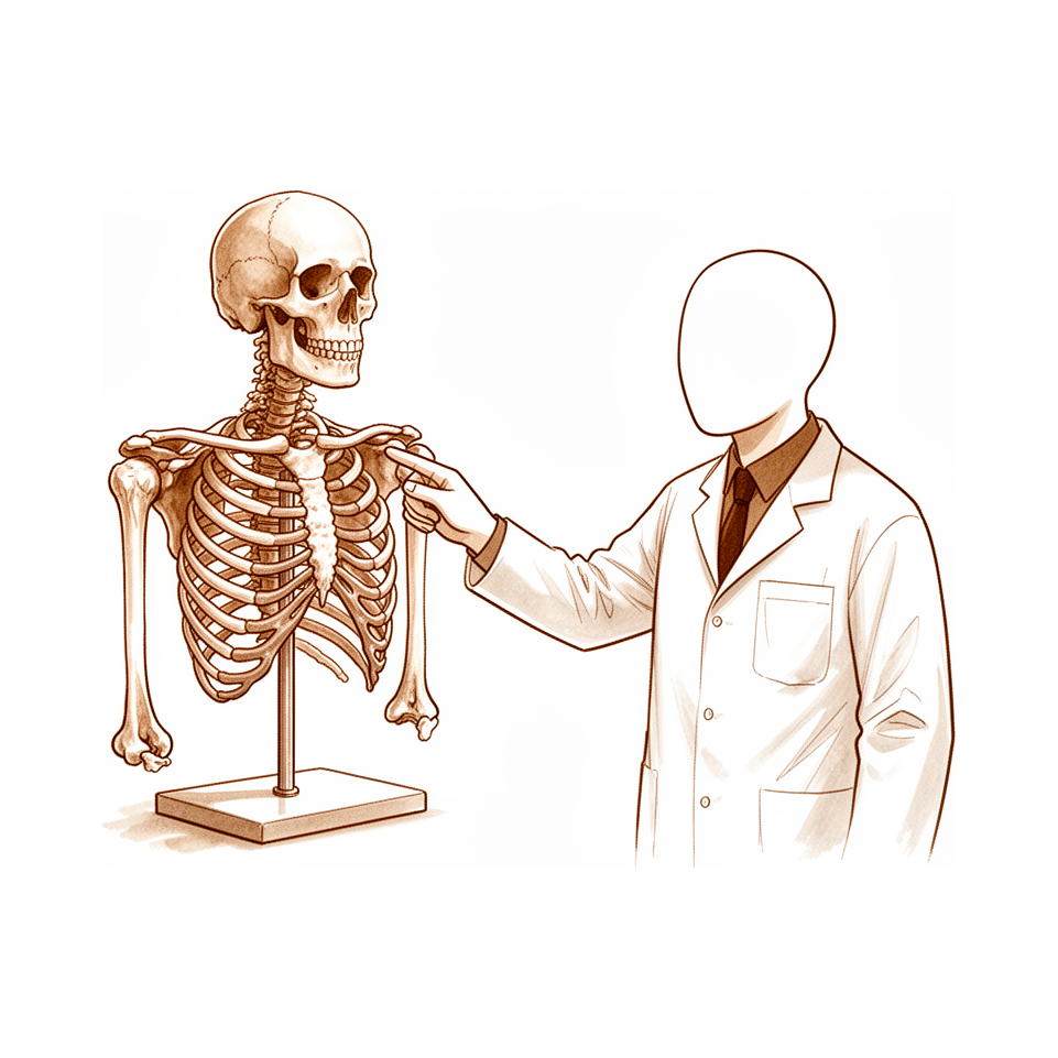
Osseous geometry of the shoulder girdle, focusing on the glenohumeral, AC, and SC joints and the concavity-compression mechanism of the glenoid.
Overview
Accurate anatomical knowledge is a prerequisite for executing upper extremity procedures with minimal functional disturbance [1]. This requirement is particularly acute in trauma management [2] and rotator cuff disease, where recent advances in basic science and surgical technology have refined anatomical understanding to guide treatment [88]. A comprehensive review of the shoulder, arm, and elbow encompasses ossification patterns, joint mechanics, ligamentous stabilizers, musculature, neurovascular structures, and surgical approaches [19]. Additionally, clinicians must be familiar with the characteristics and utility of popular upper-limb functional tests [85].
Bony anatomy presents specific considerations for surgical planning. The glenocapsular ligament is a constant structure, often comprising one or two parts, and is well vascularized along with the posterosuperior joint capsule [4]. Patient anatomy significantly influences scapular notching after reverse total shoulder arthroplasty [7], while varied descriptive terminology has created confusion regarding acromial architecture [81]. Standardized definitions of scapular landmarks are necessary for consistent measurements [5], supported by extensive references for normal 3D morphology [21]. Notably, sex-related and generational differences exist in global scapular shape [23], and implants derived from Western anatomical data may not fit the Japanese scapula and humerus well [30].
Glenoid assessment requires accurate imaging of bone defect size and relations for treatment planning [22]. The combined outer and inner circle ratio aids in appreciating paleo-glenoid morphology and estimating bone loss [27]. In elderly patients, there are no significant side-dependent differences in glenohumeral osseous anatomy [29]. Pediatric surgeons must understand the normal anatomy of the growing shoulder, including secondary ossification centers and growth plates, to avoid imaging interpretation errors [31]. Humeral anatomy shows that subscapularis footprint measurements are significantly larger in males than females, excluding the bare area [6]. While humeral head size parameters differ between genders, anatomic relationships do not [24]. Osseous adaptations at the glenohumeral joint must be considered when evaluating injured baseball players [28]. Clavicle shape can be predicted using clavicle length and patient stature [32]. Regarding measurement reliability, critical shoulder angle measurements via radiography are highly congruent, though large deviations occur between radiographs and true AP views [33]. Musculature and nerve anatomy also require attention; improved understanding of teres minor fascial anatomy and innervation may aid in treating isolated atrophy [90]. Long thoracic nerve lesions are the most common cause of scapular winging [86], and surgical anatomy of the supraclavicular brachial plexus differs from standard illustrations [20].
Ligaments and Joint Capsule
Shoulder
Accurate anatomical knowledge is required to carry out contemplated shoulder procedures with the least possible disturbance of function [1]. The coracohumeral ligament is more capsular than ligamentous based on its histologic features [61]. Two different coracoid attachments (anterior and posterior bundles) of the coracoacromial ligament were consistently identified in all specimens [69]. Coracoidal anatomic landmarks can be used intraoperatively for an anatomic reconstruction of the coracoclavicular ligaments [76].
Coracoid Surgical Considerations: Aberrant structures may be encountered in the vicinity of the coracoid during surgery, potentially prolonging the procedure or influencing technique [12]. When performing complex procedures about the coracoid where anatomy may be altered, risks are heightened and not accounted for in studies of intact specimens [3]. It is paramount to identify and protect neurovascular structures rather than relying on safe distances when performing complex procedures about the coracoid [3].
Capsular and Ligamentous Interventions: Pathologic coracoacromial anatomy is not addressed by current soft-tissue stabilization procedures and may contribute to instability recurrence [18]. Allografts are anchored to the glenoid and humeral head in a sling configuration, restoring capsular restraint and stability [54]. The "Pinch-and-Tuck" arthroscopic technique for capsular laxity in posterior shoulder instability offers efficient individualized plication [62]. This technique provides greater confidence in reduction of capsular laxity [62] and reduces risk to neurovascular structures due to control of soft tissue penetration depth [62]. Repair of anterior capsulolabral lesions combined with supraspinatus tendon tears successfully restored range of motion and increased the force required for dislocation [68].
Elbow
Recognizing complex soft tissue injuries is important to restore anatomic attachments and prevent chronic instability in global elbow instability [11]. A combination of two ligament-preserving approaches enables viewing the entirety of the joint surface and may represent an alternative to ligament-releasing approaches [40].
Muscles and Tendons
Subscapularis
Measured values of the subscapularis insertion are significantly larger in male specimens than in female specimens, with the exception of the bare area at the proximal end [6]. The tendon footprint is divided into facets, where the first facet comprises approximately 1/3 of the entire footprint area [89]. The first two facets together consist of 60% of the entire footprint [89]. Surgical visualization of the myotendinous junction is approach-dependent: the deltopectoral approach allows 100% visualization, compared to zero visualization with the anterolateral acromial approach [94].
Rotator Cuff and Superior Capsule
Prior studies overestimated rotator cuff footprint width due to a lack of discrimination between the actual cuff insertion and the capsule [96]. Shoulder anatomy affects the mechanisms of rotator cuff tear formation, with GTA, CSA, AI, LAA, and footprint dimensions playing a more significant role in L-shaped tears compared to other tear shapes [101]. An unconstrained implant approaching normal anatomy permits better function and endurance than an implant with a fixed fulcrum, provided there is meticulous reconstruction and rehabilitation of the rotator-cuff and deltoid muscles [98].
Infraspinatus and Teres Minor
The infraspinatus does not need to be released from its humeral footprint but rather can be split in line with its muscle and tendon fibers, which has a high potential for a benign postoperative healing process [93]. The posterior ridge of the greater tuberosity is a suitable landmark to locate the internervous plane between the infraspinatus and teres minor muscles and should not be crossed distally [100].
Supraspinatus
Innate anatomical features of the scapula, such as lateral overhang and coracoacromial arch angle, can predispose people to extrinsic lesions to the supraspinatus tendon by reducing the subcoracoacromial canal's surface area [97].
Biceps Tendon
Tenodesis performed between the midpoint of the pectoralis major insertion and more distal points involves a significant portion of muscle, which may not be optimal [99].
Trapezius
A specific landmark allows for accurate differentiation of the lower trapezius from the middle trapezius during tendon harvest [102]. Maximal scapular protraction increases the safety distance of the spinal accessory nerve during lower trapezius tendon harvest [102].
Surgical Exposure and Landmarks
Functional anatomical aspects and correct incision placement are critical for adequate visualization and satisfactory postoperative scarring in anterior, posterior, and superolateral surgical exposures of the shoulder [17]. The deltopectoral approach allows 100% visualization of the medial anatomic neck, compared to 20% visualization with the anterolateral acromial approach [94]. The capsulolabral footprint contributes significantly to the glenoid face, inserts directly adjacent to the articular cartilage, and extends medially along the glenoid neck [92]. The authors recommend noting the site of muscular insertion and avoiding the Bristow procedure in patients with a proximal insertion less than 2.5 centimeters from the coracoid process [95].
Neurovascular Anatomy
General Principles
Potential neurovascular compromises, even if seemingly subtle, should be thoroughly investigated [60].
Brachial Plexus and Axillary Vessels
The anatomic relationship of the axillary artery and vein to the clavicle identifies an at-risk zone between the medial quarter and midpoint of the clavicle where neurovascular structures lie beneath the bone [10].
Nerve-Specific Anatomy
The closest that the innervation point of either the upper or lower subscapular nerve came to the medial aspect of the coracoid was 11 mm [71]. Recognition of characteristic clinical features and understanding of the anatomical course of the dorsal scapular nerve are essential for accurate diagnosis [52]. Furthermore, recognition of dorsal scapular nerve anatomy and pathology may provide a more comprehensive understanding of scapular dysfunction in patients with thoracic outlet syndrome [15].
Muscular and Graft Anatomy
In muscle transfer, the neurovascular pedicle vessels are the predictable limiting factor for translation due to their shorter length compared to the nerve [57]. A length of nerve plus its accompanying artery and vein can be transplanted as a free graft, but few sites can spare them [43].
Surgical Approaches and Outcomes
A posterior surgical approach to the proximal part of the humerus exposes at least eight centimeters of the proximal part of the posterior aspect of the humeral diaphysis without endangering major blood vessels [9]. This approach utilizes the osseous origins of the lateral head of the triceps to protect the axillary and radial nerves [9]. The subscapular sling procedure can be performed without altering the anatomy of nearby structures such as the coracoid process, the conjoined tendon, and the axillary and musculocutaneous nerves [70]. There were no respiratory or neurovascular complications in the reported pediatric orthopedic case [65].
Biomechanics and Function
Scapulothoracic and Shoulder Girdle Kinematics
Consistent measurement of scapular orientation requires a clear and standardized definition of anatomical landmarks [5]. Kinematic values derived from whole cadaver studies remain applicable for investigating scapulothoracic kinematics in both normal and pathologic situations [56]. Despite these methods, a precise, easy-to-use, and low-cost non-invasive method for drawing and analyzing shoulder complex kinematics has not yet been developed [58]. Clinicians must account for small but significant kinematic asymmetries between dominant and nondominant shoulders [80].
Joint Biomechanics and Forces
Human throwing capabilities largely result from derived anatomical features that enable elastic energy storage and release at the shoulder [59]. Biomechanical studies of the sternoclavicular joint provide insights and suggest a validated methodology for further analyses [55]. In the intact clavicle, axial compressive force and torque are greatest during abduction, whereas tensile force is greatest during external rotation [84].
Glenohumeral Joint Mechanics
In-vivo measurement techniques provide clinically relevant information by calculating joint contact patterns during dynamic conditions, overcoming many limitations associated with conventional techniques for quantifying joint mechanics [63]. In the context of subscapularis deficiency and glenoid wear, alternative surgical approaches leading to normal joint biomechanics may reduce implant wear [73].
Biomechanical Studies and Methodology
Cadaveric specimens used in studies evaluating bone loss in anterior shoulder instability are not representative of the affected patient population, as males are more frequently studied and most studies are conducted in the United States [64]. Clinicians should be aware of the measurement methods used in biomechanical studies when making surgical decisions regarding glenoid bone loss quantification [77]. More advanced measurement techniques that take glenoid concavity into account are more accurate in determining the biomechanical relevance of glenoid bone loss, although the reliability of manually performed, more complex measurements was moderate [79].
Surgical Biomechanics
The triple anatomical technique for acromioclavicular joint reconstruction enables early recovery and return to sports by biologically restoring joint anatomy and kinematics [8]. For irreparable massive posterosuperior rotator cuff tears, the lower trapezius transfer demonstrated biomechanical advantages in restoring compressive forces and kinematics compared to the latissimus dorsi transfer in a cadaveric model [72]. No significant biomechanical differences in displacement or stiffness were seen between the anatomical landmark technique and the coracoid-based landmarks technique for coracoclavicular stabilization after high-grade acromioclavicular injury [83].
Obligate posterosuperior humeral head translation following the Latarjet procedure demonstrates that its triple blocking effect is real [82]. However, findings regarding post-Latarjet kinematics contradict recent in vivo CT studies, highlighting the need for further in vivo research to elucidate kinematics and mitigate late arthrosis risk [82]. There is insufficient evidence to support changing surgical techniques to preserve soft tissues based on kinematic concerns regarding scapular dyskinesia after the Latarjet procedure [67]. Further biomechanical and clinical studies are needed to validate the long-term and versatile utility of the extra-articular soft arthroscopic Latarjet technique [66]. Future research should focus on biomechanical outcomes to show the reliability of arthroscopic iliac crest bone grafting for shoulder glenoid defect augmentation using a “Three‐Pulley Four‐Point Antirotation” suture anchor technique [74].
Common Sites of Injury
Shoulder and Scapula
Acromioclavicular joint injuries require evaluation and treatment within the context of their effect on the functional integrity of the entire acromioclavicular joint complex and scapulohumeral rhythm [49]. A comprehensive clinical approach emphasizes evaluating the extent of anatomic injury and understanding its mechanical consequences regarding shoulder and arm function [34]. Anatomic placement of repair or reconstruction grafts or devices for acromioclavicular and coracoclavicular ligament injuries may help prevent common problems encountered [16]. Surgical treatment of ipsilateral Rockwood type IV acromioclavicular joint dislocation and midshaft clavicle fracture results in excellent clinical outcomes [46].
Avulsion fracture of the coracoid process at the coracoclavicular ligament attachment associated with distal clavicle fracture highlights the importance of assessing superior transverse scapular ligament integrity [36]. Anatomical reconstruction may be a reasonable treatment option for unstable injury patterns involving coracoid process avulsion fractures [36]. The majority of physeal injuries of the coracoid process can be successfully treated nonoperatively [39]. Recognition of dorsal scapular nerve anatomy and pathology may provide a more comprehensive understanding of scapular dysfunction in patients with medial scapular winging in thoracic outlet syndrome [15]. The teres major muscle has an additional distal muscle slip, which is the first macroscopic description of this anatomical variant [47].
Elbow and Forearm
Complex soft tissue injuries to the elbow require recognition to restore anatomic attachments and prevent chronic instability [11]. Excellent functional outcomes can be achieved with a conservative approach even after extensive soft tissue damage from minor injury in open antero-lateral dislocation of the elbow [44]. Traumatic subluxation of the radial head is a rather frequent injury in young children [26].
Upper Extremity General
Recognition of ulnar nerve anatomical details and variations is essential for accurate diagnosis and surgical decompression to avoid iatrogenic injury [42]. Human motor endplates in the shoulder and upper extremity persist and retain their structures even after the 6-month window of opportunity for meaningful functional recovery has elapsed [37].
Surgical Anatomy
General Principles
Functional anatomical understanding and precise incision placement are critical for achieving adequate visualization and satisfactory postoperative scarring in shoulder exposures [17].
Clavicle and Supraclavicular Region
An at-risk zone exists between the medial quarter and midpoint of the clavicle, where neurovascular structures lie beneath the bone [10]. Two or three branches of the supraclavicular nerve cross the clavicle in 97% of cases [50]. Outside of defined safe zones, there is wide variability in the location of these supraclavicular nerve branches [50].
Coracoid Process and Surrounding Structures
Risks are heightened during complex procedures about the coracoid because anatomy may be altered, a factor not accounted for in studies of intact specimens [3]. It is paramount to identify and protect neurovascular structures rather than relying on safe distances when operating around the coracoid [3]. Aberrant structures may be encountered in the vicinity of the coracoid during surgery [12]. These aberrant structures can potentially prolong the procedure or influence technique [12]. The previously described safe zone of 5 cm below the coracoid process may not be reliable to protect the musculocutaneous nerve or its twigs [35]. In 91.7% of specimens, musculocutaneous nerve entry points were within 5 cm of the coracoid process when twigs were accounted for [35].
Humerus and Scapula
A posterior surgical approach can expose at least eight centimeters of the proximal part of the posterior aspect of the humeral diaphysis without endangering major blood vessels [9]. The osseous origins of the lateral head of the triceps are utilized to protect the axillary and radial nerves during posterior humeral approaches [9]. Gender and specific scapular dimensions influence the dimensions of the safe zone for avoiding suprascapular nerve injury during shoulder arthroscopy [48]. Cerclage passage medial to lateral from the latissimus dorsi proximally to the area just distal to the inferior pectoralis major insertion distally is a safe zone for cerclage passage [51]. Palpable landmarks can be used to reduce torsional deformities in minimally invasive surgeries without the need for fluoroscopy [53].
Ligaments and Pathologic Anatomy
Anatomic placement of repair or reconstruction grafts or devices may help to prevent many of the common problems encountered in shoulder ligament injuries [16]. Pathologic coracoacromial morphology is not addressed by current soft-tissue stabilization procedures and may contribute to instability recurrence [18].
Key Evidence
- [L5] Accurate anatomical knowledge is required to carry out the contemplated procedure with the least possible disturbance of function. [1] (10.2106/00004623-194931020-00002)
- [L5] The issue emphasizes the importance of preparation and detailed anatomic knowledge in surgical procedures for upper extremity trauma, providing comprehensive descriptions of exposures and coverage with clinical examples. [2] (10.1016/j.hcl.2014.08.004)
- [L5] When performing complex procedures about the coracoid where anatomy may be altered, risks are heightened and not accounted for in studies of intact specimens; it is paramount to identify and protect neurovascular structures rather than relying on safe distances. [3] (10.1016/j.arthro.2018.11.007)
- [L5] The glenocapsular ligament is a constant anatomical structure that consists of one or two different parts. [4] (10.1007/s00167-017-4603-x)
- [L4] However, a clear and standardized definition of these landmarks is needed to ensure consistency across measurements. [5] (10.1016/j.jseint.2024.09.027)
- [L5] With the exception of the bare area at the proximal end, the measured values were significantly larger in male specimens than in female specimens. [6] (10.1016/j.arthro.2008.02.009)
- [L4] There is a high degree of significance that patient anatomy does play a role in the occurrence of notching. [7] (10.1016/j.jse.2013.09.003)
- [L5] It enables early recovery and return to sports by biologically restoring joint anatomy and kinematics. [8] (10.1016/j.eats.2025.103595)
- [L4] The described approach exposes at least eight centimeters of the proximal part of the posterior aspect of the humeral diaphysis without endangering major blood vessels, utilizing the osseous origins of the lateral head of the triceps to protect the axillary and radial nerves. [9] (10.2106/00004623-198971030-00015)
- [L5] The study defines the anatomic relationship of the axillary artery and vein to the clavicle, identifying an at-risk zone between the medial quarter and midpoint of the clavicle where neurovascular structures lie beneath the bone. [10] (10.1016/j.jse.2009.01.021)
- [Case_report] The authors highlight the importance of recognizing complex soft tissue injuries to restore anatomic attachments and prevent chronic instability. [11] (10.1016/j.xrrt.2022.08.005)
- [L4] Aberrant structures may be encountered in the vicinity of the coracoid during surgery, potentially prolonging the procedure or influencing technique. [12] (10.1111/j.1758-5740.2011.00168.x)
- [L5] The authors conclude that recognition of dorsal scapular nerve anatomy and pathology may provide a more comprehensive understanding of scapular dysfunction in these patients. [15] (10.1016/j.jhsa.2026.04.017)
- [Paper] Anatomic placement of repair or reconstruction grafts or devices may help to prevent many of the common problems encountered. [16] (10.1016/j.csm.2017.12.002)
- [L5] The article presents anatomical considerations and techniques for anterior, posterior, and superolateral surgical exposures of the shoulder, emphasizing that functional anatomical aspects and correct incision placement are critical for adequate visualization and satisfactory postoperative scarring. [17] (10.2106/00004623-196749030-00017)
- [L3] This pathologic anatomy is not addressed by current soft-tissue stabilization procedures and may contribute to instability recurrence. [18] (10.1016/j.jse.2019.01.009)
- [L5] These findings differ from the standard illustrations and descriptions of the brachial plexus. [20] (10.2106/jbjs.n.00706)
- [L5] The authors conclude that the study provides an extensive reference for normal 3D scapular morphology, offering a valuable foundation for future research. [21] (10.2106/jbjs.25.01596)
- [L4] Accurate imaging and assessment of the size and relations of bone defects are crucial for further treatment planning. [22] (10.1302/2058-5241.5.200049)
- [L4] These differences may be considered when analyzing scapular morphology. [23] (10.1016/j.xrrt.2025.04.001)
- [L4] Although there are significant differences in size parameters of the humeral head between men and women, there do not seem to be differences in anatomic relationships between genders. [24] (10.1016/j.jse.2011.03.018)
- [L4] The injury is apparently a rather frequent one and a description should be available to all orthopaedic surgeons. [26] (10.2106/00004623-195436030-00018)
- [L4] This study sets the base for the use the combined outer and inner circle and its ratio to better appreciate the paleo-glenoid morphology and thus obtain a more reliable bone loss estimation. [27] (10.1007/s00167-022-07050-y)
- [L3] Consideration must be given to osseous adaptations that occur at the glenohumeral joint when evaluating and treating this population. [28] (10.1177/23259671241260084)
- [L4] There are no significant side-dependent differences in the osseous anatomy of the glenohumeral joint. [29] (10.1016/j.jse.2015.12.024)
- [L4] Consequently, implants designed from Western anatomical data may not fit the Japanese scapula and humerus well. [30] (10.1016/j.jseint.2026.101714)
- [Case_report] Orthopedic surgeons must be familiar with the normal anatomy of the growing shoulder, its secondary ossification centers, and growth plates in order to avoid errors when interpreting imaging tests in the pediatric shoulder. [31] (10.5397/cise.2022.01151)
- [L4] This anatomic analysis shows that the clavicle shape can be predicted through the clavicle length and patients' stature. [32] (10.1016/j.jseint.2020.05.004)
- [L3] The CSA measurements using radiography were highly congruent, but a large measurement deviation occurred between radiographs and true AP views. [33] (10.1016/j.jse.2022.07.017)
- [L5] A comprehensive clinical approach emphasizing the evaluation of the extent of the anatomic injury and understanding its mechanical consequences regarding shoulder and arm function is a key in the development of guidelines for developing operative or non-operative treatment protocols and for establishing outcomes of the treatment protocols. [34] (10.1177/17585732221122335)
- [L5] The previously described safe zone of 5 cm below the coracoid process may not be reliable to protect the musculocutaneous nerve or its twigs, as 91.7% of specimens had nerve entry points within this distance when twigs were accounted for. [35] (10.1177/2325967120954417)
- [L5] This case highlights the importance of assessing SSSC integrity and suggests that anatomical reconstruction may be a reasonable treatment option in unstable injury patterns. [36] (10.1177/17585732261469144)
- [L4] Human motor endplates persist and retain their structures even after the 6-month window of opportunity for meaningful functional recovery has elapsed, suggesting limited utility of animal models for traumatic peripheral nerve injuries. [37] (10.1016/j.jse.2018.11.035)
- [L4] The majority of these injuries can be successfully treated nonoperatively. [39] (10.1177/2325967120967914)
- [L5] A combination of these two ligament-preserving approaches enables viewing the entirety of the joint surface and may represent an alternative to ligament-releasing approaches. [40] (10.1016/j.jse.2022.01.013)
- [L5] Recognition of these anatomical details and variations is essential for accurate diagnosis and surgical decompression to avoid iatrogenic injury. [42] (10.1016/j.hcl.2007.05.001)
- [L4] A length of nerve plus its accompanying artery and vein can be transplanted as a free graft, but few sites can spare them. [43] (10.1016/s0020-1383(84)80055-8)
- [Case_report] This case highlights that excellent functional outcomes can be achieved with a conservative approach even after extensive soft tissue damage from minor injury. [44] (10.1186/1471-2474-3-1)
- [Case_report] Surgical treatment of this injury pattern results in excellent clinical outcomes. [46] (10.1016/j.xrrt.2022.11.007)
- [L5] This is the first macroscopic description of an additional distal slip of the teres major muscle. [47] (10.1186/s12891-021-04227-3)
- [L5] Knowledge of the safe zone for avoiding suprascapular nerve injury is important; gender and specific scapular dimensions should be evaluated as they influence the dimensions of the safe zone. [48] (10.1016/j.jse.2011.01.033)
- [L5] Injuries to ACJ structures do not exist in isolation but must be evaluated and treated within the context of their effect on the functional integrity of the entire ACJ complex and scapulohumeral rhythm. [49] (10.5435/jaaos-d-24-00360)
- [L5] There were two or three branches of the supraclavicular nerve crossing the clavicle 97% of the time and a wide variability of the location of these branches outside the safe zones. [50] (10.1007/s11999-010-1608-x)
- [L5] Cerclage passage medial to lateral from the latissimus dorsi proximally to the area just distal to the inferior pectoralis major insertion distally is a safe zone for cerclage passage. [51] (10.1016/j.jseint.2024.08.187)
- [L4] Recognition of characteristic clinical features and understanding of the anatomical course of the nerve are essential for accurate diagnosis. [52] (10.1016/j.xrrt.2026.100807)
- [L3] Minimally invasive surgeries can be performed by using these palpable landmarks to reduce torsional deformities without the need for fluoroscopy. [53] (10.1186/s12891-020-3118-7)
- [L5] Allografts are anchored to the glenoid and humeral head in a sling configuration, restoring capsular restraint and stability. [54] (10.1002/atn2.70017)
- [L5] The study provides biomechanical insights on SCJ biomechanics, and suggests a validated methodology for further analyses. [55] (10.1016/j.jse.2026.07.025)
- [L5] These values can be used when investigating scapulothoracic kinematics during normal and pathologic situations. [56] (10.1016/j.jseint.2022.09.014)
- [L5] In muscle transfer, the neurovascular pedicle vessels are the predictable limiting factor for translation due to their shorter length compared to the nerve. [57] (10.1016/j.jse.2014.07.001)
- [L5] Despite technology innovations, a precise, easy to use and low-cost non-invasive method able to draw and analyze the kinematics of the shoulder complex has not been developed yet. [58] (10.1177/17585732221090226)
- [L4] Human throwing capabilities largely result from several derived anatomical features that enable elastic energy storage and release at the shoulder. [59] (10.1038/nature12267)
- [Case_report] Potential neurovascular compromises, even if seemingly subtle, should be thoroughly investigated. [60] (10.1016/j.xrrt.2023.03.004)
- [L5] The coracohumeral ligament is more capsular than ligamentous based on its histologic features. [61] (10.1016/j.jse.2008.07.012)
- [L5] The authors believe the technique has advantages including efficient individualized plication, greater confidence in reduction of capsular laxity, and reduced risk to neurovascular structures due to control of soft tissue penetration depth. [62] (10.1016/j.eats.2025.103794)
- [L4] This technique provides clinically relevant information by calculating in-vivo joint contact patterns during dynamic conditions and overcomes many limitations associated with conventional techniques for quantifying joint mechanics. [63] (10.1155/2010/162136)
- [L4] Males were more frequently studied, and most of these biomechanical studies were conducted in the United States. [64] (10.1016/j.asmr.2024.100996)
- [L4] There were no respiratory or neurovascular complications. [65] (10.1097/00004694-200307000-00010)
- [L5] Nevertheless, further biomechanical and clinical studies are needed to validate its long-term and versatile utility. [66] (10.1016/j.eats.2021.05.009)
- [L5] There is insufficient evidence to support changing surgical techniques to preserve soft tissues based on kinematic concerns. [67] (10.1016/j.arthro.2024.05.017)
- [L5] Repair of both pathologic conditions successfully restored range of motion and increased the force required for dislocation. [68] (10.1016/j.arthro.2013.05.031)
- [L5] Two different coracoid attachments (anterior and posterior bundles) of the coracoacromial ligament were consistently identified in all specimens. [69] (10.1016/j.arthro.2017.11.033)
- [L5] The procedure can be performed without altering the anatomy of nearby structures such as the coracoid process, the conjoined tendon, and the axillary and musculocutaneous nerves. [70] (10.1016/j.eats.2021.03.027)
- [L5] The closest that the innervation point of either the upper or lower subscapular nerve came to the medial aspect of the coracoid was 11 mm. [71] (10.1016/j.arthro.2013.02.005)
- [Abstract] The trapezius transfer demonstrated biomechanical advantages in restoring compressive forces and kinematics compared to the latissimus dorsi transfer in this cadaveric model. [72] (10.1016/j.jse.2012.12.009)
- [L5] Therefore our findings suggest that alternative surgical approaches leading to normal joint biomechanics may reduce implant wear. [73] (10.1016/j.jseint.2024.08.151)
- [L5] Future research should focus on biomechanical outcomes to show the reliability of this technique. [74] (10.1002/atn2.70233)
- [L5] Coracoidal anatomic landmarks can be used intraoperatively for an anatomic reconstruction of the coracoclavicular ligaments. [76] (10.1177/0363546508322887)
- [L5] Clinicians should be aware of the measurement methods used in biomechanical studies when making surgical decisions. [77] (10.1016/j.arthro.2015.02.020)
- [L3] While more advanced measurement techniques that take glenoid concavity into account are more accurate in determining the biomechanical relevance of glenoid bone loss, the reliability of manually performed, more complex measurements was moderate. [79] (10.1177/23259671231222938)
- [L5] Small but significant asymmetries exist between the dominant and nondominant shoulders in terms of kinematics. [80] (10.1016/j.jse.2013.08.020)
- [L4] The use of a multitude of different descriptive terminology has led to confusion in the literature concerning acromial architecture; this review aims to show the discrepancy and advise suitable terminology to prevent future confusion. [81] (10.1007/s001670050062)
- [L5] These findings contradict recent in vivo CT studies and highlight the need for further in vivo research to elucidate kinematics and mitigate late arthrosis risk. [82] (10.2106/jbjs.25.01091)
- [L5] No significant biomechanical differences in displacement or stiffness were seen between the anatomical landmark technique and the coracoid-based landmarks technique. [83] (10.1177/23259671221132541)
- [L5] Axial compressive force and torque were greatest during abduction, whereas tensile force was greatest during external rotation. [84] (10.1016/j.jse.2010.03.016)
- [L4] The current study summarizes the characteristics and the usefulness of the most popular upper-limb functional tests. [85] (10.1177/17585732221101880)
- [L4] This review explores the causes of scapula winging, with an overview of the relevant anatomy, proposed aetiology and treatment, with particular focus given to lesions of the long thoracic nerve, which is reported to be the most common aetiological factor. [86] (10.1111/sae.12033)
- [L5] Understanding the anatomy of the rotator cuff and surrounding structures is essential for treating rotator cuff disease, with recent advances in basic science and surgical technology improving knowledge of this anatomy to guide surgical management. [88] (10.1016/j.arthro.2008.07.023)
- [L5] The first facet of subscapularis tendon area consists of approximately 1/3 of the entire footprint area, and the first two facets consist of 60% of its entire footprint. [89] (10.1016/j.arthro.2013.07.104)
- [L5] An improved understanding of the fascial anatomy and innervation pattern of the teres minor muscle may help clinicians who treat patients with symptomatic isolated teres minor muscle atrophy. [90] (10.1016/j.jse.2011.12.005)
- [L5] The capsulolabral footprint contributes significantly to the glenoid face, inserts directly adjacent to the articular cartilage, and extends medially along the glenoid neck. [92] (10.1016/j.arthro.2018.03.017)
- [L5] The infraspinatus does not need to be released from its humeral footprint but rather can be split in line with its muscle and tendon fibers, which has a high potential for a benign postoperative healing process. [93] (10.1016/j.eats.2024.103288)
- [L4] The DP approach allowed better access to anterior quadrant structures, including improved ability to visualize the myotendinous junction of the subscapularis (100% versus zero) and the medial anatomic neck (100% versus 20%). [94] (10.5435/jaaosglobal-d-18-00017)
- [Case_report] The authors recommend noting the site of muscular insertion and avoiding the Bristow procedure in patients with a proximal insertion (less than 2.5 centimeters from the coracoid process). [95] (10.2106/00004623-198870030-00023)
- [L5] Prior studies overestimated the rotator cuff footprint width due to a lack of discrimination between the actual cuff insertion and the capsule. [96] (10.1016/j.jse.2011.04.034)
- [L4] Apart from acromial morphology, there could be innate anatomical features of the scapula that predispose people to extrinsic lesions to the supraspinatus tendon (lateral overhang, coracoacromial arch angle) by reducing the subcoracoacromial canal's surface area. [97] (10.1016/j.otsr.2016.08.001)
- [L4] An unconstrained implant approaching normal anatomy permits better function and endurance than an implant with a fixed fulcrum, provided there is meticulous reconstruction and rehabilitation of the rotator-cuff and deltoid muscles. [98] (10.2106/00004623-198264030-00001)
- [L5] Tenodesis performed between the midpoint of the pectoralis major insertion and more distal points involves a significant portion of muscle, which may not be optimal. [99] (10.1016/j.arthro.2015.11.048)
- [L5] The posterior ridge of the greater tuberosity is a suitable landmark to locate the internervous plane between the infraspinatus and teres minor muscles and should not be crossed distally. [100] (10.1016/j.jse.2017.10.034)
- [L3] Shoulder anatomy affects the mechanisms of rotator cuff tear formation, with GTA, CSA, AI, LAA, and footprint dimensions playing a more significant role in L-shaped tears compared to other tear shapes. [101] (10.1186/s12891-024-07829-9)
- [L5] This landmark allows for accurate differentiation of the lower trapezius from the middle trapezius during tendon harvest. [102] (10.1016/j.jseint.2026.101622)
See Also
- Rotator Cuff
- Total shoulder arthroplasty
- Shoulder Instability
- Thoracic Outlet Syndrome
- Latarjet Procedure
- Clavicle Fracture
- Fractures
- Shoulder Arthroscopy
References
[1] SURGICAL APPROACHES TO THE SHOULDER JOINT. The Journal of Bone & Joint Surgery. 1949. DOI: 10.2106/00004623-194931020-00002
[2] Options for Surgical Exposure and Soft Tissue Coverage in Upper Extremity Trauma. Hand Clinics. 2014. DOI: 10.1016/j.hcl.2014.08.004
[3] Editorial Commentary: Working Around the Coracoid—The Lighthouse to the Shoulder. Arthroscopy. 2019. DOI: 10.1016/j.arthro.2018.11.007
[4] The glenocapsular ligament and the posterosuperior part of the joint capsule of the shoulder are well vascularized. Knee Surgery, Sports Traumatology, Arthroscopy. 2017. DOI: 10.1007/s00167-017-4603-x
[5] Impact of scapula anatomical landmark positioning on scapular orientation using CT-based 3-dimensional models: an intraobserver repeatability and interobserver reproducibility study. JSES International. 2025. DOI: 10.1016/j.jseint.2024.09.027
[6] An Anatomic Study of the Subscapularis Insertion to the Humerus: The Subscapularis Footprint. Arthroscopy. 2008. DOI: 10.1016/j.arthro.2008.02.009
[7] Relationship of scapular neck length to scapular notching after reverse total shoulder arthroplasty by use of plain radiographs. Journal of Shoulder and Elbow Surgery. 2014. DOI: 10.1016/j.jse.2013.09.003
[8] The Triple Anatomical Technique for Acromioclavicular Joint Reconstruction: A True Anatomical Reconstruction Technique Using Synthetic Ligaments With Acromioclavicular Ligament Reconstruction. Arthroscopy Techniques. 2025. DOI: 10.1016/j.eats.2025.103595
[9] A posterior surgical approach to the proximal part of the humerus.. The Journal of Bone & Joint Surgery. 1989. DOI: 10.2106/00004623-198971030-00015
[10] The anatomic relationship of the axillary artery and vein to the clavicle: A cadaveric study. Journal of Shoulder and Elbow Surgery. 2009. DOI: 10.1016/j.jse.2009.01.021
[11] Global elbow instability: a case report. JSES Reviews, Reports, and Techniques. 2023. DOI: 10.1016/j.xrrt.2022.08.005
[12] Aberrant Pectoralis Minor Tendon and Surgery around the Coracoid Process. Shoulder & Elbow. 2012. DOI: 10.1111/j.1758-5740.2011.00168.x
[15] Letter regarding “The Association of Long Thoracic Nerve Variants with Medial Scapular Winging in Thoracic Outlet Syndrome: A Clinical and Anatomical Study”. The Journal of Hand Surgery. 2026. DOI: 10.1016/j.jhsa.2026.04.017
[16] Shoulder Acromioclavicular and Coracoclavicular Ligament Injuries. Clinics in Sports Medicine. 2018. DOI: 10.1016/j.csm.2017.12.002
[17] Surgical Exposure ofthe Shoulder. The Journal of Bone & Joint Surgery. 1967. DOI: 10.2106/00004623-196749030-00017
[18] Coracoacromial morphology: a contributor to recurrent traumatic anterior glenohumeral instability?. Journal of Shoulder and Elbow Surgery. 2019. DOI: 10.1016/j.jse.2019.01.009
[19] Chapter 69 Anatomy of the Shoulder, Arm, and Elbow. 2019.
[20] Surgical Anatomy of the Supraclavicular Brachial Plexus. Journal of Bone and Joint Surgery. 2015. DOI: 10.2106/jbjs.n.00706
[21] Scapular Morphology: Survival of the Fittest. Journal of Bone and Joint Surgery. 2026. DOI: 10.2106/jbjs.25.01596
[22] Clinical and radiological examination of bony-mediated shoulder instability. EFORT Open Reviews. 2020. DOI: 10.1302/2058-5241.5.200049
[23] Scapular morphological variations and sex-related and generational differences in global scapular shape: three-dimensional morphometric analysis using a homologous model. JSES Reviews, Reports, and Techniques. 2025. DOI: 10.1016/j.xrrt.2025.04.001
[24] Magnetic resonance imaging study of glenohumeral relationships between genders. Journal of Shoulder and Elbow Surgery. 2011. DOI: 10.1016/j.jse.2011.03.018
[26] TRAUMATIC SUBLUXATION OF THE RADIAL HEAD IN YOUNG CHILDREN. The Journal of Bone & Joint Surgery. 1954. DOI: 10.2106/00004623-195436030-00018
[27] High correlation between inner and outer glenoid circle diameters and its clinical relevance. Knee Surgery, Sports Traumatology, Arthroscopy. 2022. DOI: 10.1007/s00167-022-07050-y
[28] The Role of Humeral Torsion on Glenohumeral Rotation in Injured Baseball Players. Orthopaedic Journal of Sports Medicine. 2024. DOI: 10.1177/23259671241260084
[29] The osseous morphology of nondegenerated shoulders shows no side-related differences in elderly patients: an analysis of 102 computed tomography scans. Journal of Shoulder and Elbow Surgery. 2016. DOI: 10.1016/j.jse.2015.12.024
[30] Population-specific statistical shape modeling of the Japanese shoulder: a combined analysis of the scapula and humerus stratified by sex and stature. JSES International. 2026. DOI: 10.1016/j.jseint.2026.101714
[31] Normal ossification of the glenoid mimicking a glenoid fracture in an adolescent patient: a case report. Clinics in Shoulder and Elbow. 2023. DOI: 10.5397/cise.2022.01151
[32] The variance of clavicular surface morphology is predictable: an analysis of dependent and independent metadata variables. JSES International. 2020. DOI: 10.1016/j.jseint.2020.05.004
[33] Reliability and accuracy of the critical shoulder angle measured by anteroposterior radiographs: using digitally reconstructed radiograph from 3-dimensional computed tomography images. Journal of Shoulder and Elbow Surgery. 2023. DOI: 10.1016/j.jse.2022.07.017
[34] Acromioclavicular joint injuries revisited: Pathoanatomy, pathomechanics, and clinical presentation. Shoulder & Elbow. 2022. DOI: 10.1177/17585732221122335
[35] Relationship of the Musculocutaneous Nerve and Its Twigs to the Coracoid Process: An Operative Exposure. Orthopaedic Journal of Sports Medicine. 2020. DOI: 10.1177/2325967120954417
[36] Avulsion fracture of the coracoid process at the coracoclavicular ligament attachment associated with distal clavicle fracture: A case report. Shoulder & Elbow. 2026. DOI: 10.1177/17585732261469144
[37] Analysis of human muscles of the shoulder and upper extremity a temporal profile of human motor endplate degradation. Journal of Shoulder and Elbow Surgery. 2019. DOI: 10.1016/j.jse.2018.11.035
[39] Physeal Injuries of the Coracoid Process Are Closely Associated With Sports Activities: A Systematic Review. Orthopaedic Journal of Sports Medicine. 2020. DOI: 10.1177/2325967120967914
[40] Surgical Approaches To The Capitellum: A Comparative Anatomic Study. Journal of Shoulder and Elbow Surgery. 2022. DOI: 10.1016/j.jse.2022.01.013
[42] Ulnar Nerve Anatomy. Hand Clinics. 2007. DOI: 10.1016/j.hcl.2007.05.001
[43] Vascularized nerve grafts. Injury. 1984. DOI: 10.1016/s0020-1383(84)80055-8
[44] Open antero-lateral dislocation of the elbow. A case report. BMC Musculoskeletal Disorders. 2002. DOI: 10.1186/1471-2474-3-1
[46] Ipsilateral Rockwood type IV acromioclavicular joint dislocation and midshaft clavicle fracture: a case report and review of the literature. JSES Reviews, Reports, and Techniques. 2023. DOI: 10.1016/j.xrrt.2022.11.007
[47] Anatomical study of the teres major muscle: description of an additional distal muscle slip. BMC Musculoskeletal Disorders. 2021. DOI: 10.1186/s12891-021-04227-3
[48] The safe zone for avoiding suprascapular nerve injury during shoulder arthroscopy: an anatomical study on 500 dry scapulae. Journal of Shoulder and Elbow Surgery. 2011. DOI: 10.1016/j.jse.2011.01.033
[49] Effect of Acromioclavicular Joint Injuries on the Acromioclavicular Joint Complex and Scapulohumeral Rhythm: A Functional and Mechanical Perspective. Journal of the American Academy of Orthopaedic Surgeons. 2025. DOI: 10.5435/jaaos-d-24-00360
[50] The Anatomy of the Supraclavicular Nerve During Surgical Approach to the Clavicular Shaft. Clinical Orthopaedics & Related Research. 2011. DOI: 10.1007/s11999-010-1608-x
[51] Identification of an anatomical safe zone for humeral cerclage passage. JSES International. 2025. DOI: 10.1016/j.jseint.2024.08.187
[52] Dorsal scapular nerve entrapment: a systematic review. JSES Reviews, Reports, and Techniques. 2026. DOI: 10.1016/j.xrrt.2026.100807
[53] Geometrical analysis for assessing torsional alignment of humerus. BMC Musculoskeletal Disorders. 2020. DOI: 10.1186/s12891-020-3118-7
[54] Arthroscopic Anterior and Posterior Glenohumeral Capsular Augmentation With Gracilis Allograft. Arthroscopy Techniques. 2026. DOI: 10.1002/atn2.70017
[55] The biomechanics of the sternoclavicular joint: an experimental and computational study. Journal of Shoulder and Elbow Surgery. 2026. DOI: 10.1016/j.jse.2026.07.025
[56] Kinematic analysis of scapulothoracic movements in the shoulder girdle: a whole cadaver study. JSES International. 2023. DOI: 10.1016/j.jseint.2022.09.014
[57] The neurovascular anatomy of the teres major muscle. Journal of Shoulder and Elbow Surgery. 2015. DOI: 10.1016/j.jse.2014.07.001
[58] Evaluation of the range of motion of scapulothoracic, acromioclavicular and sternoclavicular joints: State of the art. Shoulder & Elbow. 2022. DOI: 10.1177/17585732221090226
[59] Elastic energy storage in the shoulder and the evolution of high-speed throwing in Homo. Nature. 2013. DOI: 10.1038/nature12267
[60] Consequences of delayed surgical intervention of a displaced midshaft clavicle fracture: a case report. JSES Reviews, Reports, and Techniques. 2023. DOI: 10.1016/j.xrrt.2023.03.004
[61] An anatomic and histologic study of the coracohumeral ligament. Journal of Shoulder and Elbow Surgery. 2009. DOI: 10.1016/j.jse.2008.07.012
[62] “Pinch‐and‐Tuck” Arthroscopic Technique for Capsular Laxity in Posterior Shoulder Instability. Arthroscopy Techniques. 2025. DOI: 10.1016/j.eats.2025.103794
[63] In Vivo Measurement of Glenohumeral Joint Contact Patterns. EURASIP Journal on Advances in Signal Processing. 2009. DOI: 10.1155/2010/162136
[64] Cadaveric Specimens Used in Studies Evaluating Bone Loss in Anterior Shoulder Instability Are Not Representative of the Affected Patient Population: A Systematic Review. Arthroscopy, Sports Medicine, and Rehabilitation. 2024. DOI: 10.1016/j.asmr.2024.100996
[65] Unknown Title. Journal of Pediatric Orthopedics. 2003. DOI: 10.1097/00004694-200307000-00010
[66] Extra‐articular Soft Arthroscopic Latarjet Technique: More Versatility and Closer Reproducibility of Classic Latarjet Procedure than Its Intra‐articular Counterpart. Arthroscopy Techniques. 2021. DOI: 10.1016/j.eats.2021.05.009
[67] Editorial Commentary: Evidence That the Shoulder Latarjet Procedure Results in Scapular Dyskinesia Is Not Compelling. Arthroscopy. 2024. DOI: 10.1016/j.arthro.2024.05.017
[68] Anterior Capsulolabral Lesions Combined With Supraspinatus Tendon Tears: Biomechanical Effects of the Pathologic Condition and Repair in Human Cadaveric Shoulders. Arthroscopy. 2013. DOI: 10.1016/j.arthro.2013.05.031
[69] Quantitative Assessment of the Coracoacromial and the Coracoclavicular Ligaments With 3‐Dimensional Mapping of the Coracoid Process Anatomy: A Cadaveric Study of Surgically Relevant Structures. Arthroscopy. 2018. DOI: 10.1016/j.arthro.2017.11.033
[70] Dynamic and Static Stabilization of Anterior Shoulder Instability With the Subscapular Sling Procedure. Arthroscopy Techniques. 2021. DOI: 10.1016/j.eats.2021.03.027
[71] Relationship of the Subscapular Nerves to the Base of the Coracoid. Arthroscopy. 2013. DOI: 10.1016/j.arthro.2013.02.005
[72] Biomechanical Comparison of the Lower Trapezius Transfer Versus Latissimus Dorsi Tendon Transfer for Irreparable Massive Posterosuperior Rotator Cuff Tears. Journal of Shoulder and Elbow Surgery. 2013. DOI: 10.1016/j.jse.2012.12.009
[73] Can Subscapularis Deficiency Explain Glenoid Wear (Thus Possibly Glenohumeral Pain). JSES International. 2024. DOI: 10.1016/j.jseint.2024.08.151
[74] Arthroscopic Iliac Crest Bone Grafting for Shoulder Glenoid Defect Augmentation Using a “Three‐Pulley Four‐Point Antirotation” Suture Anchor Technique. Arthroscopy Techniques. 2026. DOI: 10.1002/atn2.70233
[76] The Coracoidal Insertion of the Coracoclavicular Ligaments. The American Journal of Sports Medicine. 2008. DOI: 10.1177/0363546508322887
[77] Glenoid Diameter Is an Inaccurate Method for Percent Glenoid Bone Loss Quantification: Analysis and Techniques for Improved Accuracy. Arthroscopy. 2015. DOI: 10.1016/j.arthro.2015.02.020
[79] Reliability of Manual Measurements Versus Semiautomated Software for Glenoid Bone Loss Quantification in Patients With Anterior Shoulder Instability. Orthopaedic Journal of Sports Medicine. 2024. DOI: 10.1177/23259671231222938
[80] Dominance effect on scapula 3-dimensional posture and kinematics in healthy male and female populations. Journal of Shoulder and Elbow Surgery. 2014. DOI: 10.1016/j.jse.2013.08.020
[81] Acromial morphology: the enigma of terminology. Knee Surgery, Sports Traumatology, Arthroscopy. 1997. DOI: 10.1007/s001670050062
[82] Obligate Posterosuperior Humeral Head Translation Following the Latarjet Procedure Demonstrates That Its Triple Blocking Effect Is Real. Journal of Bone and Joint Surgery. 2025. DOI: 10.2106/jbjs.25.01091
[83] Comparing the Anatomical Landmarks Versus the Coracoid-Based Landmarks Techniques for Coracoclavicular Stabilization After High-Grade Acromioclavicular Injury: A Biomechanical Study. Orthopaedic Journal of Sports Medicine. 2022. DOI: 10.1177/23259671221132541
[84] Forces across the middle of the intact clavicle during shoulder motion. Journal of Shoulder and Elbow Surgery. 2010. DOI: 10.1016/j.jse.2010.03.016
[85] Upper limb functional testing in athletes: A Delphi study. Shoulder & Elbow. 2022. DOI: 10.1177/17585732221101880
[86] Scapular Winging. Shoulder & Elbow. 2014. DOI: 10.1111/sae.12033
[88] Current Perspectives on Rotator Cuff Anatomy. Arthroscopy. 2008. DOI: 10.1016/j.arthro.2008.07.023
[89] Paper #100: Subscapularis Footprint Anatomy Revisited With 3‐Dimensional Perspective and its Relationship With Supraspinatus 1st Facet. Arthroscopy. 2013. DOI: 10.1016/j.arthro.2013.07.104
[90] Teres minor muscle and related anatomy. Journal of Shoulder and Elbow Surgery. 2013. DOI: 10.1016/j.jse.2011.12.005
[92] Revisiting the Anterior Glenoid: An Analysis of the Calcified Cartilage Layer, Capsulolabral Complex, and Glenoid Bone Density. Arthroscopy. 2018. DOI: 10.1016/j.arthro.2018.03.017
[93] Humeral Head Bone Grafting of a Large Off‐Track Hill‐Sachs Lesion Using Femoral Condyle Osteochondral Allograft Through a Percutaneous Posterior Approach. Arthroscopy Techniques. 2024. DOI: 10.1016/j.eats.2024.103288
[94] Surgical Approaches to the Proximal Humerus: A Quantitative Comparison of the Deltopectoral Approach and the Anterolateral Acromial Approach. JAAOS: Global Research and Reviews. 2018. DOI: 10.5435/jaaosglobal-d-18-00017
[95] An unusual neurological complication of the Bristow procedure. A case report.. The Journal of Bone & Joint Surgery. 1988. DOI: 10.2106/00004623-198870030-00023
[96] The superior capsule of the shoulder joint complements the insertion of the rotator cuff. Journal of Shoulder and Elbow Surgery. 2012. DOI: 10.1016/j.jse.2011.04.034
[97] Anatomical and morphological study of the subcoracoacromial canal. Orthopaedics & Traumatology: Surgery & Research. 2016. DOI: 10.1016/j.otsr.2016.08.001
[98] Recent experience in total shoulder replacement.. The Journal of Bone & Joint Surgery. 1982. DOI: 10.2106/00004623-198264030-00001
[99] Qualitative Assessment and Quantitative Analysis of the Long Head of the Biceps Tendon in Relation to the Pectoralis Major Tendon Humeral Insertion: An Anatomic Study. Arthroscopy. 2016. DOI: 10.1016/j.arthro.2015.11.048
[100] The posterior ridge of the greater tuberosity of the humerus: a suitable landmark for the posterior approach to the shoulder joint?. Journal of Shoulder and Elbow Surgery. 2018. DOI: 10.1016/j.jse.2017.10.034
[101] The relationship between the shape of rotator cuff tears and shoulder anatomical parameters. BMC Musculoskeletal Disorders. 2024. DOI: 10.1186/s12891-024-07829-9
[102] Maximal scapular protraction increases the safety distance of the spinal accessory nerve during lower trapezius tendon harvest: a cadaveric study. JSES International. 2026. DOI: 10.1016/j.jseint.2026.101622