Clinicians › Elbow
Distal Triceps Tendon Rupture
Distal triceps avulsion: risk factors, flake sign and MRI grading of partial vs complete, non-operative partial tears vs early transosseous/suture-anchor repair, outcomes and pitfalls.
For patients: a plain-language version of this topic is available. See the patient guide.
Overview
Distal triceps tendon rupture is a rare injury that traditionally occurs in an active population, with repairs most frequently performed in men aged 40 to 59 years [2, 3, 19]. Complete distal triceps tendon tears are optimally treated with surgical repair, which is strongly recommended to provide active extension at the elbow [3, 4, 25]. Operative management is required in most cases, especially for active patients, laborers, and athletes wishing to return to competition [22]. In contrast, acute partial triceps tendon ruptures may have good outcomes with nonoperative management [4, 25]. Early surgical repair within three weeks after injury is the treatment of choice for distal triceps tendon ruptures [7].
Surgical repair of distal triceps tendon injuries reliably restores elbow function and strength, even in high-demand individuals, with predictable results [1, 3, 11, 31]. Primary repair of acute and chronic ruptures in a general population yields satisfactory results with a low rerupture rate [8]. Favorable functional outcomes are identified at short- to mid-term follow-up, with no cases of re-rupture reported in these periods [5, 13]. Distal triceps tendon repair provides good patient outcomes with a low complication rate [1]. However, surgical repair is associated with a heightened risk of perioperative complications [5]. Distal triceps repair for traumatic injuries is associated with 14% complication rates and 13.1% reoperation rates [17]. There is a moderate reported risk of rerupture or complication following repair [14]. Surgical repair carries a small risk of loss of elbow motion for complete triceps tendon injuries [24].
Partial ruptures of the distal triceps tendon typically occur in the superficial, lateral portion of the tendon and demonstrate a not statistically significant lower ultimate load to failure than intact tendons [9]. Endoscopic repair of partial distal triceps tendon tears leads to good clinical and radiological results after 12 months, with patients benefiting from an improvement of extension strength [15]. Endoscopic repair of superficial tears of the triceps tendon is able to restore function and strength, leading to excellent clinical results after 1 year [26]. Anatomic repair of triceps tendon ruptures demonstrated the most anatomic restoration and showed statistically significantly less repair-site motion when cyclically loaded [27].
Anatomy & Pathophysiology
Bony Anatomy
The elbow functions as a trocho-ginglymoid joint comprising medial and lateral articulations that provide inherent bony stability [52]. The ulnohumeral joint forms where the trochlea articulates with the ulna within the greater sigmoid notch, while the radiocapitellar joint involves the articulation of the capitellum with the radial head [52]. The proximal ulna articulates with the trochlea, featuring the olecranon process posteriorly and the coronoid process anteriorly [60]. The olecranon provides a broad posterior attachment site for the triceps tendon [52]. The trochlea is covered by articular cartilage over an arc of almost 270 degrees [63]. During extension, the olecranon fossa accepts the olecranon, whereas the coronoid fossa accepts the coronoid process during flexion [63].
The distal humeral articular surface is angled 30 degrees anterior to the humeral shaft axis [58] and aligned in 4 to 8 degrees of valgus relative to the humeral shaft [63]. In the coronal plane, the medial column of the distal humerus diverges approximately 45 degrees from the humeral shaft, while the lateral column diverges approximately 20 degrees [63]. The ulna medially bends approximately 8 degrees at 8 cm from the tip of the olecranon [52].
Triceps Tendon Anatomy
The triceps serves as the primary elbow extensor, inserting on the olecranon process [53]. The distal triceps tendon possesses three distinct insertional areas on the olecranon: the posterior capsular insertion, the deep muscular portion, and the superficial tendinous portion [51]. The deep muscular head corresponds to the medial head of the triceps, while the superficial tendinous portion corresponds to the long and lateral heads [51]. The tendon width at insertion is 2.6 cm, with the center of insertion located 1.1 cm from the tip of the olecranon [51]. Magnetic resonance imaging describes a bipartite insertion into the olecranon between superficial and deep components [21]. A defined interval known as the "triceps decussation" exists between the lateral triceps expansion and medial triceps tendon just proximal to the olecranon [21].
The muscle fibers of the lateral head blend imperceptibly with those of the anconeus muscle, and strong fascial connections extend over the anconeus from the lateral triceps [122]. The tendinous portion of the lateral triceps on the olecranon is slanted toward the lateral side [122].
Etiology and Mechanism of Injury
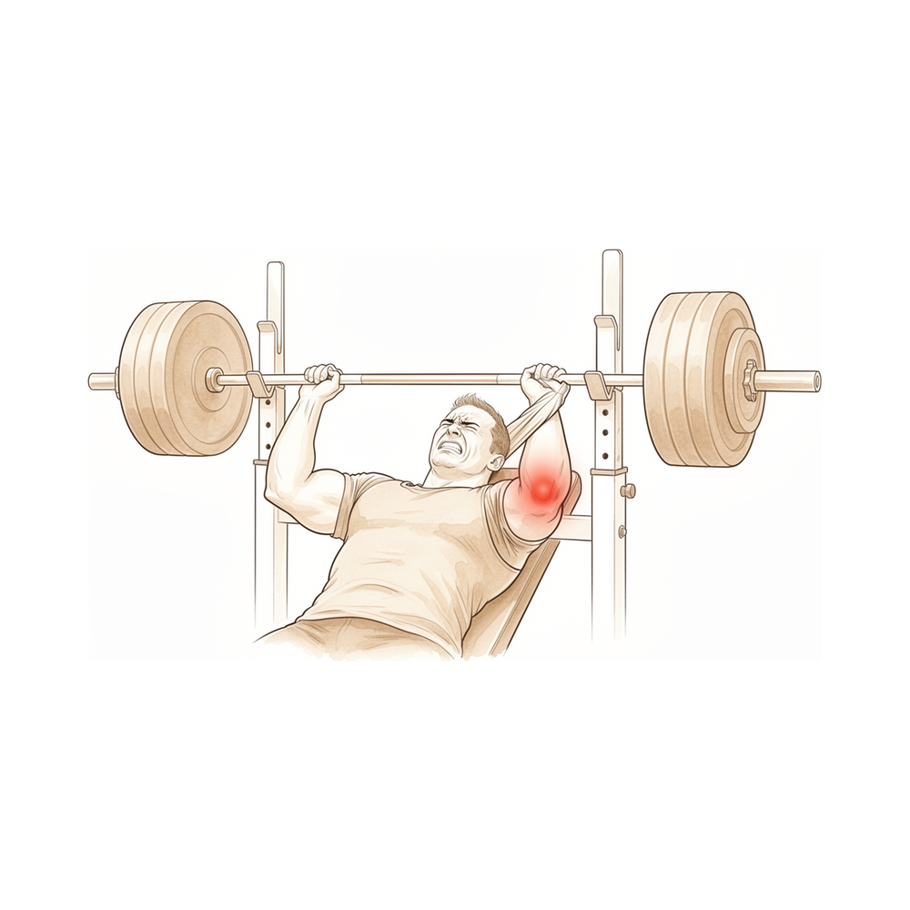
Triceps tendon injuries are possibly the rarest of all tendon injuries, with a male predominance of 2:1 [28]. The usual mechanism is a forceful sudden flexion of an extended elbow [21]. The most common mechanism involves a sudden eccentric load applied to a contracting triceps muscle, such as during weight lifting or from a fall onto an outstretched hand [47]. Direct trauma, motor vehicle accidents, and power lifting are also mechanisms of injury [28]. Lacerations and open injuries can cause distal triceps rupture [21]. Contributing etiologies include renal osteodystrophy, metabolic bone diseases, and anabolic steroid use [28]. Higher-energy mechanisms such as motor vehicle accidents or falls from a height may cause concomitant ulnar nerve injury and compartment syndrome [47].
Triceps tendon ruptures are usually seen at the osseous insertion [47]. Ruptures within the muscle belly and tears at the myotendinous junction have been reported but are less frequent [28]. Fatigue failure of the muscle has been suggested as a mechanism for intramuscular tears [47]. In fall on an outstretched hand injuries, the lateral and long heads of the triceps brachii have primary roles in eccentric contraction, resulting in only superficial tendon tears [34]. In direct injury groups, full-thickness tendon tears are more common than in fall on an outstretched hand injuries [34]. Most traumatic distal triceps tendon ruptures that develop by indirect mechanisms are partial tears [34].
Clinical Presentation and Diagnosis
Patients present with pain and swelling over the posterior aspect of the elbow [47]. Physical examination reveals tenderness to palpation, swelling, and ecchymosis [47]. A palpable defect proximal to the olecranon can confirm the clinical diagnosis of triceps rupture [47]. In the acute stage, a defect may not be palpable secondary to swelling, body habitus, or the degree of rupture [47]. Inability to actively extend against resistance is a sign of complete rupture [47]. However, not all complete tears result in the inability to actively extend against resistance [47]. This inability may be absent due to an intact lateral expansion or a compensating anconeus muscle [47]. Almost 50% of acute triceps ruptures were initially misdiagnosed in one study [47].
The diagnosis of a triceps tear is fundamentally clinical [122]. A partial rupture is frequently undiagnosed, necessitating a high index of suspicion for the diagnosis of triceps tendon rupture [122]. Care should be taken to assess the flake sign on lateral radiographs [122]. Ultrasonographic examination can be a valuable adjunct to assist in the diagnosis of a ruptured triceps tendon [122].
Classification
Epidemiology and Demographics
Distal triceps tendon rupture is a rare injury that traditionally occurs in an active population [2]. The injury has been described through a gamut of ages [28]. Adolescents who have incompletely fused or recently fused physes are susceptible to triceps tendon rupture [28]. Triceps tendon tears requiring repair are more prevalent in professional football players than previously reported and are more common than in the general population [29].
Mechanism of Injury
Laceration and open injuries with or without elbow fracture-dislocation can cause distal triceps rupture [21]. Lacerations and open injuries can also cause distal triceps rupture [47]. A direct blow may result in distal triceps injury, but this finding is less common [47]. Higher-energy mechanisms, such as a motor vehicle accident or a fall from a height, may cause concomitant ulnar nerve injury and compartment syndrome [47]. Spontaneous ruptures and ruptures at the musculotendinous junction and muscle belly have been described but are less frequent [28]. Renal osteodystrophy, metabolic bone diseases, and anabolic steroid use are contributing etiologies for triceps tendon rupture [28]. Triceps tendon ruptures may result from steroid injections [6].
Anatomical Location and Pattern
Ruptures within the muscle belly and tears at the myotendinous junction have been reported [47]. In fall on an outstretched hand injuries, all patients had superficial distal triceps tendon tears [34]. In direct injury groups, three of seven elbows had full-thickness tendon tears [34]. The lateral and long heads of the triceps brachii have primary roles in eccentric contraction in fall on an outstretched hand injuries, resulting in only superficial tendon tears [34]. Most traumatic distal triceps tendon ruptures that develop by indirect mechanisms were partial tears [34]. The medial head of the triceps has a tendon that is distinct from, and deep to, the common tendon of the long and lateral heads on gross inspection [30]. Histologic studies show the insertion of the medial head tendon and the common tendon of the long and lateral heads is confluent [30]. The triceps brachii inserts on the olecranon as a confluent tendon at the histologic level [117]. The gross proximal-to-distal tendon insertion had an average length of 13.0±1.1 mm [117]. The medial-to-lateral distance of the gross tendon insertion was an average of 20.8±2.0 mm [117]. The mean histologic dimension of the proximal-to-distal tendon insertion was 10.9±0.8 mm [117]. The mean histologic dimension of the medial-to-lateral tendon insertion was 20.9±1.2 mm [117]. The mean histologic distance from the articular tip of the olecranon to the proximal tendon insertion was 16.7±1.7 mm [117]. A defined interval located between the lateral triceps expansion and medial triceps tendon just proximal to the olecranon is known as the "triceps decussation" [21]. Magnetic resonance imaging describes a bipartite insertion between the superficial and deep components of the triceps tendon into the olecranon [21].
Clinical Presentation and Diagnosis
A palpable defect proximal to the olecranon can be apparent and may confirm the clinical diagnosis [47]. The inability to actively extend against resistance is a sign of complete rupture [47]. The ability to actively extend against resistance in the presence of a complete tear is likely secondary to an intact lateral expansion or a compensating anconeus muscle [47]. Almost 50% of acute triceps ruptures were initially misdiagnosed [47]. In the setting of chronic tears, the most common complaints are pain and weakness [47].
Clinical Presentation
Epidemiology and Demographics
Distal triceps tendon injuries are relatively rare, estimated to account for less than 1% of all upper limb tendon injuries [43].
Mechanism of Injury
The mechanism of injury is typically an acute trauma in which the tendon avulses off the olecranon [28]. Most commonly, the traumatic event is a fall on an outstretched hand in which a deceleration load is applied to the triceps while it is actively contracting [28]. Other mechanisms of injury include direct trauma, motor vehicle accidents, and power lifting [28]. Triceps tendon ruptures can occur through healthy tissue [23].
Risk Factors and Etiology
Studied risk factors include steroid use, endocrine disorders, renal failure, and metabolic bone diseases [93]. Triceps tendon ruptures may result from pathological chronic tendinitis in patients with cumulative submaximal loading [21]. Iatrogenic conditions with a history of surgery that violated the extensor mechanism are a cause of triceps tendon lesions [21].
Clinical Findings and Diagnosis
Diagnosis of a triceps rupture of the distal insertion can be readily made on physical examination, where pain, swelling, and a palpable defect can often be identified just proximal to the olecranon posteriorly [93]. Despite this, triceps tendon rupture is frequently missed in the emergency department [43]. Avulsion of the triceps tendon is easily overlooked in the acute stage, especially when combined with other lesions in the region such as fracture of the head or neck of the radius [48]. Injuries can be difficult to diagnose, potentially resulting in considerable loss of function [43].
Injury Patterns
In direct injury groups, full-thickness tendon tears are more common than in indirect mechanism groups [34].
Investigations
Clinical Presentation and Physical Examination
Distal triceps tendon ruptures are easily overlooked in the acute stage, especially when combined with other lesions in the region such as fracture of the head or neck of the radius [48]. The usual mechanism of injury is a forceful sudden flexion of an extended elbow [21]. These ruptures may result from acute traumatic events or pathological chronic tendinitis in patients with cumulative submaximal loading [21].
Imaging
Plain radiography: Plain radiographs remain the hallmark and the best screening test for elbow evaluation [41]. Most triceps tendon ruptures are accompanied by avulsion fracture of the olecranon [123]. It is important to suspect triceps tendon rupture when radiographs show a small fleck of bone avulsed from the olecranon [123].
MRI: MRI can be used to evaluate ligaments and tendons, but it is rarely indicated for elbow stiffness [66].
Other Considerations: A defined interval is located between the lateral triceps expansion and medial triceps tendon just proximal to the olecranon, known as the "triceps decussation" [21]. The distinct insertional heads of the triceps provide additional knowledge that can aid in diagnosing and treating partial triceps tears [33].
Treatment
Non-Operative
Conservative management is appropriate for partial triceps tendon ruptures, which can heal without functional deficit [18]. In cases of bilateral partial triceps tendon rupture, conservative treatment has resulted in excellent functional outcomes [16]. While complete triceps tendon ruptures require surgical repair to restore active elbow extension, partial tears may be managed conservatively [25].
Operative
Indications: Surgical repair is strongly recommended for complete distal triceps tendon ruptures [4], as these injuries must be repaired to provide active extension at the elbow [25]. Complete tears are optimally treated with surgical repair, particularly when performed acutely [3]. For partial triceps tendon ruptures in young, healthy, high-demand patients, operative repair using bone tunnels leads to a return to full function and should be utilized over conservative management [50].
Timing: Early surgical repair within three weeks after injury is the treatment of choice for distal triceps tendon ruptures [7]. Delayed repair is technically demanding and yields inferior results compared to acute repair [43]. However, for triceps tendon ruptures occurring through healthy tissue, the time of delay from injury to repair seems to have little effect on functional outcome [23].
Techniques and Outcomes: Distal triceps tendon repair reliably restores elbow function, provides good patient outcomes, and has a low complication rate [1]. Primary repair of both acute and chronic distal triceps tendon ruptures in a general population yields satisfactory results in the majority of patients with a low rerupture rate [8]. When recognized and treated surgically, triceps tendon ruptures may result in uniformly good to excellent outcomes [6]. Early surgical repair for traumatic distal triceps tendon ruptures yields good results, with the anchor group showing statistically significant earlier release from medical care [12]. Suture anchor repair of distal triceps tendon ruptures demonstrates excellent elbow function based on validated clinical outcome measures [37]. Endoscopic repair of partial distal triceps tendon tears leads to good clinical and radiological results after 12 months, with patients benefiting especially from an improvement in extension strength [15]. Endoscopic repair of superficial tears of the triceps tendon restores function and strength, leading to excellent clinical results after 1 year [26]. Arthroscopic triceps tendon repair is safe and effective, providing excellent pain relief, excellent functional scores, minimal complications, and the ability to address concomitant elbow pathologies [109].
Complications and Risks: Distal triceps repair for traumatic injuries is associated with 14% complication and 13.1% reoperation rates [17]. Regardless of repair technique, distal triceps tendon repair surgery has a relatively high complication and reoperation rate [36]. Despite the heightened risk of perioperative complications after primary repair of distal triceps tendon injuries, current series identify favorable functional outcomes and no cases of re-rupture at short- to mid-term follow-up [5]. Similarly, despite the heightened risk of perioperative complications, current series found favorable functional outcomes and no cases of reruptures at short-term to midterm follow-up [13].
Rehabilitation: At 2 weeks postoperatively, the patient is placed in a hinged elbow brace allowing 20° to 120° of assisted passive movement, followed by a further 2 weeks of unrestricted assisted passive movement in the brace [32]. Active range of motion and physiotherapy begin at 4 weeks to optimize range of movement and initiate strengthening exercises [32].
Complications
Overall Complication and Reoperation Rates: Distal triceps repair for traumatic injuries is associated with a 14% complication rate [17] and a 13.1% reoperation rate [17]. Primary repair of distal triceps tendon injuries carries a heightened risk of perioperative complications [5]. Patients undergoing distal triceps tendon rupture repair experience a moderate reported risk of rerupture or complication [14].
Rerupture: Primary repair of acute and chronic distal triceps tendon ruptures in a general population yields a low rerupture rate [8]. Primary repair of distal triceps tendon ruptures yields minimal rerupture regardless of repair construct [10]. In a series of 184 acute triceps tendon ruptures, no cases of re-rupture were identified at short- to mid-term follow-up [5]. In a series of 184 acute triceps tendon ruptures, no cases of re-rupture were identified at short-term to midterm follow-up [13].
Loss of Elbow Motion: Surgical repair of distal triceps tendon injuries offers a small risk of loss of elbow motion [24].
Recovery
Functional Outcomes: Distal triceps tendon repair reliably restores elbow function and provides good patient outcomes [1]. Early surgical management with repair of the tendon to bone can provide a satisfactory outcome in most patients [126].
Complications and Rerupture: Primary repair of distal triceps tendon injuries yields favorable functional outcomes with no cases of re-rupture at short- to mid-term follow-up [5]. This finding is consistent with other series reporting favorable functional outcomes and no cases of reruptures at short-term to midterm follow-up [13].
Return to Work: Of the patients, 69 (93.2%) returned to work at an average of 2.2±3.2 months following distal triceps repair [39]. Sixtysix patients (89.2%) were able to return to the same level of occupational intensity according to the US Department of Labor classification system [39]. Of the 5 patients who were unable to return to work in any capacity following distal triceps repair, 3 (60.0%) received disability payments [39].
Occupational Intensity and Return Timelines: Patients in higher-intensity occupations had a lower rate of return to work than patients in lower-intensity occupations [39]. Patients in higher-intensity occupations took longer to return to work than patients in lower-intensity occupations [39]. Patients who held sedentary-intensity occupations were able to return to their previous level of occupational intensity at a rate of 100.0% at a duration of 0.3±0.5 months [39]. Patients who held light-intensity occupations were able to return to their previous level of occupational intensity at a rate of 100.0% at a duration of 1.8±1.5 months [39]. Patients who held moderate-intensity occupations were able to return to their previous level of occupational intensity at a rate of 80.0% at a duration of 2.5±3.6 months [39]. Patients who held heavy-intensity occupations were able to return to their previous level of occupational intensity at a rate of 76.9% at a duration of 4.8±3.9 months [39].
Workers’ Compensation and Surgical History: At the time of surgery, 20 patients (27.0%) were covered by workers’ compensation, of whom, 15 (75%) were able to return to their previous occupation by 6.5±4.3 months postoperatively [39]. 100% of patients with non–workers’ compensation designation returned to work by 1.1±1.6 months [39]. Of the patients who underwent previous surgery on their elbow, 78% returned to work by 0.95±0.67 months following distal triceps repair [39]. Of the patients who underwent previous surgery on their elbow, 8 patients (88.9%) held light or sedentary occupations [39].
Key Evidence
- [L4] Distal triceps tendon repair reliably restores elbow function, provides good patient outcomes and has a low complication rate. [1] (10.1016/j.jse.2016.12.062)
- [L4] A distal triceps tendon rupture is a rare but potentially challenging injury, traditionally occurring in an active population, and often requires surgical repair. [2] (10.1177/03635465241283970)
- [Paper] Distal triceps tendon injuries are relatively rare, with complete tears optimally treated with surgical repair, particularly when performed acutely. [3] (10.1016/j.csm.2020.03.003)
- [L4] Acute partial triceps tendon ruptures may have good outcomes with nonoperative management, whereas surgical repair is strongly recommended for complete ruptures. [4] (10.2106/jbjs.rvw.19.00172)
- [L4] Despite heightened risk of perioperative complications after primary repair of distal triceps tendon injuries, the current series identifies favorable functional outcomes and no cases of re-rupture at short- to mid-term follow-up. [5] (10.1177/2325967118s00163)
- [L4] Triceps tendon ruptures may result in uniformly good to excellent results if recognized and treated surgically. [6] (10.1177/036354659302100327)
- [L4] Early surgical repair, within three weeks after the injury, is the treatment of choice for distal triceps tendon ruptures. [7] (10.2106/00004623-200310000-00015)
- [L4] Primary repair of acute and chronic distal triceps tendon ruptures in a general population yields satisfactory results in the majority of patients with a low rerupture rate. [8] (10.1302/0301-620x.100b5.bjj-2017-1057.r2)
- [L5] Partial ruptures of the distal triceps tendon demonstrate a not statistically significant lower ultimate load to failure than intact tendons and typically occur in the superficial, lateral portion of the tendon. [9] (10.1186/s12891-023-06720-3)
- [L3] Primary repair of distal triceps tendon ruptures yields good, durable patient outcomes with minimal rerupture regardless of repair construct. [10] (10.1016/j.jse.2017.08.006)
- [Case_report] Surgical repair of distal triceps tendon ruptures can be accomplished with predictable results. [11] (10.1016/j.jse.2006.06.002)
- [Abstract] Early surgical repair for traumatic distal triceps tendon ruptures yielded good results, with the anchor group showing statistically significant earlier release from medical care. [12] (10.1016/j.jse.2014.11.027)
- [L4] Despite the heightened risk of perioperative complications after primary repair of distal triceps tendon injuries, the current series found favorable functional outcomes and no cases of reruptures at short-term to midterm follow-up. [13] (10.1177/2325967119839998)
- [L4] Patients undergoing distal triceps tendon rupture repair experience improvements in postoperative outcomes; however, there is a moderate reported risk of rerupture or complication. [14] (10.1016/j.jse.2021.06.019)
- [L4] Endoscopic repair of partial distal triceps tendon tears leads to good clinical and radiological results after 12 months, with patients benefiting especially from an improvement of extension strength. [15] (10.1016/j.arthro.2013.03.049)
- [L4] Conservative management was entirely appropriate for this patient with bilateral partial triceps tendon rupture, resulting in excellent functional outcome. [16] (10.1177/0363546503258903)
- [L3] Distal triceps repair for traumatic injuries is associated with 14% complication and 13.1% reoperation rates. [17] (10.1016/j.xrrt.2022.05.004)
- [L4] Partial triceps tendon ruptures can heal without functional deficit. [18] (10.1177/0095399703258707)
- [L3] Distal triceps repairs in this large cohort study occur most frequently in men aged 40 to 59 years. [19] (10.1177/15589447221095114)
- [L5] [21] (10.1007/s12306-015-0359-y)
- [L5] Operative management of complete distal biceps and triceps tendon ruptures is required in most cases, especially for active patients, laborers, and athletes wishing to return to competition. [22] (10.1016/j.hcl.2016.08.019)
- [L4] Triceps tendon ruptures can occur through healthy tissue, and the time of delay from injury to repair seems to have little effect on the functional outcome. [23] (10.1177/036354658401200415)
- [L5] Most complete triceps tendon injuries should be managed with surgical repair, which offers a predictable return of function with a small risk of loss of elbow motion. [24] (10.1016/j.hcl.2015.06.010)
- [L5] Complete triceps tendon ruptures must be repaired to provide active extension at the elbow, while partial tears may be treated conservatively. [25] (10.1016/j.jhsa.2015.05.016)
- [L4] Endoscopic repair of superficial tears of the triceps tendon is able to restore function and strength and leads to excellent clinical results after 1 year. [26] (10.1016/j.arthro.2014.03.005)
- [L5] Anatomic repair of triceps tendon ruptures demonstrated the most anatomic restoration of distal triceps ruptures and showed statistically significantly less repair-site motion when cyclically loaded. [27] (10.1177/0363546509358319)
- [L5] [28] (10.1016/j.csm.2004.06.001)
- [L4] Triceps tendon tears requiring repair are more prevalent in professional football players than previously reported and are more common than in the general population. [29] (10.1177/2325967115601021)
- [L4] The medial head of the triceps has a tendon that is distinct from, and deep to, the common tendon of the long and lateral heads on gross inspection, though histologic studies show the insertion of these 2 tendons is confluent. [30] (10.1177/0363546506288752)
- [L3] Surgical repair of acute triceps tendon ruptures reliably restores strength and function even in high-demand individuals. [31] (10.1016/j.injury.2016.07.061)
- [Paper] [32] (10.1016/j.eats.2018.09.006)
- [L5] The distinct insertional heads of the triceps provide additional knowledge that can aid in diagnosing and treating partial triceps tears. [33] (10.1016/j.jses.2017.05.002)
- [L3] [34] (10.1097/corr.0000000000001550)
- [L4] Regardless of repair technique, distal triceps tendon repair surgery has a relatively high complication and reoperation rate. [36] (10.1016/j.xrrt.2024.06.008)
- [L4] This retrospective case series of suture anchor repair of distal triceps tendon ruptures showed excellent elbow function based on validated clinical outcome measures. [37] (10.1016/j.arthro.2011.12.016)
- [L4] [39] (10.1016/j.jse.2020.07.036)
- [L4] [43] (10.1177/1758573217706358)
- [L5] [47] (10.5435/00124635-201001000-00005)
- [L5] Avulsion of the triceps tendon is easily overlooked in the acute stage, especially when combined with other lesions in the region such as fracture of the head or neck of the radius. [48] (10.1016/0020-1383(86)90238-x)
- [L5] The authors conclude that operative repair of partial tricep tendon ruptures using bone tunnels in a young, healthy, high-demand patient leads to return to full function and should be utilized over conservative management. [50] (10.1093/milmed/usx075)
- [L4] [93] (10.1055/s-0038-1636729)
- [L4] In this study, we found that arthroscopic triceps tendon repair is safe and effective, with excellent pain relief, excellent functional scores, minimal complications, and the ability to address concomitant elbow pathologies. [109] (10.1016/j.asmr.2025.101164)
- [L5] [117] (10.1016/j.jhsa.2021.05.003)
- [L4] [122] (10.1016/j.jse.2005.12.010)
- [L5] Most triceps tendon ruptures are accompanied by avulsion fracture of the olecranon, and it is important to suspect this injury when radiographs show a small fleck of bone avulsed from the olecranon. [123] (10.1007/s00590-012-1127-4)
- [L4] Early surgical management with repair of the tendon to bone can provide a satisfactory outcome in most patients. [126] (10.1016/j.jse.2005.01.004)
See Also
References
[1] Outcomes following distal triceps tendon repair. Journal of Shoulder and Elbow Surgery. 2017. DOI: 10.1016/j.jse.2016.12.062
[2] Return to Work, Sport, and Sport- Related Activity After Distal Triceps Tendon Repair: A Systematic Review. The American Journal of Sports Medicine. 2025. DOI: 10.1177/03635465241283970
[3] Distal Triceps Tendon Injuries. Clinics in Sports Medicine. 2020. DOI: 10.1016/j.csm.2020.03.003
[4] Triceps Tendon Ruptures. JBJS Reviews. 2020. DOI: 10.2106/jbjs.rvw.19.00172
[5] Surgical Repair of Distal Triceps Tendon Injuries: Short-Term Clinical Outcomes and Re-Rupture Rate. Orthopaedic Journal of Sports Medicine. 2018. DOI: 10.1177/2325967118s00163
[6] Rupture of the triceps tendon associated with steroid injections. The American Journal of Sports Medicine. 1993. DOI: 10.1177/036354659302100327
[7] SURGICAL TREATMENT OF DISTAL TRICEPS RUPTURES. The Journal of Bone and Joint Surgery-American Volume. 2003. DOI: 10.2106/00004623-200310000-00015
[8] Results of primary repair of distal triceps tendon ruptures in a general population. The Bone & Joint Journal. 2018. DOI: 10.1302/0301-620x.100b5.bjj-2017-1057.r2
[9] Partial ruptures of the distal triceps tendons show only slightly lower ultimate load to failure: a biomechanical study. BMC Musculoskeletal Disorders. 2023. DOI: 10.1186/s12891-023-06720-3
[10] Functional outcomes of distal triceps tendon repair comparing transosseous bone tunnels with suture anchor constructs. Journal of Shoulder and Elbow Surgery. 2017. DOI: 10.1016/j.jse.2017.08.006
[11] Distal triceps tendon rupture and repair in an otherwise healthy pediatric patient: A case report and review of the literature. Journal of Shoulder and Elbow Surgery. 2007. DOI: 10.1016/j.jse.2006.06.002
[12] Surgical Treatment of 150 Acute Distal Triceps Tendon Ruptures. Journal of Shoulder and Elbow Surgery. 2015. DOI: 10.1016/j.jse.2014.11.027
[13] Surgical Repair of Distal Triceps Tendon Injuries: Short-term to Midterm Clinical Outcomes and Risk Factors for Perioperative Complications. Orthopaedic Journal of Sports Medicine. 2019. DOI: 10.1177/2325967119839998
[14] The surgical management of distal triceps tendon ruptures: a systematic review. Journal of Shoulder and Elbow Surgery. 2022. DOI: 10.1016/j.jse.2021.06.019
[15] The Endoscopic Repair of Partial Lesions of the Distal Triceps Tendon: First Prospective Results of 14 Cases (SS‐42). Arthroscopy. 2013. DOI: 10.1016/j.arthro.2013.03.049
[16] Bilateral Partial Rupture of Triceps Tendon. The American Journal of Sports Medicine. 2004. DOI: 10.1177/0363546503258903
[17] Complications after traumatic distal triceps tears: an analysis of 107 cases. JSES Reviews, Reports, and Techniques. 2022. DOI: 10.1016/j.xrrt.2022.05.004
[18] Triceps Tendon Ruptures in Professional Football Players. The American Journal of Sports Medicine. 2004. DOI: 10.1177/0095399703258707
[19] Surgical Outcomes, Trends, and Risk Factors of Distal Triceps Repairs. HAND. 2022. DOI: 10.1177/15589447221095114
[21] Triceps tendon rupture: the knowledge acquired from the anatomy to the surgical repair. MUSCULOSKELETAL SURGERY. 2015. DOI: 10.1007/s12306-015-0359-y
[22] Biceps and Triceps Ruptures in Athletes. Hand Clinics. 2017. DOI: 10.1016/j.hcl.2016.08.019
[23] Triceps tendon avulsion in a professional body builder. The American Journal of Sports Medicine. 2004. DOI: 10.1177/036354658401200415
[24] Distal Triceps Tendon Injuries. Hand Clinics. 2015. DOI: 10.1016/j.hcl.2015.06.010
[25] Triceps Tendon Repair. The Journal of Hand Surgery. 2015. DOI: 10.1016/j.jhsa.2015.05.016
[26] Endoscopic Repair of Tears of the Superficial Layer of the Distal Triceps Tendon. Arthroscopy. 2014. DOI: 10.1016/j.arthro.2014.03.005
[27] The Distal Triceps Tendon Footprint and a Biomechanical Analysis of 3 Repair Techniques. The American Journal of Sports Medicine. 2010. DOI: 10.1177/0363546509358319
[28] Biceps tendon and triceps tendon injuries. Clinics in Sports Medicine. 2004. DOI: 10.1016/j.csm.2004.06.001
[29] Triceps Tendon Ruptures Requiring Surgical Repair in National Football League Players. Orthopaedic Journal of Sports Medicine. 2015. DOI: 10.1177/2325967115601021
[30] Surgical Anatomy of the Triceps Brachii Tendon. The American Journal of Sports Medicine. 2006. DOI: 10.1177/0363546506288752
[31] Outcomes and complications of triceps tendon repair following acute rupture in American military personnel. Injury. 2016. DOI: 10.1016/j.injury.2016.07.061
[32] Distal Triceps Rupture Repair: The Triceps Pulley‐Pullover Technique. Arthroscopy Techniques. 2019. DOI: 10.1016/j.eats.2018.09.006
[33] The distal triceps tendon insertional anatomy—implications for surgery. JSES Open Access. 2017. DOI: 10.1016/j.jses.2017.05.002
[34] Differences in Rupture Patterns and Associated Lesions Related to Traumatic Distal Triceps Tendon Rupture Between Outstretched Hand and Direct Injuries. Clinical Orthopaedics & Related Research. 2020. DOI: 10.1097/corr.0000000000001550
[36] A comparison of distal triceps tendon repair outcomes by surgical technique. JSES Reviews, Reports, and Techniques. 2024. DOI: 10.1016/j.xrrt.2024.06.008
[37] Clinical Outcome After Suture Anchor Repair for Complete Traumatic Rupture of the Distal Triceps Tendon. Arthroscopy. 2012. DOI: 10.1016/j.arthro.2011.12.016
[39] Return to work following distal triceps repair. Journal of Shoulder and Elbow Surgery. 2021. DOI: 10.1016/j.jse.2020.07.036
[41] Orthopaedic Knowledge Update 13 Ebook Without Multimedia. Anatomy, Biomechanics, Physical Examination, and Imaging of the Elbow > Summary and Conclusions.
[43] Triceps tendon rupture: repair and rehabilitation. Shoulder & Elbow. 2017. DOI: 10.1177/1758573217706358
[47] Distal Triceps Rupture. American Academy of Orthopaedic Surgeon. 2010. DOI: 10.5435/00124635-201001000-00005
[48] Triceps tendon avulsion. Injury. 1986. DOI: 10.1016/0020-1383(86)90238-x
[50] A Case of Non-simultaneous Bilateral Partial Triceps Tendon Repair. Military Medicine. 2017. DOI: 10.1093/milmed/usx075
[51] Orthopaedic Knowledge Update 13 Ebook Without Multimedia. Anatomy, Biomechanics, Physical Examination, and Imaging of the Elbow > Annotated References.
[52] Orthopaedic Knowledge Update 13 Ebook Without Multimedia. Anatomy, Biomechanics, Physical Examination, and Imaging of the Elbow > Anatomy > Bony Anatomy.
[53] Miller S Review Of Orthopaedics. SECTION 16 PATELLAR TRACKING IN TOTAL KNEE ARTHROPLASTY > TABLE 2.3 Shoulder Spaces.
[58] Miller S Review Of Orthopaedics. ELBOW.
[60] A Lange Medical Book Current Diagnosis Treatment In Orthopedics Fifth Edition. 2Musculoskeletal Trauma Surgery > INJURIES AROUND THE ELBOW.
[63] Rockwood And Green S Fractures In Adults. 29: Principles of Nonunion and Bone Defect Treatment > Pathoanatomy and Applied Anatomy Related to Distal Humerus Fractures.
[66] Aaos Comprehensive Orthopaedic Review 3. Elbow Stiffness* > IV. Evaluation.
[93] Triceps Tendon Anatomic Repair Utilizing the “Suture Bridge” Technique. Journal of Hand and Microsurgery. 2018. DOI: 10.1055/s-0038-1636729
[109] Arthroscopic Repair of Distal Triceps Tendon Rupture Provides Excellent Functional Outcomes With Minimal Complications. Arthroscopy, Sports Medicine, and Rehabilitation. 2025. DOI: 10.1016/j.asmr.2025.101164
[117] Histologic Evaluation of the Triceps Brachii Tendon Insertion: Implications for Triceps-Sparing Surgery. The Journal of Hand Surgery. 2022. DOI: 10.1016/j.jhsa.2021.05.003
[122] Chronically ruptured triceps tendon with avulsion of the medial collateral ligament: A report of 2 cases. Journal of Shoulder and Elbow Surgery. 2007. DOI: 10.1016/j.jse.2005.12.010
[123] Triceps tendon avulsion: a case report and discussion about the olecranon ossification nucleus. European Journal of Orthopaedic Surgery & Traumatology. 2012. DOI: 10.1007/s00590-012-1127-4
[126] Acute triceps ruptures: Case report and retrospective chart review. Journal of Shoulder and Elbow Surgery. 2006. DOI: 10.1016/j.jse.2005.01.004