Clinicians › Wrist
Proximal Row Carpectomy
PRC for SLAC/SNAC II and Kienbock: radiocapitate articulation, capitate/lunate-fossa gatekeeping, PRC vs four-corner fusion, capsular interposition, long-term survivorship.
For patients: a plain-language version of this topic is available. See the patient guide.
Overview
Proximal row carpectomy is a motion-preserving salvage procedure that allows for early wrist mobilization and avoids the risk of nonunion [1]. It is indicated for advanced-stage Kienböck's disease, scaphoid nonunion, and stage II SLAC wrists with a preserved capitolunate joint [9, 10, 14]. The procedure is also selected for the concurrence of Kienbock's disease and scapholunate dissociation following acute trauma [4], and serves as a useful option for preserving joint motion in selected cases of complete amputation at the wrist joint [18]. While controversy persists regarding its use in the setting of capitate arthritis [28], the procedure is preferred for SLAC wrists with preserved capitate head cartilage due to procedural ease, lower complication rates, and socio-economic benefits [32].
Clinical outcomes demonstrate that proximal row carpectomy provides pain-free function and satisfactory range of motion, grip strength, and patient satisfaction at long-term follow-up [3, 10]. In patients younger than 45 years, the procedure offers improved motion and fewer complications compared to four-corner arthrodesis [5, 11]. Additionally, proximal row carpectomy results in slightly better wrist movement and does not require hardware removal, unlike four-corner arthrodesis [11]. For SLAC arthritis, it offers greater range of motion, fewer complications, and lower costs compared to scaphoid excision and four-corner arthrodesis [13]. Direct surgical costs for four-corner arthrodesis are 430% greater than for proximal row carpectomy, driven primarily by implant costs, despite similar clinical outcomes in the literature [30].
The procedure is reliable and durable, with good clinical results maintained out to an average of 15 years postoperatively for advanced-stage Kienböck's disease [8]. At an average follow-up of 10 years, it remains a reliable and durable option for Lichtman stage IIIA or IIIB Kienböck's disease [14]. A systematic review confirms its long-term durability for wrist arthritis [19]. Arthroscopic proximal row carpectomy is a safe and effective technique that allows for rapid mobilization compared with the open procedure [12]. Osteochondral grafting in combination with proximal row carpectomy allows for less invasive but equally effective procedures for SLAC and SNAC [22]. The incidence of the procedure has increased in recent years, even in patients under 45 years old [16].
Anatomy & Pathophysiology
Bony Anatomy
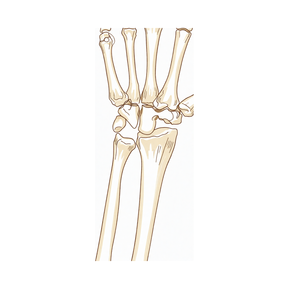
The wrist comprises the distal radioulnar, radiocarpal, and ulnocarpal joints, along with the eight carpal bones and their associated ligaments [52]. The proximal row consists of the scaphoid, lunate, triquetrum, and pisiform, while the distal row includes the trapezium, trapezoid, capitate, and hamate [52]. The pisiform is a sesamoid bone embedded within the flexor carpi ulnaris tendon, articulating with the triquetrum via the pisotriquetral joint [36]. The distal radius features two concave facets for the scaphoid and lunate, separated by the scapholunate ridge, and a sigmoid notch along the ulnar border that accommodates the ulnar head [62]. The lunate is broader palmarly than dorsally [62]. The triquetrum articulates distally with the hamate, radially with the lunate, and volarly with the pisiform [62]. The capitate head often relies on a retrograde vascular supply [62]. The hamate consists of a body and a hook (hamulus), which serves as an attachment for the transverse carpal ligament and origins for the flexor digiti minimi and opponens digiti minimi muscles [62].
The scaphoid receives its primary vascular supply from a branch of the radial artery at the dorsal ridge, with smaller vessels entering the palmar tubercle to supply the distal 30% [62]. In 80% of wrists, the lunate has both dorsal and palmar vascular supplies, whereas only a palmar supply is present in 20% [62]. The distal radius normally exhibits an average of 11 degrees of volar tilt in the sagittal plane and 23 degrees of radial inclination in the frontal plane [56]. Radial length, measured from the tip of the radial styloid to the ulnar articular surface, averages 13 mm [56]. The distal ulna has an average 20-degree inclination at its articulation with the radius [52]. The triangular fibrocartilage attaches to the base of the ulnar styloid, separating the hyaline cartilage–covered ulnar head from the styloid [52]. The sigmoid notch articular surface accommodates the ulnar head through two thirds of its arc [52]. The ulnar styloid lies dorsal to the ulnar head and extends distally [52].
The radiocarpal joints are formed by the articulation of the distal radius with the scaphoid and lunate through their respective concave facets, and with the triquetrum on the triangular fibrocartilage [52]. The distal concave articular surfaces of the proximal carpal row form the midcarpal articulations with the distal row [52]. The distal row articulates with the metacarpals, allowing mobility in the thumb, stability in the index and long finger metacarpals, and increased mobility in the ring and little finger metacarpals [52]. The interosseous membrane connects the shafts of the radius and ulna, with a thickened central portion important in force transmission between the two bones [56]. The radius possesses a lateral bow crucial for maintaining full pronation and supination, while the ulnar shaft remains fixed in rotation at the ulnohumeral joint as the radius rotates around it [56]. The boundaries of Guyon’s canal are defined by the volar carpal ligament and transverse carpal ligament, the hook of the hamate radially, and the pisiform ulnarly [56]. This canal contains the ulnar artery and nerve [56]. The flexor carpi ulnaris tendon is palpable near its insertion on the pisiform on the ulnar surface [56].
The dorsal surface of the wrist contains six compartments with wrist and digital extensor tendons, while the volar surface contains the carpal tunnel contents, including nine flexor tendons and the median nerve [56]. The radial styloid allows attachment of the brachioradialis tendon and serves as the origin for several important wrist ligaments, including the radial scapholunate and radial lunocapitate ligaments [56]. The distal ulna is covered with hyaline cartilage on its dorsal, lateral, palmar, and distal surfaces [62]. The ulnar styloid projects distally, and at its base, the fovea serves as the insertion for the triangular fibrocartilaginous complex [62]. The trapezoid has two distal facets that articulate with the index finger metacarpal [62]. The trapezium features a saddle-shaped articulation with the thumb metacarpal base and a palmar groove for the flexor carpi radialis, bordered laterally by a palmar tuberosity and the attachment for the transverse carpal ligament [62]. The pisiform serves as the origin for the abductor digiti minimi [62].
Ligamentous Anatomy
Extrinsic carpal ligaments connect the radius or ulna to the carpus, with volar ligaments generally stronger than dorsal ligaments [57]. The scapholunate interosseous ligament is C-shaped in the sagittal plane, with the dorsal third being the thickest and strongest portion [62]. This ligament consists of dorsal, palmar, and interosseous portions, with the dorsal portion being the strongest and thickest [57]. The lunotriquetral interosseous ligament is also C-shaped, where the volar portion is the thickest and strongest [57]. The triangular fibrocartilage complex (TFCC) is formed by the central meniscus homolog, dorsal and volar radioulnar ligaments, the floor of the extensor carpi ulnaris tendon sheath, and volar ulnocarpal ligaments [62]. The TFCC arises from the radial border of the distal radius and inserts into the base of the ulnar styloid and distal ulna through the ligamentum subcruentum [62]. The dorsal and volar radioulnar ligaments are the primary stabilizers of the distal radioulnar joint [62].
The space of Poirier is an area adjacent to the proximal capitate without ligamentous attachment, situated ulnar to the radioscaphocapitate ligament and radial to the long radiolunate ligament in the floor of the carpal tunnel [57]. This space is a weak area vulnerable to instability, through which the distal carpal row separates from the lunate during a perilunate dislocation [57]. The dorsal radiocarpal ligament has a trapezoidal shape, passing from the dorsal rim of the distal radius to the lunate and triquetrum [57]. The dorsal intercarpal ligament passes from the dorsal tubercle of the triquetrum to the distal pole of the scaphoid [57]. This ligament reinforces the elastic dorsal wrist capsule and helps stabilize the scapholunate articulation, with deep fibers contributing to the dorsal scapholunate interosseous ligament [57]. The radioscaphocapitate ligament connects to the waist of the scaphoid and limits ulnar translation of the carpus [57]. The long radiolunate ligament helps limit ulnar translocation of the carpus, while the short radiolunate ligament helps control lunate position [57].
The ulnocapitate ligament attaches to the ulnar head and is the most superficial or palmar of the palmar ulnocarpal ligaments [57]. The ulnotriquetral ligament attaches to the palmar radioulnar ligament and triquetrum, while the ulnolunate ligament attaches to the palmar radioulnar ligament and lunate [57]. The capitohamate ligament is a thick ligament, 5 × 5 mm in cross section, with extensions to the third or fourth metacarpals [57]. The dorsal radiocarpal ligament is associated with dorsal and volar intercalated segmental stabilities, and damage to this ligament, when in conjunction with other intrinsic ligament injuries, confers further carpal instability [57].
Vascular & Neural Anatomy
The terminal branches of the radial, ulnar, and anterior interosseous arteries provide extraosseous blood supply to the carpus through three dorsal and three palmar transverse arterial arches with longitudinal connections [65]. The dorsal intercarpal arch is the largest of the dorsal arches, supplying the distal carpal row and, through anastomoses with the radiocarpal arch, the lunate and triquetrum [65]. The palmar intercarpal arch is the most variable and does not contribute to nutrient vessels in the carpus [65]. Only the peripheral 10% to 40% of the volar, ulnar, and dorsal TFCC has a vascular supply [62].
Kinematics and Biomechanics
The wrist functions as a two-joint system linking the hand to the forearm around the highly mobile bones of the proximal carpal row [63]. The proximal carpal row has no muscular or tendinous attachments and is an intercalary segment [62]. It forms an intercalated segment between the distal carpal row and the distal radius, bound into a functional unit by the scapholunate and lunotriquetral interosseous ligaments [57]. The scapholunate interosseous ligament is the major stabilizer of the wrist and the most commonly injured wrist ligament [57]. It provides a flexion force on the lunate given its attachment to the scaphoid, while the lunotriquetral interosseous ligament provides an extension moment on the lunate given its attachment to the triquetrum [57]. The lunate, capitate, hamate, trapezium, and trapezoid function collectively as the "stable central column," controlled by the scaphoid in a two-gear, four-bar linkage system and stabilized by the triquetrum [63]. The triquetrum buffers lunate rotation and prevents ulnar translation [63]. The helicoidal joint between the triquetrum and hamate drives isotriquetral alignment in the sagittal plane during wrist radioulnar deviation [63].
The central column of the wrist (radius-lunate-capitate) is mechanically arranged as a "link joint" which "crumples" when the scaphoid is fractured or dissociated from the lunate [63]. The carpus functions as an oval ring formed by four interdependent elements (distal row, scaphoid, lunate, and triquetrum) connected to adjacent segments by ligamentous links [63]. The distal carpal row is rigid with little motion between its bones due to stout intercarpal ligaments, acting as a functional unit with the scaphoid bridging both rows [57]. Stability of the wrist link system is enhanced by strong volar ligaments and by the scaphoid, which bridges both carpal rows [71]. The central column of the wrist is responsible for flexion-extension, the medial column for rotation, and the lateral column for mobile function [71].
With axial loading through the neutral wrist, approximately 80% of forces are transmitted through the distal radius and 20% through the distal ulna [62]. The distal radius normally bears about 80% of distal radioulnar joint load, while the distal ulna bears 20% [71]. The ulna transmits 17% of the axial load in the forearm [71]. The line of the center of rotation in the forearm runs from the radial head to the distal ulna [71]. The instant center of wrist motion is usually the head of the capitate, but it varies [71]. Normal wrist range of motion is 65 degrees flexion, 55 degrees extension, 15 degrees radial deviation, and 35 degrees ulnar deviation [71]. Functional wrist range of motion is 10 degrees flexion, 35 degrees extension, 10 degrees radial deviation, and 15 degrees ulnar deviation [71]. The ratio of carpal height to third metacarpal height is normally 0.54, and the ratio of ulna-to-capitate length to third metacarpal height is normally 0.30 [71].
With wrist flexion, 60% of the motion is midcarpal and 40% is radiocarpal [62]. With wrist extension, 33% of the motion is midcarpal and 66% is radiocarpal [62]. Approximately 62° of wrist extension occurs through the radiocarpal joint and 62% of wrist flexion occurs through the midcarpal joint [57]. The midcarpal joint is mostly responsible for 20° of radial deviation and 40° of ulnar deviation [57]. More than half the motion of the carpus when the wrist was loaded in extension occurred at the midcarpal joint [82]. During wrist flexion from neutral, the proximal row translates dorsally, while during wrist extension from neutral, the proximal row translates palmarly [57]. The proximal row extends relative to the forearm/distal row with ulnar deviation and flexes relative to the forearm/distal row with radial deviation [62].
The midcarpal joint is responsible for the "dart thrower's motion," which involves moving from radial extension into ulnar flexion positioning of the wrist [57]. The dart-thrower's path of radial extension to ulnar flexion defines the transition between flexion and extension of the scaphoid and lunate, a path during which proximal row motion approaches zero [63]. This motion occurs almost exclusively through the midcarpal joint, with rotation occurring along the mechanical axis of the wrist [63]. All bones of each carpal row rotate in the same plane during any direction of global wrist motion [63]. In all but pure flexion/extension of the uninjured wrist, the proximal and distal rows move in divergent directions [63]. The wrist joint's motion planes include flexion, extension, radial deviation, ulnar deviation, and circumduction, with minimal carpal motion during pronosupination [57].
Two predominant patterns of motion exist during radioulnar deviation: the proximal row rotates mostly along the frontal plane (row pattern) or mostly along the sagittal plane (column pattern) [63]. The magnitude of out-of-plane motion during radioulnar deviation varies substantially from one individual to another [63]. In most individuals, the proximal carpal row rotates predominantly around the flexion-extension axis during radioulnar deviation [63]. The contact areas between the scaphoid and distal radius are maximized during full extension of the wrist [86]. During forearm rotation, the contact site of the scaphoid and the lunate on the distal radial articular surface changed minimally [92].
Division of either of the proximal row's interosseous ligaments in isolation does not result in a postural deformity of the lunate [63]. Similar "collapse" deformities of proximal row alignment (VISI or DISI) can occur with or without disruption of an interosseous ligament [63]. The effect of ligament sectioning on producing carpal instability may be moderated by the bone geometry of the radiocarpal joint [93]. There is a relationship between scapholunate kinematics and laxity at the level of the interosseous ligaments [104]. Computed fiber elongations of dorsal carpal ligaments vary linearly with wrist position [61]. Lunate morphology affects 3-dimensional carpal kinematics during wrist flexion and extension [58]. Hysteresis can be quantified using 4DCT wrist kinematics and is greater in wrists with scapholunate ligament injury than in healthy wrists [83]. Kinematic changes in scapholunate instability may predict the development of radioscaphoid arthritis and help identify a kinematically abnormal wrist [72]. Scaphoid nonunions have a dramatic impact on carpal kinematics, partially uncoupling the proximal and distal carpal rows [79]. SNAC wrists differ from SLAC wrists in exhibiting a decreased sagittal lunotriquetral angle, indicating a distinct pathomechanism of carpal instability [98]. Carpal distraction has a significant effect on the conformation of the carpus, especially at the radiocarpal and midcarpal joints [103].
Classification
Modified SLAC/SNAC Wrist Stages: A modified classification based on intraoperative findings subdivides stage III into IIIA and IIIB [50]. This separation describes different patterns of degeneration seen by inspection rather than a sequence of degeneration [50].
Stage I: Defined as degenerative changes limited to the radial styloid/scaphoid articulation [50].
Stage II: Defined as degenerative changes involving the entire radioscaphoid joint with mild or no changes at the capitolunate or radiolunate joint [50].
Stage IIIA: Defined as mild or no changes at the capitolunate joint and moderate to severe changes at the radiolunate joint [50]. Wrists in this stage are considered candidates for proximal row carpectomy [50].
Stage IIIB: Defined as moderate to severe changes at the capitolunate joint and mild or no changes at the radiolunate joint [50]. Wrists in this stage are considered candidates for scaphoid excision and four-corner arthrodesis [50].
Stage IV: Defined as moderate to severe changes at both the capitolunate and radiolunate joints [50].
Other Considerations: Scaphoid excision and four-corner fusion is the choice for stage III SLAC wrist [17].
Clinical Presentation
Proximal row carpectomy is a reliable motion-preserving procedure with good clinical results maintained out to an average of 15 years postoperatively [8]. Post-operative motion and grip strength values appear to remain stable over time [40]. In a study with a minimum 20-year follow-up, the flexion-extension arc was 68° and grip strength was 72% of the contralateral side [108]. In a retrospective study with a mean follow-up of 97.9 months, the mean flexion/extension arc was 93° and the mean grip strength was 25 kg [107].
At long-term follow-up, all patients older than thirty-five years of age at the time of proximal row carpectomy maintained a satisfactory range of motion, grip strength, and pain relief and were satisfied with the result [3]. However, in a long-term follow-up study, forty-six patients (74%) were not satisfied with the results of their surgery due to persistent pain or inability to return to previous occupational activities [40]. Fifty-two patients required daily pain medication for wrist pain in a long-term follow-up study [40].
Proximal row carpectomy results in a stiffened, weakened wrist when performed for static scapholunate dissociation [23]. Most muscle moment arms decreased significantly after proximal row carpectomy [26]. Radiographic follow-up beyond 2 years revealed joint narrowing and arthritic changes within the radiocapitate joint [40]. In a study with a mean follow-up of 97.9 months, fifteen patients had radiocapitate osteoarthritis [107]. There was no correlation between degenerative radiographic changes and satisfaction level in patients followed for a minimum of 20 years [108].
Proximal row carpectomy is a useful procedure to preserve joint motion of the wrist in selected cases of complete amputation at the wrist joint, achieving good functional results in young individuals [18].
Investigations
Imaging Modalities
MRI: MRI is the modality of choice for imaging radiographically occult fractures of the hand and wrist [60]. High-resolution MRI aids in the evaluation of ligament injuries, with a static magnetic field strength of at least 1.5 T using a dedicated wrist coil recommended for analyzing interosseous, intrinsic, and extrinsic ligament insertions [73]. The volar extrinsic, scapholunate interosseous, dorsal intercarpal, and lunotriquetral ligaments are best visualized using 1 mm slices with no interslice gap in the coronal plane [73]. In osteonecrosis, MRI is useful for detecting additional marrow abnormalities, as seen in the lunate in Kienböck disease [75]. Preoperative MRI may facilitate the selection of patients with more favorable capitate morphology for proximal row carpectomy based on observed variations in capitate morphology [124].
CT: CT scanning enables the 3D analysis of carpal dysfunction [73]. For Kienböck's disease, CT or MR imaging is recommended because the presence or absence of carpal collapse is important for surgical decision-making [129].
Dynamic Fluoroscopy and Cineradiography: Dynamic fluoroscopy shows abnormal motion between the scaphoid and lunate and changes in the kinematics of the midcarpal joint [73]. Cineradiography has a high diagnostic value for diagnosing scapholunate dissociations [155].
Arthroscopy: Arthroscopy is considered by many to be the diagnostic intervention of choice for determining the degree of injury to the wrist and can assess the condition of the cartilage [73].
Diagnostic Criteria and Classification
Intraoperative assessment is used to classify SLAC/SNAC wrist stages. Stage IIIA, characterized by mild or no changes at the capitolunate joint and moderate to severe changes at the radiolunate joint, is considered a candidate for proximal row carpectomy [50]. Stage IIIB, characterized by moderate to severe changes at the capitolunate joint and mild or no changes at the radiolunate joint, is considered a candidate for scaphoid excision and four-corner arthrodesis rather than proximal row carpectomy [50]. When painful scaphocapitate arthritis or inadequate wrist extension is noted, but capitolunate arthritis is not yet present, proximal row carpectomy is preferred [42]. Once Kienböck's disease extends outside of the lunate, reconstruction with proximal row carpectomy or partial or total wrist arthrodesis should be considered on the basis of which articular surfaces are affected [25].
Treatment
Non-Operative
The provided evidence does not detail specific conservative management protocols such as weight loss, physical therapy, or pharmacological interventions. Proximal row carpectomy is primarily indicated as a salvage procedure following the failure of other treatments or in the context of advanced degenerative conditions [35].
Operative
Indications: Proximal row carpectomy is indicated primarily for patients with scapholunate advanced collapse (SLAC) wrist or advanced Kienböck disease [35]. It serves as a reliable motion-preserving salvage procedure for scaphoid nonunion [10] and is a durable option for Lichtman stage IIIA or IIIB Kienböck's disease [14]. For SLAC wrists with preserved capitate head cartilage, proximal row carpectomy is preferred due to socio-economic benefits, lower complication rates, and procedural ease [32]. The procedure is also effective for patients with proximal capitate and/or lunate fossa arthritis [131]. When Kienböck disease extends outside the lunate, reconstruction with proximal row carpectomy should be considered based on the specific articular surfaces affected [25].
Contraindications and Patient Selection: Prerequisites for the standard procedure include good cartilage on the proximal pole of the capitate and in the lunate fossa of the radius [35]. Articular incongruity in the lunate fossa of the radius is a contraindication [35]. If cartilage damage exists on the proximal pole of the capitate, the modification of Salomon and Eaton is a reasonable alternative [35]. A lateral meniscal interposition allograft is supported for patients with contraindications to proximal row carpectomy, such as pre-existing arthritis in the capitate head or lunate facet of the radius [123]. Good long-term results can be achieved with careful patient selection, including in individuals who use their hands for heavy work [35].
Surgical Approach / Technique: The most important surgical step is the preservation of the radioscaphocapitate ligament [35]. Arthroscopic proximal row carpectomy is a safe, effective, and reliable procedure that allows for rapid mobilization compared with open techniques [12]. The wrist arthroscope can serve as an adjunct for preoperative staging to assess the condition of the proximal capitate and distal radius [41], and arthroscopy can generally assist with the procedure [133]. A palmar approach allows early rehabilitation with better recovery of wrist motility [24]. Combining proximal row carpectomy with rigid internal fixation for wrist arthrodesis is a highly predictable operation with less morbidity and fewer complications than older techniques using distant bone graft [29]. Proximal row carpectomy with interposition arthroplasty improves pain and function in patients with advanced wrist arthritis [131]. New individualized options, such as osteochondral grafting in combination with proximal row carpectomy, allow for less invasive but equally effective procedures for SLAC and scaphoid nonunion advanced collapse [22].
Rehabilitation: Postoperative immobilisation is not necessary after proximal row carpectomy [20]. The procedure allows patients to have early motion, avoids the risk of complications such as nonunion, and enables potentially improved overall wrist motion for young and high-demand patients [1].
Outcomes and Comparison: Proximal row carpectomy provides pain-free function that compares favorably with total or limited carpal fusions [10]. Patients report better function during activities of daily living compared to wrist four-corner fusion [2]. At long-term follow-up, all patients older than thirty-five years of age at the time of surgery maintained satisfactory range of motion, grip strength, and pain relief, and were satisfied with the result [3]. In patients younger than age 45 years, proximal row carpectomy offers improved motion and fewer complications compared to 4-corner arthrodesis [5]. Overall, the procedure results in slightly better wrist movement with fewer surgical complications and no need for hardware removal compared to four-corner arthrodesis [11]. Existing evidence suggests advantages of greater range of motion, fewer complications, and lower costs compared to scaphoid excision and four-corner arthrodesis [13]. Direct surgical costs were 430% greater for 4-corner arthrodesis than proximal row carpectomy, driven primarily by implant costs, despite similar clinical outcomes reported in the literature [30]. Three-corner arthrodesis results are comparable with 4-corner arthrodesis and proximal row carpectomy [27].
Other Considerations: When performed for static scapholunate dissociation, proximal row carpectomy results in a stiffened, weakened wrist [23]. The procedure has gained recent support and its incidence has increased, even in patients under 45 years old [16]. A systematic review confirms the long-term durability of proximal row carpectomy when used for the treatment of wrist arthritis [19]. Traditional surgical procedures such as proximal row carpectomy have been shown to be reliable treatment options for relieving pain and improving function in Kienböck disease [15]. It is a reliable motion-preserving procedure for advanced-stage Kienböck's disease [8].
Biomechanics: After proximal row carpectomy, contact pressure increases, contact area decreases, and translational motion increases [47].
Complications
Comparison with Four-Corner Arthrodesis
Proximal row carpectomy is associated with fewer overall surgical complications than four-corner arthrodesis [5, 11, 13]. In comparative cohorts, the overall complication incidence in the four-corner arthrodesis group was double that of the proximal row carpectomy group [43]. While the rates of postoperative sepsis and reflex sympathetic dystrophy are equal between the two procedures [132], patients undergoing four-corner arthrodesis face a significantly higher rate of secondary operations. These secondary interventions address nonunion, symptomatic hardware, and other implant-related issues, often culminating in conversion to wrist arthrodesis [161].
Specific Complication Rates and Risks
Nonunion: Four-corner arthrodesis carries specific risks including nonunion at 5.5% [132], with one cohort reporting a 6% nonunion rate [43]. Proximal row carpectomy allows patients to avoid this specific risk of nonunion [1]. Hardware and Impingement: Hardware-related problems occur in 3.3% of four-corner arthrodesis cases [132], and dorsal impingement is observed in 2.6% [132] to 3% [43] of patients. Arthritis and Conversion: Proximal row carpectomy is associated with a significantly higher risk of developing further osteoarthritis than four-corner fusion [43]. However, there is no significantly higher rate of conversion to total wrist arthrodesis after proximal row carpectomy compared to four-corner fusion [43]. Conversion to total wrist arthrodesis was significantly more frequent after partial arthrodesis (19%) than after proximal row carpectomy (5%) [43].
Long-Term Degeneration and Outcomes
Radiographic Degeneration: Radiographic follow-up beyond 2 years after proximal row carpectomy reveals joint narrowing and arthritic changes within the radiocapitate joint [40]. Patient Satisfaction and Pain: In a long-term proximal row carpectomy cohort, forty-six patients (74%) were not satisfied with the results due to persistent pain or inability to return to previous occupational activities [40]. Additionally, fifty-two patients in this cohort required daily pain medication for wrist pain [40]. Surgical Failure: Surgical failure rates with conversion to wrist fusion following proximal row carpectomy occurred early within the post-operative follow-up [40]. Twelve patients in a long-term proximal row carpectomy cohort had undergone a wrist arthrodesis [40].
Other Considerations
Arthroscopic proximal row carpectomy appears to be a safe, effective, and reliable procedure for a variety of wrist conditions [12].
Recovery
Other Considerations: Proximal row carpectomy facilitates early motion [1]. In the context of hand replantation combined with proximal row carpectomy, postoperative wrist recovery was rapid [45]. However, finger extension remained poor for over 3 months following this specific procedure [45].
Key Evidence
- [L5] Proximal row carpectomy allows patients to have early motion, avoids the risk of complications such as nonunion, and enables potentially improved overall wrist motion for young and high-demand patients. [1] (10.2106/jbjs.st.19.00054)
- [L3] The patients after proximal row carpectomy reported better function during activities of daily living. [2] (10.1177/1753193416638812)
- [L4] At the time of long-term follow-up, all patients older than thirty-five years of age at the time of a proximal row carpectomy had maintained a satisfactory range of motion, grip strength, and pain relief and were satisfied with the result. [3] (10.2106/00004623-200411000-00001)
- [L4] Proximal row carpectomy was selected as the most reliable procedure for this concurrence, and surgeons should remain vigilant for these conditions after wrist trauma. [4] (10.1007/s11552-012-9477-2)
- [L4] Proximal row carpectomy has improved motion and fewer complications. [5] (10.1016/j.jhsa.2017.03.015)
- [L3] Proximal row carpectomy is a motion-preserving salvage procedure with a low rate of conversion to wrist arthrodesis. [6] (10.1016/j.jhsa.2021.09.031)
- [L4] This study demonstrates proximal row carpectomy to be a reliable motion-preserving procedure with good clinical results maintained out to an average of 15 years postoperatively. [8] (10.1016/j.jhsa.2007.12.010)
- [L4] For stage II SLAC wrist with a preserved capitolunate joint, proximal row carpectomy is preferred because it is technically less demanding and yields durable results. [9] (10.5435/00124635-200307000-00007)
- [L4] Proximal row carpectomy has proven to be a reliable motion-preserving salvage procedure that can provide pain-free function that compares favorably with total or limited carpal fusions. [10] (10.1016/s0749-0712(21)01450-5)
- [L3] Proximal row carpectomy seems to result in slightly better movement of the wrist with fewer surgical complications and no need for hardware removal. [11] (10.1016/j.jhsa.2014.12.035)
- [L4] Arthroscopic proximal row carpectomy appears to be a safe, effective, and reliable procedure for a variety of wrist conditions, and it allows for rapid mobilization of the wrist compared with the open procedure. [12] (10.1016/j.jhsa.2011.01.009)
- [L4] Existing evidence suggests proximal row carpectomy has advantages of greater range of motion, fewer complications and lower costs. [13] (10.1177/17531934241265838)
- [L4] At an average follow-up of 10 years, proximal row carpectomy is a reliable and durable procedure for patients with Lichtman stage IIIA or IIIB Kienböck's disease. [14] (10.1016/j.jhsa.2008.02.031)
- [L5] Traditional surgical procedures such as radial shortening osteotomy and proximal row carpectomy have been shown to be reliable treatment options for relieving pain and improving function. [15] (10.1016/j.jhsa.2012.06.029)
- [L2] Surgical management of wrist arthritis remains a controversial issue, but proximal row carpectomy has gained recent support and its incidence has increased, even in patients under 45 years old. [16] (10.1016/j.jhsa.2023.11.009)
- [L4] The procedure is the choice for stage III SLAC wrist, whereas proximal row carpectomy is indicated in stage II SLAC where the capitolunate joint is intact. [17] (10.1053/otor.2003.36321)
- [L4] Proximal row carpectomy is a useful procedure to preserve joint motion of the wrist in selected cases of complete amputation at the wrist joint, achieving good functional results in young individuals. [18] (10.1007/s11552-008-9141-z)
- [L4] This systematic review confirms the long-term durability of proximal row carpectomy when used for the treatment of wrist arthritis. [19] (10.1055/s-0032-1329547)
- [L3] The authors conclude that postoperative immobilisation is not necessary after proximal row carpectomy. [20] (10.1177/1753193408092490)
- [L4] New individualized options, like osteochondral grafting in combination with proximal row carpectomy or distal resection of the scaphoid, allow for less invasive but equally effective procedures. [22] (10.1177/1753193420973322)
- [L4] Proximal row carpectomy, when performed for static scapholunate dissociation, results in a stiffened, weakened wrist. [23] (10.1177/1753193410382719)
- [L4] The proximal row carpectomy by palmar approach allows early rehabilitation with better recovery of wrist motility, while the midcarpal tenodesis shows progressive carpal collapse over time even in the absence of symptoms. [24] (10.1177/1753193418775067)
- [L4] Once disease extends outside of the lunate, reconstruction with proximal row carpectomy or partial or total wrist arthrodesis should be considered on the basis of which articular surfaces are affected. [25] (10.2106/jbjs.24.01090)
- [Paper] Most muscle moment arms decreased significantly after proximal row carpectomy. [26] (10.1016/j.clinbiomech.2011.03.002)
- [L4] Three-corner arthrodesis results are comparable with 4-corner arthrodesis and proximal row carpectomy. [27] (10.1016/j.jhsa.2015.07.032)
- [L5] Controversy persists over the relative merits of proximal row carpectomy versus 4-corner arthrodesis and whether proximal row carpectomy may be performed in the setting of capitate arthritis. [28] (10.1016/j.jhsa.2011.01.018)
- [L4] This technique of wrist arthrodesis combining proximal row carpectomy and rigid internal fixation has proved to be a highly predictable operation with much less morbidity and fewer complications than with older techniques using distant bone graft. [29] (10.1016/j.jhsa.2012.11.010)
- [L3] Direct surgical costs were 430% greater for 4-corner arthrodesis than proximal row carpectomy, driven primarily by implant costs, despite similar clinical outcomes reported in the literature. [30] (10.1016/j.jhsa.2018.06.101)
- [L3] The authors prefer proximal row carpectomy for SLAC wrists with preserved capitate head cartilage due to socio-economic benefits, lower complication rates, and procedural ease. [32] (10.1177/1753193408087116)
- [L4] [35] (10.1016/j.jhsa.2015.04.033)
- [L4] [36] (10.1007/s00402-014-2002-z)
- [L3] [40] (10.1007/s11552-011-9368-y)
- [L4] [41] (10.1016/j.hcl.2005.08.006)
- [L5] When painful scaphocapitate arthritis or inadequate wrist extension is noted, but capitolunate arthritis is not yet present, the author prefers proximal row carpectomy. [42] (10.1016/s0749-0712(21)01451-7)
- [L4] [43] (10.1177/1753193419876063)
- [L5] Postoperatively recovery of the wrist was rapid, though extension of the fingers remained poor for over 3 months. [45] (10.1016/0020-1383(94)90161-9)
- [Paper] [47] (10.1007/s11552-008-9086-2)
- [L4] [50] (10.1016/j.jhsa.2014.03.032)
- [L5] This study describes the effect of lunate morphology on 3-dimensional carpal kinematics during wrist flexion and extension. [58] (10.1016/j.jhsa.2014.09.019)
- [L5] Despite complex carpal bone anatomy and kinematics, computed fiber elongations were found to vary linearly with wrist position. [61] (10.1016/j.jhsa.2012.04.025)
- [L3] These kinematic changes may predict the development of radioscaphoid arthritis and help identify a kinematically abnormal wrist. [72] (10.1177/17531934241242676)
- [L4] Scaphoid nonunions have a dramatic impact on carpal kinematics, partially uncoupling the proximal and distal carpal rows. [79] (10.1016/j.jhsa.2008.03.008)
- [L4] More than half the motion of the carpus when the wrist was loaded in extension occurred at the midcarpal joint. [82] (10.1016/j.jhsa.2012.10.035)
- [L3] This study demonstrated that hysteresis can be quantified using 4DCT wrist kinematics and is greater in wrists with scapholunate ligament injury than in healthy wrists. [83] (10.1177/17531934261468199)
- [L5] The contact areas between the scaphoid and distal radius are maximized during full extension of the wrist, which helps stabilize the radiocarpal joint and potentially reduces the risk of injury to the carpus and the distal radius. [86] (10.1177/1753193413507810)
- [L5] During forearm rotation, the contact site of the scaphoid and the lunate on the distal radial articular surface changed minimally. [92] (10.1016/j.jhsa.2013.01.021)
- [L5] The effect of ligament sectioning on producing carpal instability may be moderated by the bone geometry of the radiocarpal joint. [93] (10.1016/j.jhsa.2006.10.018)
- [L4] SNAC wrists differ from SLAC wrists in exhibiting a decreased sagittal lunotriquetral angle, indicating a distinct pathomechanism of carpal instability. [98] (10.1186/s12891-025-08652-6)
- [L4] Carpal distraction has a significant effect on the conformation of the carpus, especially at the radiocarpal and midcarpal joints. [103] (10.1016/j.jhsa.2009.11.013)
- [L5] Our findings support the theory that there is a relationship between scapholunate kinematics and laxity at the level of the interosseous ligaments. [104] (10.1016/j.jhsa.2019.10.024)
- [Paper] [107] (10.1016/j.otsr.2020.03.038)
- [L4] [108] (10.1016/j.jhsa.2013.04.028)
- [L5] These results support the clinical trial of a lateral meniscal interposition allograft in patients with contraindications for proximal row carpectomy, such as pre-existing arthritis in the capitate head or lunate facet of the radius. [123] (10.1016/j.jhsa.2008.10.030)
- [L4] Based on observed variations in capitate morphology and the potential for associated alterations in joint contact forces after proximal row carpectomy, preoperative MRI may facilitate the selection of patients with more favorable capitate morphology. [124] (10.1016/j.jhsa.2008.02.021)
- [L3] CT or MR imaging is recommended as the presence or absence of carpal collapse is important for surgical decision-making. [129] (10.1177/17531934231153966)
- [L4] Proximal row carpectomy with interposition arthroplasty is an effective motion-sparing procedure for patients with proximal capitate and/or lunate fossa arthritis, improving pain and function. [131] (10.1177/15589447241298721)
- [L4] [132] (10.1016/j.jhsa.2009.06.020)
- [L4] [133] (10.1016/j.otsr.2021.103161)
- [L3] Cineradiography has a high diagnostic value for diagnosing scapholunate dissociations. [155] (10.1177/1753193413489056)
- [L4] In contrast, the FCA patient group had a significantly higher rate of secondary operations, including those for nonunion, symptomatic hardware, and other implant-related issues, when combined with wrist arthrodesis conversion. [161] (10.1055/s-0037-1604395)
See Also
- Kienböck's Disease
- Scapholunate Ligament Injury
References
[1] Proximal Row Carpectomy in Young Patients. JBJS Essential Surgical Techniques. 2021. DOI: 10.2106/jbjs.st.19.00054
[2] Comparison of activities of daily living after proximal row carpectomy or wrist four-corner fusion. Journal of Hand Surgery (European Volume). 2016. DOI: 10.1177/1753193416638812
[3] Proximal Row Carpectomy: Study with a Minimum of Ten Years of Follow-up. The Journal of Bone and Joint Surgery-American Volume. 2004. DOI: 10.2106/00004623-200411000-00001
[4] Kienbock's Disease and Scapholunate Dissociation after Acute Wrist Trauma. HAND. 2012. DOI: 10.1007/s11552-012-9477-2
[5] Proximal Row Carpectomy and 4-Corner Arthrodesis in Patients Younger Than Age 45 Years. The Journal of Hand Surgery. 2017. DOI: 10.1016/j.jhsa.2017.03.015
[6] Risk of Total Wrist Arthrodesis Following Proximal Row Carpectomy: An Analysis of 1,070 Patients. The Journal of Hand Surgery. 2023. DOI: 10.1016/j.jhsa.2021.09.031
[8] Treatment of Advanced-Stage Kienböck's Disease With Proximal Row Carpectomy: An Average 15-Year Follow-Up. The Journal of Hand Surgery. 2008. DOI: 10.1016/j.jhsa.2007.12.010
[9] Proximal Row Carpectomy and Intercarpal Arthrodesis for the Management of Wrist Arthritis. Journal of the American Academy of Orthopaedic Surgeons. 2003. DOI: 10.5435/00124635-200307000-00007
[10] PROXIMAL ROW CARPECTOMY FOR THE TREATMENT OF SCAPHOID NONUNION. Hand Clinics. 2001. DOI: 10.1016/s0749-0712(21)01450-5
[11] Four-Corner Arthrodesis Versus Proximal Row Carpectomy: A Retrospective Study With a Mean Follow-Up of 17 Years. The Journal of Hand Surgery. 2015. DOI: 10.1016/j.jhsa.2014.12.035
[12] Arthroscopic Proximal Row Carpectomy. The Journal of Hand Surgery. 2011. DOI: 10.1016/j.jhsa.2011.01.009
[13] Proximal row carpectomy or scaphoid excision and four-corner arthrodesis for treatment of scapholunate advanced collapse arthritis. Journal of Hand Surgery (European Volume). 2024. DOI: 10.1177/17531934241265838
[14] Proximal Row Carpectomy for Advanced Kienböck's Disease: Average 10-Year Follow-Up. The Journal of Hand Surgery. 2008. DOI: 10.1016/j.jhsa.2008.02.031
[15] Kienböck Disease. The Journal of Hand Surgery. 2012. DOI: 10.1016/j.jhsa.2012.06.029
[16] A Changing Landscape in the Surgical Management of Wrist Arthritis: An Analysis of National Trends From 2009 to 2019. The Journal of Hand Surgery. 2024. DOI: 10.1016/j.jhsa.2023.11.009
[17] Scaphoid excision and four-corner fusion in the SLAC/SNAC wrist. Operative Techniques in Orthopaedics. 2003. DOI: 10.1053/otor.2003.36321
[18] Hand Replantation with Proximal Row Carpectomy. HAND. 2008. DOI: 10.1007/s11552-008-9141-z
[19] Long-Term Outcomes of Proximal Row Carpectomy: A Systematic Review of the Literature. Journal of Wrist Surgery. 2012. DOI: 10.1055/s-0032-1329547
[20] Proximal Row Carpectomy With or Without Postoperative Immobilisation. Journal of Hand Surgery (European Volume). 2008. DOI: 10.1177/1753193408092490
[22] Management of scapholunate advanced collapse and scaphoid nonunion advanced collapse without proximal row carpectomy or four corner fusion. Journal of Hand Surgery (European Volume). 2020. DOI: 10.1177/1753193420973322
[23] Proximal row carpectomy for scapholunate dissociation. Journal of Hand Surgery (European Volume). 2010. DOI: 10.1177/1753193410382719
[24] Proximal row carpectomy, scaphoidectomy with midcarpal arthrodesis or midcarpal tenodesis: when and how to use. Journal of Hand Surgery (European Volume). 2018. DOI: 10.1177/1753193418775067
[25] Kienböck Disease: Recent Advances in Understanding and Management. Journal of Bone and Joint Surgery. 2025. DOI: 10.2106/jbjs.24.01090
[26] Effects of proximal row carpectomy on wrist biomechanics: A cadaveric study. Clinical Biomechanics. 2011. DOI: 10.1016/j.clinbiomech.2011.03.002
[27] Three-Corner Arthrodesis With Scaphoid and Triquetrum Excision for Wrist Arthritis. The Journal of Hand Surgery. 2015. DOI: 10.1016/j.jhsa.2015.07.032
[28] Scapholunate Advanced Collapse and Scaphoid Nonunion Advanced Collapse Arthritis—Update on Evaluation and Treatment. The Journal of Hand Surgery. 2011. DOI: 10.1016/j.jhsa.2011.01.018
[29] Modified AO Arthrodesis of the Wrist (With Proximal Row Carpectomy). The Journal of Hand Surgery. 2013. DOI: 10.1016/j.jhsa.2012.11.010
[30] Comparison of Direct Surgical Costs for Proximal Row Carpectomy and 4-Corner Arthrodesis. The Journal of Hand Surgery. 2018. DOI: 10.1016/j.jhsa.2018.06.101
[32] Proximal Row Carpectomy Versus Four-Corner Arthrodesis as a Treatment for SLAC (Scapholunate Advanced Collapse) Wrist. Journal of Hand Surgery (European Volume). 2008. DOI: 10.1177/1753193408087116
[35] Proximal Row Carpectomy. The Journal of Hand Surgery. 2015. DOI: 10.1016/j.jhsa.2015.04.033
[36] Two cases of pisiform bone impingement syndrome after proximal row carpectomy. Archives of Orthopaedic and Trauma Surgery. 2014. DOI: 10.1007/s00402-014-2002-z
[40] Long-Term Outcomes of Proximal Row Carpectomy: A Minimum of 15-Year Follow-up. HAND. 2011. DOI: 10.1007/s11552-011-9368-y
[41] Proximal Row Carpectomy. Hand Clinics. 2005. DOI: 10.1016/j.hcl.2005.08.006
[42] INTERCARPAL FUSION FOR THE TREATMENT OF SCAPHOID NONUNION. Hand Clinics. 2001. DOI: 10.1016/s0749-0712(21)01451-7
[43] Functional outcomes after salvage procedures for the destroyed wrist: an overview. Journal of Hand Surgery (European Volume). 2019. DOI: 10.1177/1753193419876063
[45] An unusual carpometacarpal fracture—dislocation. Injury. 1994. DOI: 10.1016/0020-1383(94)90161-9
[47] AAHS Abstracts. HAND. 2008. DOI: 10.1007/s11552-008-9086-2
[50] Proximal Row Carpectomy Versus Scaphoid Excision and Intercarpal Arthrodesis: Intraoperative Assessment and Procedure Selection. The Journal of Hand Surgery. 2014. DOI: 10.1016/j.jhsa.2014.03.032
[52] Campbell S Operative Orthopaedics 4 Volume Set. NERVE INJURIES AT THE LEVEL OF THE HAND AND WRIST > ANATOMY.
[56] A Lange Medical Book Current Diagnosis Treatment In Orthopedics Fifth Edition. 2Musculoskeletal Trauma Surgery > FRACTURES AND DISLOCATIONS OF THE DISTAL AND MID-FOREARM.
[57] Aaos Comprehensive Orthopaedic Review 3. Carpal Instability* > II. Anatomy and Biomechanics (See Chapter 92).
[58] The Effect of Lunate Morphology on the 3-Dimensional Kinematics of the Carpus. The Journal of Hand Surgery. 2015. DOI: 10.1016/j.jhsa.2014.09.019
[60] Orthopaedic Knowledge Update 13 Ebook Without Multimedia. Anatomy, Evaluation, Clinical Examination, and Imaging > Imaging: Advances in Imaging of the Hand and Upper Extremity > Magnetic Resonance Imaging.
[61] Elongation of the Dorsal Carpal Ligaments: A Computational Study of In Vivo Carpal Kinematics. The Journal of Hand Surgery. 2012. DOI: 10.1016/j.jhsa.2012.04.025
[62] Aaos Comprehensive Orthopaedic Review 3. Anatomy of the Hand and Wrist > VII. The Wrist.
[63] Green S Operative Hand Surgery. WRIST BIOMECHANICS > Carpal Kinematics.
[65] Campbell S Operative Orthopaedics 4 Volume Set. NERVE INJURIES AT THE LEVEL OF THE HAND AND WRIST > CIRCULATION.
[71] Miller S Review Of Orthopaedics. Genetics of musculoskeletal conditions and abnormalities are summarized in Table 1.27 > Elbow biomechanics > Arthrodesis.
[72] Radiocarpal and midcarpal kinematics in scapholunate instability: a four-dimensional CT study in vivo. Journal of Hand Surgery (European Volume). 2024. DOI: 10.1177/17531934241242676
[73] Green S Operative Hand Surgery. Advanced Imaging.
[75] Campbell S Operative Orthopaedics 4 Volume Set. WRIST AND ELBOW.
[79] Interfragmentary Motion in Patients With Scaphoid Nonunion. The Journal of Hand Surgery. 2008. DOI: 10.1016/j.jhsa.2008.03.008
[82] In Vivo Kinematics of the Scaphoid, Lunate, Capitate, and Third Metacarpal in Extreme Wrist Flexion and Extension. The Journal of Hand Surgery. 2013. DOI: 10.1016/j.jhsa.2012.10.035
[83] Hysteresis in dynamic wrist kinematics: a 4DCT comparison between healthy and scapholunate ligament-injured wrists. Journal of Hand Surgery (European Volume). 2026. DOI: 10.1177/17531934261468199
[86] Contact areas of the scaphoid and lunate with the distal radius in neutral and extension: correlation of falling strategies and distal radial anatomy. Journal of Hand Surgery (European Volume). 2013. DOI: 10.1177/1753193413507810
[92] Changes in Contact Site of the Radiocarpal Joint and Lengths of the Carpal Ligaments in Forearm Rotation: An In Vivo Study. The Journal of Hand Surgery. 2013. DOI: 10.1016/j.jhsa.2013.01.021
[93] Severity of Scapholunate Instability Is Related to Joint Anatomy and Congruency. The Journal of Hand Surgery. 2007. DOI: 10.1016/j.jhsa.2006.10.018
[98] Computer-aided three-dimensional analysis of carpal alignment in scaphoid nonunion advanced collapse wrists: A comparative study with scapholunate advanced collapse and healthy wrists. BMC Musculoskeletal Disorders. 2025. DOI: 10.1186/s12891-025-08652-6
[103] Conformational Changes in the Carpus During Finger Trap Distraction. The Journal of Hand Surgery. 2010. DOI: 10.1016/j.jhsa.2009.11.013
[104] The Relationship Between the Tensile and the Torsional Properties of the Native Scapholunate Ligament and Carpal Kinematics. The Journal of Hand Surgery. 2020. DOI: 10.1016/j.jhsa.2019.10.024
[107] Long-term radiological changes and functional outcomes after proximal row carpectomy: Retrospective study with 3 years’ minimum follow-up. Orthopaedics & Traumatology: Surgery & Research. 2020. DOI: 10.1016/j.otsr.2020.03.038
[108] Proximal Row Carpectomy: Minimum 20-Year Follow-Up. The Journal of Hand Surgery. 2013. DOI: 10.1016/j.jhsa.2013.04.028
[123] Proximal Row Carpectomy: Role of a Radiocarpal Interposition Lateral Meniscal Allograft. The Journal of Hand Surgery. 2009. DOI: 10.1016/j.jhsa.2008.10.030
[124] MRI Study of the Capitate, Lunate, and Lunate Fossa With Relevance to Proximal Row Carpectomy. The Journal of Hand Surgery. 2008. DOI: 10.1016/j.jhsa.2008.02.021
[129] Diagnostic performance of traditional radiographic indices in detection of carpal collapse in Kienböck’s disease. Journal of Hand Surgery (European Volume). 2023. DOI: 10.1177/17531934231153966
[131] Outcomes of Proximal Row Carpectomy With Interposition Arthroplasty for Advanced Wrist Arthritis. HAND. 2024. DOI: 10.1177/15589447241298721
[132] Management of Scapholunate Advanced Collapse Pattern of Degenerative Arthritis of the Wrist. The Journal of Hand Surgery. 2009. DOI: 10.1016/j.jhsa.2009.06.020
[133] Kienböck's disease in 2021. Orthopaedics & Traumatology: Surgery & Research. 2022. DOI: 10.1016/j.otsr.2021.103161
[155] The diagnostic accuracy of wrist cineradiography in diagnosing scapholunate dissociation. Journal of Hand Surgery (European Volume). 2013. DOI: 10.1177/1753193413489056
[161] Long-Term Outcome and Secondary Operations after Proximal Row Carpectomy or Four-Corner Arthrodesis. 2017. DOI: 10.1055/s-0037-1604395