Scaphoid Fixation PDF Evidence
Percutaneous and open compression-screw fixation of scaphoid fractures.
Why this operation has been suggested
This operation uses a small incision or a needle through the skin to hold your broken wrist bone in place with a screw. Your surgeon suggests this for you because your fracture is unstable, displaced, or located in the upper part of the bone where healing is difficult. While most people start with a cast for nine to twelve weeks, surgery is recommended when the bone is broken by more than one millimeter or if treatment is delayed longer than 28 days. Waiting too long raises the chance of the bone failing to heal from 5% to 28%. The main goal of this operation is to help your wrist heal fully so you can return to normal activities without pain or weakness.
Before the operation
You will need to arrange a ride home and wear comfortable clothing. Bring a list of all current medications to your visit. Your surgeon may order blood tests and an anaesthetic review to ensure you are safe for surgery. You might also need X-rays, an MRI, or a CT scan. These images help confirm the fracture and plan the treatment. Please fast as instructed by your care team and stop any medications your surgeon advises. This preparation helps your surgery go smoothly.
On the day
You will arrive at the hospital and check in with your surgeon's team. Your anaesthetist will meet you to discuss your care plan. This operation is done under general anaesthetic. You will be fully asleep for the operation. Some patients may also have a regional nerve block for post-operative pain relief — the anaesthetist decides on the day based on your individual circumstances.
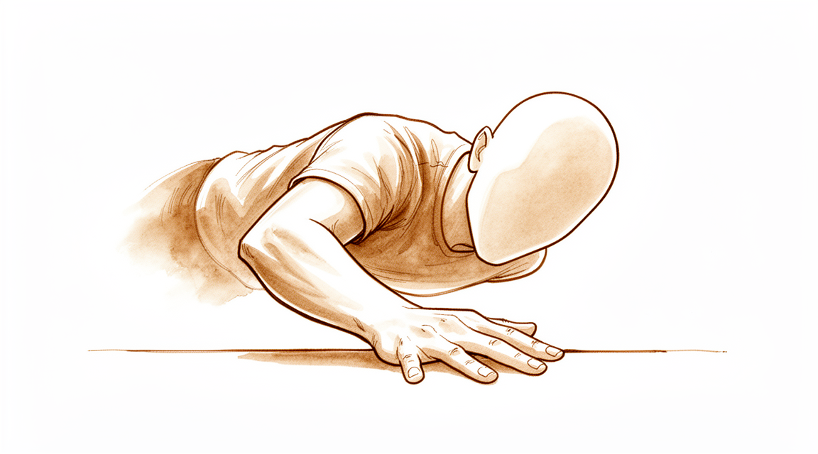
After you are asleep, your surgeon will perform the procedure through a single cut over the wrist or through tiny punctures in the skin. You will wake up in the recovery area once the surgery is finished. You will rest there while the team monitors you before you go home.
What the operation involves
Your surgeon will treat your scaphoid fracture, a small bone in your wrist. Because this bone has a unique shape with a narrow middle section called the waist, your surgeon will plan the repair carefully to match its complex structure. In some cases, the injury also involves damaged ligaments or cartilage, which your surgeon will address during the procedure.
For nondisplaced fractures, your surgeon may use a percutaneous approach. This means placing a screw through the skin without making a large cut. This method holds the bone in place to allow it to heal. If the fracture is more complex, your surgeon might use an open approach with a single incision to access the bone directly. The goal is to secure the fracture firmly so you can begin moving your wrist early.
Your surgeon will use screws or plates to stabilize the bone. These metal devices keep the broken pieces aligned while healing occurs. Once the fixation is secure, the skin is closed with stitches or glue, and a dressing is applied. Because scaphoid fractures can be hard to spot at first, your surgeon has used specific imaging to ensure the repair targets the correct area.
After the operation
You will wake up in a recovery ward where your pain will be managed. Your surgeon may use a small incision or a percutaneous approach through the skin. You will have a dressing, and possibly a sling or brace, to protect your wrist. You can usually go home the same day, but an overnight stay is possible. Because you will be groggy, someone must stay with you for the first 24 hours. You will start moving gently as soon as you are comfortable.
Recovery
Your hand may feel sore and swollen for the first few days. This is normal as your body heals. You will likely wear a cast, splint, or brace to keep your wrist steady. Keep your hand elevated above your heart when resting to help reduce the swelling.
You will begin gentle movement exercises as soon as your surgeon says it is safe. These exercises help restore motion without stressing the healing bone. If you have a cast or brace, you will wear it during daily tasks like eating or dressing. You can usually sleep with your hand supported on a pillow to stay comfortable.
Your surgeon may use a small incision or a needle through the skin to place the fixation. This approach helps protect the skin while securing the bone. As your pain settles and movement returns, you will gradually do more activities. Your timeline may differ from others; your surgeon and physiotherapist will guide your specific steps.
What can go wrong
Most patients do well, but problems can occasionally happen. Your surgeon and the team monitor you closely to spot any issue early.
Sometimes, a break in a nearby wrist bone is missed when you first come in. You might notice that pain in your wrist does not get better as expected. If this happens, tell your surgeon right away so they can check for other injuries.
In some cases, you may have damage to the soft tissues or cartilage inside the wrist along with the break. You might feel a deep ache or a sense of instability that does not match the injury you thought you had. Bring this up at your next review so your surgeon can look closer.
If you have metal hardware inside your wrist, sharp edges or screw heads can sometimes rub against nearby areas. You might feel a new, sharp pain or notice a lump that moves under the skin. If you feel something poking or rubbing where it should not, contact the clinic to have it checked.
The complications table on this page lists typical rates if you want the specifics.
When to call us
Call us if you have a fever, increasing redness, or discharge from your wound. Go to emergency if you feel sudden severe pain, new numbness, or cannot move your hand. Seek urgent help for calf swelling or shortness of breath. If you had a wrist injury but standard X-rays were normal, call us if pain persists. We may need to repeat imaging in 10 to 14 days to find a hidden fracture.
Evidence & references
title: "Scaphoid Fixation" slug: scaphoid-fixation region: wrist audience: patient mesh_terms: ["Scaphoid Bone/surgery", "Fracture Fixation", "Internal", "Bone Screws", "percutaneous scaphoid fixation", "Herbert screw"] article_count: 21 model_used: qwen3.5-35b-a3b-q8 generated_at: '2026-05-18T13:42:31+00:00' key_articles: - title: "Evaluation and Management of Carpal Fractures Other Than the Scaphoid" ref_num: 1 evidence_tier: paper evidence_level: 5 doi: 10.5435/jaaos-d-20-00062 year: 2020 - title: "Percutaneous Screw Fixation or Cast Immobilization for Nondisplaced Scaphoid Fractures" ref_num: 2 evidence_tier: paper evidence_level: 1 doi: 10.2106/00004623-200104000-00001 year: 2001 - title: "Coronal plane partial articular fractures of the distal femoral condyle" ref_num: 3 evidence_tier: paper evidence_level: 4 doi: 10.1302/0301-620x.95b9.30656 year: 2013 - title: "3D computational anatomy of the scaphoid and its waist for use in fracture treatment" ref_num: 4 evidence_tier: paper evidence_level: 4 doi: 10.1186/s13018-021-02330-8 year: 2021 - title: "Incidence of Ligamentous and Other Injuries Associated With Scaphoid Fractures During Arthroscopically Assisted Reduction and Percutaneous Fixation" ref_num: 5 evidence_tier: paper evidence_level: 4 doi: 10.1016/j.arthro.2008.01.003 year: 2008 - title: "Arthroscopic treatment of a juvenile tillaux fracture" ref_num: 6 evidence_tier: paper evidence_level: 4 doi: 10.1007/s00167-006-0234-3 year: 2006 - title: "Fixation of Anterior Pelvic Ring Injuries" ref_num: 7 evidence_tier: paper evidence_level: 4 doi: 10.5435/jaaos-d-17-00839 year: 2019 - title: "Recurrent episodes of micturition with expulsion of symphyseal plate screws following pelvic ring fixation: case report" ref_num: 8 evidence_tier: case_report evidence_level: 5 doi: 10.1186/s12891-015-0581-7 year: 2015 - title: "Locking Plate Placement with Unicortical Screw Fixation Adjunctive to Intramedullary Rodding in Long Bones of Patients with Osteogenesis Imperfecta" ref_num: 9 evidence_tier: paper evidence_level: 4 doi: 10.2106/jbjs.n.01185 year: 2015 - title: "A Biomechanical Comparison of Periprosthetic Femoral Fracture Fixation in Normal and Osteoporotic Cadaveric Bone" ref_num: 10 evidence_tier: paper evidence_level: 5 doi: 10.1016/j.arth.2011.08.019 year: 2012 - title: "Biomechanical characteristics of fixation methods for floating pubic symphysis" ref_num: 11 evidence_tier: paper evidence_level: 5 doi: 10.1186/s13018-017-0541-z year: 2017 - title: "Surgical Hip Dislocation Is a Reliable Approach for Treatment of Femoral Head Fractures" ref_num: 15 evidence_tier: paper evidence_level: 4 doi: 10.1007/s11999-015-4352-4 year: 2015 - title: "Effect of different multiplanar reformation algorithms on image quality of intraoperative three-dimensional fluoroscopy" ref_num: 37 evidence_tier: paper evidence_level: 4 doi: 10.1177/1753193419848963 year: 2019 synthesis_version: "v2" verifier_status: skipped
Overview
- Fractures of the carpus other than the scaphoid are frequently missed on initial presentation [1].
- Diagnosis of carpal fractures other than the scaphoid requires a high index of suspicion with tailored imaging [1].
- The specific indications for percutaneous screw fixation of nondisplaced scaphoid fractures must be determined in larger randomized, prospective studies [2].
- The risks and benefits of percutaneous screw fixation of nondisplaced scaphoid fractures must be determined in larger randomized, prospective studies [2].
- The complex scaphoid anatomy with its waist might alter the strategy of fracture fixation [4].
- The complex scaphoid anatomy with its waist might alter education regarding fracture treatment [4].
- The complex scaphoid anatomy with its waist might alter research regarding fracture treatment [4].
- In a series of patients with acute scaphoid fractures, 15 of 24 presented with associated ligamentous and/or chondral/osteochondral injuries [5].
Anatomy & Pathophysiology
- Scaphoid fractures account for almost 75% of all carpal fractures [13].
- Scaphoid fractures are rare in children and in the elderly [13].
- The scaphoid is a small, irregular S-shaped tubular bone located in the proximal carpal row on the radial aspect of the wrist [18].
- The scaphoid lies entirely within the wrist joint at a 45-degree plane to the longitudinal and horizontal axis of the wrist [18].
- The scaphoid articulates with the trapezium and trapezoid on its distal surface, the radius on its proximal/lateral surface, and the capitate and lunate on its medial surface [18].
- The proximal articular surface of the scaphoid is convex and articulates with the radius [18].
- The capitate head articulates with a sulcus on the scaphoid located across the radial articular surface, providing a socket-like fit [18].
- The scaphoid gently pronates and flexes distally such that the distal pole sits ulnarly angulated relative to the proximal pole [18].
- Two distinct articular facets for the trapezium and trapezoid are present at the distal articular surface, forming the scaphotrapeziotrapezoid (STT) joint [18].
- Over 80% of the scaphoid surface is covered with articular cartilage [18].
- The scaphoid has a reduced capacity for periosteal healing due to extensive articular cartilage coverage and an increased tendency for delayed union and nonunion [18].
- The scaphoid is ridged across its nonarticular dorsoradial surface, along which critical dorsal ridge vessels traverse [18].
- The ridge on the scaphoid is the insertion point for both the dorsal component of the scapholunate and intercarpal ligaments [18].
- Ligamentous attachments of the scaphoid are predominantly found on the nonarticular dorsoradial surface [18].
- Short intrinsic ligaments provide stability to the scaphoid through attachments to other carpal bones, particularly the lunate, and merge with extrinsic ligaments and the wrist capsule [18].
- The radioscapocapitate ligament does not attach to the scaphoid bone itself but crosses the waist, acting as a sling allowing rotation [18].
- There are no tendon attachments to the scaphoid [18].
- The scaphoid acts as a midcarpal joint "bridge" linking and synchronizing the motions of the proximal and distal carpal rows [18].
- Motion of the scaphoid includes rotation proximally and gliding distally while providing stability to the midcarpal joint [18].
- The blood supply of the scaphoid arises from the dorsal distal pole, resulting in a poor blood supply to the proximal pole [13].
- The proximal pole has a poor blood supply, is less likely to heal than the distal pole, and may undergo avascular necrosis [13].
- The vascular supply of the scaphoid originates from two vascular pedicles from the scaphoid branches of the radial artery [18].
- The dorsal branch enters via small foramina along the spiral groove and dorsal ridge of the scaphoid and supplies 70% to 80% of the scaphoid proximally, including the proximal pole [18].
- The volar branch enters via the scaphoid tubercle and supplies the remaining 20% to 30% of the distal scaphoid [18].
- The waist of the scaphoid has been shown to have minimal or no perforating vasculature [18].
- No vessels perforate the proximal dorsal cartilaginous area or through the scapholunate ligament [18].
- Proximal fractures are associated with at least temporary disruption of the interosseous blood supply to the proximal pole [18].
- Only 67% of scaphoid bones have arterial foramina throughout their length, including the distal, middle, and proximal thirds [23].
- In 13% of scaphoid bones, blood supply is predominantly in the distal third [23].
- In 20% of scaphoid bones, most arterial foramina are in the waist area with no more than a single foramen near the proximal third [23].
- One third of scaphoid fractures occurring in the proximal third may be without adequate blood supply [23].
- The prevalence of osteonecrosis can be 35% in fractures at the proximal pole level [23].
- Fractures in the proximal pole take longer to heal and usually have higher rates of nonunion [23].
- Vessels enter the scaphoid from the radial artery laterovolarly, dorsally, and distally [23].
- The laterovolar and dorsal systems share in the blood supply to the proximal two thirds of the scaphoid [23].
- Vascularity of the proximal pole and 70% to 80% of the interosseous circulation are provided through branches of the radial artery entering through the dorsal ridge [23].
- In the distal tuberosity region, 20% to 30% of the bone receives its blood supply from volar branches of the radial artery [23].
- The usual mechanism of scaphoid fracture is forced hyperextension of the wrist [13].
- Scaphoid fractures are caused by a fall on the outstretched palm, resulting in severe hyperextension and slight radial deviation of the wrist [23].
- Hyperextension past 95 degrees is the usual position of injury for scaphoid fractures [29].
- Other mechanisms postulated to produce scaphoid fractures include axial loading and hyperflexion of the wrist [29].
- With the hyperextension mechanism, a fracture of the scaphoid usually begins at the volar waist with a tensile failure [29].
- Forces in hyperextension fractures propagate to the dorsal surface with compression loading until failure occurs [29].
- In cadaveric studies, wrists placed in extreme dorsiflexion and ulnar deviation produced fractures through the scaphoid waist as the scaphoid impinged on the dorsal rim of the radius [29].
- Proximal scaphoid fractures resulted from dorsal subluxation during forced hyperextension [29].
- Of scaphoid fractures, 60% to 80% occur at the scaphoid waist or midportion [23].
- Seventeen percent of patients with scaphoid fractures have other fractures of the carpus and forearm [23].
- Associated injuries include transscaphoid perilunar dislocations, fractures of the trapezium, Bennett fractures, fractures of the radial head, dislocations of the lunate, and fractures at the distal end of the radius [23].
- Fractures tend to occur at the waist partly because the radioscapocapitate (RSC) ligament acts as a fulcrum over which the scaphoid waist fractures [12].
- Snuffbox tenderness applies predominantly to waist fractures, which represent 70% of scaphoid fractures [12].
- The second most common type of scaphoid fracture is a proximal pole fracture, at 20% [12].
- The least common type of scaphoid fracture is a distal pole fracture, at 10% [12].
- Nonunion occurs in 10% to 15% of all scaphoid fractures [29].
- Nonunion rates for nondisplaced waist fractures treated with casting are 5% to 12% [29].
- Nonunion rates for displaced scaphoid fractures treated nonoperatively reach 50% [29].
- The risk of nonunion increases with a delay of treatment for more than 4 weeks [29].
- The risk of nonunion increases with proximal pole fractures [29].
- The risk of nonunion increases with fracture displacement greater than 1 mm [29].
- The risk of nonunion increases with osteonecrosis [29].
- The risk of nonunion increases with tobacco use [29].
- The risk of nonunion increases with associated carpal instability, specifically dorsal intercalated segmental instability (DISI) with a scapholunate angle greater than 60 degrees and a capitolunate angle greater than 15 degrees [29].
- DISI is secondary to humpback deformity, defined as flexion with an intrascaphoid angle greater than 45 degrees, whereas the normal intrascaphoid angle is 24 degrees [29].
- Untreated displaced fractures of the waist will usually angulate as the volar bone is reabsorbed, yielding a "humpback" flexion deformity of the scaphoid [29].
- The resultant radial column shortening and extension of the proximal scaphoid pole releases the lunate to rotate into DISI under the influence of the attached triquetrum [29].
- Untreated scaphoid nonunion will predictably progress to arthritic change, termed scaphoid nonunion advanced collapse (SNAC) [29].
- In SNAC, arthritic change arises at the radial styloid articulation with the distal scaphoid pole (stage I) [29].
- In SNAC, degeneration of the scaphocapitate joint follows (stage II) [29].
- In SNAC, degeneration of the midcarpal joint occurs ultimately (stage III) [29].
- Arthritic changes have been found in 97% of patients assessed at least 5 years after injury [29].
- The degree of arthritic changes in SNAC is proportionate to the duration of nonunion [29].
- Patients with untreated scaphoid nonunion generally present with escalating mechanical pain and limitations in range of motion [29].
- In a review of 30-year follow-up results, 10% of patients treated with thumb spica short-arm casts developed nonunion [29].
- Of those who developed nonunion in the 30-year review, 60% demonstrated radiographic evidence of radiocarpal osteoarthritis [29].
- In the 30-year review, only 2% of the healed group demonstrated degenerative change [29].
- The scaphoid bone is located in the proximal carpal row on the radial aspect of the wrist and is a small, irregular S-shaped tubular bone [18].
- The scaphoid spans the proximal and distal carpal rows and acts as a "tie-rod" to coordinate smooth carpal motion [29].
- The scaphoid derives its name from its peculiar boat- or skiff-shaped contour [29].
- Fractures of the carpus other than the scaphoid are frequently missed on initial presentation and require a high index of suspicion with tailored imaging for diagnosis [1].
- In a series of 24 patients with acute scaphoid fractures, 15 presented with associated ligamentous and/or chondral/osteochondral injuries [5].
- The complex scaphoid anatomy with its waist might alter the strategy of fracture fixation, education, and research [4].
- The patient usually presents with pain on the radial side of the wrist [12].
- There may be swelling on the radial side of the wrist in scaphoid fractures [12].
- There is usually a history of trauma, such as falling on an outstretched hand, collision of the wrist against a person or heavy obstacle, or a direct blow against an object [12].
- There may be limited range of motion and pain when applying extended wrist loading or positioning the wrist in extreme positions of flexion or extension [12].
- Wrists with acute scaphoid fractures may have swelling and bruising in the radial aspect of the wrist [12].
- Wrists with chronic scaphoid injury may have swelling in the dorsoradial wrist [12].
- To palpate the anatomic snuffbox for the waist examination, palpate just distal to the radial styloid in the "soft spot" [12].
- The distal pole of the scaphoid should be palpated at the scaphoid tubercle on the palmar aspect of the wrist [12].
- The proximal pole is palpated dorsally in line with the second ray just distal to the dorsal radius lip [12].
- The scapholunate ligament is in line between the second and third rays just distal to the dorsal radius lip and corresponds to the 3-4 wrist arthroscopy portal [12].
- The proximal pole is just radial to the scapholunate ligament/3-4 portal area [12].
- Pain on longitudinal compression of the thumb (scaphoid axial compression test) is a sign of scaphoid fracture [12].
- If all three tests of anatomic snuffbox tenderness, scaphoid tubercle tenderness, and scaphoid axial compression test are positive, there is 87% to 100% sensitivity and 74% specificity for scaphoid fracture [12].
- There may be slight fullness in the anatomical snuffbox in scaphoid fractures [13].
- Precisely localized tenderness in the anatomical snuffbox is an important diagnostic sign for scaphoid fracture [13].
- Examination for scaphoid fracture must include pressure backwards over the scaphoid tubercle, palpation over the proximal pole, and telescoping of the thumb base [13].
- If any of the specific examination signs for scaphoid fracture are positive, the suspicion for a scaphoid fracture should be high [13].
- X-rays for scaphoid fractures should include AP, lateral, and two oblique views [13].
- Even with standard X-rays, the fracture may not be seen in the first few days after injury [13].
- Two weeks later, the scaphoid fracture break is usually much clearer due to bone resorption at the fracture site and slight displacement of fragments [13].
- The scaphoid fracture crack is usually transverse through the narrowest part of the bone (the waist), but it may be more proximal or more distal [13].
- One should always look for signs of associated carpal displacement when evaluating scaphoid fractures [13].
- A CT scan is more sensitive for diagnosing a scaphoid fracture than X-rays [13].
- A CT scan is particularly useful in confirming the alignment of bone fragments if surgery is planned [13].
- A CT scan is useful to confirm whether a scaphoid fracture has united [13].
- MRI is the definitive way to confirm or exclude a diagnosis of scaphoid fracture if the technique is available [13].
- If X-rays look normal but clinical features are suggestive of a fracture, the patient must not be discharged [13].
- The usual advice for suspected scaphoid fracture with normal initial X-rays is to return for a second X-ray 2 weeks later [13].
- Meanwhile, the wrist should be immobilized in a cast extending from the upper forearm to just short of the metacarpophalangeal joints of the fingers, but incorporating the proximal phalanx of the thumb [13].
- The wrist should be held dorsiflexed and the thumb forwards in the "glass-holding" position (the so-called scaphoid plaster) [13].
- An alternative to casting for suspected scaphoid fracture is to arrange an MRI scan or, if not available, a CT scan [13].
- At least four X-rays of possible scaphoid fractures should be taken [13].
- Even then, X-rays might be normal initially in scaphoid fractures [13].
- If any doubt exists regarding a scaphoid fracture, the patient should be placed in plaster and either re-X-rayed in 2 weeks or an MRI scan obtained [13].
- The initial AP view of a scaphoid fracture often fails to show the fracture [13].
- A CT scan is useful for showing the configuration of a scaphoid fracture [13].
- The structures causing pain on the ulnar side of the wrist include the distal radioulnar joint (DRUJ), the distal ulnocarpal joint, and the triangular fibrocartilage complex (TFCC) [19].
- The TFCC includes the dorsal and volar radioulnar ligaments, ulnar collateral ligament, meniscal homologue, articular disc, ulnolunate ligaments, ulnotriquetral ligaments, and extensor carpi ulnaris sheath [19].
- The deep and superficial fibers of the TFCC begin on the ulnar side of the lunate fossa of the radius [19].
- The deep fibers of the TFCC attach ulnarly at the head of the ulna called the "fovea" [19].
- The superficial fibers of the TFCC attach to the ulnar styloid tip where it joins with the ulnar collateral ligaments [19].
- Articular surface contact in the shallow sigmoid notch accounts for about 20% of DRUJ stability [19].
- Articular surface contact in the shallow sigmoid notch allows dorsopalmar translation of about 1 cm with the forearm in neutral position [19].
- During forearm rotation, the ulnar head at its articulation with the sigmoid notch appears to move from dorsal and distal in full pronation to proximal and palmar in full supination [19].
- Additional DRUJ stability is provided through the dorsal and palmar margins and their attachments to the radioulnar ligaments [19].
- The extensor carpi ulnaris sheath and part of the distal radioulnar ligaments attach to the ulnar styloid [19].
-
The ulnar styloid extends 2 to 6 mm distal to the ulnar head [19].
Classification
- Fractures of the carpus other than the scaphoid are frequently missed on initial presentation [1].
- Diagnosis of carpal fractures other than the scaphoid requires a high index of suspicion with tailored imaging [1].
- The specific indications for percutaneous screw fixation of nondisplaced scaphoid fractures must be determined in larger randomized, prospective studies [2].
- The specific risks and benefits of percutaneous screw fixation of nondisplaced scaphoid fractures must be determined in larger randomized, prospective studies [2].
- The complex scaphoid anatomy with its waist might alter the strategy of fracture fixation [4].
- The complex scaphoid anatomy with its waist might alter the strategy of education and research [4].
- In a series of acute scaphoid fractures, 15 of 24 patients presented with associated ligamentous and/or chondral/osteochondral injuries [5].
Clinical Presentation
- Fractures of the carpus other than the scaphoid are frequently missed on initial presentation and require a high index of suspicion with tailored imaging for diagnosis [1].
- Scaphoid fractures account for almost 75% of all carpal fractures but are rare in children and in the elderly [13].
- Scaphoid fractures are the most common carpal injury in the pediatric population, accounting for approximately 3% of hand and carpal fractures and 0.34% of all fractures in children [34].
- The usual mechanism of scaphoid fracture is forced hyperextension of the wrist, often following a fall onto an outstretched hand [13].
- Almost 90% of patients with scaphoid fractures recall a hyperextension injury [31].
- Patients classically present with radial-sided wrist pain [12].
- Swelling may be present on the radial side of the wrist in acute fractures [12].
- Chronic scaphoid injuries may present with swelling in the dorsoradial wrist [12].
- There may be limited range of motion and pain when applying extended wrist loading or positioning the wrist in extreme positions of flexion or extension [12].
- Fractures tend to occur at the waist partly because the radioscaphocapitate (RSC) ligament acts as a fulcrum over which the scaphoid waist fractures [12].
- Waist fractures represent 70% of scaphoid fractures [12].
- The second most common type of scaphoid fracture is a proximal pole fracture, at 20% [12].
- The least common type of scaphoid fracture is a distal pole fracture, at 10% [12].
- "Snuffbox tenderness" applies predominantly to waist fractures [12].
- To palpate the anatomic snuffbox for the waist examination, palpate just distal to the radial styloid in the "soft spot" [12].
- The distal pole of the scaphoid is palpated at the scaphoid tubercle on the palmar aspect of the wrist [12].
- With radial deviation of the wrist, the distal pole prominence should move palmarly toward the examiner's thumb [12].
- The proximal pole is palpated dorsally in line with the second ray just distal to the dorsal radius lip [12].
- The proximal pole is just radial to the scapholunate ligament/3-4 portal area [12].
- Pain on longitudinal compression of the thumb (scaphoid axial compression test) is a sign of scaphoid fracture [12].
- If all three tests of anatomic snuffbox tenderness, scaphoid tubercle tenderness, and scaphoid axial compression test are positive, there is 87% to 100% sensitivity and 74% specificity for scaphoid fracture [12].
- There may be slight fullness in the anatomical snuffbox [13].
- Precisely localized tenderness in the anatomical snuffbox is an important diagnostic sign [13].
- Examination for scaphoid fracture must include pressure backwards over the scaphoid tubercle, palpation over the proximal pole, and telescoping of the thumb base [13].
- If any of these specific examination maneuvers are positive, the suspicion for a scaphoid fracture should be high [13].
- Standard four-view radiographs are subsequently used to confirm the diagnosis of scaphoid fracture [31].
- Up to 30% to 40% of scaphoid fractures are not identified on initial assessment and investigation with standard four-view radiographs [31].
- Patients subsequently found to have a fracture confirmed on repeated assessment and radiologic imaging, most frequently at 10 to 14 days after injury, are said to have had an occult fracture of the scaphoid [31].
- No single clinical sign has been found to be adequately sensitive or specific for scaphoid fracture diagnosis [31].
- Anatomical snuffbox tenderness has a sensitivity of 87–100% and specificity of 3–98% [31].
- Axial compression of the thumb has a sensitivity of 48–100% and specificity of 22–97% [31].
- Scaphoid tubercle tenderness has a sensitivity of 82–100% and specificity of 17–57% [31].
- Pain on ulnar deviation has a sensitivity of 67–100% and specificity of 17–60% [31].
- Pain on radial deviation has a sensitivity of 67–90% and specificity of 31–42% [31].
- Reduced range of movement of the thumb has a sensitivity of 65–66% and specificity of 38–59% [31].
- Thumb–index finger pinch has a sensitivity of 75–79% and specificity of 44–76% [31].
- In a study of 246 patients with a suspected fracture of the scaphoid, anatomical snuffbox tenderness was found to have a sensitivity of 90% and a specificity of 40% [31].
- In the same study, scaphoid tubercle tenderness had a sensitivity of 87% and specificity of 57% [31].
- A prospective analysis of 73 patients with a suspected scaphoid fracture found that pain on ulnar deviation of the pronated wrist had a negative predictive value (NPV) of 100% [31].
- A combination of anatomical snuffbox tenderness, scaphoid tubercle tenderness, and anatomical snuffbox pain on longitudinal compression of the thumb generated a sensitivity of 100% and a specificity of 74% [31].
- The combination of three clinical signs yielding 100% sensitivity and 74% specificity was valid only for the first 24 hours after injury [31].
- Pain on thumb–index finger pinch and anatomical snuffbox pain on pronation of the forearm were most suggestive of a true scaphoid fracture [31].
- The best predictors of fracture within 72 hours of injury were the absence of pain on ulnar deviation of the wrist and pain on thumb–index finger pinch [31].
- Scaphoid tubercle tenderness was most predictive at week 2 [31].
- A clinical scaphoid score (CSS) using three clinical tests (tenderness in the ASB with the wrist in ulnar deviation, tenderness over the scaphoid tubercle, and pain upon longitudinal compression of the thumb) identified that patients with a CSS of 4 or higher require an MRI [31].
- Radiographs are often negative at initial presentation in approximately 25% of scaphoid fracture cases [35].
- In chronic injuries, athletes may complain of an inability to perform a push-up [35].
- Tenderness over the anatomic snuffbox or pain with resisted pronation prevents the surgeon from ruling out a scaphoid fracture in athletes [35].
- A scaphoid view, with the wrist in 30° of extension and 20° of ulnar deviation, or a clenched-fist PA view should be obtained in addition to standard wrist radiographs [35].
- MRI is useful if radiographs are inconclusive in athletes [35].
- MRI is used to assess osteonecrosis of the proximal pole of the scaphoid [35].
- MRI can help assess for a scapholunate ligament injury, another common cause of radial-sided wrist pain in the athlete after a fall [35].
- Associated ligamentous and/or chondral/osteochondral injuries were present in 15 of 24 patients with acute scaphoid fractures [5].
- Associated injuries like distal radius fracture, transscaphoid perilunate dislocations, ulnar styloid fractures, capitate fractures, and bilateral injuries can be present in up to 10% of patients [34].
- Scaphoid fractures are an often missed injury [35].
- Fractures treated in less than 28 days from injury result in a 5% nonunion rate [35].
- If treatment is delayed longer than 28 days, the nonunion rate increases to 28% [35].
- X-rays should include AP, lateral and two oblique views; even then, the fracture may not be seen in the first few days after the injury [13].
- Two weeks later, the break is usually much clearer on X-ray due to bone resorption at the fracture site and slight displacement of fragments [13].
- The crack is usually transverse through the narrowest part of the bone (the waist), but it may be more proximal or more distal [13].
- A CT scan is more sensitive for diagnosing a scaphoid fracture than X-rays [13].
- A CT scan is particularly useful in confirming the alignment of the bone fragments if surgery is planned [13].
- A CT scan is useful to confirm whether the fracture has united or not [13].
- MRI is the definitive way to confirm or exclude a diagnosis of scaphoid fracture if the technique is available [13].
- If radiographs are equivocal, ultrasonography can be used to diagnose scaphoid fracture [34].
- Radiography can be repeated after 2 weeks of immobilization to assess for evidence of healing fracture [34].
- An examination by a specialist after the injury has become less painful allows for a more accurate physical examination and thus substantially increases the sensitivity of detecting a scaphoid fracture [32].
- If the probability of a fracture remains unacceptable and new scaphoid specific radiographs are also normal, advanced imaging (typically CT or MRI) can be used to attempt to exclude a fracture [32].
- The higher the pretest odds of a fracture, the more likely an imaging diagnosis of a fracture will correlate with a true fracture [32].
- The lower the pretest odds (i.e., "rule out" rather than "confirm"), the less likely that a radiologic diagnosis of a fracture will correspond with a true fracture [32].
- In children, traditional thinking was that scaphoid fractures involved the distal pole with excellent healing rates, but now the majority of fractures occur at the waist [34].
- Children with scaphoid fractures may present late due to subtle pain and swelling in the anatomic snuffbox [34].
- Nondisplaced, acute scaphoid fractures treated in short arm thumb spica casts for 6 to 12 weeks have a reported union rate of 90% [34].
- Chronic fractures and osteonecrosis are independent predictors of worse functional outcomes in pediatric scaphoid fractures [34].
- 95% of all pediatric patients with scaphoid fractures reported functional status better than or equal to the general population per median DASH score [34].
- The median Modified Mayo Wrist Score (MMWS) for both surgical and nonsurgical pediatric patients represented excellent functional outcome with no difference in outcomes for the two groups [34].
Investigations
- Fractures of the carpus other than the scaphoid are frequently missed on initial presentation and require a high index of suspicion with tailored imaging for diagnosis [1].
- Scaphoid fractures account for almost 75% of all carpal fractures [13].
- Scaphoid fractures are rare in children and in the elderly [13].
- The usual mechanism of scaphoid fracture is forced hyperextension of the wrist [13].
- The most common mechanism of injury for scaphoid fractures is a fall onto the outstretched hand with the forearm pronated [26].
- Scaphoid fractures occur in three anatomical locations: distal tubercle, waist, and proximal pole [13].
- Fractures at the waist represent 70% of scaphoid fractures [12].
- Proximal pole fractures represent 20% of scaphoid fractures [12].
- Distal pole fractures represent 10% of scaphoid fractures [12].
- Fractures tend to occur at the waist partly because the radioscaphocapitate (RSC) ligament acts as a fulcrum over which the scaphoid waist fractures [12].
- In children, fractures of the distal third of the scaphoid (transverse distal pole and tuberosity) are the most common [26].
- In children, peak age for scaphoid fracture incidence is 15 to 19 years [26].
- Type I scaphoid injuries in children younger than 8 years are usually chondral [26].
- Type II scaphoid injuries in children between 8 and 11 years are usually osteochondral [26].
- Type III scaphoid injuries in children older than 12 years are more "adult-like" because the scaphoid is ossified [26].
- The blood supply of the scaphoid arises from the dorsal distal pole [13].
- The proximal pole has a poor blood supply and is less likely to heal than the distal pole [13].
- Avascular necrosis may occur in the proximal pole of the scaphoid [13].
- Patients with scaphoid fractures usually present with pain on the radial side of the wrist [12].
- Swelling may be present on the radial side of the wrist in acute scaphoid fractures [12].
- Limited range of motion is common in scaphoid fractures [12].
- Pain may occur when applying extended wrist loading or positioning the wrist in extreme positions of flexion or extension [12].
- Wrists with chronic scaphoid injury may have swelling in the dorsoradial wrist [12].
- "Snuffbox tenderness" applies predominantly to waist fractures [12].
- To palpate the anatomic snuffbox for the waist, palpate just distal to the radial styloid in the "soft spot" [12].
- The distal pole of the scaphoid is palpated at the scaphoid tubercle on the palmar aspect of the wrist [12].
- The proximal pole of the scaphoid is palpated dorsally in line with the second ray just distal to the dorsal radius lip [12].
- The proximal pole is just radial to the scapholunate ligament/3-4 portal area [12].
- Pain on longitudinal compression of the thumb (scaphoid axial compression test) is a sign of scaphoid fracture [12].
- If all three tests of anatomic snuffbox tenderness, scaphoid tubercle tenderness, and scaphoid axial compression test are positive, there is 87% to 100% sensitivity and 74% specificity for scaphoid fracture [12].
- Clinical signs of scaphoid fracture in children include dorsal swelling of the wrist, tenderness in the anatomic snuffbox, swelling of the distal part of the radius, and painful dorsiflexion of the wrist or extension of the thumb [26].
- Plain radiography is approximately 50% sensitive for the detection of a scaphoid fracture [26].
- Plain radiography is less than 50% sensitive for the detection of other carpal bones fractures [26].
- Up to 30% of patients with suspected scaphoid fracture may have positive follow-up radiographs after 2 weeks [26].
- X-rays for scaphoid fractures should include AP, lateral, and two oblique views [13].
- X-rays should include anteroposterior, lateral, and scaphoid views with the wrist in ulnar deviation [26].
- Fractures may not be seen on X-rays in the first few days after injury [13].
- Two weeks later, the fracture is usually much clearer on X-ray due to bone resorption at the fracture site and slight displacement of fragments [13].
- The crack is usually transverse through the narrowest part of the bone (the waist) on X-ray [13].
- A CT scan is more sensitive for diagnosing a scaphoid fracture than X-rays [13].
- A CT scan is particularly useful in confirming the alignment of bone fragments if surgery is planned [13].
- A CT scan is useful to confirm whether a scaphoid fracture has united [13].
- MRI is the definitive way to confirm or exclude a diagnosis of scaphoid fracture if available [13].
- MRI is more sensitive than CT for diagnosing scaphoid fractures [26].
- A normal MRI study as early as 2 days after injury has a negative predictive value of 100% for scaphoid fracture [26].
- In a series of 24 patients with acute scaphoid fractures, 15 presented with associated ligamentous and/or chondral/osteochondral injuries [5].
- Neglected scaphoid nonunion is associated with osteonecrosis and progressive radiocarpal and midcarpal arthritis [28].
- A diagnosis of osteonecrosis can be challenging because of the limited sensitivity of imaging modalities, including contrast-enhanced MRI [28].
- The presence of large cavitary lesions or cysts with bone resorption around the midwaist to proximal pole suggests compromised blood supply [28].
- A recent study found that the healing potential of a scaphoid nonunion is not dependent on the presence of proximal pole vascularity [28].
- In a series of 35 scaphoid nonunions with more than half found to have impaired vascularity on intraoperative histopathologic analysis, 33 of 35 nonunions healed with curettage, nonvascularized autogenous bone grafting, and headless screw fixation [28].
- Twelve of 14 patients with fibrous scaphoid nonunions treated with screw fixation alone experienced healing at 4.4-month follow-up [28].
- The two persistent nonunions in the fibrous nonunion series occurred in proximal pole fractures more than 1 year after injury [28].
- A stable scaphoid nonunion without deformity or osteonecrosis can be successfully managed without bone grafting [28].
- An unstable nonunion requires bone grafting to restore height and correct carpal malalignment, principally dorsal intercalated segment instability [28].
- Twelve scaphoid waist nonunions with humpback deformity were successfully managed with a retrograde screw and ipsilateral distal radius cancellous bone graft [28].
- A systematic review found that both cancellous and corticocancellous bone grafting led to reliable union (95% or 92%, respectively) [28].
- Cancellous grafting required less time to union than corticocancellous grafting [28].
- Corticocancellous grafting led to more consistent deformity correction than cancellous grafting [28].
- The 1,2 intercompartmental supraretinacular artery was commonly used as a vascularized pedicled bone graft for scaphoid nonunion [28].
- The Mathoulin pedicled graft from the volar distal radius has been used with good clinical results for scaphoid nonunion [28].
- A free medial femoral condyle vascularized graft has a reported union rate of 94% for scaphoid nonunion [28].
- The free medial femoral condyle vascularized graft demonstrated a union rate of 84% in revision scenarios [28].
- A retrospective review found CT-confirmed healing in 15 of 16 scaphoids consecutively treated with a medial femoral trochlea flap for proximal one-fifth nonunions [28].
- Acutrak screw fixation led to a significantly higher union rate than Herbert screw fixation (94% versus 71%) in a review of 132 scaphoid nonunions [28].
- Acutrak screw fixation led to more accurate central axis screw placement than Herbert screw fixation [28].
- Optimization of post-processing algorithms for intraoperative three-dimensional fluoroscopy may increase image quality for assessing implant positioning [37].
- Limitations in evaluating fracture reduction quality still exist with intraoperative three-dimensional fluoroscopy [37].
- The complex scaphoid anatomy with its waist might alter the strategy of fracture fixation, education, and research [4].
Treatment
- Fractures of the carpus other than the scaphoid are frequently missed on initial presentation and require a high index of suspicion with tailored imaging for diagnosis [1].
- The specific indications for and the risks and benefits of percutaneous screw fixation of nondisplaced scaphoid fractures must be determined in larger randomized, prospective studies [2].
- The complex scaphoid anatomy with its waist might alter the strategy of fracture fixation, education and research [4].
- In a series of patients with acute scaphoid fractures, 15 of 24 presented with associated ligamentous and/or chondral/osteochondral injuries [5].
- Arthroscopic treatment allows accurate reconstruction of the weight bearing surface of the joint and secure internal fixation of the fracture [6].
- Unicortical locking plate fixation effectively supplements intramedullary rod fixation in selected cases of osteogenesis imperfecta [9].
- Constructs with locking and nonlocking screws demonstrated equivalent loads at failure and were superior in load at failure compared with cables [10].
- Subcutaneous fixation had satisfactory outcomes, with sub-rod offering good anti-compression and sub-plate providing favorable anti-rotational capacity [11].
Complications
- Fractures of the carpus other than the scaphoid are frequently missed on initial presentation [1].
- In a series of 24 patients with acute scaphoid fractures, 15 presented with associated ligamentous and/or chondral/osteochondral injuries [5].
- The presence of protruding metal prominences, even smooth ones like a plate corner or screw head, might endanger the bladder [8].
- Surgical dislocation for femoral head fractures presents a higher risk of heterotopic ossification compared to common approaches [15].
Recovery
- Fractures of the carpus other than the scaphoid are frequently missed on initial presentation and require a high index of suspicion with tailored imaging for diagnosis [1].
- The aim of treatment for coronal plane partial articular fractures of the distal femoral condyle is to obtain anatomical reduction and rigid fixation in order to allow early mobilisation and restoration of function [3].
- The complex scaphoid anatomy with its waist might alter the strategy of fracture fixation, education and research [4].
- In a series of patients with acute scaphoid fractures, 15 of 24 presented with associated ligamentous and/or chondral/osteochondral injuries [5].
- Arthroscopic treatment allows accurate reconstruction of the weight bearing surface of the joint and secure internal fixation of the fracture [6].
- Regardless of fixation strategy, posterior ring reduction and stabilization is crucial for anterior pelvic ring injuries [7].
- The presence of protruding metal prominences, even smooth ones like a plate corner or screw head, might endanger the bladder [8].
- Unicortical locking plate fixation effectively supplements intramedullary rod fixation in selected cases of osteogenesis imperfecta [9].
Key Evidence
- [L5] Fractures of the carpus other than the scaphoid are frequently missed on initial presentation and require a high index of suspicion with tailored imaging for diagnosis. (10.5435/jaaos-d-20-00062)
- [L1] The specific indications for and the risks and benefits of percutaneous screw fixation of such fractures must be determined in larger randomized, prospective studies. (10.2106/00004623-200104000-00001)
- [L4] The aim of treatment is to obtain anatomical reduction and rigid fixation in order to allow early mobilisation and restoration of function. (10.1302/0301-620x.95b9.30656)
- [L4] The complex scaphoid anatomy with its waist might alter the strategy of fracture fixation, education and research. (10.1186/s13018-021-02330-8)
- [L4] In this series, 15 of 24 patients with acute scaphoid fractures presented with associated ligamentous and/or chondral/osteochondral injuries. (10.1016/j.arthro.2008.01.003)
- [L4] The procedure is technically feasible, allows accurate reconstruction of the weight bearing surface of the joint and secure internal fixation of the fracture. (10.1007/s00167-006-0234-3)
- [L4] Regardless of fixation strategy, posterior ring reduction and stabilization is crucial. (10.5435/jaaos-d-17-00839)
- [Case_report] The presence of protruding metal prominences, even smooth ones like a plate corner or screw head, might endanger the bladder. (10.1186/s12891-015-0581-7)
- [L4] Unicortical locking plate fixation effectively supplements intramedullary rod fixation in selected cases of osteogenesis imperfecta. (10.2106/jbjs.n.01185)
- [L5] Constructs with locking and nonlocking screws demonstrated equivalent loads at failure and were superior in load at failure compared with cables. (10.1016/j.arth.2011.08.019)
- [L5] Subcutaneous fixation had satisfactory outcomes, with sub-rod offering good anti-compression and sub-plate providing favorable anti-rotational capacity. (10.1186/s13018-017-0541-z)
- [L4] Our experience with surgical dislocation shows clinical results comparable to previously reported outcomes in femoral head fractures treated with common approaches; we also present a similar rate of AVN and a lower rate of posttraumatic arthritis, but a higher risk of heterotopic ossification. (10.1007/s11999-015-4352-4)
- [L4] Optimization of post-processing algorithms, rather than modifications of image acquisition, may increase the image quality for assessing implant positioning, but limitations in evaluating fracture reduction quality still exist. (10.1177/1753193419848963)
References
[1] Evaluation and Management of Carpal Fractures Other Than the Scaphoid. Journal of the American Academy of Orthopaedic Surgeons. 2020. DOI: 10.5435/jaaos-d-20-00062 [2] Percutaneous Screw Fixation or Cast Immobilization for Nondisplaced Scaphoid Fractures. The Journal of Bone and Joint Surgery-American Volume. 2001. DOI: 10.2106/00004623-200104000-00001 [3] Coronal plane partial articular fractures of the distal femoral condyle. The Bone & Joint Journal. 2013. DOI: 10.1302/0301-620x.95b9.30656 [4] 3D computational anatomy of the scaphoid and its waist for use in fracture treatment. Journal of Orthopaedic Surgery and Research. 2021. DOI: 10.1186/s13018-021-02330-8 [5] Incidence of Ligamentous and Other Injuries Associated With Scaphoid Fractures During Arthroscopically Assisted Reduction and Percutaneous Fixation. Arthroscopy. 2008. DOI: 10.1016/j.arthro.2008.01.003 [6] Arthroscopic treatment of a juvenile tillaux fracture. Knee Surgery, Sports Traumatology, Arthroscopy. 2006. DOI: 10.1007/s00167-006-0234-3 [7] Fixation of Anterior Pelvic Ring Injuries. Journal of the American Academy of Orthopaedic Surgeons. 2019. DOI: 10.5435/jaaos-d-17-00839 [8] Recurrent episodes of micturition with expulsion of symphyseal plate screws following pelvic ring fixation: case report. BMC Musculoskeletal Disorders. 2015. DOI: 10.1186/s12891-015-0581-7 [9] Locking Plate Placement with Unicortical Screw Fixation Adjunctive to Intramedullary Rodding in Long Bones of Patients with Osteogenesis Imperfecta. The Journal of Bone and Joint Surgery-American Volume. 2015. DOI: 10.2106/jbjs.n.01185 [10] A Biomechanical Comparison of Periprosthetic Femoral Fracture Fixation in Normal and Osteoporotic Cadaveric Bone. The Journal of Arthroplasty. 2012. DOI: 10.1016/j.arth.2011.08.019 [11] Biomechanical characteristics of fixation methods for floating pubic symphysis. Journal of Orthopaedic Surgery and Research. 2017. DOI: 10.1186/s13018-017-0541-z [12] Green S Operative Hand Surgery. Examination and Imaging of the Scaphoid. [13] Apley And Solomon S Concise System Of Orthopaedics And Trauma. FRACTURES OF THE DISTAL RADIUS IN CHILDREN > FRACTURE OF THE SCAPHOID. [15] Surgical Hip Dislocation Is a Reliable Approach for Treatment of Femoral Head Fractures. Clinical Orthopaedics & Related Research. 2015. DOI: 10.1007/s11999-015-4352-4 [18] Rockwood And Green S Fractures In Adults. 42: Fractures of the Distal Radius and Ulna > Pathoanatomy and Applied Anatomy Related to Scaphoid Fractures. [19] Campbell S Operative Orthopaedics 4 Volume Set. MALPOSITIONED NONUNION OF SCAPHOID FRACTURES ("HUMPBACK" DEFORMITY) > DISTAL RADIOULNAR AND ULNOCARPAL JOINT INJURIES. [23] Campbell S Operative Orthopaedics 4 Volume Set. NERVE INJURIES AT THE LEVEL OF THE HAND AND WRIST > FRACTURES OF THE SCAPHOID. [26] Campbell S Operative Orthopaedics 4 Volume Set. OVERCORRECTION OSTEOTOMY AND LIGAMENTOUS REPAIR OR RECONSTRUCTION > SCAPHOID AND CARPAL FRACTURES. [28] Orthopaedic Knowledge Update Trauma. Hand/Carpal Fractures and Dislocations > Scaphoid Fractures > Scaphoid Nonunion. [29] Green S Operative Hand Surgery. Biomechanics of Scaphoid Fractures and Implications of Nonunion. [31] Rockwood And Green S Fractures In Adults. 42: Fractures of the Distal Radius and Ulna > Signs and Symptoms of Scaphoid Fractures. [32] Rockwood And Green S Fractures In Adults. 42: Fractures of the Distal Radius and Ulna > Suspected Scaphoid Fractures. [34] Orthopaedic Knowledge Update 13 Ebook Without Multimedia. Pediatric Forearm, Wrist, and Hand Trauma > Scaphoid Fractures. [35] Orthopaedic Knowledge Update Sports Medicine 6. Hand and Wrist Injuries > Hand Injuries > Scaphoid Fractures. [37] Effect of different multiplanar reformation algorithms on image quality of intraoperative three-dimensional fluoroscopy. Journal of Hand Surgery (European Volume). 2019. DOI: 10.1177/1753193419848963