Clinicians › Wrist
Arthroscopy and Surgical Techniques
Wrist arthroscopy for intra-articular pathology, focusing on TFCC repair (Palmer I vs II) and surgical techniques for DRUJ stabilization.
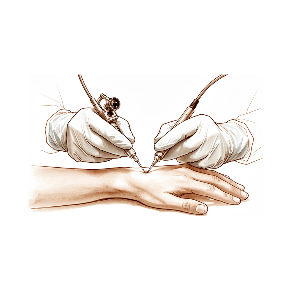
Overview
Therapeutic arthroscopy is the logical extension of diagnostic arthroscopy, representing a practical reality for surgery under endoscopic control [3]. As an essential diagnostic and therapeutic tool for orthopaedic surgeons, wrist arthroscopy is characterized by an ever-expanding list of indications and procedures [9]. Arthroscopic procedures generally demonstrate better results and improved localization of the injury with a low complication rate compared with open techniques [20]. In special situations, minimally invasive procedures can be performed through the arthroscope [5]. However, diagnostic arthroscopy performed in the setting of an unclear preoperative diagnosis yielded limited diagnostic benefit [2].
The technique emphasizes the use of traction for joint distention and the availability of various portals and instruments for diagnostic and therapeutic procedures [17]. Subtalar arthroscopy is a standardised and reproducible procedure with new diagnostic and minimally invasive therapeutic options [13]. Needle arthroscopy is a simple, safe, and well tolerated technique with promise as a diagnostic, scientific, and possibly therapeutic tool in rheumatic diseases, but arthroscopic experience is necessary for this procedure [14]. The described arthroscopic Hill-Sachs filling technique using an absorbable interference screw is safe, easily reproducible, does not require a long learning curve, and allows restoration of joint stability [12]. If results stand over time, the advantages of arthroscopic Bankart suture repair—namely less morbidity, lower cost, less pain, and preservation of motion—would show that arthroscopic shoulder stabilization has great potential for the future [53].
Arthroscopic treatment of talus bipartitus can be a safe and effective option with excellent short- and long-term outcomes [8]. Good to excellent outcomes can be consistently reached in greater than 80% of patients with arthroscopic debridement and microfracture for osteochondral lesions of the talar dome [15]. Arthroscopic and open approaches have comparable outcome profiles regarding recurrence and complications for dorsal ganglion cysts [6]. Additional long-term comparative studies are needed to accurately differentiate the efficacy of open and arthroscopic techniques for dorsal ganglion excision [4]. More prospective studies comparing open and arthroscopic excision are needed to delineate if there is a true functional benefit for ganglion cysts [7]. The most common indications for repeat wrist arthroscopy after failed primary treatment were ligamentous instability and osteoarthritis from dynamic impaction [25]. It is appropriate to base decisions regarding salvage operative procedures for displaced intra-articular fractures of the distal radius on the presence of severe symptoms or loss of function rather than on radiographic evidence of osteoarthrosis [11]. Suggested guidelines for the practice of arthroscopic surgery cover privileges, training, practice standards, continuing education, and performance review to ensure patient safety and surgeon competence [21]. The journals Arthroscopy and Arthroscopy Techniques are complementary, with the former publishing clinically relevant research and the latter focusing on surgical techniques [10]. The Foundations of Arthroscopy Techniques Collection was introduced to fill the gap for trainees and practitioners seeking essential fundamentals across multiple joints [24]. Smaller studies that include second-look arthroscopy provide the most convincing evidence for the efficacy of combined procedures, according to the authors [1].
Anatomy & Pathophysiology
Bony Anatomy
The wrist is the anatomic region between the forearm and the hand, encompassing the distal radioulnar, radiocarpal, and ulnocarpal joints and the eight carpal bones [63]. These bones comprise the scaphoid, lunate, triquetrum, and pisiform in the proximal row, and the trapezium, trapezoid, capitate, and hamate in the distal row [63]. Carpal bone sizes vary from the smallest (pisiform and trapezoid) to the largest (capitate) [63]. The radiocarpal joints form where the distal radius articulates with the scaphoid and lunate via their concave facets, and with the triquetrum on the triangular fibrocartilage [63]. The distal concave articular surfaces of the proximal row form the midcarpal articulations with the distal row [63].
The distal radius articular surface features two concave facets, the scaphoid and lunate facets, separated by the scapholunate or anterior-posterior ridge [70]. The sigmoid notch along the ulnar border of the distal radius is a shallow concavity for the articulating ulnar head at the distal radioulnar joint [70]. This notch is a convex structure that allows the radius to rotate around the distal ulna [67]. The distal ulnar convexity articulates at the lesser sigmoid notch of the distal radius, which accommodates the ulnar head through two thirds of its arc [63]. There is about a 20-degree inclination of the distal ulna at its articulation with the radius [63]. The distal ulna is covered with hyaline cartilage on its dorsal, lateral, palmar, and distal surfaces [70]. The ulnar styloid projects distally and lies dorsal to the ulnar head [63, 70]. At its base, the fovea serves as the insertion for the triangular fibrocartilaginous complex (TFCC) [70].
The distal radius has three articular components: distally the scaphoid and lunate fossae, and the sigmoid notch which allows articulation with the ulna medially [67]. Between the scaphoid and lunate fossa is a ridge that corresponds with the scapholunate interval [67]. The radial styloid allows attachment of the brachioradialis tendon and is the origin of several important wrist ligaments, including the radial scapholunate and radial lunocapitate ligaments [67]. The concave elliptical distal radius is oriented in the sagittal plane with an average of 11 degrees of volar tilt [67]. In the frontal plane, the average radial inclination of the distal radius is 23 degrees [67]. Radial length is measured from the tip of the radial styloid to the ulnar articular surface and averages 13 mm [67]. The space available for manipulation of the arthroscope and instrumentation in the wrist is substantially smaller than that available for arthroscopy of larger joints [19].
Individual carpal bone anatomy includes specific vascular and articulation patterns. The scaphoid's primary vascular supply is a branch of the radial artery at the dorsal ridge, with a group of smaller vessels entering the palmar tubercle to supply the distal 30% [70]. A dorsal and a palmar vascular supply are found in the lunate in 80% of wrists; in 20% of wrists, only a palmar supply is found [70]. The lunate is broader palmarly than dorsally [70]. The triquetrum articulates with the hamate distally, the lunate radially, and the pisiform volarly [70]. The triquetrum is stabilized to the fovea of the ulna through the ulnotriquetral ligament [70]. The hamate consists of the body and the hook (hamulus) of the hamate, which serves as an attachment for the transverse carpal ligament and for the origins of the flexor digiti minimi and opponens digiti minimi [70]. The head of the capitate often relies on a retrograde vascular supply [70]. Two ridges separate the distal articular surface of the capitate into three facets for articulation with the metacarpals of the index, long, and ring fingers [70]. The trapezoid has two distal facets, which articulate with the metacarpal of the index finger [70]. The trapezium has a saddle-shaped articulation with the base of the thumb metacarpal [70]. The trapezium has a palmar groove for the flexor carpi radialis (FCR), bordered laterally by a palmar tuberosity and the attachment for the transverse carpal ligament [70]. The pisiform is a sesamoid bone within the flexor carpi ulnaris (FCU) tendon and is the origin for the abductor digiti minimi [70].
Ligaments
The chondroligamentous supports attaching the distal radius and ulnar side of the carpus to the distal ulna are designated as the triangular fibrocartilage complex (TFCC) [63]. The TFCC includes the ulnar collateral ligament, the dorsal and volar radioulnar ligaments, the articular disc, the meniscal homologue, the extensor carpi ulnaris sheath, and the ulnolunate and ulnotriquetral ligament [63]. The TFCC is formed by the central meniscus homolog, the dorsal and volar radioulnar ligaments, the floor of the extensor carpi ulnaris (ECU) tendon sheath, and the volar ulnocarpal ligaments [70]. The TFCC arises from the radial border of the distal radius and inserts into the base of the ulnar styloid and distal ulna through the ligamentum subcruentum [70]. The dorsal and volar radioulnar ligaments are the primary stabilizers of the distal radioulnar joint [70]. Only the peripheral 10% to 40% of the volar, ulnar, and dorsal TFCC has a vascular supply [70]. The triangular fibrocartilage attaches to the base of the ulnar styloid and separates the hyaline cartilage–covered ulnar head from the styloid [63]. The distal ulna has an ulnar styloid which contains attachments to the triangular fibrocartilage complex, including the meniscus homolog, the volar and dorsal ulnar carpal ligaments, and the ulnar collateral ligament at the wrist [67].
Extrinsic carpal ligaments connect the radius or the ulna to the carpus [68]. In general, the volar ligaments are stronger than the dorsal ligaments [68]. The extrinsic or crossing ligaments include the radial collateral ligament from the radial styloid to the scaphoid waist [63]. The radial collateral ligament originates at the radius (0 mm from radial styloid) and inserts at the scaphoid waist and distal palmar trapezium [70]. The extrinsic or crossing ligaments include the ulnar collateral ligament from the base of the ulnar styloid attaching to the pisiform [63]. The extrinsic or crossing ligaments include the transverse carpal ligament [63]. The volar extrinsic or crossing ligaments include the radioscapocapitate ligament, the radiolunotriquetral ligament, and the radioscapolunate ligament on the radial side [63]. The radioscaphocapitate (RSC) ligament connects to the waist of the scaphoid, around which the scaphoid rotates, and limits ulnar translation of the carpus [68]. The radioscaphocapitate ligament originates at the radius (4 mm from radial styloid) and inserts at the scaphoid waist and midpalmar capitate [70]. The long radiolunate ligament helps to limit ulnar translocation of the carpus [68]. The short radiolunate ligament helps control lunate position [68]. The short radiolunate ligament originates at the volar-ulnar margin of the radius and inserts at the lunate [70]. The radioscapholunate ligament is a vascular conduit, not a true ligament, also known as the ligament of Testut [68]. The radioscapholunate ligament originates at the mesocapsule with termination of AIN and AIA and inserts at the ligament of Testut and Kuenz [70]. The radiolunatotriquetral ligament originates at the radius (10 mm from radial styloid) and inserts at the lunate ± triquetrum [70].
On the ulnar side, the volar extrinsic or crossing ligaments include the ulnolunate and ulnotriquetral components of the TFCC [63]. The ulnolunate ligament attaches to the palmar radioulnar ligament and lunate [68]. The ulnolunate ligament originates at the volar radioulnar ligament and inserts at the lunate [70]. The ulnotriquetral ligament attaches to the palmar radioulnar ligament and triquetrum [68]. The ulnotriquetral ligament originates at the volar radioulnar ligament and inserts at the triquetrum [70]. The ulnocapitate ligament attaches to the ulnar head, originates from the volar margin of the ulnar fovea, and is the most superficial or palmar [68]. The ulnocapitate ligament originates at the volar margin of the ulnar head and inserts at the capitate [70].
Dorsally, the identifiable extrinsic ligaments include the dorsal radiocarpal and the dorsal intercarpal ligaments [63]. The dorsal radiocarpal ligament (DRC) or dorsal radiotriquetral ligament has a trapezoidal shape and passes from the dorsal rim of the distal radius to the lunate and the triquetrum [68]. The dorsal radiocarpal ligament originates at the dorsal radius at the Lister tubercle and inserts at the lunate and triquetrum [70]. The dorsal radiocarpal ligament originates at the dorsal lip of the distal radius, adjacent to the dorsal radial tubercle (Lister tubercle), and traverses the radiocarpal joint obliquely to insert into the lunate and triquetrum [70]. The trapezoidal dorsal radiocarpal ligament attaches along the dorsal radial articular margin of the lunate fossa, from the Lister tubercle to the lesser sigmoid notch [63]. The dorsal radiocarpal ligament spans the lunotriquetral joint and inserts on the dorsal surface of the triquetrum [63]. Fibers of the dorsal radiocarpal ligament insert onto the dorsal lunotriquetral interosseous ligament (LTIL) [68]. Damage to the dorsal radiocarpal ligament, when in conjunction with other intrinsic ligament injuries, confers further carpal instability [68]. The dorsal intercarpal ligament is attached to the distal, dorsal surface of the triquetrum and passes across the midcarpal joint to attach to the dorsal surfaces of the scaphoid waist and the trapezoid [63]. The dorsal intercarpal ligament (DIC) passes from the dorsal tubercle of the triquetrum to the distal pole of the scaphoid [68]. The dorsal intercarpal ligament originates at the triquetrum and inserts at the scaphoid, trapezoid, and capitate [70]. With the DRC ligament, the DIC ligament reinforces the elastic dorsal wrist capsule and helps stabilize the scapholunate articulation with a contribution to the dorsal SLIL from its deep fibers [68]. The extrinsic wrist ligaments include the dorsal intercarpal ligament and the dorsal radiocarpal ligament [70].
Intrinsic carpal ligaments originate and insert within the carpus [68]. The interosseous ligaments include the scapholunate and lunotriquetral interosseous ligaments connecting the proximal carpal row [63]. The intrinsic wrist ligaments include the scapholunate interosseous ligament and the lunotriquetral interosseous ligament [70]. The scapholunate interosseous ligament (SLIL) is a major stabilizer of the wrist and the most commonly injured wrist ligament [68]. The scapholunate interosseous ligament is C-shaped, consisting of dorsal, palmar, and interosseous portions, with the dorsal portion being the strongest/thickest [68]. The scapholunate interosseous ligament is C-shaped in the sagittal plane, with the dorsal third being the thickest, strongest portion of the ligament [70]. The scapholunate interosseous ligament provides a flexion force on the lunate given its attachment to the scaphoid [68]. The lunotriquetral interosseous ligament (LTIL) is C-shaped, where the volar portion is the thickest/strongest [68]. The volar portion of the lunotriquetral ligament is the thickest [70]. The lunotriquetral interosseous ligament provides an extension moment on the lunate given its attachment to the triquetrum [68]. The interosseous ligaments also include the ligaments connecting the trapezium to the trapezoid, the trapezoid to the capitate, and the capitate to the hamate in the distal carpal row [63]. The capitohamate ligament is a thick ligament, 5 × 5 mm in cross section, with extensions to the third or fourth metacarpals [68].
On the palmar side of the carpus, between the radiolunotriquetral ligament and the radioscapocapitate ligament, is a relatively thin area, the space of Poirier, overlying the palmar surface of the lunate [63]. The space of Poirier is an area adjacent to the proximal capitate without ligamentous attachment, situated ulnar to the RSC ligament and radial to the long radiolunate in the floor of the carpal tunnel [68]. The space of Poirier is a weak area that is vulnerable to instability; the distal carpal row separates from the lunate through this space during a perilunate dislocation [68].
Vascular Anatomy
The terminal branches of the radial, ulnar, and anterior interosseous arteries provide extraosseous blood supply to the carpus through three dorsal and three palmar transverse arterial arches with longitudinal connections [73]. The dorsal radiocarpal arch is located at the radiocarpal joint and supplies the lunate and triquetrum [73]. The dorsal intercarpal arch is the largest, located between the proximal and distal carpal rows, and supplies the distal carpal row and, through anastomoses with the radiocarpal arch, the lunate and triquetrum [73]. The basal metacarpal arch is located at the base of the metacarpals, is the most variable, and supplies the distal carpal row [73]. The palmar radiocarpal arch is located at the level of the radiocarpal joint on the palmar surfaces of the lunate and triquetrum [73]. The intercarpal arch is located between the proximal and distal carpal rows, is the most variable, and does not contribute to nutrient vessels in the carpus [73]. The deep palmar arch is located at the level of the metacarpal bases, is consistent, and communicates with the dorsal basal metacarpal arch and the palmar metacarpal arteries [73].
Biomechanics and Kinematics
The eight carpal bones that comprise the wrist joint represent the most complex articular system in the human body [64]. The human wrist allows precise positioning of the hand and optimization of power and prehensile tasks throughout a nearly hemispherical arc of wrist motion [64]. Injuries to the wrist mechanism can lead to instability, resulting in a painful lack of motion, strength, and function [64]. The wrist can essentially be considered to be a two-joint system linking the hand (distal carpal row and metacarpals) to the forearm (radius and ulna) around the highly mobile bones of the proximal carpal row [71]. The two principle articulations are the radiocarpal and midcarpal joints, situated proximal and distal to the mobile proximal carpal row [71]. The bones of the wrist are composed of multiple articulations: the radiocarpal (RC) joint, midcarpal (MC) joint, pisotriquetral joint, trapeziometacarpal joint, and carpometacarpal joints [68]. The wrist joint’s motion planes include flexion, extension, radial deviation, ulnar deviation, and circumduction; there is minimal carpal motion with pronosupination [68]. Approximately 62° of wrist extension occurs through the RC joint and 62% of wrist flexion occurs through
Classification
Palmer: The Palmer classification describes acute distal surface triangular fibrocartilage complex (TFCC) tears by location [41]. Central TFCC tears (Palmer 1A) are avascular, and débridement is recommended [41]. Ulnar or dorsal/ulnar TFCC tears (Palmer 1B) are the most amenable to arthroscopic repair due to good blood supply in this region [41]. Repair techniques for ulnar or dorsal/ulnar TFCC tears include inside-out, outside-in, and all-arthroscopic techniques [41]. Volar distal (Palmer 1C) and radial (Palmer 1D) TFCC tears are commonly débrided, although some are repaired [41]. Foveal TFCC tears affect the deep TFCC and bony insertion and are not included in the Palmer classification [41]. Foveal TFCC tears may be associated with distal radioulnar joint (DRUJ) instability and may be diagnosed with the arthroscopic hook test [41]. Bone tunnel repair is one commonly used technique for foveal TFCC tears [41]. The central two-thirds of the TFCC may be débrided without affecting DRUJ stability because the dorsal and volar radiocarpal ligaments are maintained [41]. Ulnocarpal impaction is often associated with a chronic, central TFCC tear (Palmer class 2) [41]. The diagnosis of ulnocarpal impaction may be confirmed arthroscopically with cartilage changes on the proximal, ulnar aspect of the lunate [41]. The distal aspect of the ulna may be removed arthroscopically to prevent impaction in ulnocarpal impaction [41]. An open, extra-articular ulnar shortening osteotomy is an alternative to arthroscopic distal ulna removal for ulnocarpal impaction [41].
Geissler: Geissler grading classifies interosseous ligament injury based on findings in the radiocarpal and midcarpal spaces [147]. Geissler Grade I is defined as attenuation or hemorrhage of the interosseous ligament seen from the radiocarpal space with no incongruency of carpal alignment in the midcarpal space [147]. Geissler Grade II is defined as attenuation or hemorrhage of the interosseous ligament seen from the radiocarpal space with incongruency or step-off in the midcarpal space and a possible gap less than the width of a probe [147]. Geissler Grade III is defined as incongruency or step-off of carpal alignment seen from both radiocarpal and midcarpal spaces where a probe may be passed through the gap between carpal bones [147]. Geissler Grade IV is defined as incongruency or step-off of carpal alignment seen from both radiocarpal and midcarpal spaces [147]. Scapholunate ligament lesions are classified according to Geissler and graded as partial or complete [145].
Outerbridge: Modified Outerbridge grading is used to grade chondral lesions in wrist arthroscopy [147]. Cartilage lesions are classified according to Outerbridge [145].
Other Considerations: Arthroscopy is the benchmark for the identification of scapholunate (SL) and lunotriquetral (LT) ligament injuries [41]. Magnetic resonance arthrogram has high sensitivity and high specificity for the identification of scapholunate and lunotriquetral ligament injuries [41]. Débridement alone or débridement with pinning may be efficacious in patients with partial ligament tears without dissociation [41]. TFCC lesions are categorized according to Palmer [145].
Clinical Presentation
A thorough wrist examination remains integral to any arthroscopic assessment [26]. Diagnostic arthroscopy serves as a useful adjunct in the diagnosis and treatment of intra-articular wrist pathology, provided it follows a careful history and physical examination [27]. The arthroscopic approach focuses on the patient's symptoms and is a feasible method to alleviate them [23]. Surgeons must be vigilant during arthroscopy to avoid overlooking concomitant peripheral tears, as physical examination and MRI provide little diagnostic information for these specific lesions [40]. In children and adolescents with chronic wrist pain, most lesions have not been correctly identified by MRI before arthroscopy [43]. Given the considerable disagreement between and within experienced observers, reconsideration should be given to arthroscopy as the reference standard in the diagnosis of triangular fibrocartilage complex tears [30].
It is appropriate to base decisions regarding salvage operative procedures on the presence of severe symptoms or loss of function rather than on radiographic evidence of osteoarthrosis [11]. The most common indications for repeat wrist arthroscopy were ligamentous instability and osteoarthritis from dynamic impaction [25]. Diagnostic arthroscopy yields evidence of redundant dorsal capsular tissue in dorsal wrist capsular impingement [33]. Arthroscopy allows for the simultaneous treatment of ganglions and other pathologies [39].
Participants who underwent arthroscopic investigation for persistent wrist pain improved on average by approximately 50% at one year [18]. However, most patients who underwent arthroscopic investigation for persistent wrist pain continued to have some pain and disability at one year [18]. The most common procedure performed among American Board of Orthopaedic Surgery Part II candidates was to address excision or repair of the triangular fibrocartilage complex, followed by synovectomy, and diagnostic arthroscopy [46].
Investigations
Plain radiography: Four standard views are required for wrist radiography: posteroanterior with the wrist in ulnar deviation, lateral, semi-pronated oblique, and semi-supinated oblique [82]. An anteroposterior view with the fist clenched is added when scapholunate injury is suspected [82]. On the lateral view, the axes of the radius, lunate, capitate, and third metacarpal are co-linear, while the scaphoid projects at an angle of approximately 45 degrees to this line [82]. Dorsal intercalated segmental instability is characterized by the lunate tilting backwards and the axes of the capitate and metacarpals lying dorsal to that of the radius [82]. Conversely, volar intercalated segment instability presents with the lunate and scaphoid tilting volarwards, and the capitate and metacarpals lying anterior to the radius [82]. Ten to fifteen percent of scaphoid fractures are not visible on initial X-rays [82]. If initial X-rays are normal, the clinical diagnosis should be treated and the wrist immobilized [82]. If MRI is unavailable, repeated X-rays are needed two weeks later to detect undisplaced scaphoid fractures [82].
MRI: MRI is the modality of choice for imaging radiographically occult fractures of the hand and wrist [69]. Its primary advantages over CT and radiography include improved tissue characterization of soft tissues such as wrist ligaments and hand synovium, and the lack of ionizing radiation [69]. Modern MRI is generally at 1.5T or 3T, with 3T preferred for hand and wrist imaging, especially for small fields of view [69]. A static magnetic field strength of at least 1.5 T using a dedicated wrist coil is recommended for analyzing interosseous, intrinsic, and extrinsic ligament insertions [78]. The volar extrinsic, scapholunate interosseous, dorsal intercarpal, and lunotriquetral ligaments are best visualized using 1 mm slices with no interslice gap in the coronal plane [78]. The dorsal radiocarpal and intercarpal ligaments are best viewed on both coronal and sagittal images [78]. Oblique axial views along the longitudinal axes of ligaments allow further analysis, especially when an injury is suspected [78]. Concomitant cartilage-sensitive imaging is integrative to influence assessment and surgical management, as cartilage integrity influences clinical and surgical management, especially in the setting of scapholunate advanced collapse wrist [78]. Real-time MRI has been used to investigate dynamic instabilities, although its routine use in clinical practice is yet to be further determined [78]. MRI with contrast enhancement is most commonly used to determine whether soft-tissue lesions are solid or cystic or, in rheumatologic imaging, to better visualize erosions and synovial burden [69]. Dynamic contrast enhancement has been used with inconsistent results to assess for the presence of avascular necrosis in the lunate or scaphoid after injury [69]. MR arthrography can be performed for evaluation of the triangular fibrocartilage and intercarpal ligament tears, but this is generally unnecessary with the increasing availability of high field MRI [69]. With proper technique, injuries to the triangular fibrocartilage complex can be demonstrated with MRI [81]. Perforations in the triangular fibrocartilage complex appear as linear defects or gaps filled with hyperintense fluid on coronal gradient-echo or T2-weighted pulse sequences [81]. The addition of arthrographic contrast improves the visualization of scapholunate and lunotriquetral ligaments on MR images [81]. MRI is useful in detecting additional marrow abnormalities in osteonecrosis, as seen in the lunate in Kienböck disease or in the scaphoid after fracture [81]. Asymmetry of marrow signal in proximal and distal fragments of a fractured scaphoid is suggestive of proximal pole ischemia [81]. MRI provides earlier detection of synovitis and erosive bone changes associated with rheumatoid arthritis than do radiographs [81]. The use of arthroMRI together with high resolution has improved sensitivity but still requires technical improvements to compare with arthroscopy in the diagnosis of tears of the triangular fibrocartilage complex [129]. Early MRI reduces uncertainty and streamlines care for suspected scaphoid fractures [82].
CT: CT scanning enables the 3D analysis of carpal dysfunction [78]. By adding motion in real time (4D CT), this modality may hold promise in the future to potentially quantify the location and degree of injury noninvasively and help surgeons plan their surgical treatment [78].
Diagnostic Arthroscopy: Arthroscopy is considered by many to be the diagnostic intervention of choice for determining the degree of injury to the wrist and can assess the condition of the cartilage, ability to reduce the carpus, and any other associated injuries [78]. The degree of intrinsic and extrinsic ligament injury can be identified from arthroscopic evaluation [78]. Geissler grade II scapholunate interosseous ligament injuries tend to be isolated, whereas grade IV injuries often involve complete dorsal extrinsic ligament disruption [78]. Surgeons must be vigilant during arthroscopy to avoid overlooking concomitant peripheral tears of the triangular fibrocartilage complex, as physical examination and MRI provide little diagnostic information [40]. Most lesions found in children and adolescents with chronic wrist pain have not been correctly identified by MRI before arthroscopy [43]. A thorough understanding of the 3-D anatomy of the wrist is essential to optimize efficiency and to minimize complications associated with improper instrument placement during diagnostic arthroscopy [27]. With only a fair correlation between arthroscopy and MRI, it cannot be concluded that the two methods are equivalent for assessing wrist cartilage [130]. Wrist arthroscopy still has an important role to play in the assessment of a painful degenerative wrist [130].
Other Imaging and Techniques: Dynamic fluoroscopy shows abnormal motion between the scaphoid and lunate and changes in the kinematics of the midcarpal joint [78]. In scapholunate dissociation patients with dorsal intercalated segmental instability, the triquetrum-hamate relationship remains permanently engaged, whereas normally it changes from full engagement in ulnar deviation to complete disengagement in radial deviation [78]. Live imaging shows whether dorsal intercalated segmental instability is reducible, giving the physician valuable information as treatment is planned [78]. The diagnostic test accuracy of X-ray arthrography is limited [109]. Computerized tomographic arthrography and arthroscopy enabled accurate definition of an unusual anomaly of the scapula [29]. The high overall level of anatomic accuracy (96%) for lesser metatarsal phalangeal joint arthroscopy allows consideration of this resource as a valuable tool in the diagnosis and treatment of these joints [52]. The needle arthroscopy is a simple, safe, and well tolerated technique with promise as a diagnostic tool in rheumatic diseases, but arthroscopic experience is necessary for this procedure [14].
Treatment
General Principles and Indications
Therapeutic arthroscopy is a practical reality and the logical extension of diagnostic arthroscopy [3]. Wrist arthroscopy serves as an essential diagnostic and therapeutic tool for orthopaedic surgeons, with an expanding list of indications and procedures [9]. Arthroscopic procedures demonstrate better results and improved localization of injury with a low complication rate compared with open techniques [20]. The place of wrist arthroscopy in daily practice is related to the background of the hand surgeon [16]. Surgical intervention for thoracobiliary fistulae should only be indicated once conservative measures have failed [133].
Wrist Arthroscopy
Outcomes and Safety: Participants who underwent arthroscopic investigation for persistent wrist pain improved on average by approximately 50% at one year, but most continued to have some pain and disability [18]. Arthroscopic TFCC treatment in paediatric patients is safe and yielded favourable subjective and objective outcomes and patient/parent satisfaction [84]. Wrist arthroscopy in children and adolescents referred for chronic wrist pain was observed to have no intra- or postoperative complications [127]. Generally, the temperatures measured with dry wrist arthroscopy are safe [103].
Specific Procedures: Two groups have proposed the use of arthroscopic synovectomy as a day-case procedure, as it does not require an open arthrotomy and there is less joint capsule and ligament damage, allowing immediate postoperative mobilization [127]. The recurrence rate for arthroscopic resection of dorsal wrist ganglia was 12.3%, and patient satisfaction was high [102]. Arthroscopic management of dorsal wrist impingement is associated with a lower risk of flexion loss compared to open dorsal wrist capsulectomy [31]. Arthroscopic debridement of redundant dorsal capsular tissue offers a safe and effective treatment to improve pain and functional scores [33]. There was no statistical difference in clinical outcomes after open versus arthroscopic TFCC repair [34].
Complex Pathology and Adjuncts: The authors believe that smaller studies that include second-look arthroscopy provide the most convincing evidence for the efficacy of combined procedures [1]. The current arthroscopic rationale shows no superiority over conventional management for the osteoarticular or carpal ligament components of distal radius fractures [154]. Surgical stabilization provides durable, favorable outcomes for nontraumatic midcarpal instability, making it a reliable treatment option for patients who do not achieve sufficient relief from nonsurgical treatment [152]. Radial shortening is effective for patient satisfaction and functional improvement in Litchman stage 3B Kienbock’s disease [100].
Ankle and Foot Arthroscopy
Arthroscopic treatment for talus bipartitus can be a safe and effective option with excellent short- and long-term outcomes [8]. Arthroscopic bone marrow stimulation techniques provided satisfactory clinical outcomes for osteochondral lesions of the talus [98]. Clinical outcomes assessed 2 years postoperatively were superior in patients who underwent arthroscopic microfracture with atelocollagen augmentation compared to those who underwent arthroscopic microfracture alone, although the differences were not statistically significant [36]. The arthroscopic approach for late complications of calcaneal fractures focuses on the patient's symptoms and is a feasible approach to alleviate them [23]. Arthroscopic management serves as a minimally invasive alternative to open surgery for diagnosis and treatment of early-stage tuberculosis of the ankle [131].
Shoulder and Elbow Arthroscopy
Arthroscopic debridement of the elbow addresses the pathologic processes associated with arthritis of the elbow and was safe and effective in the reported series [99]. The literature on clinical outcomes using sliding knots or nonsliding knots for shoulder procedures is limited to level 4 evidence [134]. This series represents the first report of arthroscopic management of the failed SLAP repair [138].
Hand and Finger Arthroscopy
The needle arthroscopy is a simple, safe, and well tolerated technique with promise as a diagnostic, scientific, and possibly therapeutic tool in rheumatic diseases, but arthroscopic experience is necessary for this procedure [14]. After a preferred minimally invasive treatment with stable reconstruction of the articular surface for difficult intra-articular fractures of the proximal interphalangeal joint, sufficient aftercare is necessary to improve surgical outcomes [48].
Complications
General Complication Rates and Outcomes: A systematic review suggests that previously documented rates of wrist arthroscopy complications may underestimate the true incidence [122]. Arthroscopic and open approaches demonstrate comparable outcome profiles regarding recurrence and complications [6]. Four-year follow-up data support the use of arthroscopy for dorsal wrist ganglion, with favorable outcomes, recurrence, and complication rates [54]. In patients undergoing arthroscopic investigation for persistent wrist pain, average improvement at one year is approximately 50%; however, most patients continue to experience some pain and disability [18]. Meticulous attention to detail and anticipation of potential problems largely avoid complications associated with wrist arthroscopy [42].
Portal Placement and Anatomical Risks: A fundamental understanding of wrist anatomy is critical to avoiding complications secondary to portal placement [32]. Inappropriate portal placement, either too distal or too proximal along the wrist, may cause injury to the articular cartilage or the triangular fibrocartilage [19].
Vascular Complications: Vascular injuries after arthroscopy are rare [151]. The Committee on Complications of the Arthroscopy Association of North America identified only twelve vascular complications in more than 375,000 knee arthroscopies [151]. Nine of these twelve complications involved injury to the popliteal vessel [151]. The anterolateral portal, located one centimeter proximal to the joint line, is in direct proximity to the lateral inferior genicular artery [151]. The frequency of anterolateral portal use in arthroscopy probably results in a number of injuries to the lateral inferior genicular artery [151].
Infection: Post-arthroscopy septic arthritis is a recognized complication for which current data and practical recommendations exist [28].
Other Considerations: The author’s experience with more than 150 laser-assisted wrist arthroscopies using the Ho:YAG laser has been excellent, with no laser-related complications [136]. Aggressive early arthroscopic debridement after discovery, combined with postoperative radiation therapy, has proved effective in addressing moderate to severe heterotopic ossification of the elbow [58]. Arthroscopic subtalar arthrodesis is associated with fewer complications compared to open surgery [61]. Arthroscopy-assisted absorbable screw combined with Kirschner wire internal fixation for Sanders type III displaced intra-articular calcaneal fractures is associated with a low incidence of postoperative complications [142]. Long-term follow-up of forearm shortening and volar radiocarpal capsulotomy for wrist flexion deformity in children with amyoplasia shows that the initial improvement in wrist position is not maintained [35].
Recovery
Light activity (weeks): The provided evidence does not specify a typical week range for desk work, driving, or light activities of daily living.
Full activity (months): The provided evidence does not specify a month range for the return to manual work, sport, or full range of motion and strength.
Complete recovery / outcome plateau (months): The provided evidence does not specify a month range for when pain, strength, and final functional outcomes stabilise.
Rehabilitation protocol: The provided evidence does not detail specific physiotherapy phasing, immobilisation duration, weight-bearing or range-of-motion progression schedules, or sling/brace removal timing.
Functional milestones: Patient-reported outcome measures improved after medium-term follow-up following 3D planning and patient specific instrumentation for intraarticular corrective osteotomy [126]. Range of motion and grip strength improved postoperatively comparable to the healthy contralateral side following 3D planning and patient specific instrumentation for intraarticular corrective osteotomy [126].
Other Considerations: Arthroscopic treatment of talus bipartitus is associated with excellent short- and long-term outcomes [8]. Good to excellent outcomes are consistently reached in greater than 80% of patients following arthroscopic debridement and microfracture for osteochondral lesions of the talar dome [15]. Clinical outcomes at 2 years postoperatively were superior in patients who underwent arthroscopic microfracture with atelocollagen augmentation compared to those who underwent arthroscopic microfracture alone, although the differences were not statistically significant [36]. Arthroscopic thermal shrinkage is effective for the majority of patients with mild to moderate chronic distal radioulnar joint instability in long-term follow-up [56]. Arthroscopic subtalar arthrodesis is associated with bone fusion in over 90% of cases, a shorter time to healing, a simpler postoperative course, and fewer complications compared to open surgery [61]. At an average follow-up of 4.5 years, 28% of hands had persistent symptoms following carpal tunnel release by the Agee endoscopic technique [62]. Good clinical results observed in patients 10 years after radial shortening osteotomy are likely to remain stable at 20 years after surgery [155]. Proximal row carpectomy is a reliable and durable procedure for patients with Lichtman stage IIIA or IIIB Kienböck's disease at an average follow-up of 10 years [156]. Ninety-seven percent of patients had resolution of their preoperative biceps symptoms following arthroscopic transfer of the long head of the biceps tendon [157]. Most participants who underwent arthroscopic investigation for persistent wrist pain continued to have some pain and disability at one year [18]. Endoscopic surgical repair of abductor tendon tears generally shows good or excellent results [132]. Patient-reported outcome measures may not fully capture objective improvements in gait and strength following endoscopic surgical repair of abductor tendon tears [132].
Key Evidence
- [L5] The authors believe that smaller studies that include second-look arthroscopy provide the most convincing evidence for the efficacy of these combined procedures. [1] (10.1016/j.arthro.2017.01.005)
- [L4] Diagnostic arthroscopy performed in the setting of an unclear preoperative diagnosis yielded limited diagnostic benefit. [2] (10.1177/1558944716661993)
- [L5] Therapeutic arthroscopy has become the logical extension of diagnostic arthroscopy, and surgery under endoscopic control is now a practical reality. [3] (10.2106/00004623-198365030-00027)
- [L1] Additional long-term comparative studies are needed to accurately differentiate the efficacy of open and arthroscopic techniques. [4] (10.1016/j.jhsa.2008.01.009)
- [L5] In special situations, minimally invasive procedures can be performed through the arthroscope. [5] (10.1016/j.hcl.2016.08.005)
- [L2] Arthroscopic and open approaches have comparable outcome profiles regarding recurrence and complications. [6] (10.1177/1753193417734428)
- [L4] More prospective studies comparing open and arthroscopic excision are needed to delineate if there is a true functional benefit. [7] (10.1016/j.hcl.2013.08.020)
- [L4] Arthroscopic treatment can be a safe and effective option with excellent short- and long-term outcomes. [8] (10.1007/s00167-017-4613-8)
- [L5] Wrist arthroscopy is an essential diagnostic and therapeutic tool for the orthopaedic surgeon with an ever-expanding list of indications and procedures. [9] (10.1016/j.arthro.2007.11.002)
- [L5] The editors state that Arthroscopy and Arthroscopy Techniques are complementary, with the former publishing clinically relevant research and the latter focusing on surgical techniques. [10] (10.1016/j.arthro.2015.05.001)
- [L4] It is appropriate to base decisions regarding salvage operative procedures on the presence of severe symptoms or loss of function rather than on radiographic evidence of osteoarthrosis. [11] (10.2106/00004623-199709000-00003)
- [L5] The authors believe the described arthroscopic procedure is safe, is easily reproducible, does not require a long learning curve, and allows restoration of joint stability. [12] (10.1016/j.eats.2022.08.046)
- [L4] Subtalar arthroscopy is a standardised and reproducible procedure with new diagnostic and minimally invasive therapeutic options. [13] (10.1007/s001670050084)
- [L4] The needle arthroscopy is a simple, safe, and well tolerated technique, with promise as a diagnostic, scientific, and possibly therapeutic tool in rheumatic diseases, but arthroscopic experience is necessary for this procedure. [14] (10.1007/s00167-002-0329-4)
- [L4] Good to excellent outcomes can be consistently reached in greater than 80% of patients with arthroscopic debridement and microfracture. [15] (10.1016/j.arthro.2012.04.055)
- [L4] The place of wrist arthroscopy in daily practice is related to the background of the hand surgeon. [16] (10.1055/s-0033-1351355)
- [L2] Participants who underwent arthroscopic investigation for persistent wrist pain improved on average by approximately 50% at one year; however, most continued to have some pain and disability. [18] (10.1016/j.jht.2012.03.001)
- [L5] [19] (10.2106/00004623-199908000-00015)
- [L5] Arthroscopic procedures demonstrate better results and improved localization of the injury with a low complication rate compared with open techniques. [20] (10.5435/00124635-200105000-00006)
- [L5] This statement outlines suggested guidelines for the practice of arthroscopic surgery, covering privileges, training, practice standards, continuing education, and performance review to ensure patient safety and surgeon competence. [21] (10.1016/s0749-8063(11)00686-4)
- [L4] The arthroscopic approach focuses on the patient's symptoms and is a feasible approach to alleviate them. [23] (10.1007/s00167-012-2086-3)
- [L5] The authors introduce the Foundations of Arthroscopy Techniques Collection to fill the gap for trainees and practitioners seeking essential fundamentals across multiple joints. [24] (10.1016/j.arthro.2024.09.002)
- [L4] The most common indications for repeat wrist arthroscopy were ligamentous instability and osteoarthritis from dynamic impaction. [25] (10.1055/s-0033-1364090)
- [L5] A thorough wrist examination remains integral to any arthroscopic assessment. [26] (10.1016/j.jhsa.2008.07.015)
- [L5] [27] (10.1016/j.hcl.2017.06.004)
- [Paper] [28] (10.1016/j.otsr.2015.09.004)
- [L4] Computerized tomographic arthrography and arthroscopy enabled accurate definition of the anomaly. [29] (10.2106/00004623-198870030-00021)
- [L3] Given the considerable disagreement between and within experienced observers, reconsideration should be given to arthroscopy as the reference standard in the diagnosis of these tears. [30] (10.1016/j.jhsa.2018.02.031)
- [L4] The procedure is associated with a lower risk of flexion loss compared to open dorsal wrist capsulectomy. [31] (10.1016/j.jhsa.2008.06.020)
- [L5] A fundamental understanding of wrist anatomy is critical in the avoidance of complications secondary to portal placement. [32] (10.1016/s0749-0712(21)00187-6)
- [L4] Diagnostic arthroscopy yields evidence of redundant dorsal capsular tissue, and arthroscopic debridement of this tissue offers a safe and effective treatment to improve pain and functional scores. [33] (10.1016/j.jhsa.2016.12.012)
- [L3] There was no statistical difference in clinical outcomes after open versus arthroscopic TFCC repair. [34] (10.1016/j.jhsa.2008.01.020)
- [L4] Long-term follow-up of the procedure shows that the initial improvement in wrist position is not maintained. [35] (10.1016/j.jhsa.2011.10.013)
- [L1] Clinical outcomes assessed 2 years postoperatively were superior in patients who underwent arthroscopic microfracture with atelocollagen augmentation compared to those who underwent arthroscopic microfracture alone, although the differences were not statistically significant. [36] (10.1186/s12891-020-03730-3)
- [L4] Arthroscopy allows for the simultaneous treatment of ganglions and other pathologies. [39] (10.1016/j.jhsa.2012.04.042)
- [L4] Surgeons must be vigilant during arthroscopy to avoid overlooking concomitant peripheral tears, as physical examination and MRI provide little diagnostic information. [40] (10.1177/1753193413479479)
- [L5] As with any surgical procedure, complications associated with wrist arthroscopy may largely be avoided by paying meticulous attention to detail and by anticipating potential problems before they occur. [42] (10.1016/s0749-0712(21)00029-9)
- [L3] However, most of these lesions have not been correctly identified by MRI before arthroscopy. [43] (10.1016/j.arthro.2012.04.152)
- [L4] The most common procedure performed was to address excision or repair of the TFCC, followed by synovectomy, and diagnostic arthroscopy. [46] (10.5435/jaaosglobal-d-25-00158)
- [L5] After a preferred minimally invasive treatment with stable reconstruction of the articular surface, sufficient aftercare is necessary to improve surgical outcomes. [48] (10.1177/1753193414559464)
- [L5] The high overall level of anatomic accuracy (96%) allows consideration of this resource as a valuable tool in the diagnosis and treatment of these joints. [52] (10.1016/j.arthro.2014.03.018)
- [L4] The authors stated that if the results stood over time, the advantages of this arthroscopic technique, namely less morbidity, lower cost, less pain, and preservation of motion, would show that an arthroscopic shoulder stabilization would have great potential for the future. [53] (10.1016/j.arthro.2010.04.009)
- [L4] The outcomes, recurrence, and complications rates after 4 years of follow-up presented in this study support the use of arthroscopy as a treatment for dorsal wrist ganglion. [54] (10.1177/1558944717743601)
- [L4] Arthroscopic thermal shrinkage is effective for the majority of the patients with mild to moderate chronic distal radioulnar joint instability in long-term follow-up. [56] (10.1177/1753193420927882)
- [L4] Aggressive early arthroscopic debridement after discovery with the addition of postoperative radiation therapy has proved effective in addressing this potentially serious complication. [58] (10.1016/j.arthro.2013.03.050)
- [L4] Arthroscopic subtalar arthrodesis is gaining in popularity based on evidence of bone fusion in over 90% of cases, with a shorter time to healing, a simpler postoperative course, and fewer complications compared to open surgery. [61] (10.1016/j.otsr.2016.08.002)
- [L4] At an average follow-up of 4.5 years, 28% of hands had persistent symptoms, but results were scarcely different from the conventional technique with no patient requiring reoperation. [62] (10.1054/jhsb.1999.0226)
- [L4] Arthroscopic TFCC treatment in paediatric patients is safe and yielded favourable subjective and objective outcomes and patient/parent satisfaction. [84] (10.1177/1753193418825070)
- [L4] Arthroscopic bone marrow stimulation techniques provided satisfactory clinical outcomes. [98] (10.1177/0363546512472979)
- [L4] Arthroscopic debridement of the elbow addresses the pathologic processes associated with arthritis of the elbow and was safe and effective in this series. [99] (10.1016/j.arthro.2007.03.084)
- [L3] The procedure is effective for patient satisfaction and functional improvement. [100] (10.1007/s00264-007-0428-4)
- [L4] The recurrence rate was 12.3%, and patient satisfaction was high, supporting the use of arthroscopy as primary treatment for DWG resection. [102] (10.1016/j.arthro.2010.05.008)
- [L4] Generally, the temperatures measured with dry arthroscopy are safe. [103] (10.1016/j.jhsa.2026.01.026)
- [L1] The diagnostic test accuracy of X-ray arthrography is limited. [109] (10.1177/1753193411402762)
- [L4] This systematic review suggests that the previously documented rate of wrist arthroscopy complications may be underestimating the true incidence. [122] (10.1016/j.arthro.2012.01.008)
- [L4] At a mean follow-up of 6 years, pain was significantly reduced and wrist function was significantly improved compared with preoperative status. [125] (10.1177/1753193420930587)
- [L4] ROM and grip strength improved postoperatively comparable to the healthy contralateral side and patient-reported outcome measures improved after medium-term follow-up. [126] (10.1186/s12891-022-05946-x)
- [L4] [127] (10.1055/s-0038-1639508)
- [L4] The use of arthroMRI together with high resolution has improved sensitivity but still technical improvements to compare with arthroscopy in diagnostic of tears of the triangular fibrocartilage complex. [129] (10.1016/j.arthro.2009.04.027)
- [L3] With only a fair correlation between arthroscopy and MRI, it cannot be concluded that the two methods are equivalent for assessing wrist cartilage and, as such, wrist arthroscopy still has an important role to play in the assessment of a painful degenerative wrist. [130] (10.1177/1753193408090395)
- [L4] It serves as a minimally invasive alternative to open surgery for diagnosis and treatment. [131] (10.1186/s13018-018-1048-y)
- [L5] Endoscopic surgical repair of abductor tendon tears generally shows good or excellent results, though patient-reported outcome measures may not fully capture objective improvements in gait and strength. [132] (10.1016/j.arthro.2024.02.028)
- [L5] Surgical intervention should only be indicated once these conservative measures have failed. [133] (10.1016/s0020-1383(02)00072-4)
- [L4] The literature on clinical outcomes using sliding knots or nonsliding knots for shoulder procedures is limited to level 4 evidence. [134] (10.1177/2325967120911646)
- [L4] The author's experience with more than 150 laser-assisted wrist arthroscopies using the Ho:YAG laser has been excellent, with no laser-related complications. [136] (10.1016/s0749-0712(21)00185-2)
- [L3] [138] (10.1016/j.arthro.2008.04.015)
- [L3] This approach is associated with a low incidence of postoperative complications and a quick return to sports activities. [142] (10.1186/s12891-025-08438-w)
- [Paper] [145] (10.1016/s0363-5023(09)60080-0)
- [Paper] [147] (10.1055/s-0039-1692929)
- [L4] [151] (10.2106/00004623-199072050-00019)
- [L2] Surgical stabilization provides durable, favorable outcomes for nontraumatic MCI, making it a reliable treatment option for patients who do not achieve sufficient relief from nonsurgical treatment. [152] (10.1016/j.jhsa.2025.07.037)
- [Letter] The authors emphasize that the current arthroscopic rationale shows no superiority over conventional management for the osteoarticular or carpal ligament components of distal radius fractures, and establishing the benefits of arthroscopic management remains complex. [154] (10.1016/j.arthro.2023.08.071)
- [L4] Good clinical results observed in patients 10 years after radial shortening osteotomy are likely to remain stable at 20 years after surgery. [155] (10.1016/j.jhsa.2025.04.018)
- [L4] At an average follow-up of 10 years, proximal row carpectomy is a reliable and durable procedure for patients with Lichtman stage IIIA or IIIB Kienböck's disease. [156] (10.1016/j.jhsa.2008.02.031)
- [L3] Ninety-seven percent of patients had resolution of their preoperative biceps symptoms. [157] (10.1177/2325967113s00090)
See Also
- Wrist Arthroscopy
- Wrist Ganglia
- Carpal Tunnel Release
- Proximal Row Carpectomy
- Kienböck's Disease
References
[1] Authors' Reply. Arthroscopy. 2017. DOI: 10.1016/j.arthro.2017.01.005
[2] Diagnostic Wrist Arthroscopy for Nonspecific Wrist Pain. HAND. 2016. DOI: 10.1177/1558944716661993
[3] Arthroscopic surgery.. The Journal of Bone & Joint Surgery. 1983. DOI: 10.2106/00004623-198365030-00027
[4] Arthroscopic Versus Open Dorsal Ganglion Excision: A Prospective, Randomized Comparison of Rates of Recurrence and of Residual Pain. The Journal of Hand Surgery. 2008. DOI: 10.1016/j.jhsa.2008.01.009
[5] Wrist Arthroscopy for Athletic Injuries. Hand Clinics. 2017. DOI: 10.1016/j.hcl.2016.08.005
[6] Arthroscopic versus open excision of dorsal ganglion cysts: a systematic review. Journal of Hand Surgery (European Volume). 2017. DOI: 10.1177/1753193417734428
[7] Arthroscopic Excision of Ganglion Cysts. Hand Clinics. 2014. DOI: 10.1016/j.hcl.2013.08.020
[8] Talus bipartitus: a systematic review and report of two cases with arthroscopic treatment. Knee Surgery, Sports Traumatology, Arthroscopy. 2017. DOI: 10.1007/s00167-017-4613-8
[9] New Advances in Wrist Arthroscopy. Arthroscopy. 2008. DOI: 10.1016/j.arthro.2007.11.002
[10] Our Journals Arthroscopy and Arthroscopy Techniques Are Complementary. Arthroscopy. 2015. DOI: 10.1016/j.arthro.2015.05.001
[11] Displaced Intra-Articular Fractures of the Distal Aspect of the Radius. The Journal of Bone and Joint Surgery (American Volume). 1997. DOI: 10.2106/00004623-199709000-00003
[12] Arthroscopic Hill‐Sachs Filling Technique Using an Absorbable Interference Screw. Arthroscopy Techniques. 2022. DOI: 10.1016/j.eats.2022.08.046
[13] Subtalar arthroscopy – indications and surgical technique. Knee Surgery, Sports Traumatology, Arthroscopy. 1998. DOI: 10.1007/s001670050084
[14] Technique of synovial biopsy of metacarpophalangeal joints using the needle arthroscope. Knee Surgery, Sports Traumatology, Arthroscopy. 2002. DOI: 10.1007/s00167-002-0329-4
[15] Outcome of Arthroscopic Debridement and Microfracture as the Primary Treatment for Osteochondral Lesions of the Talar Dome. Arthroscopy. 2012. DOI: 10.1016/j.arthro.2012.04.055
[16] Trends in Wrist Arthroscopy. Journal of Wrist Surgery. 2013. DOI: 10.1055/s-0033-1351355
[17] Wrist Arthroscopy. 2021.
[18] Prognosis and Prognostic Factors for Patients with Persistent Wrist Pain Who Proceed to Wrist Arthroscopy. Journal of Hand Therapy. 2012. DOI: 10.1016/j.jht.2012.03.001
[19] Techniques of Wrist Arthroscopy . The Journal of Bone and Joint Surgery-American Volume*. 1999. DOI: 10.2106/00004623-199908000-00015
[20] Wrist Arthroscopy: Principles and Clinical Applications. Journal of the American Academy of Orthopaedic Surgeons. 2001. DOI: 10.5435/00124635-200105000-00006
[21] Suggested Guidelines for the Practice of Arthroscopic Surgery. Arthroscopy. 2011. DOI: 10.1016/s0749-8063(11)00686-4
[23] Arthroscopic management of late complications of calcaneal fractures. Knee Surgery, Sports Traumatology, Arthroscopy. 2012. DOI: 10.1007/s00167-012-2086-3
[24] Introducing Foundations of Arthroscopy Techniques. Arthroscopy. 2024. DOI: 10.1016/j.arthro.2024.09.002
[25] Revision Wrist Arthroscopy after Failed Primary Arthroscopic Treatment. Journal of Wrist Surgery. 2014. DOI: 10.1055/s-0033-1364090
[26] Wrist Arthroscopy: Current Concepts. The Journal of Hand Surgery. 2008. DOI: 10.1016/j.jhsa.2008.07.015
[27] Diagnostic Wrist Arthroscopy. Hand Clinics. 2017. DOI: 10.1016/j.hcl.2017.06.004
[28] Post-arthroscopy septic arthritis: Current data and practical recommendations. Orthopaedics & Traumatology: Surgery & Research. 2015. DOI: 10.1016/j.otsr.2015.09.004
[29] Unusual anomaly of the scapula defined by arthroscopy and computerized tomographic arthrography. Report of a case.. The Journal of Bone & Joint Surgery. 1988. DOI: 10.2106/00004623-198870030-00021
[30] An Evaluation of the Reliability of Wrist Arthroscopy in the Assessment of Tears of the Triangular Fibrocartilage Complex. The Journal of Hand Surgery. 2018. DOI: 10.1016/j.jhsa.2018.02.031
[31] Arthroscopic Management of Dorsal Wrist Impingement. The Journal of Hand Surgery. 2008. DOI: 10.1016/j.jhsa.2008.06.020
[32] COMPLICATIONS OF WRIST ARTHROSCOPY. Hand Clinics. 1999. DOI: 10.1016/s0749-0712(21)00187-6
[33] Diagnosis and Arthroscopic Management of Dorsal Wrist Capsular Impingement. The Journal of Hand Surgery. 2017. DOI: 10.1016/j.jhsa.2016.12.012
[34] Clinical Comparison of Arthroscopic Versus Open Repair of Triangular Fibrocartilage Complex Tears. The Journal of Hand Surgery. 2008. DOI: 10.1016/j.jhsa.2008.01.020
[35] Long-Term Results of Forearm Shortening and Volar Radiocarpal Capsulotomy for Wrist Flexion Deformity in Children With Amyoplasia. The Journal of Hand Surgery. 2012. DOI: 10.1016/j.jhsa.2011.10.013
[36] Arthroscopic microfracture with atelocollagen augmentation for osteochondral lesion of the talus: a multicenter randomized controlled trial. BMC Musculoskeletal Disorders. 2020. DOI: 10.1186/s12891-020-03730-3
[39] Ganglions of the Wrist and Associated Triangular Fibrocartilage Lesions: A Prospective Study in Arthroscopically-treated Patients. The Journal of Hand Surgery. 2012. DOI: 10.1016/j.jhsa.2012.04.042
[40] Incidence and diagnosis of ‘the double lesion’ of the triangular fibrocartilage complex. Journal of Hand Surgery (European Volume). 2013. DOI: 10.1177/1753193413479479
[41] Chapter 39 Wrist Arthroscopy. 2019.
[42] COMPLICATIONS OF WRIST ARTHROSCOPY AND HOW TO PREVENT THEM. Hand Clinics. 1995. DOI: 10.1016/s0749-0712(21)00029-9
[43] Pathomorphologic Findings of Wrist Arthroscopy in Children and Adolescents With Chronic Wrist Pain. Arthroscopy. 2012. DOI: 10.1016/j.arthro.2012.04.152
[46] Trends in Volume of Wrist Arthroscopy Among American Board of Orthopaedic Surgery Part II Candidates. JAAOS: Global Research and Reviews. 2025. DOI: 10.5435/jaaosglobal-d-25-00158
[48] Management of difficult intra-articular fractures or fracture dislocations of the proximal interphalangeal joint. Journal of Hand Surgery (European Volume). 2014. DOI: 10.1177/1753193414559464
[52] Lesser Metatarsal Phalangeal Joint Arthroscopy: Anatomic Description and Comparative Dissection. Arthroscopy. 2014. DOI: 10.1016/j.arthro.2014.03.018
[53] Arthroscopic Bankart Suture Repair: Technique and Early Results. Arthroscopy. 2010. DOI: 10.1016/j.arthro.2010.04.009
[54] Arthroscopic Resection of Dorsal Wrist Ganglion: Results and Rate of Recurrence Over a Minimum Follow-up of 4 Years. HAND. 2017. DOI: 10.1177/1558944717743601
[56] Arthroscopic thermal stabilization for distal radioulnar joint instability: 3 to 19 years follow-up. Journal of Hand Surgery (European Volume). 2020. DOI: 10.1177/1753193420927882
[58] Early Arthroscopic Management Strategies for Patients Developing Moderate to Severe Heterotopic Ossification of the Elbow (SS‐43). Arthroscopy. 2013. DOI: 10.1016/j.arthro.2013.03.050
[61] Arthroscopic subtalar arthrodesis. Orthopaedics & Traumatology: Surgery & Research. 2016. DOI: 10.1016/j.otsr.2016.08.002
[62] Carpal Tunnel Release by the Agee Endoscopic Technique. Journal of Hand Surgery. 1999. DOI: 10.1054/jhsb.1999.0226
[63] Campbell S Operative Orthopaedics 4 Volume Set. NERVE INJURIES AT THE LEVEL OF THE HAND AND WRIST > ANATOMY.
[64] Green S Operative Hand Surgery. INTRODUCTION.
[67] A Lange Medical Book Current Diagnosis Treatment In Orthopedics Fifth Edition. 2Musculoskeletal Trauma Surgery > FRACTURES AND DISLOCATIONS OF THE DISTAL AND MID-FOREARM.
[68] Aaos Comprehensive Orthopaedic Review 3. Carpal Instability* > II. Anatomy and Biomechanics (See Chapter 92).
[69] Orthopaedic Knowledge Update 13 Ebook Without Multimedia. Anatomy, Evaluation, Clinical Examination, and Imaging > Imaging: Advances in Imaging of the Hand and Upper Extremity > Magnetic Resonance Imaging.
[70] Aaos Comprehensive Orthopaedic Review 3. Anatomy of the Hand and Wrist > VII. The Wrist.
[71] Green S Operative Hand Surgery. WRIST BIOMECHANICS > Carpal Kinematics.
[73] Campbell S Operative Orthopaedics 4 Volume Set. NERVE INJURIES AT THE LEVEL OF THE HAND AND WRIST > CIRCULATION.
[78] Green S Operative Hand Surgery. Advanced Imaging.
[81] Campbell S Operative Orthopaedics 4 Volume Set. WRIST AND ELBOW.
[82] Apley And Solomon S Concise System Of Orthopaedics And Trauma. FRACTURES OF THE DISTAL RADIUS IN CHILDREN > Imaging.
[84] Arthroscopic treatment of triangular fibrocartilage complex injuries in paediatric and adolescent patients. Journal of Hand Surgery (European Volume). 2019. DOI: 10.1177/1753193418825070
[98] Arthroscopic Bone Marrow Stimulation Techniques for Osteochondral Lesions of the Talus. The American Journal of Sports Medicine. 2013. DOI: 10.1177/0363546512472979
[99] Decreasing Risk to the Posterior Interosseous Nerve During Endoscopic Repair of the Distal Biceps Tendon (SS‐70). Arthroscopy. 2007. DOI: 10.1016/j.arthro.2007.03.084
[100] Is radial shortening useful for Litchman stage 3B Kienbock’s disease?. International Orthopaedics. 2007. DOI: 10.1007/s00264-007-0428-4
[102] Arthroscopic Resection of Dorsal Wrist Ganglia: 114 Cases With Minimum Follow‐Up of 2 Years. Arthroscopy. 2010. DOI: 10.1016/j.arthro.2010.05.008
[103] Are There Thermal Risks Associated With Dry Arthroscopy? A Simulated Comparison of Temperatures in Wet Versus Dry Wrist Arthroscopy. The Journal of Hand Surgery. 2026. DOI: 10.1016/j.jhsa.2026.01.026
[109] The diagnostic accuracy of X-ray arthrography for triangular fibrocartilaginous complex injury: a systematic review and meta-analysis. Journal of Hand Surgery (European Volume). 2011. DOI: 10.1177/1753193411402762
[122] Complications of Wrist Arthroscopy. Arthroscopy. 2012. DOI: 10.1016/j.arthro.2012.01.008
[125] Four-corner arthrodesis with a dorsal locking plate: 4–9-year follow-up. Journal of Hand Surgery (European Volume). 2020. DOI: 10.1177/1753193420930587
[126] 3D planning and patient specific instrumentation for intraarticular corrective osteotomy of trapeziometacarpal-, metacarpal and finger joints. BMC Musculoskeletal Disorders. 2022. DOI: 10.1186/s12891-022-05946-x
[127] Wrist Arthroscopy in Juvenile Idiopathic Arthritis: A Review of Current Literature and Future Implications. Journal of Wrist Surgery. 2018. DOI: 10.1055/s-0038-1639508
[129] Wrist Arthroscopy: Evaluating the Effectiveness of Clinical Tests and the Standard MRI in the Detection of Tears of the Triangular Fibrocartilage Complex of the Wrist (SS‐27). Arthroscopy. 2009. DOI: 10.1016/j.arthro.2009.04.027
[130] Comparison of MRI and Wrist Arthroscopy for Assessment of Wrist Cartilage. Journal of Hand Surgery (European Volume). 2008. DOI: 10.1177/1753193408090395
[131] Arthroscopic management for early-stage tuberculosis of the ankle. Journal of Orthopaedic Surgery and Research. 2019. DOI: 10.1186/s13018-018-1048-y
[132] Editorial Commentary: Endoscopic Repair of Abductor Tendon Tears Results in Variable Patient Reported Outcomes and Generally Good to Excellent Results. Arthroscopy. 2024. DOI: 10.1016/j.arthro.2024.02.028
[133] Traumatic thoracobiliary fistulae: a case report with a review of the current management options. Injury. 2002. DOI: 10.1016/s0020-1383(02)00072-4
[134] Sliding or Nonsliding Arthroscopic Knots for Shoulder Surgery: A Systematic Review. Orthopaedic Journal of Sports Medicine. 2020. DOI: 10.1177/2325967120911646
[136] LASER-ASSISTED WRIST ARTHROSCOPY. Hand Clinics. 1999. DOI: 10.1016/s0749-0712(21)00185-2
[138] Outcomes of Failed SLAP Repairs (SS‐14). Arthroscopy. 2008. DOI: 10.1016/j.arthro.2008.04.015
[142] Arthroscopy-assisted absorbable screw combined with Kirschner wire internal fixation for Sanders type III displaced intra-articular calcaneal fractures: a retrospective study. BMC Musculoskeletal Disorders. 2025. DOI: 10.1186/s12891-025-08438-w
[145] Interobserver Reliability and Intraobserver Reproducibility of Digital Photograph Documentation of Wrist Arthroscopy. The Journal of Hand Surgery. 2009. DOI: 10.1016/s0363-5023(09)60080-0
[147] Role of Wrist Arthroscopy in the Management of Established Scaphoid Nonunion. Journal of Hand and Microsurgery. 2020. DOI: 10.1055/s-0039-1692929
[151] False aneurysm after arthroscopic meniscectomy. A report of two cases.. The Journal of Bone & Joint Surgery. 1990. DOI: 10.2106/00004623-199072050-00019
[152] Long-Term Surgical Outcomes of Dorsal Capsulodesis and Three-Ligament Tenodesis in Midcarpal Instability: A Mean Follow-Up of 8 Years. The Journal of Hand Surgery. 2025. DOI: 10.1016/j.jhsa.2025.07.037
[154] Regarding “Adjuvant Arthroscopy Does Not Improve the Functional Outcome of Volar Locking Plate for Distal Radius Fractures: A Randomized Clinical Trial”. Arthroscopy. 2024. DOI: 10.1016/j.arthro.2023.08.071
[155] Long-Term Outcomes of Radial Shortening Osteotomy for Kienböck Disease: Minimum 20-Year Follow-Up Study. The Journal of Hand Surgery. 2026. DOI: 10.1016/j.jhsa.2025.04.018
[156] Proximal Row Carpectomy for Advanced Kienböck's Disease: Average 10-Year Follow-Up. The Journal of Hand Surgery. 2008. DOI: 10.1016/j.jhsa.2008.02.031
[157] Arthroscopic Transfer of the Long Head of the Biceps Tendon. Orthopaedic Journal of Sports Medicine. 2013. DOI: 10.1177/2325967113s00090