Clinicians › Hand
Neuropathy
Hand neuropathies: compressive etiologies (CTS, CuTS) diagnosis, systemic causes, and surgical/non-operative management options.
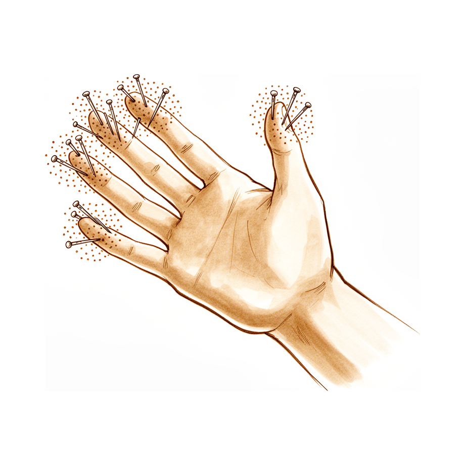
Overview
Peripheral neuropathy presents as a heterogeneous group of conditions where early diagnosis is critical to initiate treatments that slow progression [1]. While multifocal neuropathy requires careful diagnosis and conservative management first [2], the term "double crush syndrome" is considered inadequate for describing its multiple variables, and "multifocal neuropathy" is already reserved for a distinct autoimmune disease [2]. Peripheral nerve injuries are common and debilitating, yet available treatments remain suboptimal [4]. Complications of managing compressive neuropathy include iatrogenic injury, treatment failure, and pathologic pain syndromes; preventing these outcomes relies on a solid understanding of normal anatomy and anatomic variations [7]. Because missed diagnoses result in unsatisfactory outcomes, approaching peripheral nerves with a multifocal technique requires a more detailed initial assessment [8].
Current diagnostic and prognostic standards emphasize that the sole use of monofilament testing to diagnose peripheral neuropathy is not recommended [6]. Only 4.1% of patients who had electrodiagnostic testing ordered by a surgeon had no measurable neuropathy [5]. Comparison of sensory nerve responses is more effective than the use of an absolute median nerve latency in documenting median nerve abnormalities [34]. The most important prognostic factor for nerve recovery is patient age [38]. In studies of ulnar neuropathy at the elbow with cubital bowing, a custom hand dynamometer provides a better indication of disease severity, progression, or improvement in strength [61]. Outcomes assessment tools in peripheral nerve surgery evaluate aspects relating to function, pain, and patient perception of outcomes [9].
Despite major microsurgical improvements, the clinical outcome of peripheral nerve surgery is still regarded as suboptimal [33]. Results of nerve repair to date have been no better than fair, with only 50% of patients regaining useful function [13]. Significant advances using microsurgical techniques for nerve repair have led to improved results after peripheral nerve surgery and have extended the types of nerve repair that can be accomplished [63]. The literature demonstrates positive functional outcomes after nerve transfer operations, leading to increased utility and variations [35]. However, the clinical role for end-to-side nerve repair without deliberate nerve injury is limited and should currently only be used to reconstruct noncritical sensory deficits [37]. A unique and common protocol for the treatment of hand neuropathy in cyclists is lacking despite the range of treatment available for peripheral neuropathies [3]. Study findings on home intervention for chemotherapy-induced peripheral neuropathy could better define conservative interventions for its treatment [12].
Anatomy & Pathophysiology
General Nerve Injury & Pathophysiology
Understanding the classification, pathophysiology, and electrodiagnosis of nerve lesions is critical for the appropriate diagnosis, localization, and management of nerve trauma in the hand [17]. Peripheral nerve injuries induce both structural and functional brain changes, which may be associated with significant sensorimotor abnormalities and pain [74]. Treatment outcomes in these patients are impacted by maladaptive neuroplasticity as well as psychological and personality characteristics [74]. In pediatric populations, surgical repair combined with long-term hand therapy results in excellent functional outcomes following peripheral nerve injury [24].
Compressive Neuropathies
Chronic nerve compression injuries are frequent, particularly in the upper extremity, and can degrade quality of life and limit work capacity [18]. Symptoms range from initial sensory abnormalities, paresthesias, and pain to motor disruption and permanent sensory loss as the injury progresses [18]. Recent evidence indicates a distinct pathophysiology involving Schwann cell mechanosensitivity for chronic nerve compression injuries, rather than a mild form of Wallerian degeneration [18]. Clinical examination and electrodiagnostic studies remain the gold standard for diagnosis, although diagnosis can be difficult in patients with confounding factors such as diabetes [18]. Treatment ranges from medical to surgical management depending on the nerve involved and the severity and duration of compression [18].
The pathogenesis of carpal tunnel syndrome focuses on the anatomy of the carpal tunnel, underlying systemic or physiologic disorders, and patterns of hand use associated with symptoms [121]. Acute carpal tunnel syndrome is a compartment syndrome of the carpal tunnel characterized by a rapid rise and sustained increase in interstitial pressure within the carpal canal [177]. Chronic carpal tunnel syndrome is a compressive neuropathy involving an insidious rise in carpal tunnel interstitial pressure of a moderate degree [177]. In chronic cases, the increase in interstitial pressure may initially be intermittent but is likely to be elevated constantly with time and increased symptoms [177].
Multifocal Neuropathy & Double Crush Syndrome
The underlying pathophysiology of double crush syndrome is widely debated [22]. Upton and McCombs theorized that compression at one location on a nerve's axon predisposes that same axon to injury elsewhere due to disrupted bidirectional transport of essential nutrients along the axon [22]. In this context, more proximal lesions closer to the cell body have a greater effect on nerve function [22]. The term "double crush" is considered misleading because it does not account for conditions where three or more sites of a given nerve are affected [22]. Furthermore, the term "crush" is limiting because it implies compression and excludes other mechanical stresses such as stretch that may produce the same adverse outcome [22]. It also limits the scope of the disease to purely mechanical factors, whereas multiple medical and pharmacological factors likely contribute to multifocal neuropathy [22].
Diabetic Neuropathy
Neuropathy is present in a substantial number of diabetic patients, with around 10% having some degree of neuropathy at the time of diagnosis [26]. Forty percent of diabetic patients develop neuropathy within 10 years of diagnosis, and elderly patients with diabetes have a higher prevalence [26]. Neuropathy in diabetes is caused by sustained hyperglycemia, which leads to the formation of advanced glycation end products (AGE) [26]. AGE cause direct neuronal injury and microvascular damage, both of which negatively affect peripheral nerve function [26]. Sustained hyperglycemia also causes excess production of reactive oxygen species, resulting in direct damage to intracellular proteins and membrane lipids [26]. There is significant overlap between the development of neuropathy and vasculopathy in diabetic patients [26]. Excess production of superoxide species disrupts nitric oxide function, causing vasoconstriction and subsequent ischemia and end-organ damage [26].
Neuromas & Neuralgia
Upper extremity trauma often results in peripheral nerve injury, which can lead to neuroma formation when axonal nerve fibres are damaged [55]. When a nerve is severed and left in discontinuity or inadequately repaired, an end-bulb neuroma forms, comprising a disorganized tangle of regenerating nerve fibres [55]. A partial nerve laceration or severe crush injury can result in a neuroma-in-continuity [55]. Many, but not all, neuromas result in neuropathic pain [55]. The reported incidence of painful neuroma formation after upper extremity amputations is in the range of 4%–25% [55]. Specific symptoms manifesting from traumatic neuromas include tenderness, spontaneous pain, allodynia, hyperalgesia, and mechanical or thermal hypersensitivity in the innervated area [55].
Neuralgia can be conceptualized into four categories: * Compression neuropathy: Amenable to surgery [148]. * Neuroma: Amenable to surgery [148]. * Painful hyperalgesia: Not amenable to surgery [148]. * Phantom nerve pain: Not amenable to surgery [148].
Complex Regional Pain Syndrome
Complex regional pain syndrome (CRPS) represents autonomic and pain transmission dysregulation, resulting in peripheral sensitization with allodynia, dysesthesia, hyperpathia, and a reduced tolerance for pain when using the affected area for basic function [113]. CRPS occurs most commonly after a traumatic injury or iatrogenic insult [113]. * CRPS I: Theoretically includes patients with a musculoskeletal injury without a defined neural injury [113]. * CRPS II: Includes patients who fulfill the same criteria as CRPS I but have evidence of a neural injury [113].
Electrodiagnostic Pathophysiology
Electrodiagnostic studies essentially detect only two types of pathophysiology in peripheral nerve system fibers: axon loss (axonotmetic lesion) and focal demyelination (neuropraxic lesion) [105]. Axon loss is manifested as conduction failure [105]. Focal demyelination can cause either conduction slowing or conduction block, depending on the severity of the process [105]. The severity of conduction slowing has no correlation with the severity of clinical symptoms, such as weakness or static large-fiber sensory loss [105]. If weakness or static large-fiber sensory loss is present, substantial amounts of either conduction block, axon loss, or a combination of both must be present [105]. There is no correlation between the severity of conduction delay on nerve conduction velocity testing and the severity of either symptoms or impairment rating [105]. Electrodiagnostic procedures assess only the largest, most heavily myelinated axons; the lightly myelinated and unmyelinated axons that transmit pain are not directly evaluated [105].
Factors affecting nerve recovery in compression lesions include nerve fiber pathology, level of injury, duration of injury, and status of the end organs [105]. Age is not a prognostic factor for nerve recovery in compression lesions [105]. Sensory function usually returns before motor function in compression lesions [105]. High axonotmetic lesions may take 1 to 2 years for maximal recovery, whereas lesions at the wrist may take 6 to 9 months for maximal recovery of nerve function [105]. An advancing Tinel's sign can be monitored and is a good prognostic sign during nerve recovery [105].
Hand Anatomy & Functional Units
The hand is both an organ designed to obtain information and an organ of execution [42]. It functions efficiently only if the proximal joints of the limb are stable and yet mobile [42]. The hand moves within a large volume of space, the shoulder being the apex [42]. The shoulder is the most mobile joint in the body and allows orientation of the upper limb as required [42]. The movements of the clavicle amplify those of the shoulder [42]. The arm assures projection of the limb from the trunk [42]. The elbow, through flexion–extension movements, brings the hand closer to or moves it away from the body [42]. Distal to the elbow, there is in effect only one physiological unit [42]. The combined movements of the wrist and forearm place the hand in a position for grasping [42]. For gripping, the wrist is usually in flexion when close to the trunk and in extension when placed at a distance [42]. Forearm rotation (pronation–supination) plays an important role, particularly for bringing food to the mouth [42].
The hand’s blood and nerve supplies are continuous with those of the rest of the limb [42]. Some of the hand's muscles, the extrinsic muscles, arise in the arm and forearm [42]. The open hand, with fingers extended and in contact, forms a balanced graceful oval in its longitudinal axis [42]. The proximal “carpometacarpal” half of the hand is flattened, presenting two faces, each with a unique anatomical and functional significance [42]. The posterior or dorsal aspect of the hand is convex, and the anterior, palmar or volar aspect is concave [42]. The distal half of the hand is separated into five digits, which flex toward the palm [42]. The digits converge in closing—that is, they flex and adduct—and diverge in opening—that is, they extend and abduct [42]. The thumb has a more proximal and lateral position, allowing movement inward and outward from the palm [42]. The four fingers are the distal extension of the carpometacarpal part of the hand [42]. The hinges of finger movements are not at the bases of the digits, but at the thenar crease and at the transverse distal palmar crease [42]. When the fingers are extended and separated, the tips of the fingers lie on the circumference of a circle whose center is the head of the third metacarpal [42]. The hand consists of 19 bones, 17 articulations, and 19 muscles situated entirely within the hand, and about the same number of tendons activated by the forearm muscles [42].
The skeleton of the hand and wrist consists of 27 bones, of which 19 are long bones [45]. The skeleton is divided into five rays, each ray making up a polyarticulated chain comprising the metacarpals and phalanges [45]. The base of each metacarpal articulates with the distal row of the carpus [45]. The carpus articulates with the skeleton of the forearm through its proximal row [45]. The radioulno-carpal articulation has two axes of movement to which is added a third—pronation and supination from the forearm [45]. The wrist has three axes of movement, permitting the hand to be positioned in any spatial configuration [45]. The radial ray or first ray is the shortest and is made up of only three bones—a metacarpal and two phalanges [45]. The first ray continues the external column of the carpus formed by the scaphoid and trapezium [45]. The trapezium is clearly angled out in front of the carpal plane so that the first metacarpal makes an angle of about 45 degrees with the second metacarpal in the sagittal plane [45]. The thumb metacarpal is the shortest, the index finger metacarpal is the longest, and the others decrease in length from the third to the fifth digits [45]. The proximal and particularly the middle phalanges of the middle and ring fingers are longer than those of the index finger [45]. The skeleton of the hand presents a longitudinal and transverse concavity, giving it the shape of a cup with a palmar concavity when the thumb is placed next to the index finger [45]. The transverse axis of the palm is oblique, more distal at the metacarpophalangeal joint of the index finger and more proximal at the fifth metacarpophalangeal joint [45]. The transverse axis forms an acute angle of approximately 75 degrees with the longitudinal axis [45]. The epiphyseal plates are located at the proximal ends of the phalanges and the first metacarpal, whereas they are located at the distal ends of the other metacarpals [45].
The metacarpal arch is endowed with a great deal of adaptability because of the mobility of the peripheral metacarpals [81]. The thumb metacarpal is independent and articulates with the trapezium [81]. The middle metacarpals are united to the carpus by the intrinsic interlocking encasement of the bones themselves [81]. The index metacarpal is the most firmly fixed [81]. The ring metacarpal is a transitional element to the fifth metacarpal and has about 10 degrees of mobility in flexion and extension [81]. The fifth metacarpal is semi-independent; it articulates with the hamate and is restrained on its radial side by its articulation with the base of the fourth metacarpal [81]. The fifth metacarpal has a range of flexion–extension of approximately 20 degrees [81]. The second to fifth metacarpals are all bound together by various fibrous structures, the most distal of which is the deep transverse intermetacarpal ligament [81]. The deep transverse intermetacarpal ligament is better named the interglenoid ligament, because it ties together the anterior “glenoid ligaments” of the metacarpophalangeal articulations, known as the “volar plates” [81]. The longitudinal arches are composed of a fixed portion, the carpometacarpal, and a mobile portion, the digits [81]. The keystones of the longitudinal arches are the metacarpophalangeal articulations, whose thick anterior glenoid capsules, the volar plates, prevent hyperextension [81]. The stability of the metacarpophalangeal joints is essential to the support of the longitudinal arch as well as of the transverse metacarpal arch [81]. The five rays of the hand differ in mobility and independence, with considerable mobility for the thumb, much less for the fifth ray, and even less for the others [81]. The index ray has a certain degree of independence at the phalangeal level, owing to the arrangement of its flexor and extensor muscles [81].
There are “functional cutaneous units” in the hand similar to the ones customarily described in the face [78]. One cutaneous unit on the dorsum of the hand extends from the wrist to the proximal interphalangeal joints of the fingers and the interphalangeal joint of the thumb [78]. The dorsal covering of the interphalangeal articulations of the digits forms a unique cutaneous unit characterized by a considerable excess of skin when the digits are in extension [78]. The fine tight skin of the dorsal aspect of the middle phalanx forms another cutaneous unit [78]. The dorsal integument of the distal phalanx is very special because of the nail bed with its matrix [78]. The palm forms a cutaneous unit extending from the distal transverse crease of the wrist up to the transverse crease at the base of the digits [78]. The palmar integument may be subdivided into two separate zones by the oppositional crease of the thumb, which constitutes the oblique axis of the hand [78]. The skin of the radial portion of the palm covers the thenar eminence and the external part of the palm and is the mobile portion [78]. The skin of the ulnar and distal portion covers the hypothenar eminence where the skin has poor mobility [78]. The distal part of the palm beyond the transverse distal palmar crease is a true hinge just at the level of the metacarpophalangeal articulations [78]. The central triangular part of the palm has skin that is fixed and poorly vascularized, covering almost directly the superficial palmar aponeurosis, which inserts into it [78]. The integument of the palmar face of the digits may be subdivided into phalangeal units separated by the digital flexion folds: three for the digits and two for the thumb [78]. When a digit is completely flexed, the integument of the adjacent phalanges comes into contact in the zones of the flexion creases, establishing areas of cutaneous contact in the form of a diamond [78]. The sides of this diamond do not undergo variations in length during the movements of flexion and extension, and incisions made along their level present a minimal chance of retraction [78]. The web spaces are formed from the union of two nonsymmetrical cutaneous surfaces [78]. The dorsal slope of the web space has a gradual incline and its supple skin is not adherent to the subjacent
Classification
Nerve Injury Grading Systems
Seddon: Seddon first classified peripheral nerve injuries into three grades: neuropraxia, axonotmesis, and neurotmesis [193]. This system serves as the foundational framework for understanding the severity of peripheral nerve damage [191].
Sunderland: Sunderland expanded the Seddon classification into five degrees of injury, further subdividing nerve injuries according to the discontinuity of different layers of connective tissue in the peripheral nerve [191, 193]. * Grade 1 (Neuropraxia): Involves compression or mild crush that damages the Schwann cell sheath while axons and connective tissue remain in continuity [191]. This results in a local conduction block in the absence of axonal injury, producing transient disturbance of motor and sensory function lasting from hours up to a few weeks, with complete recovery expected within six weeks [191, 193]. * Grade 2 (Axonotmesis): Denotes disconnection of only the axon and its surrounding Schwann cell sheath, with continuity of all connective tissue layers preserved [191]. This involves disruption of the axon while the connective tissue endoneurial sheath remains intact [193]. The injury results in denervation of targets and disturbed motor and sensory function, with functional recovery taking weeks or months [191]. * Grade 3: Involves disconnection of the axon, its sheaths, and the endoneurial layer, but not other connective tissue layers [191]. This corresponds to axonal disruption associated with a partial tear of the endoneurial tube, resulting in distal axonal degeneration and fibrosis leading to incomplete recovery [193]. * Grade 4: Involves disconnection of the axon, its sheaths, the endoneurium, and the perineurium, with only the epineurium remaining in continuity [191]. This involves complete axonal and endoneurial disruption with only the epineurium remaining intact, leading to fibrosis that blocks regeneration [193]. * Grade 5 (Neurotmesis): Corresponds to complete division of the nerve, with minimal potential for recovery without surgical intervention [191, 193].
Mackinnon and Dellon: Mackinnon and Dellon added a sixth category representing a combination of any of the previous five injury types [193].
Compressive Neuropathy Severity
McGowan–Golberg and Dellon: These classifications are traditionally used to describe disease severity in cubital tunnel syndrome [185]. Grading is hierarchical, based on sensory symptoms alone, sensory symptoms with mild weakness, and sensory symptoms with severe weakness and muscle wasting or paralysis [185]. The McGowan classification applies to chronic ulnar neuropathy and may not be suitable for acute-onset neuropathies where muscle atrophy has not been observed [188]. These classifications are based on symptoms and/or clinician opinion without objective measures of function [185].
Akahori and Gu: This classification incorporates electrodiagnostic criteria in the form of conduction velocities [185].
Modified Neuropathy Grading Scale (NGS): The NGS is a modification of the McGowan classification used in place of the original for acute-onset ulnar neuritis [188, 192]. Clinical presentations of delayed-onset ulnar neuritis are classified into three groups: * Rapidly progressive: Characterized by rapidly progressive sensorimotor ulnar neuropathy, increasing posteromedial elbow pain at the cubital tunnel during end-range flexion and/or extension, and rapidly deteriorating range of motion within the first week after surgery [192]. * Non-progressive: Characterized by mild sensory ulnar neuropathy with no motor weakness and no significant pain at the cubital tunnel or elbow motion loss [192]. * Slowly progressive: Characterized by sensory or mild sensorimotor ulnar neuropathy of insidious onset between weeks 2 and 6 [192].
There is no reliable grading system that directs treatment and predicts outcome for cubital tunnel syndrome [185].
Diagnostic Classification and Terminology
Double Crush Syndrome: The term "double crush" is misleading because it does not account for conditions where three or more sites of a given nerve are affected [22]. Furthermore, the term "crush" limits the scope of the disease to purely mechanical compression, excluding other mechanical stresses such as stretch [22]. The term "double crush syndrome" does not address the multiple variables involved in multifocal neuropathy [2].
Multifocal Neuropathy (MFN): The term "multifocal neuropathy" is already used for a different autoimmune disease [2]. However, the term MFN is proposed to emphasize that mechanical compression is only one factor among complex medical and pharmacological contributors [22].
Other Considerations: The frequency of polyneuropathy subtypes depends on the type and number of criteria applied in a diagnostic model [46]. Ultrasound scrutiny of nerve abnormalities beyond assessment of nerve calibre may allow for more accurate diagnostic classification of peripheral neuropathy [15]. Symptoms of compressive neuropathies range from sensory abnormalities, paresthesias, and pain in initial stages to motor disruption and permanent sensory loss as the injury proceeds [18]. Carpal tunnel syndrome is a common peripheral neuropathy resulting from median nerve compression in the carpal canal, with incidence and prevalence varying by occupation, age, and gender [19]. The demographics of patients with various compressive neuropathies were not homogeneous, suggesting different etiologies [11]. Symptomatic neural loops are a rare diagnostic challenge and diagnosis of exclusion; clinicians must be aware of symptomatic neural loops as an anatomic variation and consider microvascular decompression if other etiologies are absent [32].
Clinical Presentation
General Principles and Diagnostic Approach
Approaching peripheral nerves with a multifocal technique requires a detailed initial assessment, as missed diagnoses result in unsatisfactory outcomes [8]. Clinical evaluation of the injured or dysfunctional hand and wrist can be daunting because patients often have difficulty accurately describing symptoms and may incorrectly attribute pathology to a perceived deficit [25]. A careful physical examination is essential to direct care and future testing, as diagnostic tests such as imaging and serum laboratory studies can be expensive, time consuming, and often nonspecific [25]. Clinical assessment by way of history taking and examination, with an algorithmic approach to the interpretation of signs and symptoms of nerve function and the plausible pathology, remains at the core of nerve assessment and treatment planning [27]. Operative exploration remains the gold standard for nerve assessment where clinical and current diagnostic tests are inconclusive, particularly when nerve disruption is suspected clinically [27].
Outcomes assessment tools for peripheral nerve surgery evaluate several aspects relating to function, pain, and patient perception of outcomes [9]. Diagnosis of chronic nerve compression (CNC) injuries can be difficult in patients with confounding factors such as diabetes [18]. Carpal tunnel syndrome is a condition that requires attention to service delivery, awareness of systemic disease, and long-term nerve biology, rather than just surgical competence [29]. Necrotizing fasciitis of an extremity with underlying vasculitis neuropathy can lead to a poor functional prognosis of the limb [30]. High median nerve entrapments are an obscure but increasingly recognized cause of painful upper extremity syndromes that are often overlooked due to their elusive diagnosis and non-classic presentation [92]. Neuromuscular impairments in focal hand dystonia and upper extremity compression neuropathies are distinguishable through differences in patient history, clinical presentation, and physical examination findings [103].
Carpal Tunnel Syndrome
Carpal tunnel syndrome is the most common compression neuropathy of the upper extremity [28] and a common peripheral neuropathy resulting from median nerve compression in the carpal canal, with incidence and prevalence varying by occupation, age, and gender [19]. The mean age at diagnosis is 50 years [28]. The condition is more common in women than men by nearly four times [28]. By the age of 65 years, the prevalence is approximately 5.1% for women and 1.3% for men [28]. Risk factors include obesity, pregnancy, hypothyroidism, diabetes mellitus, and menopause [28]. The American Academy of Orthopaedic Surgeons (AAOS) guidelines list body mass index (BMI) and high hand repetition rate as factors with strong evidence of increased risk for development of carpal tunnel syndrome [28].
Classically, carpal tunnel syndrome presents with nocturnal paresthesias in a median nerve distribution that gradually worsen as nerve injury progresses, leading to sensory loss and thenar muscle atrophy late in the disease course [28]. Many patients report pain in the hand and may even report symptoms that are not directly referable to the median nerve [28]. Symptoms of pain, numbness, and tingling in the hands are common in the general population [93]. The diagnosis is primarily made on the basis of the clinical symptoms presented by the patient [101] and is made by clinical history, physical examination, and supportive diagnostic testing with exclusion of other possible disorders [28].
Physical Examination: A positive Tinel sign at the wrist or development of symptoms after provocative Phalen maneuver can aid in the diagnosis [28]. The reported specificity of Tinel sign and Phalen maneuver can vary from 55% to 100% [28]. Although the specificity of Phalen and Tinel sign tests is high, their sensitivity is low, which limits their diagnostic value [101]. Carpal tunnel syndrome evaluation includes the presence of numbness or tingling, Tinel’s sign at the wrist, Phalen’s test, Durkan’s compression test, and wrist flexion test [100]. Thenar muscle bulk and abductor pollicis brevis (APB) strength are assessed during evaluation, with muscle strength graded on an ordinal scale from 0 to 5 using the Medical Research Council (MRC) grading system [100]. Sensory function is evaluated using Semmes-Weinstein monofilament testing across standard threshold levels [100].
Diagnostic Testing: Compression of the median nerve is the most frequently encountered compressive neuropathy [31]. Only 4.1% of the patients who had EDX testing ordered by a surgeon had no measurable neuropathy [5]. Symptoms diagrammed within the median nerve distribution are associated with abnormal nerve conduction among workers [50]. Extramedian symptoms are not related to nerve conduction studies or nerve ultrasonography in patients with carpal tunnel syndrome [47]. Ultrasonography is a very useful method in the diagnostic evaluation, capable of discovering the cause of median nerve compression, especially in cases with an atypical clinical presentation [51].
Controversy persists regarding the need for electrodiagnostic studies to confirm the diagnosis [49]. Some surgeons consider nerve conduction studies to be superfluous in typical cases, believing a confident diagnosis can be made based on history and clinical findings alone [49]. Some studies have shown that preoperative nerve conduction studies did not improve the outcomes of surgery [49]. Nerve conduction studies are suggested to be desirable only in doubtful cases, in patients with atypical symptoms or unclear course of the disease [49].
Case Definitions and Criteria: When defining cases for epidemiological purposes, a balance must be drawn between sensitivity and specificity, and the choice depends in part on the purpose of the research [97]. Where information is limited to symptoms, the most specific case definition would be numbness/tingling with an extensive median distribution, and also affecting part-median regions of the hand, but with no involvement of non-median regions [97]. A more sensitive, but somewhat less specific definition would be numbness/tingling with an extensive median distribution, irrespective of other symptoms [97]. Inclusion criteria for mild to moderate carpal tunnel syndrome in a case-control study included the presence of 2 or more of the following positive symptoms: numbness and tingling in the area of the median nerve, night-time paresthesia, positive Phalen test, positive Tinel sign, and pain in the wrist area radiating to the shoulder [94]. Inclusion criteria also included diminished nerve conduction values (below 50 m/s) and/or increased motor latency (above 4 m/s) in an NCS [94]. Inclusion criteria further included a score of 1-3 on the Hi-Ob scale [94].
Patients presenting with a clinical diagnosis of carpal tunnel syndrome with pain as a frequently experienced and predominant symptom require consideration for urgent investigation and surgical treatment to prevent chronic motor fascicle compression with permanent functional deficits [102].
Ulnar Neuropathy and Cubital Tunnel Syndrome
The diagnosis of ulnar neuropathy at the elbow (UNE) is based on findings from presenting symptoms, examination, use of provocative tests and electromyography [53]. Clinical findings of localized pain or tenderness at the medial elbow, night-time paraesthesias, aggravation of symptoms with elbow flexion and motor or sensory changes of the ulnar nerve in the absence of previous injury should alert the clinician to the presence of UNE [53]. The most widely used provocative tests for determining the presence of UNE are Tinel's sign, the elbow flexion test, the pressure test and the combined pressure and flexion test [53]. Provocative tests for ulnar neuropathy have been criticized for their lack of sensitivity, specificity and predictive value [53]. A diagnosis of ulnar nerve compression merits a comprehensive workup by the treating surgeon and a high suspicion for concomitant median nerve compression [56].
Physical examination for cubital tunnel syndrome includes standardized provocative tests (elbow flexion compression test, Tinel sign), plus assessment of 2-point discrimination and Patient-Reported Outcomes Measurement Information System (PROMIS) surveys [99].
Other Neuropathies and Diagnostic Modalities
Despite the range of treatment available for peripheral neuropathies, a unique and common protocol is lacking on the specific topic of hand neuropathy in cyclists [3]. This small-scale study of a clinical case series suggests the association of symptoms to focal neuropathy with specific locations in computer operators [10]. Study findings from a home intervention program could better define conservative interventions for treatment of chemotherapy induced peripheral neuropathy [12]. Closer scrutiny of nerve abnormalities beyond assessment of nerve calibre may allow for more accurate diagnostic classification of peripheral neuropathy [15] and may contribute to the understanding of the intersection of structure and function in peripheral neuropathy [15].
Most diagnoses of distal peripheral neuropathy after shoulder surgery likely represent previously asymptomatic disease rather than surgical injuries, as the surgery is likely coincidental or merely brings the problem to the patient's attention [16]. The burden of peripheral neuropathy defined by abnormal monofilament testing among older adults is substantial, even among adults without diabetes [21]. The diagnosis and treatment of compressive neuropathies continue to evolve with technology, shifting towards preoperative imaging with ultrasound and MRN [20]. The management of failed decompressions for compressive neuropathies remains challenging [20]. While peripheral nerve injuries can be managed without electromyography, the diagnostic aid has gained additional confidence in both diagnosis and prognosis [52].
Neurogenic Thoracic Outlet Syndrome: Cervical and brachial plexus MRI can exclude demyelinating lesions, tumours, radiculopathy and rarely demonstrate deviation, fibrosis or compression of the plexus [54]. Radiographs of the cervical spine will demonstrate the presence of a cervical rib or prolonged transverse process, which may indicate an increased likelihood of anatomical differences [54]. Neurophysiology is important to demonstrate the absence of CTS, cubital tunnel or other neuropathies, or rarely brachial plexopathy [54]. The ability to diagnose and understand the pathophysiology is limited by the tools available and their inherent inability to measure subtle physiologic effects on nerves [54]. Standard diagnostic questionnaires with pain diagrams, radiographs, MRI of the cervical spine and brachial plexus, neurophysiological studies and ultrasound-guided diagnostic local anesthetic injections are utilized in the care of patients [54]. All diagnostic tests have significant limitations [54].
Painful Mononeuropathies: Upper extremity trauma often results in peripheral nerve injury, which can lead to the formation of a neuroma when axonal nerve fibres are damaged [55]. An algorithm to diagnose painful mononeuropathies is based on a history of known or suspected nerve injury, pain with neuropathic features within the distribution of the affected nerve, combined with either a positive Tinel sign, positive response on a local anaesthetic block or confirmation of a neuroma of neuronal discontinuity with ultrasound or magnetic resonance imaging [55]. The intensity and nature of the pain arising from a painful mononeuropathy may vary, depending on different pathophysiological mechanisms [55].
Investigations
Clinical Assessment and History
Clinical evaluation of the injured or dysfunctional hand and wrist requires combining patient history with a careful physical examination to pinpoint or narrow the scope of possible pathologic processes [25]. Patients often have difficulty accurately describing their symptoms and may incorrectly attribute pathology to a perceived deficit, whether real or imagined [25]. A systematic method to approaching the physical examination is essential due to the number of structures in a small space [25]. Knowledge of relevant differential diagnoses is required for clinical assessment and treatment planning, including exploratory surgery [27]. A small-scale study of a clinical case series suggests the association of symptoms to focal neuropathy with specific locations in computer operators [10]. Symptomatic neural loops are a rare, diagnostic challenge and diagnosis of exclusion; clinicians must be aware of this anatomic variation and consider microvascular decompression if other etiologies are absent [32]. In case of peripheral nerve localization and when a clear cleavage plane is absent, the correct management should be simple nerve decompression followed by biopsy, along with an adequate and prolonged follow-up period [41]. Early surgical exploration based on unequivocal clinical findings may be appropriate in the setting of normal preoperative testing and imaging in patients with spontaneous peripheral nerve palsy [39]. Adequate clinical diagnosis through predominantly motor symptoms and electroneuromyography alterations can allow for early decompression with good postoperative functional outcomes for isolated compression of the recurrent motor branch of the median nerve [152]. Peripheral neuropathy due to direct nerve infiltration of tubercular infection is unusual yet must be taken into consideration in such cases [165].
Electrodiagnostic Studies
The diagnostic accuracy of nerve conduction studies for ulnar neuropathy at the elbow may be lower than 80%–90% and depends on the severity of the neuropathy; short segment testing is suggested to improve accuracy [184]. Extramedian symptoms are not related to nerve conduction studies or nerve ultrasonography [47].
Imaging Modalities
Diagnostic tests such as imaging and serum laboratory studies are useful in determining pathologic processes but can be expensive, time consuming, and often nonspecific [25]. Closer scrutiny of nerve abnormalities beyond assessment of nerve calibre may allow for more accurate diagnostic classification of peripheral neuropathy, as well as contribute to the understanding of the intersection of structure and function in peripheral neuropathy [15]. The diagnosis and treatment of compressive neuropathies continue to evolve with technology, shifting towards preoperative imaging with ultrasound and MRN, while the management of failed decompressions remains challenging [20]. Current best practice supports the use of history, examination, and electro-diagnostic studies for diagnosis, while ultrasound and MRI have specific but limited roles [68].
MRI: MR imaging can depict the peripheral nerves in the extremities effectively [115]. Magnetic resonance imaging confirmed inflammation of the nerve in the forearm for posterior interosseous neuropathy [125]. MRI is an excellent diagnostic modality to evaluate rapidly growing upper extremity masses and distinguish ganglions from malignant processes [172]. Magnetic resonance neurography detects diabetic neuropathy early and with proximal predominance [66]. Total burden of nerve lesion voxels was significantly increased in diabetic polyneuropathy with strong spatial predominance at thigh level [66]. MRI is probably most useful in identifying additional pathology such as flexor tendon bowstringing in Dupuytren's disease [95].
Other Considerations: Emerging diagnostic modalities, including simultaneous positron emission tomography and magnetic resonance imaging, nerve-specific fluorescence imaging, quantitative ultrasound and magnetic resonance imaging, peripheral nerve diffusion tensor imaging, magnetic resonance neurography, polarization-sensitive optical coherence tomography, and serum biomarkers, offer promising advances to better localize and define injury severity in peripheral nerve injury [129]. An 8-MHz Doppler tone assessment may be used to identify superficially displaced neurovascular bundles when Dupuytren cords lie beneath soft fleshy prominences, but false-negatives are possible [95].
Specific Diagnostic Tests and Limitations
Operative exploration remains the reference standard for studies of diagnostic accuracy and of continued importance in nerve disorders [27].
Treatment
General Principles and Prognosis
Despite major microsurgical improvements, the clinical outcome of peripheral nerve surgery remains suboptimal [33], as current management techniques for major peripheral nerve injuries fail to achieve adequate functional neural regeneration [141]. Suboptimal treatment can lead to devastating consequences due to pain and dysfunction, with 24% to 41% of patients with a major peripheral nerve injury unable to return to their previous line of work [104]. There remains a lack of consensus on optimal treatment strategies for many clinical scenarios [104]. The goals for treating surgeons are to accurately describe the injury, assess the likelihood of improvement without intervention, and choose the optimal intervention at the earliest time point when clarity is available [104]. In diabetic patients, neuropathy is present in a substantial number, with around 10% having some degree of neuropathy at diagnosis and 40% developing it within 10 years [26]. This neuropathy is caused by sustained hyperglycemia leading to the formation of AGE, which cause direct neuronal injury and microvascular damage [26]. Sustained hyperglycemia also causes excess production of reactive oxygen species, resulting in direct damage to intracellular proteins and membrane lipids [26]. Orthopaedic treatments for neuromuscular disorders often treat symptoms of the disease, making it important to be aware of the overall prognosis and natural history to devise an appropriate treatment plan [106]. An interdisciplinary approach with regular longitudinal follow-ups is recommended for patients with neuromuscular diseases to provide improved day-to-day satisfaction alongside optimal clinical outcomes [106].
Non-Operative
A comprehensive, conservative treatment program has a positive and lasting effect on pain and disability scores in patients with non-radicular peripheral neuropathic pain [112]. Patients with early carpal tunnel syndrome can be managed with conservative treatment [147], and non-surgical management is favourable compared with no treatment while the patient awaits surgery [118]. Early self-management for the prevention of neuropathies may reduce the incidence of bilateral symptoms and the number of patients requiring decompressive surgery [118]. A splinting/exercise combination improves hand strength over time, but it may take more than 4 weeks to achieve typical strength in patients with carpal tunnel syndrome [160]. Patients with carpal tunnel syndrome demonstrated significant deficits in hand strength in comparison to normative data at both baseline and after a 4-week conservative treatment [160]. The appropriate duration of a conservative trial of treatment of a compressive neuropathy is unclear [117]. Acute compressive neurapraxias may resolve in a period of weeks [117], and 90% of cases with mild symptoms followed for a mean of 22 months recovered spontaneously [117]. One-half of the patients suffering from mild compression of the ulnar nerve could be expected to recover without surgery if treated vigorously by minimising the pressure on the nerve [117]. For moderate compression of the ulnar nerve, non-operative treatment is completely unsuccessful [117]. Only mild symptoms, and only then in the acute stage, would resolve solely with information [117]. Patients with more pronounced and longer duration symptoms may benefit from non-operative treatment for cubital tunnel syndrome [117]. Patients with mild or moderate cubital tunnel syndrome have a good likelihood of symptom reduction or recovery without surgical treatment [117]. Patients given only an information programme improved as much as those treated with an elbow brace or nerve gliding exercises for cubital tunnel syndrome [117]. The best treatment for neuralgic amyotrophy remains unknown, though early corticosteroid therapy may shorten the duration of active symptoms and hasten recovery [36]. Since no successful treatment is available for radiation-induced brachial plexus neuropathy, restricted use of radiation therapy to the brachial plexus is warranted [136]. Few patients with neurogenic thoracic outlet syndrome in the pediatric and young adult population were successfully managed with nonoperative activity modification and physical therapy [155]. Study findings could better define conservative interventions for treatment of chemotherapy induced peripheral neuropathy [12]. Despite the range of treatment available for peripheral neuropathies, a unique and common protocol is lacking for hand neuropathy in cyclists [3]. Tactile stimulation programs are evaluated in patients following peripheral nerve injury for therapies for reducing touch induced dysesthesia [156]. Treatment of neuropathic pain and accompanying nerve tension disorders presents a challenge to the practicing clinician [143]. Research has provided an encouraging foundation for the development of treatment intervention for neuropathic pain and nerve tension disorders via mechanical means [143]. Current treatment of compressive neuropathy has used methods to decrease compression such as splinting [143]. Exercise and electrical stimulation to assist the specificity of end organ reinnervation and axonal progression hold promise in nerve injury treatment [143]. Sensory and motor reeducation techniques assist in reorganization of the somatosensory cortex [143]. These findings suggest the applicability of cannabis-based medicines for peripheral neuropathy [59]. Pharmacological intervention as an adjunctive therapy to surgical peripheral nerve repair may prove to be not only beneficial but also necessary for satisfactory long-term functional recovery [60].
Operative
Indications: Carpal tunnel release should be recommended to patients who have failed nonsurgical treatment [147]. Surgery on neural structures compromised by neuromas, neuroma-in-continuity, or compression is indicated if symptoms persist after nonoperative modalities, including sympatholytic medications as long as the symptoms can be controlled medically in the perioperative period [111]. With evidence of motor symptoms and intrinsic wasting in neurogenic thoracic outlet syndrome, most surgeons (79%) would recommend surgery as a first-line treatment option [161]. The remainder of the group would consider surgery after a failed trial of physical therapy (21%) for neurogenic thoracic outlet syndrome with motor symptoms and intrinsic wasting [161].
Surgical Approach / Technique: Most carpal tunnel releases address moderate or severe median neuropathy [5]. This method of carpal tunnel release using the Paine Retinaculotome has demonstrated itself to be efficient and safe, with symptoms and signals relieved in more than 95% of patients [135]. The clear majority of well-designed prospective, randomized studies have failed to demonstrate any benefit to adjunctive internal neurolysis in the setting of primary carpal tunnel release [157]. Surgical options for neural injury in complex regional pain syndrome include neurolysis, neurorrhaphy, or neural relocation [111]. Modification of the neural bed is also an option for neural injury in complex regional pain syndrome [111]. For the treatment of compression neuropathy in complex regional pain syndrome, complete release of the involved nerve is important [111]. If there is damage to the neural bed or the neural bed is compromised, modification of the neural environment is appropriate for compression neuropathy [111]. The authors recommend microneurolysis as a treatment option for patients with chronic neuralgic amyotrophy who have failed to improve with nonsurgical treatment [149]. Numerous clinical case series demonstrated the effectiveness and safety of local/rotational flaps and autologous vein wrapping when used in the presence of recurrent compressive neuropathy [132]. With surgery on the brachial plexus for neurogenic thoracic outlet syndrome, the preferred surgical approach is a supraclavicular approach with brachial plexus neu [161]. The most popular surgical option for neurogenic thoracic outlet syndrome with concomitant distal double crush compression neuropathies is a comprehensive approach where all potential compression sites are explored in the brachial plexus and distal extremity [161]. A smaller proportion of experts preferred a distal-first approach for neurogenic thoracic outlet syndrome with concomitant distal double crush compression neuropathies in the absence of positive findings on imaging [161]. A smaller proportion of experts preferred a proximal-first approach for neurogenic thoracic outlet syndrome with concomitant distal double crush compression neuropathies targeted at surgical decompression of the brachial plexus [161].
Nerve Repair, Grafting, and Transfers: Options for treatment of peripheral nerve injuries include direct repair, single or cabled autografts, allografts, conduit-assisted repairs, nerve transfers, and tendon- and muscle-based salvage options [104]. Nerve autograft, processed nerve allograft, nerve transfers, and tendon transfers each have distinct pros and cons regarding functional recovery, recovery time, and ability to address neuropathic pain [23]. The literature demonstrates positive functional outcomes after nerve transfer operations, and thus, the utility and variations have increased [35]. The management philosophy for nerve transfers is summarized in one concept: converting a high nerve injury into a low nerve injury [69]. Cross-palm nerve grafting may be a useful adjunct to enhance sensory recovery in severe ulnar neuropathy [130]. Stabilization of the neuromuscular junction may be an important adjunct in peripheral nerve repair that should be explored [141]. This review summarizes treatment options for peripheral nerve injuries with current techniques and concepts, covering developments in research and clinical application including tolerance induction, cell-based supportive therapies, and bioengineering of nerve conduits [58]. A comprehensive and individualized treatment plan is crucial for optimizing patient outcomes with painful neuromas [131]. The painful neuroma is a debilitating sequela of nerve injury with poorly understood pathophysiology involving fascicular escape and scarring [150]. Treatment for painful neuromas must be tailored to the individual patient as there are a number of approaches available [150]. Neuromas of the upper extremity are common and challenging to treat, with a multitude of operative and nonoperative techniques described with varying degrees of efficacy [151]. Nerve transfers are performed without tension at the coaptation site [140]. The immobilization required after nerve transfer is variable and dependent upon the location of the nerve transfers and concomitant procedures that have been performed [140]. Typically, the nerve transfer coaptation is protected for 7e10 days [140]. Initially a bulky dressing is used for 2e3 days following nerve transfer surgery [140]. Avoid tension on the repair site or sites by performing nerve grafts from the sural nerve or, a branch of the medial or lateral antebrachial, cutaneous nerve in complex regional pain syndrome cases [111]. Manage adhesions between the skin and nerve by Z-plasty local flaps or distant flaps in complex regional pain syndrome cases [111]. If excessive scarring or adhesions develop, modify the neural bed with the use of autologous fat, rotational muscle flaps, pedicled muscle or fascial flaps, free muscle transfer, autologous or allograft venous wraps, or nerve conduits [111]. Minimize internal neurolysis in complex regional pain syndrome cases [111].
Postoperative Care and Rehabilitation: Outcomes assessment tools and the current choices of measurements in outcomes research of peripheral nerve surgery evaluate several aspects relating to function, pain, and patient perception of outcomes [9]. Patient understanding of the nerve transfer procedures is important to ensure initial treatment strategies are correctly implemented [140]. Pre-operatively, patients are described the nerve transfers that will be performed with a focus on providing realistic expectations with regards to return of function and timing [140]. Patients are described the short and long term rehabilitation that will be necessary post-operatively for nerve transfers [140]. In some cases, patients are instructed preoperatively in the motor retraining exercises by utilizing the contralateral arm and normal movement patterns for nerve transfers [140]. The early perioperative care for nerve transfers is directed toward protection of the nerve coaptation site, edema, proximal and distal joint range of motion, and pain control [140]. Postoperative pain following the surgical procedure may be controlled in most cases with analgesics, edema control, early range of motion and hand therapy [140]. There are some patients who continue to experience ongoing neuropathic pain that will negatively impact therapy and outcome after nerve transfers [140]. In cases of ongoing neuropathic pain after nerve transfers, referral to a multi-disciplinary pain management team may be necessary [140]. Nonoperative, conservative treatment and postoperative care are discussed for carpal and cubital tunnel syndromes [123]. Postoperative management of CRPS patients is dependent on the type of surgical procedure [111]. Sympatholytic intervention with parenteral or oral agents is important in postoperative management of CRPS patients [111]. Exacerbation of symptoms is rare in postoperative management of CRPS patients [111]. Patients should expect prolonged rehabilitation, continued use of oral nonnarcotic agents for 3 to 6 months, and some residual disability after surgery for CRPS [111]. Surgical release of the intrinsic muscles by myotomy or tenotomy about the MCP or PIP joints will decrease “stiffness” but will not restore full range of motion, with 50% improvement being average [111]. Early recognition, preferably during the first or at least the second week and attention to patients with extraordinary pain, finger stiffness, swelling, allodynia, or paresthesia may prevent the development of full-blown CRPS [109]. Removal or splitting of a dressing or cast to relieve pressure, elevation of an edematous hand, and intensive hand therapy are frequently helpful in preventing the development of full-blown CRPS [109]. These patients should be followed frequently until the acute stage is resolved for CRPS prevention [109]. Frequently, an irritated or entrapped median nerve is the cause of a CRPS [109]. The surgeon should have a low threshold for performing electrodiagnostic studies and/or surgical decompression for suspected nerve entrapment in CRPS [109]. A thorough anamnesis may reveal previous pain history or ongoing musculoskeletal pain as risk factor for developing pain behavior and CRPS [109]. These patients should be monitored frequently in the early phase, and adequate pain management is important for CRPS [109]. For patients who have a history of CRPS, there may be value in preemptive treatment with a long-acting sympathetic block or indwelling catheter for a regional nerve blockade for any proposed surgical procedure [109].
Complications
Iatrogenic and Surgical Complications
Most diagnoses of distal peripheral neuropathy following shoulder surgery likely represent previously asymptomatic disease rather than surgical injuries [16]. Brachial plexus blockade causes subtle subclinical decreases in sensibility at short-term follow-up, without any clinically relevant manifestations [65]. Local complications such as delayed neuropathy can occur due to local toxicity from organophosphate insecticide injection [164].
Treatment Failure and Recurrence
Factors associated with poorer or delayed recovery from recurrent carpal tunnel syndrome include early recurrence (<1 year), involvement of the non-dominant hand, intra-operatively fibrosis and scar tissue around the nerve, and associated co-morbidities of obesity, diabetes mellitus, and cervical spine problems [40]. Patients with a history of ulnar nerve lesions are at a significantly increased risk of developing carpal tunnel syndrome, especially within the first 2 years [70]. Dissatisfaction and disability after surgery in patients with advanced ulnar nerve neuropathy may stem from problems with coping or mood disturbances [183]. Depression was found to be the dominant predictor of both patient dissatisfaction and disability in patients who underwent carpal tunnel release [183]. Patients with concomitant pain syndromes or mood disorders rated the result of endoscopic carpal tunnel release less satisfactory [183]. Static numbness, weakness, and atrophy may be permanent, and incomplete understanding of this may lead to dissatisfaction and additional treatment [183]. Dissatisfaction after ulnar nerve decompression may be inappropriately ascribed to technical shortcomings [183].
Neuroplasticity and Pain Syndromes
Brain changes following peripheral nerve injury contribute to common symptoms including hypoesthesia, hyperalgesia, cold sensitivity, and chronic neurogenic pain [74]. Young children are less likely to develop long-term chronic neuropathic pain syndromes than adults following nerve injury [64].
Diagnostic and Prognostic Complications
There is a risk of incorrectly crediting earlier treatment as more effective due to lead time bias in neuropathies with a high likelihood of recovery without treatment [76]. The distinction between recurrent and persistent ulnar neuropathy is based on symptoms, but it can be difficult to distinguish true changes from misperception [183]. Without a control group, both objective and subjective outcome can be misinterpreted in the setting of persistent or recurrent ulnar neuropathy [183]. A perceived response to treatment may be the placebo effect, self-limiting course of the disease, or regression to the mean [183].
Pathophysiological Complications
Sustained hyperglycemia leads to the formation of AGE, which cause direct neuronal injury and microvascular damage that negatively affect peripheral nerve function [26]. Compression at one location on a nerve's axon may predispose that same axon to injury elsewhere due to disrupted bidirectional transport of essential nutrients [22]. The resultant clinical effect of disrupted axonic flow due to compression remains a topic of significant debate [22].
Recovery
Prognosis and Natural History: Follow-up time and age significantly influence the outcome following nerve repair, with significant improvements in the total score seen throughout the follow-up period [57]. Young children show better sensory recovery and are less likely to develop long-term chronic neuropathic pain syndromes than adults following nerve injury [64].
Surgical Outcomes and Limitations: Outcomes are comparable to nerve autograft and exceed those for nerve conduit in historical and registry controls [75]. Although the patients in this study are still within the early follow-up period, initial results for collagen nerve tubes compare favorably with those reported in the existing literature for various types of nerve repair and reconstruction, suggesting that collagen nerve tubes might offer a clinically effective option for restoration of sensory function [163]. The rate of axon progression along the nerve transplant was greater than the expected 1 mm/day, averaging 2.4 mm/day in the author's four illustrative patients [181]. Both long-term satisfaction and QuickDASH scores in a cohort of 108 hands undergoing flexor tenosynovectomy for recurrent carpal tunnel syndrome are consistent with or better than published results from nerve-shielding procedures [67]. Factors associated with poorer or delayed recovery in recurrent carpal tunnel syndrome include early recurrence (<1 year), involvement of non-dominant hand, intra-operatively fibrosis and scar tissue around the nerve, and associated co-morbidities of obesity, diabetes mellitus and cervical spine problems [40].
Adjuvant Therapies and Management: Treatment options for "long" nerve gaps include nerve autograft, processed nerve allograft, nerve transfers, and tendon transfers, each with distinct pros and cons regarding functional recovery, recovery time, and ability to address neuropathic pain [23].
Assessment and Methodology: Hand abduction tracings are a quantitative outcome measure to follow recovery over time for intrinsic hand function and can be used in patients with severe ulnar neuropathy following surgical intervention [73]. There is a risk of lead time bias in early peripheral nerve exploration for neuropathies with a high likelihood of recovery without treatment, where earlier diagnosis may incorrectly credit treatment as more effective [76].
Key Evidence
- [L5] It emphasizes that early diagnosis is critical because treatments are available to help slow the progression of neuropathy. [1] (10.1007/s00415-019-09688-0)
- [L5] Multifocal neuropathy is a challenging entity requiring careful diagnosis and conservative treatment first; a new terminology is needed as 'double crush syndrome' does not address the multiple variables involved and 'multifocal neuropathy' is already used for a different autoimmune disease. [2] (10.1016/j.jhsa.2016.09.010)
- [L4] Despite the range of treatment available for peripheral neuropathies, a unique and common protocol is lacking on this specific topic. [3] (10.1016/j.jht.2021.11.003)
- [Paper] Peripheral nerve injuries are common and debilitating, with available treatments remaining suboptimal. [4] (10.1016/j.hcl.2013.04.002)
- [L2] Only 4.1% of the patients who had EDX testing ordered by a surgeon had no measurable neuropathy. [5] (10.1177/15589447241284776)
- [L2] The sole use of monofilament testing to diagnose peripheral neuropathy is not recommended. [6] (10.1370/afm.1016)
- [L5] Complications of compressive neuropathy management include iatrogenic injury, treatment failure, and pathologic pain syndromes, with prevention relying on a solid understanding of normal anatomy and anatomic variations. [7] (10.1016/j.hcl.2015.01.012)
- [L5] Approaching peripheral nerves with a multifocal technique requires a more detailed initial assessment, as missed diagnoses result in unsatisfactory outcomes. [8] (10.1016/j.jhsa.2016.09.011)
- [L5] Outcomes assessment tools and the current choices of measurements in outcomes research of peripheral nerve surgery evaluate several aspects relating to function, pain, and patient perception of outcomes. [9] (10.1016/j.hcl.2013.04.004)
- [L4] This small-scale study of a clinical case series suggests the association of symptoms to focal neuropathy with specific locations. [10] (10.1186/1471-2474-5-26)
- [L3] The demographics of patients with various compressive neuropathies were not homogeneous, suggesting different etiologies. [11] (10.1177/15589447221107701)
- [L4] Study findings could better define conservative interventions for treatment of chemotherapy induced peripheral neuropathy. [12] (10.1016/j.jht.2017.11.033)
- [L5] Results of nerve repair to date have been no better than fair, with only 50% of patients regaining useful function. [13] (10.5435/00124635-200007000-00005)
- [L5] Future efforts in peripheral nerve injury clinical research should focus on aligning multiple core outcome sets in a reproducible fashion across similar conditions while integrating the patient experience. [14] (10.1016/j.jhsg.2025.100939)
- [L4] Closer scrutiny of nerve abnormalities beyond assessment of nerve calibre may allow for more accurate diagnostic classification of peripheral neuropathy, as well as contribute to the understanding of the intersection of structure and function in peripheral neuropathy. [15] (10.1136/jnnp-2014-309599)
- [Letter] Most diagnoses of distal peripheral neuropathy after shoulder surgery likely represent previously asymptomatic disease rather than surgical injuries, as the surgery is likely coincidental or merely brings the problem to the patient's attention. [16] (10.1016/j.jse.2015.03.020)
- [L5] An understanding of the classification, pathophysiology, and electrodiagnosis of these lesions is critical to the appropriate diagnosis, localization, and management of nerve trauma in the hand. [17] (10.1016/s0749-0712(21)00497-2)
- [L5] [18] (10.1016/j.jhsa.2010.01.007)
- [L5] Carpal tunnel syndrome is a common peripheral neuropathy resulting from median nerve compression in the carpal canal, with incidence and prevalence varying by occupation, age, and gender. [19] (10.1016/s0749-0712(21)00354-1)
- [L5] The diagnosis and treatment of compressive neuropathies continue to evolve with technology, shifting towards preoperative imaging with ultrasound and MRN, while the management of failed decompressions remains challenging. [20] (10.1016/j.jhsg.2022.10.009)
- [L4] The burden of peripheral neuropathy defined by abnormal monofilament testing among older adults is substantial, even among adults without diabetes. [21] (10.1038/s41598-021-98565-w)
- [L5] [22] (10.1016/j.jhsa.2016.09.009)
- [L5] Treatment options include nerve autograft, processed nerve allograft, nerve transfers, and tendon transfers, each with distinct pros and cons regarding functional recovery, recovery time, and ability to address neuropathic pain. [23] (10.1016/j.jhsg.2024.01.012)
- [Paper] Surgical repair with long-term hand therapy results in excellent functional outcomes following pediatric peripheral nerve injury. [24] (10.1055/s-0039-1692928)
- [L4] [27] (10.1177/17531934241238736)
- [L5] Carpal tunnel syndrome is a condition that requires attention to service delivery, awareness of systemic disease, and long-term nerve biology, rather than just surgical competence. [29] (10.1177/17531934261433793)
- [L5] Necrotizing fasciitis of an extremity with underlying vasculitis neuropathy can lead to a poor functional prognosis of the limb. [30] (10.1055/s-0039-1683945)
- [Paper] This article provides a historical overview and pathophysiological review of carpal tunnel syndrome, noting that compression of the median nerve is the most frequently encountered compressive neuropathy. [31] (10.1016/s0749-0712(01)00006-3)
- [Case_report] Symptomatic neural loops are a rare, diagnostic challenge and diagnosis of exclusion; clinicians must be aware of this anatomic variation and consider microvascular decompression if other etiologies are absent. [32] (10.1016/j.jhsa.2012.07.012)
- [L5] Despite major microsurgical improvements, the clinical outcome of peripheral nerve surgery is still regarded as suboptimal. [33] (10.1177/1753193411420348)
- [L5] Comparison of sensory nerve responses is more effective than the use of an absolute median nerve latency in documenting median nerve abnormalities. [34] (10.1002/mus.22208)
- [L4] The literature demonstrates positive functional outcomes after nerve transfer operations, and thus, the utility and variations have increased. [35] (10.2106/jbjs.rvw.24.00150)
- [L5] The best treatment for neuralgic amyotrophy remains unknown, though early corticosteroid therapy may shorten the duration of active symptoms and hasten recovery. [36] (10.1016/j.jhsa.2010.09.010)
- [L4] The clinical role for end-to-side nerve repair without deliberate nerve injury is limited and should currently only be used to reconstruct noncritical sensory deficits. [37] (10.1016/j.hcl.2008.04.006)
- [L4] Early surgical exploration based on unequivocal clinical findings may be appropriate in the setting of normal preoperative testing and imaging in patients with spontaneous peripheral nerve palsy. [39] (10.1016/j.jhsg.2024.12.008)
- [Paper] Factors associated with poorer/delayed recovery include early recurrence (<1 year), involvement of non-dominant hand, intra-operatively fibrosis and scar tissue around the nerve, and associated co-morbidities of obesity, diabetes mellitus and cervical spine problems. [40] (10.1007/s12593-011-0051-x)
- [L4] In case of peripheral nerve localization and when a clear cleavage plane is absent, the correct management should be simple nerve decompression followed by biopsy, along with an adequate and prolonged follow-up period. [41] (10.1177/1558944719828008)
- [L4] The frequency of polyneuropathy subtypes depends on the type and number of criteria applied in a diagnostic model. [46] (10.1111/jns.12424)
- [L4] Extramedian symptoms are not related to nerve conduction studies or nerve ultrasonography. [47] (10.1016/j.jelekin.2017.08.003)
- [L1] [49] (10.1177/1753193412445162)
- [L2] Symptoms diagrammed within the median nerve distribution are associated with abnormal nerve conduction among workers. [50] (10.1016/j.jhsa.2011.08.016)
- [L5] Ultrasonography is a very useful method in the diagnostic evaluation of carpal tunnel syndrome, capable of discovering the cause of median nerve compression, especially in cases with an atypical clinical presentation. [51] (10.1007/s11552-012-9435-z)
- [L4] The authors state that while peripheral nerve injuries can be managed without electromyography, the diagnostic aid has gained them additional confidence in both diagnosis and prognosis. [52] (10.2106/00004623-196244060-00001)
- [L5] [53] (10.1258/ht.2011.011007)
- [L5] [54] (10.1177/17531934251361644)
- [L5] [55] (10.1177/17531934241240389)
- [L3] A diagnosis of ulnar nerve compression merits a comprehensive workup by the treating surgeon and a high suspicion for concomitant median nerve compression. [56] (10.1177/1558944718813669)
- [L3] Follow-up time and age significantly influence the outcome following nerve repair, with significant improvements in the total score seen throughout the follow-up period. [57] (10.1054/jhsb.2001.0567)
- [L5] This review summarizes treatment options for peripheral nerve injuries with current techniques and concepts, covering developments in research and clinical application including tolerance induction, cell-based supportive therapies, and bioengineering of nerve conduits. [58] (10.1155/2016/4175293)
- [L2] These findings suggest the applicability of cannabis-based medicines for peripheral neuropathy. [59] (10.1016/j.jhsa.2024.09.015)
- [L5] Pharmacological intervention as an adjunctive therapy to surgical peripheral nerve repair may prove to be not only beneficial but also necessary for satisfactory long-term functional recovery. [60] (10.1016/j.jhsa.2018.01.023)
- [L4] Therefore, a custom HD would provide a better indication of disease severity, progression, or improvement in strength in studies of ulnar neuropathy at the elbow with CB. [61] (10.1097/phm.0b013e31822415b6)
- [L5] Successful results following revision neurectomy range from 60% to 80%. [62] (10.5435/00124635-200808000-00016)
- [L5] Significant advances using microsurgical techniques for nerve repair have led to improved results after peripheral nerve surgery and have extended the types of nerve repair that can be accomplished. [63] (10.1016/j.hcl.2007.02.003)
- [L3] Young children show better sensory recovery and are less likely to develop long-term chronic neuropathic pain syndromes than adults following nerve injury. [64] (10.1177/1753193408087029)
- [L2] Brachial plexus blockade causes subtle subclinical decreases in sensibility at short-term follow-up, without any clinically relevant manifestations. [65] (10.1177/1558944716650411)
- [L3] [66] (10.1002/ana.24524)
- [L4] Both long-term satisfaction and QuickDASH scores in our cohort are consistent with or better than published results from nerve-shielding procedures. [67] (10.1177/1558944719840735)
- [L4] Current best practice supports the use of history, examination, and electro-diagnostic studies for diagnosis, while ultrasound and MRI have specific but limited roles. [68] (10.1007/s11552-010-9263-y)
- [L5] The management philosophy is summarized in one concept: converting a high nerve injury into a low nerve injury. [69] (10.1016/s0749-0712(21)00507-2)
- [L2] Patients with a history of ulnar nerve lesions are at a significantly increased risk of developing carpal tunnel syndrome, especially within the first 2 years. [70] (10.1016/j.jhsg.2026.100970)
- [L4] Hand abduction tracings are a quantitative outcome measure to follow recovery over time for intrinsic hand function and can be used in patients with severe ulnar neuropathy following surgical intervention. [73] (10.1016/j.jht.2023.09.005)
- [L5] [74] (10.1016/j.jht.2018.01.011)
- [L3] Outcomes are comparable to nerve autograft and exceed those for nerve conduit in historical and registry controls. [75] (10.1016/j.jhsa.2015.06.016)
- [L5] [76] (10.1177/17531934251329292)
- [L5] High median nerve entrapments are an obscure but increasingly recognized cause of painful upper extremity syndromes that are often overlooked due to their elusive diagnosis and non-classic presentation. [92] (10.1016/s0749-0712(21)00358-9)
- [L4] Symptoms of pain, numbness, and tingling in the hands are common in the general population. [93] (10.1001/jama.282.2.153)
- [L3] [94] (10.1016/j.jht.2016.10.003)
- [L3] [97] (10.1186/1471-2474-14-242)
- [L3] [99] (10.1177/15589447221127334)
- [L3] [100] (10.1177/15589447261475385)
- [Paper] [101] (10.1016/j.apmr.2014.06.022)
- [L3] This study suggests that patients presenting with a clinical diagnosis of carpal tunnel syndrome with pain as a frequently experienced and predominant symptom require consideration for urgent investigation and surgical treatment to prevent chronic motor fascicle compression with permanent functional deficits. [102] (10.1016/j.otsr.2010.01.009)
- [L5] Neuromuscular impairments in focal hand dystonia and upper extremity compression neuropathies are distinguishable through differences in patient history, clinical presentation, and physical examination findings, which should guide accurate diagnosis and therapeutic intervention. [103] (10.1016/j.jht.2008.12.004)
- [L3] A comprehensive, conservative treatment program has a positive and lasting effect on pain and disability scores in patients with non-radicular PNP. [112] (10.1016/j.jht.2014.02.003)
- [L5] MR imaging can depict the peripheral nerves in the extremities effectively. [115] (10.1007/s00330-006-0180-y)
- [L2] [117] (10.1177/1753193408098480)
- [L4] [118] (10.1258/ht.2009.010002)
- [L5] Future progress in nerve surgery requires rigorous empirical evidence to define effective rehabilitation protocols, clarify the role of allografts versus autografts, and determine the clinical utility of nerve wraps and connectors. [119] (10.1016/j.jhsg.2025.100938)
- [L5] The study of the pathogenesis of carpal tunnel syndrome focuses on the anatomy of the carpal tunnel, underlying systemic or physiologic disorders, and the patterns of hand use associated with symptoms. [121] (10.1016/s0749-0712(21)00309-7)
- [L5] Nonoperative, conservative treatment and postoperative care are discussed. [123] (10.1016/s0749-0712(21)00306-1)
- [L5] Magnetic resonance imaging confirmed inflammation of the nerve in the forearm. [125] (10.1007/s11420-011-9238-8)
- [L5] Emerging diagnostic modalities, including simultaneous positron emission tomography and magnetic resonance imaging, nerve-specific fluorescence imaging, quantitative ultrasound and magnetic resonance imaging, peripheral nerve diffusion tensor imaging, magnetic resonance neurography, polarization-sensitive optical coherence tomography, and serum biomarkers, offer promising advances to better localize and define injury severity in peripheral nerve injury. [129] (10.1016/j.jhsg.2025.100940)
- [L4] Cross-palm nerve grafting may be a useful adjunct to enhance sensory recovery in severe ulnar neuropathy. [130] (10.1177/1558944718822851)
- [L5] A comprehensive and individualized treatment plan is crucial for optimizing patient outcomes with painful neuromas. [131] (10.5435/jaaos-d-24-00581)
- [L4] Numerous clinical case series demonstrated the effectiveness and safety of local/rotational flaps and autologous vein wrapping when used in the presence of recurrent compressive neuropathy. [132] (10.1016/j.jhsa.2018.01.013)
- [L4] This method has demonstrated itself to be efficient and safe in the treatment of carpal tunnel syndrome, with symptoms and signals relieved in more than 95% of patients. [135] (10.1007/s11552-013-9566-x)
- [L2] Since no successful treatment is available, restricted use of RT to the brachial plexus is warranted, especially when administered concomitantly with cytotoxic therapy. [136] (10.3109/02841869009096384)
- [L5] [140] (10.1016/j.jht.2013.12.007)
- [L5] Current management techniques for major peripheral nerve injuries fail to achieve adequate functional neural regeneration, and stabilization of the neuromuscular junction may be an important adjunct in peripheral nerve repair that should be explored. [141] (10.5435/00124635-201102001-00006)
- [L5] [143] (10.1016/j.jht.2011.12.004)
- [Paper] Patients with early carpal tunnel syndrome can be managed with conservative treatment, but carpal tunnel release should be recommended to patients who have failed nonsurgical treatment. [147] (10.1016/b978-0-12-385157-4.00652-7)
- [L5] [148] (10.1016/j.jhsg.2021.11.004)
- [L4] The authors recommend microneurolysis as a treatment option for patients with chronic neuralgic amyotrophy who have failed to improve with nonsurgical treatment. [149] (10.1016/j.jhsa.2020.07.015)
- [L5] The painful neuroma is a debilitating sequela of nerve injury with poorly understood pathophysiology involving fascicular escape and scarring; treatment must be tailored to the individual patient as there are a number of approaches available. [150] (10.1016/j.jhsa.2009.12.019)
- [L5] Neuromas of the upper extremity are common and challenging to treat, with a multitude of operative and nonoperative techniques described with varying degrees of efficacy. [151] (10.1016/j.hcl.2013.04.007)
- [Case_report] Adequate clinical diagnosis through predominantly motor symptoms and electroneuromyography alterations can allow for early decompression with good postoperative functional outcomes. [152] (10.1177/1558944721990779)
- [L4] Few patients were successfully managed with nonoperative activity modification and physical therapy. [155] (10.1016/j.jhsa.2023.12.013)
- [L1] [156] (10.1016/j.jht.2020.05.003)
- [L4] The clear majority of well-designed prospective, randomized studies have failed to demonstrate any benefit to adjunctive internal neurolysis in the setting of primary carpal tunnel release. [157] (10.1016/s0749-0712(02)00027-6)
- [L3] [160] (10.1002/acr.22089)
- [L5] [161] (10.1016/j.jhsa.2026.03.014)
- [L4] Although the patients in this study are still within the early follow-up period, our initial results compare favorably with those reported in the existing literature for various types of nerve repair and reconstruction, suggesting that collagen nerve tubes might offer a clinically effective option for restoration of sensory function. [163] (10.1016/j.jhsa.2008.03.015)
- [L4] While systemic toxicity is rare in injection-type suicides, local complications such as delayed neuropathy can occur due to local toxicity. [164] (10.1007/s11552-008-9126-y)
- [L4] Peripheral neuropathy due to direct nerve infiltration of tubercular infection is unusual yet must be taken into consideration in such cases. [165] (10.1016/j.jhsa.2023.02.002)
- [L4] MRI is an excellent diagnostic modality to evaluate rapidly growing upper extremity masses and distinguish ganglions from malignant processes. [172] (10.1007/s11552-007-9083-x)
- [L5] [177] (10.1016/s0749-0712(21)00307-3)
- [L4] The rate of axon progression along the nerve transplant was greater than the expected 1 mm/day, averaging 2.4 mm/day in the author's four illustrative patients. [181] (10.1016/s0749-0712(21)00487-x)
- [L4] [183] (10.1016/j.jhsa.2011.11.024)
- [L5] The diagnostic accuracy of nerve conduction studies for ulnar neuropathy at the elbow may be lower than 80%–90% and depends on the severity of the neuropathy; short segment testing is suggested to improve accuracy. [184] (10.1177/17531934241288802)
- [L5] [185] (10.1302/2058-5241.6.200129)
- [L3] [188] (10.1177/0363546514540448)
- [L5] [191] (10.1302/2058-5241.2.160071)
- [L2] [192] (10.1016/j.arthro.2014.03.022)
- [L5] [193] (10.1177/175899839900400102)
See Also
- Compression Neuropathies
- Dupuytren's Disease
References
[1] Expert consensus recommendations to improve diagnosis of ATTR amyloidosis with polyneuropathy. Journal of Neurology. 2020. DOI: 10.1007/s00415-019-09688-0
[2] First commentary on “Multifocal Neuropathy: Expanding the Scope of Double Crush Syndrome”. The Journal of Hand Surgery. 2016. DOI: 10.1016/j.jhsa.2016.09.010
[3] Preventive strategies, exercises and rehabilitation of hand neuropathy in cyclists: A systematic review. Journal of Hand Therapy. 2022. DOI: 10.1016/j.jht.2021.11.003
[4] Nerve Physiology. Hand Clinics. 2013. DOI: 10.1016/j.hcl.2013.04.002
[5] Most Carpal Tunnel Releases Address Moderate or Severe Median Neuropathy. HAND. 2024. DOI: 10.1177/15589447241284776
[6] Accuracy of Monofilament Testing to Diagnose Peripheral Neuropathy: A Systematic Review. The Annals of Family Medicine. 2009. DOI: 10.1370/afm.1016
[7] Complications of Compressive Neuropathy. Hand Clinics. 2015. DOI: 10.1016/j.hcl.2015.01.012
[8] Second commentary on “Multifocal Neuropathy: Expanding the Scope of Double Crush Syndrome”. The Journal of Hand Surgery. 2016. DOI: 10.1016/j.jhsa.2016.09.011
[9] How to Measure Outcomes of Peripheral Nerve Surgery. Hand Clinics. 2013. DOI: 10.1016/j.hcl.2013.04.004
[10] Upper limb neuropathy in computer operators? A clinical case study of 21 patients. BMC Musculoskeletal Disorders. 2004. DOI: 10.1186/1471-2474-5-26
[11] Demographics of Common Compressive Neuropathies in the Upper Extremity. HAND. 2022. DOI: 10.1177/15589447221107701
[12] Home Intervention for Chemotherapy Induced Peripheral Neuropathy, Sensorimotor Program. Journal of Hand Therapy. 2018. DOI: 10.1016/j.jht.2017.11.033
[13] Peripheral Nerve Injury and Repair. Journal of the American Academy of Orthopaedic Surgeons. 2000. DOI: 10.5435/00124635-200007000-00005
[14] Challenges in Clinical Research for Nerve Injuries (Nerve SPACE 2025). Journal of Hand Surgery Global Online. 2026. DOI: 10.1016/j.jhsg.2025.100939
[15] Ultrasound in the diagnosis of peripheral neuropathy: structure meets function in the neuromuscular clinic. Journal of Neurology, Neurosurgery & Psychiatry. 2015. DOI: 10.1136/jnnp-2014-309599
[16] Regarding “Distal peripheral neuropathy after open and arthroscopic shoulder surgery: an under-recognized complication”. Journal of Shoulder and Elbow Surgery. 2015. DOI: 10.1016/j.jse.2015.03.020
[17] ELECTRODIAGNOSTIC EVALUATION OF TRAUMATIC NERVE INJURIES. Hand Clinics. 2000. DOI: 10.1016/s0749-0712(21)00497-2
[18] Compressive Neuropathies of the Upper Extremity: Update on Pathophysiology, Classification, and Electrodiagnostic Findings. The Journal of Hand Surgery. 2010. DOI: 10.1016/j.jhsa.2010.01.007
[19] CARPAL TUNNEL SYNDROME. Hand Clinics. 1996. DOI: 10.1016/s0749-0712(21)00354-1
[20] Future Considerations in the Diagnosis and Treatment of Compressive Neuropathies of the Upper Extremity. Journal of Hand Surgery Global Online. 2023. DOI: 10.1016/j.jhsg.2022.10.009
[21] Prevalence of peripheral neuropathy defined by monofilament insensitivity in middle-aged and older adults in two US cohorts. Scientific Reports. 2021. DOI: 10.1038/s41598-021-98565-w
[22] Multifocal Neuropathy: Expanding the Scope of Double Crush Syndrome. The Journal of Hand Surgery. 2016. DOI: 10.1016/j.jhsa.2016.09.009
[23] Management of “Long” Nerve Gaps. Journal of Hand Surgery Global Online. 2024. DOI: 10.1016/j.jhsg.2024.01.012
[24] Long-Term Outcomes following Pediatric Peripheral Nerve Injury Repair. Journal of Hand and Microsurgery. 2020. DOI: 10.1055/s-0039-1692928
[25] Orthopaedic Knowledge Update 13 Ebook Without Multimedia. Anatomy, Evaluation, Clinical Examination, and Imaging > Evaluation and Clinical Examination: Current Concepts.
[26] Rockwood And Green S Fractures In Adults. 21: Psychosocial Aspects of Recovery After Trauma > Peripheral Neuropathy.
[27] Updates in diagnostic tools for diagnosing nerve injury and compressions. Journal of Hand Surgery (European Volume). 2024. DOI: 10.1177/17531934241238736
[28] Chapter 31 Neuropathies, Vascular Conditions: Buerger’s, Raynaud’s; Degenerative Conditions. 2020.
[29] Editorial. The simplicity and complexity of carpal tunnel syndrome. Journal of Hand Surgery (European Volume). 2026. DOI: 10.1177/17531934261433793
[30] Severe Functional Loss of the Hand due to Necrotizing Fasciitis with Underlying Vasculitis Neuropathy. Journal of Hand and Microsurgery. 2019. DOI: 10.1055/s-0039-1683945
[31] Medical history of carpal tunnel syndrome. Hand Clinics. 2002. DOI: 10.1016/s0749-0712(01)00006-3
[32] Symptomatic Neural Loop Causing Hemidigital Anesthesia: Case Report. The Journal of Hand Surgery. 2012. DOI: 10.1016/j.jhsa.2012.07.012
[33] Nerve surgery and gene therapy: a neurobiological and clinical perspective. Journal of Hand Surgery (European Volume). 2011. DOI: 10.1177/1753193411420348
[34] Electrodiagnostic evaluation of carpal tunnel syndrome. Muscle & Nerve. 2011. DOI: 10.1002/mus.22208
[35] Review of Outcomes After Peripheral Nerve Transfers for Motor Nerve Injury in the Upper Extremity. JBJS Reviews. 2024. DOI: 10.2106/jbjs.rvw.24.00150
[36] Neuralgic Amyotrophy: Parsonage-Turner Syndrome. The Journal of Hand Surgery. 2010. DOI: 10.1016/j.jhsa.2010.09.010
[37] End-to-Side Nerve Repair: Review of the Literature and Clinical Indications. Hand Clinics. 2008. DOI: 10.1016/j.hcl.2008.04.006
[38] Chapter 28 Nerve Injuries and Nerve Transfers. 2019.
[39] Early Surgical Decompression for the Treatment of Hourglass-Like Constriction of the Posterior Interosseus Nerve. Journal of Hand Surgery Global Online. 2025. DOI: 10.1016/j.jhsg.2024.12.008
[40] Recurrent Carpal Tunnel Syndrome—Analysis of the Impact of Patient Personality in Altering Functional Outcome Following a Vascularised Hypothenar Fat Pad Flap Surgery. Journal of Hand and Microsurgery. 2012. DOI: 10.1007/s12593-011-0051-x
[41] Sleeve-Shaped Neurothekeoma of the Ulnar Nerve: A Unique Case of a Still Unclear Pathological Entity. HAND. 2019. DOI: 10.1177/1558944719828008
[42] Exam Of The Hand Wrist 2Ed. INTRODUCTION.
[45] Exam Of The Hand Wrist 2Ed. 1.1 SKELETON OF THE HAND > The osseous skeleton.
[46] Small and large fiber sensory polyneuropathy in type 2 diabetes: Influence of diagnostic criteria on neuropathy subtypes. Journal of the Peripheral Nervous System. 2020. DOI: 10.1111/jns.12424
[47] Are extramedian symptoms associated with peripheral causes in patient with carpal tunnel syndrome? Electrodiagnostic and ultrasonographic study. Journal of Electromyography and Kinesiology. 2018. DOI: 10.1016/j.jelekin.2017.08.003
[49] The results of carpal tunnel release for carpal tunnel syndrome diagnosed on clinical grounds, with or without electrophysiological investigations: a randomized study. Journal of Hand Surgery (European Volume). 2012. DOI: 10.1177/1753193412445162
[50] Performance of Simplified Scoring Systems for Hand Diagrams in Carpal Tunnel Syndrome Screening. The Journal of Hand Surgery. 2012. DOI: 10.1016/j.jhsa.2011.08.016
[51] Elongated muscle belly of the flexor digitorum superficial causing carpal tunnel syndrome. HAND. 2012. DOI: 10.1007/s11552-012-9435-z
[52] Use of the Electromyogram in Evaluating Upper-Extremity Peripheral Nerve Lesions. The Journal of Bone & Joint Surgery. 1962. DOI: 10.2106/00004623-196244060-00001
[53] Ulnar neuropathy at the elbow: a review and single case cadaveric study. Hand Therapy. 2011. DOI: 10.1258/ht.2011.011007
[54] Round table discussion. Neurogenic thoracic outlet syndrome. Journal of Hand Surgery (European Volume). 2025. DOI: 10.1177/17531934251361644
[55] Multidisciplinary strategies to treat painful mononeuropathies in the upper extremity: from lab to bedside. Journal of Hand Surgery (European Volume). 2024. DOI: 10.1177/17531934241240389
[56] The Association Between Concomitant Ulnar Nerve Compression at the Elbow and Carpal Tunnel Syndrome. HAND. 2018. DOI: 10.1177/1558944718813669
[57] The Long Term Recovery Curve in Adults after Median or Ulnar Nerve Repair: A Reference Interval. Journal of Hand Surgery. 2001. DOI: 10.1054/jhsb.2001.0567
[58] The Role of Current Techniques and Concepts in Peripheral Nerve Repair. Plastic Surgery International. 2016. DOI: 10.1155/2016/4175293
[59] The Use of Cannabinoids in the Treatment of Peripheral Neuropathy and Neuropathic Pain: A Systematic Review. The Journal of Hand Surgery. 2025. DOI: 10.1016/j.jhsa.2024.09.015
[60] Emerging Strategies on Adjuvant Therapies for Nerve Recovery. The Journal of Hand Surgery. 2018. DOI: 10.1016/j.jhsa.2018.01.023
[61] Assessing Weakness in Patients with Ulnar Neuropathy. American Journal of Physical Medicine & Rehabilitation. 2011. DOI: 10.1097/phm.0b013e31822415b6
[62] Morton Neuroma: Primary and Secondary Neurectomy. Journal of the American Academy of Orthopaedic Surgeons. 2008. DOI: 10.5435/00124635-200808000-00016
[63] The Role of Microsurgery in Nerve Repair and Nerve Grafting. Hand Clinics. 2007. DOI: 10.1016/j.hcl.2007.02.003
[64] Age-Dependent Development Of Chronic Neuropathic Pain, Allodynia and Sensory Recovery after Upper Limb Nerve Injury in Children. Journal of Hand Surgery (European Volume). 2008. DOI: 10.1177/1753193408087029
[65] Brachial Plexus Blockade Causes Subclinical Neuropathy. HAND. 2016. DOI: 10.1177/1558944716650411
[66] Magnetic resonance neurography detects diabetic neuropathy early and with Proximal Predominance. Annals of Neurology. 2015. DOI: 10.1002/ana.24524
[67] Flexor Tenosynovectomy for Recurrent Carpal Tunnel Syndrome: A Retrospective Case Series of 108 Hands. HAND. 2019. DOI: 10.1177/1558944719840735
[68] Is there Light at the End of the Tunnel? Controversies in the Diagnosis and Management of Carpal Tunnel Syndrome. HAND. 2010. DOI: 10.1007/s11552-010-9263-y
[69] NERVE TRANSFERS IN THE UPPER EXTREMITY. Hand Clinics. 2000. DOI: 10.1016/s0749-0712(21)00507-2
[70] Incidence of Carpal Tunnel Syndrome After the Diagnosis of Ulnar Neuropathy. Journal of Hand Surgery Global Online. 2026. DOI: 10.1016/j.jhsg.2026.100970
[73] The hand diagram: A novel outcome measure following supercharged end-to-side anterior interosseous nerve to ulnar nerve transfer in severe compressive ulnar neuropathy. Journal of Hand Therapy. 2024. DOI: 10.1016/j.jht.2023.09.005
[74] Peripheral nerve injuries, pain, and neuroplasticity. Journal of Hand Therapy. 2018. DOI: 10.1016/j.jht.2018.01.011
[75] Evaluating Nerve Repair Outcomes in Upper Extremity Nerve Injuries Utilizing Processed Nerve Allografts, Tube Conduit, and Nerve Autograft. The Journal of Hand Surgery. 2015. DOI: 10.1016/j.jhsa.2015.06.016
[76] The potential for lead time bias in early peripheral nerve exploration. Journal of Hand Surgery (European Volume). 2025. DOI: 10.1177/17531934251329292
[78] Exam Of The Hand Wrist 2Ed. Functional cutaneous units.
[81] Exam Of The Hand Wrist 2Ed. The arches of the hand > The metacarpal arch.
[92] HIGH MEDIAN NERVE ENTRAPMENTS. Hand Clinics. 1996. DOI: 10.1016/s0749-0712(21)00358-9
[93] Prevalence of Carpal Tunnel Syndrome in a General Population. JAMA. 1999. DOI: 10.1001/jama.282.2.153
[94] Overall health status in patients with mild to moderate carpal tunnel syndrome: A case-control study. Journal of Hand Therapy. 2017. DOI: 10.1016/j.jht.2016.10.003
[95] Dupuytren S Disease And Related Hyperproliferative Disorders. 54. The Future of Dupuytren’s Research and Treatment > 54.4 Mechanical Measurements and Procedures > 54.4.4 Imaging.
[97] Symptoms, signs and nerve conduction velocities in patients with suspected carpal tunnel syndrome. BMC Musculoskeletal Disorders. 2013. DOI: 10.1186/1471-2474-14-242
[99] Gray-Scale and Power Doppler Ultrasound Findings Predictive of Cubital Tunnel Syndrome Severity. HAND. 2022. DOI: 10.1177/15589447221127334
[100] Clinical Characteristics of Concomitant Versus Isolated Carpal and Cubital Tunnel Syndromes: A Prospective Cohort Study. HAND. 2026. DOI: 10.1177/15589447261475385
[101] Carpal Tunnel Syndrome: Hand Surgeons, Hand Therapists, and Physical Medicine and Rehabilitation Physicians Agree on a Multidisciplinary Treatment Guideline—Results From the European HANDGUIDE Study. Archives of Physical Medicine and Rehabilitation. 2014. DOI: 10.1016/j.apmr.2014.06.022
[102] Diagnosis of motor fascicle compression in carpal tunnel syndrome. Orthopaedics & Traumatology: Surgery & Research. 2010. DOI: 10.1016/j.otsr.2010.01.009
[103] Clinical Relevance of Neuromuscular Findings and Abnormal Movement Patterns: A Comparison between Focal Hand Dystonia and Upper Extremity Entrapment Neuropathies. Journal of Hand Therapy. 2009. DOI: 10.1016/j.jht.2008.12.004
[104] Orthopaedic Basic Science Fifth Edition Print Ebook. Diagnosis and Repair of Peripheral Nerve Injuries > Introduction.
[105] AMA Guides to the Evaluation of Permanent Impairment, 5th edition. AMA Guides to the Evaluation of Permanent Impairment, 5th edition > Chapter 16 — The Upper Extremities > Table 16-15, p. 492.
[106] Orthopaedic Basic Science Fifth Edition Print Ebook. Rare Genetic Bone Diseases of Orthopaedic Significance > Summary.
[109] Green S Operative Hand Surgery. Chronic Regional Pain Syndrome.
[111] Green S Operative Hand Surgery. A Practical Guide for Complex Regional Pain Syndrome in the Acute Stage and Late Stage > Surgical Management of Neural Injury in Complex Regional Pain Syndrome.
[112] Outcomes following the conservative management of patients with non-radicular peripheral neuropathic pain. Journal of Hand Therapy. 2014. DOI: 10.1016/j.jht.2014.02.003
[113] Campbell S Operative Orthopaedics 4 Volume Set. PERIPHERAL NERVE INJURIES OF THE UPPER AND LOWER EXTREMITIES > COMPLEX REGIONAL PAIN SYNDROME (REFLEX SYMPATHETIC DYSTROPHY).
[115] Role of magnetic resonance imaging in entrapment and compressive neuropathy—what, where, and how to see the peripheral nerves on the musculoskeletal magnetic resonance image: part 2. Upper extremity. European Radiology. 2006. DOI: 10.1007/s00330-006-0180-y
[117] Conservative Treatment of the Cubital Tunnel Syndrome. Journal of Hand Surgery (European Volume). 2009. DOI: 10.1177/1753193408098480
[118] Service development and evaluation of a new carpal tunnel syndrome education class. Hand Therapy. 2010. DOI: 10.1258/ht.2009.010002
[119] Ongoing Clinical Challenges in Nerve Surgery (Nerve SPACE 2025). Journal of Hand Surgery Global Online. 2026. DOI: 10.1016/j.jhsg.2025.100938
[121] ANATOMY OF THE MEDIAN NERVE AT THE WRIST. Hand Clinics. 1996. DOI: 10.1016/s0749-0712(21)00309-7
[123] THE ROLE OF SPLINTING AND REHABILITATION IN THE TREATMENT OF CARPAL AND CUBITAL TUNNEL SYNDROMES. Hand Clinics. 1996. DOI: 10.1016/s0749-0712(21)00306-1
[125] Posterior Interosseous Neuropathy: Electrodiagnostic Evaluation. HSS Journal®: The Musculoskeletal Journal of Hospital for Special Surgery. 2012. DOI: 10.1007/s11420-011-9238-8
[129] Localizing Nerve Injury, Defining Injury Severity, and Estimating Prognosis (Nerve SPACE 2025). Journal of Hand Surgery Global Online. 2026. DOI: 10.1016/j.jhsg.2025.100940
[130] Cross-Palm Nerve Grafts to Enhance Sensory Recovery in Severe Ulnar Neuropathy. HAND. 2019. DOI: 10.1177/1558944718822851
[131] Current Concepts of the Management of Painful Traumatic Peripheral Nerve Neuromas. Journal of the American Academy of Orthopaedic Surgeons. 2024. DOI: 10.5435/jaaos-d-24-00581
[132] Barriers to Epineural Scarring: Role in Treatment of Traumatic Nerve Injury and Chronic Compressive Neuropathy. The Journal of Hand Surgery. 2018. DOI: 10.1016/j.jhsa.2018.01.013
[135] Carpal Tunnel Release Using the Paine Retinaculotome Inserted through a Palmar Incision. HAND. 2013. DOI: 10.1007/s11552-013-9566-x
[136] Radiation-Induced Brachial Plexus Neuropathy in Breast Cancer Patients. Acta Oncologica. 1990. DOI: 10.3109/02841869009096384
[140] Advances in nerve transfer surgery. Journal of Hand Therapy. 2014. DOI: 10.1016/j.jht.2013.12.007
[141] Limb Salvage With Major Nerve Injury: Current Management and Future Directions. Journal of the American Academy of Orthopaedic Surgeons. 2011. DOI: 10.5435/00124635-201102001-00006
[143] Interventions in the Disturbances in the Motor and Sensory Environment. Journal of Hand Therapy. 2012. DOI: 10.1016/j.jht.2011.12.004
[147] Carpal Tunnel Syndrome. Encyclopedia of the Neurological Sciences. 2014. DOI: 10.1016/b978-0-12-385157-4.00652-7
[148] What is Operative? Conceptualizing Neuralgia: Neuroma, Compression Neuropathy, Painful Hyperalgesia, and Phantom Nerve Pain. Journal of Hand Surgery Global Online. 2023. DOI: 10.1016/j.jhsg.2021.11.004
[149] Outcomes of Microneurolysis of Hourglass Constrictions in Chronic Neuralgic Amyotrophy. The Journal of Hand Surgery. 2021. DOI: 10.1016/j.jhsa.2020.07.015
[150] Neuromas of the Hand and Upper Extremity. The Journal of Hand Surgery. 2010. DOI: 10.1016/j.jhsa.2009.12.019
[151] Management of Neuromas of the Upper Extremity. Hand Clinics. 2013. DOI: 10.1016/j.hcl.2013.04.007
[152] Isolated Compression of the Recurrent Motor Branch of the Median Nerve: A Case Report. HAND. 2022. DOI: 10.1177/1558944721990779
[155] Thoracic Outlet Syndrome in the Pediatric and Young Adult Population. The Journal of Hand Surgery. 2024. DOI: 10.1016/j.jhsa.2023.12.013
[156] Tactile stimulation programs in patients with hand dysesthesia after a peripheral nerve injury: A systematic review. Journal of Hand Therapy. 2021. DOI: 10.1016/j.jht.2020.05.003
[157] Role of ancillary procedures in surgical management of carpal tunnel syndrome: epineurotomy, internal neurolysis, tenosynovectomy, and tendon transfers. Hand Clinics. 2002. DOI: 10.1016/s0749-0712(02)00027-6
[160] Effect of Carpal Tunnel Syndrome on Grip and Pinch Strength Compared With Sex‐ and Age‐Matched Normative Data. Arthritis Care & Research. 2013. DOI: 10.1002/acr.22089
[161] Strategies for Work-up and Treatment of Case Scenarios in Neurogenic Thoracic Outlet Syndrome. The Journal of Hand Surgery. 2026. DOI: 10.1016/j.jhsa.2026.03.014
[163] Early Clinical Experience With Collagen Nerve Tubes in Digital Nerve Repair. The Journal of Hand Surgery. 2008. DOI: 10.1016/j.jhsa.2008.03.015
[164] Delayed Neuropathy Due to Organophosphate Insecticide Injection in an Attempt to Commit Suicide. HAND. 2008. DOI: 10.1007/s11552-008-9126-y
[165] Peripheral Tubercular Neuritis. The Journal of Hand Surgery. 2023. DOI: 10.1016/j.jhsa.2023.02.002
[172] Compression Neuropathy of the Radial Nerve Due to Ganglion Cysts. HAND. 2008. DOI: 10.1007/s11552-007-9083-x
[177] THE PATHOPHYSIOLOGY OF CARPAL TUNNEL SYNDROME. Hand Clinics. 1996. DOI: 10.1016/s0749-0712(21)00307-3
[181] FREE VASCULARIZED NERVE TRANSFER IN THE UPPER EXTREMITY. Hand Clinics. 1999. DOI: 10.1016/s0749-0712(21)00487-x
[183] Recurrent or Persistent Cubital Tunnel Syndrome. The Journal of Hand Surgery. 2012. DOI: 10.1016/j.jhsa.2011.11.024
[184] Re: Bourke G, Wade R, van Alfen N. Updates in diagnostic tools for diagnosing nerve injuries and compressions. J Hand Surg Eur. 2024, 49: 668–80. Journal of Hand Surgery (European Volume). 2024. DOI: 10.1177/17531934241288802
[185] Cubital tunnel syndrome. EFORT Open Reviews. 2021. DOI: 10.1302/2058-5241.6.200129
[188] Delayed-Onset Ulnar Neuritis After Release of Elbow Contractures. The American Journal of Sports Medicine. 2014. DOI: 10.1177/0363546514540448
[191] Nerve injuries of the upper extremity and hand. EFORT Open Reviews. 2017. DOI: 10.1302/2058-5241.2.160071
[192] Delayed‐Onset Ulnar Neuritis After Release of Elbow Contracture: Preventive Strategies Derived From a Study of 563 Cases. Arthroscopy. 2014. DOI: 10.1016/j.arthro.2014.03.022
[193] Nerve Repair and Regeneration. The British Journal of Hand Therapy. 1999. DOI: 10.1177/175899839900400102