Clinicians › Hand
Contractures and Deformities
Dupuytren’s, trigger finger, and fixed flexion deformities: etiology, staging, and surgical/non-surgical management options.
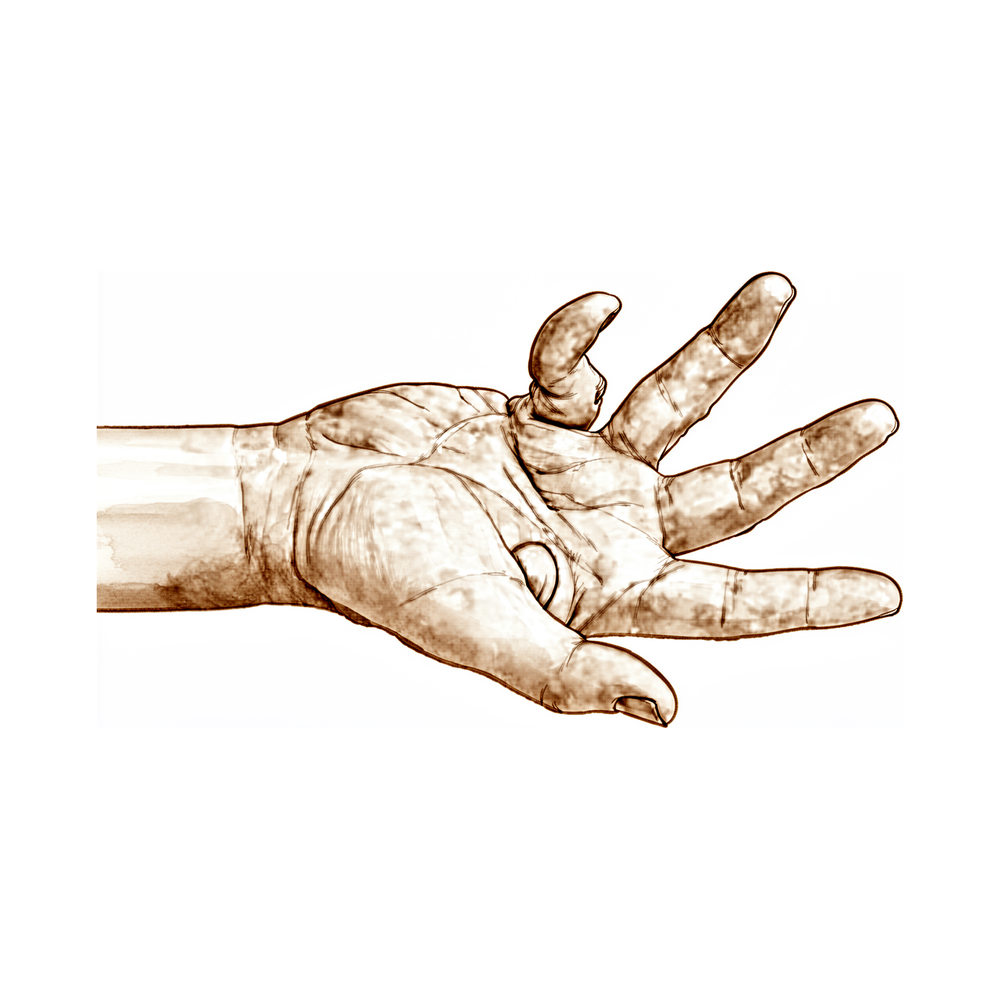
Overview
Contractures and deformities of the upper extremity encompass a diverse spectrum of pathologies, including post-traumatic stiffness, congenital conditions, and degenerative diseases such as Dupuytren’s contracture. Severe supination contracture of the forearm typically presents before age 10 in obstetric brachial plexus injury [1], while intrinsic thumb contractures require careful evaluation to distinguish among multiple etiologies [2]. In pediatric populations, elbow contracture release can yield significant range-of-motion improvements comparable to adult outcomes [11], with anterior approach surgery for flexion contractures providing satisfactory results and low complication rates [16]. For posttraumatic PIP joint flexion contractures, surgical release generally improves the deformity by 25° to 30° and shifts the flexion/extension arc into a more functional range [10]. Patients undergoing open surgical elbow contracture release after trauma must be counseled that recurrence is possible and multiple procedures may be required to achieve functional motion [4].
Management strategies vary by condition and severity. Passive stretching is a straightforward, complication-free method for improving flexion contracture in camptodactyly in infants younger than three years [3]. Initial nonsurgical treatment is warranted for the stiff digit, though results may be poorer in severe or chronic cases; surgery is considered if limited improvement occurs after 3 to 6 months of conservative management [6]. For Dupuytren’s contracture, surgery aims to correct contractures rather than cure the disease [8], with postoperative outcomes for fasciectomy and fasciotomy being successful despite common complications and likely recurrence within a few years [5]. Collagenase Clostridium histolyticum treatment has shown that most successfully treated joints maintained contractures well below surgical thresholds three years post-treatment [17], with clinical practice outcomes similar to trial findings despite lower injection rates [58]. However, older age and increased contracture correction are risk factors for skin tearing during collagenase treatment [18].
Outcomes assessment and specific procedural considerations are critical. The ACE-EB hand assessment monitors changes and serves as a post-operative outcome measure in epidermolysis bullosa [7], while patient-rated measures should accompany contracture severity in Dupuytren’s cases [25]. Preoperative deformity significantly predicts complete intraoperative and follow-up correction in Dupuytren’s disease [47]. For Boutonniere deformity, nonoperative treatment may achieve one to two grades of ROM improvement, though deformity can persist [21]. Immediate postoperative active mobilization is safe and yields similar deformity correction outcomes compared to immobilization after tendon transfer for claw deformity [55]. In pediatric patients under 13, flexor carpi ulnaris to extensor carpi radialis brevis tendon transfer requires caution due to the risk of postoperative extension deformity [75]. The choice of surgical approach for malunited hand fractures depends on deformity location, soft tissue compliance, tendon balance, and joint contracture presence [74].
Anatomy & Pathophysiology
General Principles of Deformity
Joint deformity arises from contracture of overlying skin, subcutaneous fascia, muscle, muscle imbalance, joint instability, and joint destruction [108]. Unbalanced muscle weakness or spasticity results in joint deformity which, if not corrected, will eventually become fixed [108]. Fibrosis and contracture of muscles that cross a joint cause a fixed deformity of the joint [108]. Neuromuscular conditions share commonalities in musculoskeletal manifestations, including muscular weakness or imbalance that may result in diminished function, contracture, deformity, gait disturbance, and scoliosis [22]. The hand is both an organ designed to obtain information and an organ of execution [49]. The hand functions efficiently only if the proximal joints of the limb are stable and yet mobile [49].
Skeletal Architecture
The skeleton of the hand and wrist consists of 27 bones, of which 19 are long bones [53]. The radial ray or first ray is the shortest, made up of a metacarpal and two phalanges, and possesses great freedom of movement [53]. The trapezium is angled out in front of the carpal plane so that the first metacarpal makes an angle of about 45 degrees with the second metacarpal in the sagittal plane [53]. The lengths of the metacarpals vary, with the thumb metacarpal being the shortest and the index finger metacarpal being the longest [53]. The proximal and particularly middle phalanges of the middle and ring fingers are longer than those of the index finger [53]. The skeleton of the hand presents a longitudinal and transverse concavity, giving it the shape of a cup with a palmar concavity when the thumb is placed next to the index finger [53]. The transverse axis of the palm is oblique, more distal at the metacarpophalangeal joint of the index finger and more proximal at the fifth metacarpophalangeal joint, forming an acute angle of approximately 75 degrees with the longitudinal axis [53]. The epiphyseal plates are located at the proximal ends of the phalanges and the first metacarpal, whereas they are located at the distal ends of the other metacarpals [53].
The metacarpal arch is endowed with adaptability because of the mobility of the peripheral metacarpals [85]. The index metacarpal is the most firmly fixed, while the fifth metacarpal is semi-independent with a range of flexion–extension of approximately 20 degrees [85]. The second to fifth metacarpals are bound together by various fibrous structures, the most distal of which is the deep transverse intermetacarpal ligament [85]. The deep transverse intermetacarpal ligament is also known as the interglenoid ligament because it ties together the anterior glenoid ligaments of the metacarpophalangeal articulations, known as the volar plates [85]. The longitudinal arches are composed of a fixed carpometacarpal portion and a mobile digital portion [85]. The keystones of the longitudinal arches are the metacarpophalangeal articulations, whose thick anterior glenoid capsules, the volar plates, prevent hyperextension [85]. The stability of the metacarpophalangeal joints is essential to the support of the longitudinal arch as well as of the transverse metacarpal arch [85]. An imbalance between the three muscular systems, especially an "intrinsic-minus" situation, results in a break in the continuity of the curves at the level of the metacarpophalangeal joint [85].
Musculotendinous Anatomy
Control of digital posture requires a complex balance of extrinsic and intrinsic muscle forces [52]. Extrinsic muscles have their origin outside of the hand and their insertion on the hand or carpus, whereas intrinsic muscles have both origin and insertion within the hand [52]. The extrinsic extensors run through six different fibroosseous retinacular compartments at the wrist level [52]. The first extensor compartment contains the abductor pollicis longus and the extensor pollicis brevis [52]. The second extensor compartment contains the extensor carpi radialis longus and the extensor carpi radialis brevis [52]. The third compartment contains the extensor pollicis longus, which turns abruptly radialward about Lister tubercle [52]. The fourth extensor compartment contains the extensor indicis proprius lying deep to the four tendons of the extensor digitorum communis [52]. The fifth compartment contains the extensor digiti quinti [52]. The sixth compartment contains the extensor carpi ulnaris [52].
The extensor digitorum communis tendons of the middle, ring, and little fingers are tethered together by juncturae tendinum over the dorsum of the hand proximal to the metacarpophalangeal joint [52]. The digital extensor tendons are stabilized over the mid-line of the metacarpophalangeal joint by their attachment to sagittal band fibers [52]. The sagittal band fibers insert onto the volar proximal phalanx and onto the lateral borders of the volar plate [52]. The sagittal bands normally keep the extrinsic extensor as far as possible away from the center of rotation of the metacarpophalangeal joint, thereby giving it the greatest mechanical efficiency [52]. With rupture or attenuation of the sagittal band fibers, the extrinsic extensor tendon can sublux to the ulnar side of the metacarpal head causing ulnar deviation of the finger [52]. The extrinsic finger flexors are the flexor digitorum profundus and the flexor digitorum superficialis [52]. The flexor digitorum profundus inserts on the proximal volar aspect of the distal phalanx, flexing the distal interphalangeal joint as well as the proximal interphalangeal and metacarpophalangeal joints [52]. The flexor digitorum superficialis acts as a flexor of the proximal interphalangeal and metacarpophalangeal joints [52].
There are seven interosseous muscles, four dorsal and three volar [83]. The dorsal interossei are abductors and lie to the radial side of the index and middle fingers and the ulnar side of the middle and ring fingers [83]. The volar interossei are adductors and lie to the ulnar side of the index finger and the radial side of the ring and little fingers [83]. The middle finger has two dorsal interossei (abductors) and no volar interossei (adductors) because the central axis of the hand lies within it [83]. Each dorsal interosseous muscle, with the exception of the third, has two muscle heads: a superficial head and a deep head [83]. The superficial head of the dorsal interosseous abducts and weakly flexes the proximal phalanx [83]. The deep head of the dorsal interosseous flexes and weakly abducts the proximal phalanx while extending the middle and distal phalanges [83]. Transverse fibers arch dorsally from each lateral band to join each other over the dorsum of the finger, flexing the proximal phalanx [83]. Oblique fibers (spiral fibers) from the lateral bands sweep over the distal third of the proximal phalanx to insert onto the lateral tubercles at the base of the middle phalanx, extending the middle phalanx [83]. The lateral bands are joined by the lateral slips of the extensor tendon to form the conjoined lateral band [83]. The two conjoined lateral bands to each finger unite at the distal third of the middle phalanx to form the terminal tendon, which inserts at the base of the distal phalanx to extend it [83]. The volar interossei form the ulnar lateral band of the index finger and the radial lateral band of the ring and little fingers [83]. The abductor digiti quinti and flexor digiti quinti brevis are similar in structure and function to the superficial and deep heads of the dorsal interossei, respectively [83]. The opponens digiti quinti arises from the pisohamate ligament and the hook of the hamate and inserts onto the ulnar side of the diaphysis of the fifth metacarpal, which it flexes and supinates [83].
The flexor digitorum profundus originates from the proximal ulna and the interosseous membrane [89]. The flexor digitorum superficialis has two heads: a radial head originating from the proximal shaft of the radius, and a humeral ulnar head originating from the medial humeral epicondyle and coronoid process of the ulna [89]. The flexor digitorum superficialis tendon bifurcates around the flexor digitorum profundus at the beginning of the A2 pulley [89]. The flexor digitorum superficialis tendon slips reunite distally at the Camper chiasm, with approximately half of the fibers staying on the ipsilateral side and half crossing to the contralateral side of the finger [89]. The flexor pollicis longus originates from two heads: a radial head from the proximal radius and interosseous membrane, and an accessory head from the coronoid process of the ulna and medial epicondyle of the humerus [89]. The fibroosseous tunnel, or digital flexor sheath, consists of annular pulleys which provide mechanical stability, and cruciate pulleys which provide flexibility [89]. The A2 and A4 pulleys are the most essential in maintaining the mechanical advantage of the flexor tendons [89]. The tenosynovium that lines the fibroosseous tunnel supplies both nutrition and lubrication to the poorly vascularized flexor tendons [89]. Within the sheath, tendon vascularity is supplied via the vincula system: the vinculum longus and brevis [89].
Soft Tissue and Skin Anatomy
The dorsal skin is thin, lined by a horny layer that is only 0.02 mm thick, and possesses a normal pilosebaceous system [91]. The dorsal skin has loose connections with the deeper planes, allowing free gliding and full flexion at the digital joints [91]. Flexion of the fingers produces a significant lengthening of the dorsal skin; in the middle finger, the distance between the wrist and the ungual fold shows an average increase of 3 cm as the finger goes from extension to full flexion [91]. Flexion at the metacarpophalangeal joint alone requires an average skin lengthening of 1.25 cm [91]. The dorsal and palmar areas of skin are independent because of a system of adhesions that anchors their common boundary to the underlying plane [91].
The superficial palmar fascia lies in a coronal plane beneath the palmar subcutaneous tissue and covers a triangular area of the central palm [92]. Four central bands of fascia extend distally from the proximal corner of the superficial palmar fascia toward each of the fingers, with no central band for the thumb [92]. At the distal palmar crease level, the central bands are bridged transversely by the superficial transverse palmar ligament [92]. Superficial fibers of the central bands merge with vertical retinacular fibers at the dermis’s undersurface and are more often involved with nodules than other fiber groups [92]. Intermediate fibers split transversely into two sections extending toward the lateral border of the digit base, forming the spiral band that tracks around the neurovascular bundle [92]. Deep fibers continue dorsally to merge with sagittal interosseous fascia fibers and pierce the transverse deep intermetacarpal ligament to merge with fibers of the sagittal bands of the extensor mechanism [92]. Fibers dorsal to the digital neurovascular bundle are collectively called Cleland ligament, and those palmar to the neurovascular bundle are called Grayson ligament [92]. A subdermal fascial layer borders the periphery of the web spaces from roughly the radial thumb sesamoid to the ulnar side of the small finger’s proximal flexion crease [92].
The dorsal covering of the interphalangeal articulations of the digits forms a unique cutaneous unit characterized by a considerable excess of skin when the digits are in extension [82]. The skin of the radial portion of the palm is relatively well vascularized and is the mobile portion [82]. The skin of the ulnar and distal portion of the palm has poor mobility and covers the hypothenar eminence [82]. The central triangular part of the palm has fixed, poorly vascularized skin covering almost directly the superficial palmar aponeurosis [82]. When a digit is completely flexed, the integument of the adjacent phalanges comes into contact in the zones of the flexion creases, establishing areas of cutaneous contact in the form of a diamond [82]. The sides of the diamond-shaped cutaneous contact zones do not undergo variations in length during the movements of flexion and extension [82]. The dorsal slope of the web spaces has a gradual incline and its supple skin is not adherent to the subjacent region [82]. The palmar surface of the web spaces is flat and precipitously interrupted, with skin densely adherent to the commissural skeleton [82].
Pathophysiology of Specific Deformities
Intrinsic contracture of the hand results from adherent, fibrotic, or contracted interosseous and lumbrical muscles causing an imbalance of viscoelastic forces about the extensor mechanism [34]. Severe intrinsic contracture deforms the finger into a posture in which the metacarpophalangeal joint is flexed while the proximal interphalangeal joint is extended [34]. Dislocation of the metacarpophalangeal joint may occur in rheumatoid patients with concurrent synovitis [34].
Recontracture in Dupuytren disease is a rate, not an event, and may represent Dupuytren-related soft tissue shortening, scar contracture, tendon imbalance, or combinations of these factors [33]. Early recontracture in Dupuytren disease develops during the first three postoperative months and is due to residual secondary effects of a chronic flexed posture on tendons, ligaments, skin, and other soft tissues [33]. Progressive recontracture in Dupuytren disease is a steady worsening of contracture that continues to progress after the first 3 months postoperative and is due to residual active disease [33]. Late recontracture in Dupuytren disease begins after an initial period of stability lasting a year or more and represents new disease activity in previously quiescent tissues [33]. Incomplete correction and early recontracture are more likely for PIP contractures greater than 40 degrees to 60 degrees, MCP contractures greater than 50 degrees, and composite contractures greater than 110 degrees [33]. Myofibroblast-rich cellular histology at the time of a corrective procedure correlates with progressive recontracture in Dupuytren disease [33]. Imbalances of the extensor mechanism are caused by Dupuytren's disease [78].
The pennation angle of the extensor digitorum increased with the severity of forearm and wrist and finger deformities in spastic cerebral palsy [24]. In patients with cerebral palsy, flexion deformity of the digits in the early stages is due to muscle spasticity and in late stages is due to muscle and sometimes joint contractures [155]. Severe flexion deformity of the metacarpophalangeal joint from combined forces of extrinsic digital flexors and intrinsic muscles results in great tension in the retinacular ligaments of the extensor mechanism, particularly the sagittal bands [155]. Attenuation of the sagittal bands allows the extensor tendon to dislocate with subsequent loss of extension at the metacarpophalangeal joint [155]. When sagittal bands are disrupted, extensor tendons become unstable and may sublux or dislocate ulnarily, impairing or losing active metacarpophalangeal extension [152]. Subluxation of the extensor tendons after sagittal band injury causes the intrinsic muscles to rest in a shortened position, which eventually results in myostatic contracture [152]. Contracture of the intrinsic muscles leads to swan neck deformity [152]. Adaptive shortening of the extrinsic finger flexors is required for the development of claw finger deformity [136]. Loss of intrinsic function of the foot leads to an imbalance allowing the extensor digitorum long
Classification
General Principles and Assessment
A useful classification for stiff joints directs treatment based on the specific structures responsible for the contracture, including skin, joint capsule, ligaments, tendons, and bones [153]. Contractures affecting only one anatomical structure carry a better prognosis than those involving combinations of structures [153]. It is important to distinguish a joint contracture, defined by a loss of passive motion, from a lag in motion where passive motion is largely intact but active motion is deficient [153]. Two main types of contractures exist: flexion contractures with limited passive extension and extension contractures with limited passive flexion [153].
Camptodactyly
Camptodactyly is a nontraumatic, often progressive flexion contracture of the proximal interphalangeal joint that usually affects the little finger [41]. The true incidence of camptodactyly is unclear but is estimated to affect less than 1% of the population [41].
Type I: Classic camptodactyly presents in infancy and is frequently confined to one or both little fingers [41]. It is usually sporadic but may be inherited as an autosomal dominant trait with incomplete penetrance [41].
Type II: This type shares similar clinical characteristics to type I but presents in adolescence, with females more often affected than males [41]. The deformity tends to worsen with the adolescent growth spurt [41].
Type III: This type occurs in the setting of an underlying syndrome and follows the inheritance pattern of that specific disorder [41].
An abnormal lumbrical insertion and adherent or hypoplastic flexor digitorum superficialis are the most commonly cited etiologies for camptodactyly [154]. Other potential etiologies include skin contracture, an absent central slip/extensor tendon, articular incongruity, an abnormal volar plate, and an abnormal oblique retinacular ligament [154].
Dupuytren’s Disease
Tubiana and Michon: This classification system scores contracture in a complete finger regardless of the contribution of the metacarpophalangeal joint or the proximal interphalangeal joint [139]. Stage 1 is defined as contraction of 0–45°, Stage 2 as 46–90°, Stage 3 as 91–135°, and Stage 4 as contraction over 135° [139]. The system does not allow deduction of whether a finger should be treated conservatively, minimally invasively, or with open surgery [139]. A needle fasciotomy is an alternative to surgical treatment for a 90° metacarpophalangeal joint contraction in Stage 2 Dupuytren’s disease but will be of little success for an exclusive 90° proximal interphalangeal joint contraction [139].
Revised Tubiana: This staging system incorporates total flexion deformity and additional clinical risk factors to provide a more objective and precise method for assessing disease severity [104]. It may predict surgical outcomes in Dupuytren’s disease [104].
There is a lack of standardization in the recording of data for Dupuytren’s disease, making critical comparison of techniques or results impossible due to major inconsistencies in reporting [114]. In the proliferative stage, there are no contractures, although there may be pain and tenderness at the sites of nodules [48]. A good postoperative result in the proliferative stage is defined as no recurrence of a nodule in an operative area and no impairment of function [48]. A good postoperative result in the involutional and residual stages is defined as a painless hand that is nearly normal in mobility, strength, and appearance [48]. A fair postoperative result is given to a hand whose function has been substantially improved and in which the contractures have been corrected to at least 50% [48]. A poor postoperative result is classed as a hand in which the contractures have been improved less than 50% or there is permanent stiffness [48].
Elbow Contractures
In arthroscopic arthrolysis for elbow contracture, minimal contracture is defined as an arc greater than 90°, moderate as 61°–90°, severe as 31°–60°, and very severe as less than 31° [66]. Functional range of motion for the elbow is defined as flexion ≥130° and extension ≤30° [66]. Almost functional range of motion for the elbow is defined as flexion ≥120° and extension ≤40° [66]. Flexion contracture in obstetric brachial plexus injury is not primarily related to bony changes of the elbow [19].
First Web Space
Sandzen: This classification system for first web space contractures includes mild, moderate, and severe categories [70]. Mild first web space contractures involve a localized area of skin [70].
Congenital Anomalies
Swanson Classification: First presented in the 1960s based on the understanding of limb development and causation of limb anomalies present at the time, this system was recommended by the International Federation of Societies for Surgery of the Hand Committee on Congenital Conditions as an appropriate system for use by hand and upper limb surgeons [145].
Congenital contracture of the ulnar digits is a congenital flexion deformity involving the middle, ring, and small fingers [35]. It is distinguished from ischemic contracture by a bony prominence on the proximal ulna [35]. Classification and understanding of congenital hand and upper extremity disorders has improved since the 1970s, with a primary focus on achieving optimal function through recognizing deformities, identifying surgical options, and managing patient expectations [37].
Other Considerations
Patient-rated measures should be used alongside contracture severity to assess outcomes in Dupuytren’s contracture [25]. The relationship between the severity of flexion contracture and functional disability measured by the DASH questionnaire appears to be very weak or even absent in patients with Dupuytren’s contracture [39]. A novel hand assessment tool can be used to monitor changes, track the natural history of hand contractures, guide practice, and serve as a post-operative outcome measure in children and adults with epidermolysis bullosa [7].
Clinical Presentation
General Principles and Neuromuscular Manifestations
Muscular weakness or imbalance in neuromuscular conditions may result in diminished function, contracture, deformity, gait disturbance, and scoliosis [22]. These orthopaedic manifestations are no longer confined to the pediatric population [22]. Correct diagnosis of the etiology of deformity is imperative for successful treatment [12]. Because one technique does not treat all deformities uniformly, the true etiology must be determined before surgical intervention [63]. Patient-rated measures should be used alongside contracture severity to assess outcomes [25].
Intrinsic Hand Contractures
In severe cases of intrinsic hand contracture, the hand assumes a posture with hyperflexed metacarpophalangeal joints and hyperextended proximal interphalangeal joints [34]. In mild cases, the patient may perceive a feeling of weakness or tightness when attempting to grasp a large object because the contracture hinders PIP joint motion [34]. Intrinsic contracture of the hand may result from trauma, spasticity, ischemia, rheumatologic disorders, or iatrogenic causes [34]. Considerable disability may result from intrinsic hand contracture due to weakness in grip strength, difficulty with grasping larger objects, and troubles with maintenance of hygiene [34]. Dislocation of the MCP joint may occur in rheumatoid patients with concurrent synovitis [34].
Dupuytren’s Contracture
Classic Dupuytren disease and atypical non-Dupuytren disease are distinct clinical entities that differ in presentation, etiology, treatment, and prognosis [54]. Dupuytren's disease may remain non-progressive without developing contracture, as demonstrated by individuals with palm thickening but no functional impairment over 20 to 30 years [60]. Recurrence of the contracture is always a possibility in Dupuytren's disease, with early age, Dupuytren diathesis, multifocal disease, PIP joint disease, and small finger contracture identified as potential predictive factors [36]. The relationship between severity of flexion contracture and functional disability measured by the DASH questionnaire appears to be very weak or even absent in patients with Dupuytren’s contracture [39].
Camptodactyly and Congenital Deformities
The true incidence of camptodactyly is estimated to affect less than 1% of the population [41]. Type II camptodactyly presents in adolescence, with females seeming to be more often affected than males [41]. The deformity of Type II camptodactyly tends to worsen with the adolescent growth spurt [41]. Type III camptodactyly follows the inheritance pattern of the specific disorder with which it is associated [41]. Congenital contracture of the ulnar digits is a new congenital flexion deformity involving the middle, ring, and small fingers, distinguished from ischemic contracture by a bony prominence on the proximal ulna [35]. Congenital flexion deformity with an aberrant origin of the flexor digitorum profundus may be a potentially ignored disease often mistaken for Volkmann contracture [38]. Many hand and upper extremity deformities secondary to amniotic constriction bands are encountered [57].
Elbow Contractures
Pseudo-Volkmann contracture is well described but uncommon [13]. In a cohort of surgically managed traumatic elbow injuries, 270 patients underwent a subsequent contracture release, representing a 1.4% rate [115]. The median time from index fracture treatment to contracture release was 7 months, with a range of 2 months to 10 years [115]. The mean age for patients undergoing elbow contracture release was 43 years, compared to 56 years for noncontracture patients [115].
Postburn and Posttraumatic Contractures
Most deep dermal and full-thickness burns involve the dorsum of the hand due to the natural response to use the palms to shield the face and body [45]. Postburn flexion contractures are more commonly seen in the young population [45]. The most severe dorsal hand burns result in a claw hand deformity with the MCP joints in rigid hyperextension and PIP joint flexion contracture [45]. A residual flexion contracture was described in 24% of children operated on the fingers for carpal tunnel syndrome and finger deformities, most often reducible and predominated in the distal interphalangeal joint [67].
Burn contractures are classified by severity: * Type I: Involve more than 30 degrees of passive flexion with the wrist in maximum extension but are limited to less than 30 degrees with the wrist in maximal flexion, and are limited to the skin [45]. * Type II: Fail to reach 30 degrees of passive flexion even with the wrist in maximal extension and involve scarring of both the skin and MCP joint capsule [45]. * Type III: Are the most severe, with the MCP joint position fixed in extension regardless of wrist position, often including bony or articular abnormalities [45].
Specific Finger Deformities
The swan neck deformity can progress significantly with time because of increasing DIPJ flexion contracture [30]. The presence of a proximal interphalangeal joint contracture was associated with a reduced clinical response to corticosteroid injection for trigger fingers [59]. First web space contracture classically presents with adduction and supination of the thumb metacarpal with metacarpophalangeal joint flexion [70]. The mean first web space angle normally measures 100 degrees [70].
Investigations
Physical Examination: A careful physical examination is essential to direct care and future testing if indicated [50]. The clinician must combine the patient history with this examination to pinpoint or at least narrow the scope of possible pathologic processes [50]. The primary purpose of evaluation for intrinsic contractures is to distinguish among the many causes [2].
Doppler Ultrasound: An 8-MHz Doppler tone assessment may be used to identify superficially displaced neurovascular bundles when Dupuytren cords lie beneath soft fleshy prominences [98]. However, false-negatives are possible with this assessment for identifying neurovascular bundles in Dupuytren's disease [98].
MRI: MRI is probably most useful in identifying additional pathology such as flexor tendon bowstringing in Dupuytren's disease [98]. It may also provide a quantitative noninvasive measure of cellularity of affected areas, which serves as an index of biologic activity [98]. The potential staging tool of MRI for cellularity in Dupuytren's disease has not been investigated yet on a large scale [98]. MR assessment of Dupuytren's is hindered by the resolution of current equipment, orientation issues due to multiplanar deformities of the fingers, and lack of intraoperative availability [98].
Laboratory: Diagnostic tests such as imaging and serum laboratory studies are useful in determining pathology but can be expensive, time consuming, and often nonspecific [50].
Other Considerations: The modified Terrono classification for Type 1 thumb deformity in rheumatoid arthritis could detect advanced deformity earlier and was more strongly correlated with hand function [42]. The ACE-EB hand assessment can be used to monitor changes, track natural history of hand contractures, guide practice and serve as a post-operative outcome measure in children and adults [7]. Patient-rated measures should be used alongside contracture severity to assess outcomes in patients with Dupuytren's contracture [25].
Treatment
Non-Operative
Initial nonsurgical treatment is warranted for the stiff digit, though results may be poorer in patients with more severe or chronic contractures [6]. The duration of orthotic use in weeks is significantly associated with the extent of contracture resolution for the stiff proximal interphalangeal joint [29]. Nonoperative treatment of the Boutonniere deformity can achieve one to two grades of range of motion improvement, although deformity can persist even after dedicated conservative management [21]. For supple congenital clasped thumbs, nonoperative treatments such as splinting or casting are usually preferred to allow relatively weak extensor muscles to become functional [79]. Nonoperative treatment using serial casting and splints should be tried before attempting open surgical release for posttraumatic proximal interphalangeal joint contracture [135]. Physical and occupational therapy in conjunction with bracing can help improve function in many children with arthrogryposis, potentially mitigating or avoiding the need for surgical management [96]. Nonsurgical treatment modalities for upper extremity cerebral palsy include therapy, splinting, assisted devices, and neuromuscular agents aimed at preserving joint motion and preventing contractures [97]. Intramuscular administration of botulinum toxin A is used for spasticity in upper extremity cerebral palsy and can assist with aggressive therapy or serve as a diagnostic tool for preoperative planning [97]. In the short term, severe proximal interphalangeal joint contractures caused by Dupuytren disease treated with collagenase injection appear to benefit from specific postinjection orthotic intervention and targeted exercises [65]. For camptodactyly, 20% of patients improve with nonoperative treatment, and good results have been obtained with dynamic splinting, though some flexion deformity recurred when splinting was discontinued [105]. A passive stretching protocol of 5-minute stretches 20 times daily for children younger than 3 years improved mild camptodactyly from 20 degrees to 1 degree, moderate camptodactyly from 39 to 12 degrees, and severe camptodactyly from 75 to 28 degrees [105]. It is reasonable to advise patients with mild camptodactyly deformities to live with their deformities [105]. For upper limb amyoplasia, stretching and splinting are the mainstays of early intervention, with stretching exercises initiated as early as possible to achieve passive elbow flexion past 90 degrees for hand-to-mouth activity [106].
Operative
Indications: Surgical intervention for intrinsic contractures of the hand, such as distal intrinsic release, is reserved for cases where therapy fails and must be coupled with an effective hand therapy program [14]. For upper limb amyoplasia, elbow extension contractures that fail to resolve sufficiently to allow for hand-to-mouth activities are managed with surgical release [106]. Patients with weakness of extension or snapping with persistent pain in chronic (> 6 weeks after injury) or spontaneous extensor tendon subluxations are candidates for surgical treatment, while joint contracture and arthritic deformity are considered contraindications [151]. Surgical treatment for arthrogryposis should be reserved for enhancing function, not just improving the appearance of the deformity [96]. For upper extremity cerebral palsy, surgical goals focus on optimizing function to assist with activities of daily living and hygiene, although improved aesthetics is frequently important to patients and families [97]. The earliest timing for surgical intervention in patients with upper extremity cerebral palsy is thought to be between 5 and 7 years of age [97].
Surgical Approach / Technique: For a minimal uncomplicated elbow flexion contracture (<30 to 40 degrees), extension will usually improve to 5 to 10 degrees of residual contracture without loss of flexion [99]. For moderate elbow contractures with localized heterotopic ossification and motion limited in both flexion and extension (50 to 105 degrees), a final range of motion of 20 to 130 degrees is a reasonable expectation [99]. For patients with less than 30 degrees of total elbow motion, a reasonable goal is 25 to 130 degrees, though results are variable [99]. Several techniques are often combined in a single stage to provide an improved elbow position in patients with upper motor neuron syndrome, with or without volitional control [64]. For upper limb amyoplasia, elbow extension contractures that fail to resolve sufficiently to allow for hand-to-mouth activities are managed with surgical release involving triceps tendon lengthening and posterior capsule release [106]. Muscle transfer procedures to achieve active elbow flexion in amyoplasia are usually performed when the patient is older than 5 years to allow participation in an intensive, active rehabilitation protocol [106]. Most clinical series of patients who have undergone surgical release for posttraumatic proximal interphalangeal joint flexion contractures document improvement in flexion contracture between 25° to 30° and a shift of the flexion/extension arc into a more functional range [10]. The goal of surgery for camptodactyly of the lesser digits is to achieve a more extended resting position to allow improved grasp, not to obtain a normal digit [76]. For young children with camptodactyly where the deformity disappears with wrist flexion, release of the sublimis tendon may correct the deformity and prevent worsening during growth, usually performed by 4 years of age [105]. In older children and young adults with camptodactyly who have weak extension at the proximal interphalangeal joint, release of the flexor digitorum sublimis muscle and transfer into the extensor apparatus is advised [105]. A volar release, including local skin flap and volar plate release, has been used before tendon transfer to allow passive correction of the flexion deformity in camptodactyly [105]. With Smith’s approach for camptodactyly, the postoperative arc of motion was 85 degrees (range 45 to 100 degrees) [105]. Recent studies evaluating the effectiveness of early release for camptodactyly showed no improvement between preoperative and postoperative total proximal interphalangeal joint motion, though digits tended to rest in a more extended position [105]. If the flexor digitorum superficialis tendon is normal proximally in camptodactyly, a lengthening and transposition of the insertions at a chiasma level is performed [105]. Bony abnormalities at the neck of the proximal phalanx or involving the articular surfaces of the proximal interphalangeal joint should not be explored during camptodactyly surgery because there does not seem to be any benefit [105]. The proximal interphalangeal joint is maintained in extension for 4 weeks after camptodactyly surgery in a cast, followed by a splint allowing active-resisted flexion unless the flexor digitorum superficialis was lengthened, in which case this is delayed another 8 weeks [105]. Immediate postoperative active mobilization is safe and has similar outcomes of deformity correction compared to immobilization following tendon transfer for claw deformity correction [55]. Correct diagnosis of the etiology of deformity is imperative for successful treatment of swan neck deformity in cerebral palsy [12]. Surgical reconstruction for thumb deformity in cerebral palsy aims to create a stable thumb capable of satisfactory grasp and release by decreasing deformity, balancing muscle forces, and stabilizing joints [73]. Mainstays of surgical treatment in upper extremity cerebral palsy include tendon lengthening and transfers, neurectomies, contracture releases, and joint stabilization procedures [97]. Appropriate interventions and management of expectations will optimize limb appearance and function while avoiding unexpected sequelae in the spastic upper extremity [23]. Most arthrogrypotic hand deformity surgeries result in better outcomes if performed during the early years of life using bony and/or soft-tissue interventions [32]. For upper limb amyoplasia, surgical correction of the wrist is based on the severity of the deformity and functional capacity, with children demonstrating active extension to neutral often responding well to a volar wrist fascial release with or without wrist flexor lengthening [106]. Children with amyoplasia and more severe wrist flexion contracture frequently require a bony procedure, commonly a dorsal carpal wedge osteotomy, to attain neutral wrist extension [106]. Surgical correction of thumb-in-palm deformity in amyoplasia often requires a formal volar thenar release as well as release of the adductor pollicis and first dorsal interossei muscles within the first web space [106]. For Larsen syndrome, the elbow may be fixed by webbing in the antecubital space with a flexion contracture varying from 60 to 90 degrees [56]. Despite recurrence, the mean postoperative deformity after osteotomy of the thumb delta phalanx in Rubinstein–Taybi Syndrome was significantly better than preoperatively, and the majority of patients' families subjectively reported good function [28]. Therapy or surgical correction plays a role for most children with congenital hand differences, but some will adapt to their hand difference with no intervention [44]. Severe supination contracture of the forearm in obstetric brachial plexus injury usually presents before the age of 10 years [1]. Forearm/wrist anomalies significantly compromise functional results but are not a contraindication for pollicization for congenital thumb hypoplasia [130].
Other Considerations: Postoperative outcomes for fasciectomy and fasciotomy in Dupuytren’s contracture were successful, but surgical complications were common and recurrence of a contracture was likely within a few years [5]. A simple staged procedure using a central slip facilitation device is a valid alternative for severe Dupuytren's proximal interphalangeal joint contracture, demonstrating reliable correction and acceptable patient outcomes [9]. Percutaneous needle fasciotomy for primary Dupuytren’s contracture showed a significant change in total passive extension deficit after 2 years, with the best results in correction of metacarpophalangeal joint contractures [103]. Collagenase Clostridium histolyticum (CCH) is a safe and effective treatment to improve hand function in Dupuytren's contracture, with most adverse events being minor and self-resolving [111]. Most successfully treated joints with CCH had a contracture well below the threshold for surgical intervention 3 years after treatment [17]. Long-term recurrence rates for CCH suggest disease recurrence or progression in 4 out of 6 patients with metacarpophalangeal contractures and 2 patients with proximal interphalangeal contractures, though recurrence was generally less severe than the initial contracture in the metacarpophalangeal group [20]. Older age and increased contracture correction are risk factors for skin tearing, a common complication of CCH treatment [18]. Recurrent contracture in joints previously successfully treated with CCH may be effectively retreated with up to 3 injections of CCH at a short-term follow-up of 1 year [40]. Skin grafts should possibly not be a contraindication for enzyme treatments for recurrent Dupuytren contracture [132]. There is low level of evidence that both surgical and nonsurgical treatments provide clinically important improvements for recurrent Dupuytren contracture [110]. Recontracture is defined as a rate (percent per year) rather than a single event, and definitions vary based on the need for repeat treatment, loss of a defined percent of initial correction, or loss of a specified number of correction degrees [33]. Early recontracture begins 1–2 weeks posttreatment, plateaus after 6–12 weeks, and is caused by residual secondary pathology [33]. Progressive recontracture begins 6–12 weeks post-treatment, continues to progress, and is caused by residual primary pathology [33]. Late recontracture begins after a posttreatment plateau lasting 12 months or longer and represents new disease activity or true recurrence [33]. Incomplete correction and early recontracture are more likely for proximal interphalangeal contractures greater than 40 to 60 degrees, metacarpophalangeal contractures greater than 50 degrees, and composite contractures greater than 110 degrees [33]. Myofibroblast-rich cellular histology at the time of a corrective procedure correlates with progressive recontracture [33]. The author questions the safety of Xiaflex (CCH) for patients with contractures less than 20 degrees because no published prospective trials included such patients [123]. Despite a lower injection rate in clinical practice, correction of joint contracture and range of motion with CCH was similar to findings from clinical trials [58]. Currently there remains limited evidence to guide the management of patients with Dupuytren's contracture [27]. Dupuytren's disease may remain non-progressive without developing contracture, as demonstrated by case reports of individuals with palm thickening but no functional impairment over 20 to 30 years [60].
Complications
Dupuytren Contracture
Recurrence: Recurrence of contracture is a constant possibility following treatment [36], with long-term rates suggesting recurrence in 67% of MCP joint contractures and 100% of PIP joint contractures [69]. Recurrence is likely within a few years after fasciectomy or fasciotomy [5] and was common in prospectively collected cohorts with a mean 3.8 years of follow-up [68]. Recurrences were frequent in younger patients and for PIP contractures following needle aponeurotomy [43]. Disease recurrence or progression occurred in 4 out of 6 patients with MCP contractures and 2 patients with PIP contractures at 8-year follow-up after collagenase treatment [20].
Risk Factors: Early age, Dupuytren diathesis, multifocal disease, PIP joint disease, and small finger contracture are identified as potential predictive factors for recurrence [36]. Aggressive or treatment-resistant Dupuytren’s is more common in patients with age of onset less than 50, involvement requiring treatment of the small finger, bilateral involvement, thumb or index finger involvement, Garrod’s knuckle pads, Ledderhose disease, or more than two fingers involved in one hand [147]. Patients with more aggressive Dupuytren’s have earlier clinical recurrences [147]. The frequency of repeated operations was much lower among women than men [142].
Timing and Mechanism: Recurrence does not usually occur within 12 to 18 months of surgery [142]. Recurrent contracture (not disease recurrence) could be predicted as early as 6 months after surgery [71]. Reccontracture is a rate (i.e., percent per year), not an event [33]. Early recontracture begins 1–2 weeks posttreatment and plateaus after 6–12 weeks, due to residual secondary effects of a chronic flexed posture on tendons, ligaments, skin, and other soft tissues [33]. Progressive recontracture begins 6–12 weeks post-treatment and continues to progress due to residual active disease [33]. Late recontracture begins after an initial period of stability lasting a year or more and represents new disease activity in previously quiescent tissues [33].
Severity and Management: Recurrence was generally less severe than the initial contracture in the MCP group [20] and generally less severe than the initial contracture overall [69]. Incomplete correction and early recontracture are more likely for PIP contractures greater than 40 degrees to 60 degrees, MCP contractures greater than 50 degrees, and composite contractures greater than 110 degrees [33]. Recurrent contracture in joints previously successfully treated with collagenase may be effectively retreated with up to 3 injections of collagenase at a short-term follow-up of 1 year [40]. Recurrent contracture will occur if the flexor tendon moment arms are not reconstructed after skeletal extension torque reverses the contracture [117].
Secondary Deformities: More severe contractures may result in secondary anatomic changes including proximal interphalangeal joint capsular contractures, central slip attenuation with boutonniere deformity, sagittal band attenuation with extensor subluxation, and intrinsic muscle tightness [147].
Elbow Contracture
Recurrence and Outcomes: Contracture may reoccur after open surgical elbow contracture release [4]. Some patients may require multiple procedures to achieve functional motion after elbow contracture release [4]. Improved, but not normal, elbow motion can be expected in many but not all cases of operative treatment for elbow contracture in patients twenty-one years of age or younger [81].
Prognostic Indicators: Range of motion continues to improve up to 1 year after injury, and failure to progress 3 months after injury suggests impending elbow joint contractures [72].
Hand and Finger Contractures
Mucopolysaccharidoses and Mucolipidoses: A residual flexion contracture was described in 24% of the children operated on the fingers for carpal tunnel syndrome and finger deformities in mucopolysaccharidoses and mucolipidoses [67]. Residual flexion contractures in these children were most often reducible and predominated in the distal interphalangeal joint [67].
Other Syndromic Deformities: The swan neck deformity progressed significantly with time because of increasing DIPJ flexion contracture [30]. Recurrence of deformity occurred after osteotomy of the thumb delta phalanx in Rubinstein–Taybi Syndrome [28].
Recovery
Natural History and Prognosis: Elbow flexion contracture in patients with obstetric brachial plexus injury is not primarily related to bony changes of the elbow [19]. Following traumatic elbow injury, range of motion continues to improve up to 1 year [72]. However, failure to progress in range of motion 3 months after traumatic elbow injury suggests impending elbow joint contractures [72]. The swan neck deformity associated with distal interphalangeal joint flexion contractures progresses significantly with time due to increasing contracture [30]. Additionally, contractures can occur in muscles subjected to frequent injections and may develop in adult life as well as in childhood [15].
Dupuytren's Disease Recurrence: Recurrence of a contracture is likely within a few years following fasciectomy or fasciotomy for Dupuytren’s contracture [5]. Recurrence is always a possibility, with early age, Dupuytren diathesis, multifocal disease, PIP joint disease, and small finger contracture identified as potential predictive factors [36]. In prospectively collected cohorts with a mean 3.8 years of follow-up, recurrence after Dupuytren contracture treatment was common [68]. Long-term recurrence rates suggest recurrence in 67% of MCP joint contractures and 100% of PIP joint contractures, though recurrence was generally less severe than the initial contracture [69]. Most successfully treated joints had a contracture well below the threshold for surgical intervention 3 years after treatment with collagenase Clostridium histolyticum [17]. Initial evaluation of long-term recurrence rates suggests disease recurrence or progression in 4 out of 6 patients with MCP contractures and 2 patients with PIP contractures treated with collagenase injection, though recurrence was generally less severe than the initial contracture in the MCP group [20]. Recurrences were frequent in younger patients and for PIP contractures following needle aponeurotomy for Dupuytren contracture [43]. Preoperative deformity is a significant predictor of complete intraoperative correction and complete correction at follow-up for Dupuytren’s disease [47].
Post-Operative Outcomes and Complications: Forty-seven (96 per cent) of the forty-nine shoulders had a good clinical result after distal release of deltoid muscle contracture [157]. Despite recurrence, the mean postoperative deformity was significantly better than preoperatively following osteotomy of the thumb delta phalanx in Rubinstein–Taybi Syndrome, and the majority of patients' families subjectively reported good function [28].
Non-Operative Recovery Factors: The duration of orthotic use (weeks of treatment) is significantly associated with the extent of contracture resolution for the stiff proximal interphalangeal joint [29]. In the short term, severe PIP joint contractures caused by Dupuytren disease and treated with collagenase injection benefit from specific, postinjection orthotic intervention and targeted exercises [65]. Passive stretching is a straightforward, complication-free method of improving flexion contracture in camptodactyly of different severities in infants younger than three years of age [3].
Key Evidence
- [L3] Severe supination contracture of the forearm usually presents before the age of 10 years. [1] (10.1177/17531934221121912)
- [L5] The purpose of this article is to describe the many causes of intrinsic contracture, detail how they are evaluated to distinguish among them, and delineate the surgical and nonsurgical treatment options currently in use. [2] (10.1016/j.hcl.2011.09.008)
- [L4] It is a straightforward, complication-free method of improving flexion contracture in camptodactyly of different severities in infants. [3] (10.1016/j.jhsa.2010.07.032)
- [L4] Patients must be counseled that contracture may reoccur, and some may require multiple procedures to achieve functional motion. [4] (10.1016/j.jse.2017.10.023)
- [L2] Postoperative outcomes were successful, but surgical complications were common and recurrence of a contracture was likely within a few years. [5] (10.1177/1753193410397971)
- [L5] Initial nonsurgical treatment is warranted but may have poorer results in patients with more severe or chronic contractures; surgery should be considered for limited improvement after 3 to 6 months of conservative management. [6] (10.5435/jaaos-d-18-00310)
- [L4] It can be used to monitor changes, track natural history of hand contractures, guide practice and serve as a post-operative outcome measure in children and adults. [7] (10.1177/17589983261444959)
- [Textbook] Surgery is performed to correct contractures, not to cure the disease. [8] (10.1007/978-3-642-22697-7_32)
- [L4] The simple staged procedure is a valid alternative in the management of severe Dupuytren's PIPJ contracture, demonstrating reliable, reproducible correction of the deformity and acceptable patient outcomes. [9] (10.1177/1753193412439673)
- [L4] Most clinical series of patients who have undergone surgical release document improvement in flexion contracture between 25° to 30° and a shift of the flexion/extension arc into a more functional range. [10] (10.5435/00124635-200609000-00002)
- [L4] Elbow contracture release in the pediatric and adolescent population can provide significant improvements in range of motion similar to that achieved in adults. [11] (10.1016/j.jse.2016.09.008)
- [L4] Correct diagnosis of the etiology of deformity is imperative for successful treatment. [12] (10.1016/j.jhsa.2014.01.039)
- [L4] Pseudo-Volkmann contracture is well described but uncommon. [13] (10.5435/jaaosglobal-d-18-00031)
- [L5] Surgical intervention, such as distal intrinsic release, is reserved for cases where therapy fails and must be coupled with an effective hand therapy program. [14] (10.1016/j.hcl.2011.10.001)
- [L4] Contractures can occur in muscles subjected to frequent injections and may develop in adult life as well as in childhood. [15] (10.2106/00004623-197456040-00019)
- [L4] Surgical correction of elbow flexion contractures in pediatric patients through an anterior approach leads to a satisfactory result in the majority of cases, with a low incidence of complications. [16] (10.1016/j.jse.2020.01.081)
- [L4] Most successfully treated joints had a contracture well below the threshold for surgical intervention 3 years after treatment. [17] (10.1016/j.jhsa.2012.09.028)
- [L2] Patients can be counseled before CCH treatment that older age and increased contracture correction are risk factors for this common complication. [18] (10.1016/j.jhsa.2019.06.010)
- [L4] This indicates that flexion contracture is not primarily related to bony changes of the elbow. [19] (10.5435/jaaos-d-17-00110)
- [L4] Initial evaluation of long-term recurrence rates suggests disease recurrence or progression in 4 out of 6 patients with MCP contractures and 2 patients with PIP contractures; however, recurrence was generally less severe than the initial contracture in the MCP group. [20] (10.1016/j.jhsa.2010.01.003)
- [L3] One to two grades of ROM improvement can be achieved, although deformity can persist even after dedicated conservative management. [21] (10.1016/j.jht.2025.02.013)
- [L5] Appropriate interventions and management of expectations will optimize limb appearance and function while avoiding unexpected sequelae. [23] (10.5435/jaaos-d-20-00719)
- [L3] In contrast, the pennation angle of the extensor digitorum increased with the severity of forearm and wrist and finger deformities. [24] (10.1177/17531934261480554)
- [L3] These results suggest that patient-rated measures should be used alongside contracture severity to assess outcomes. [25] (10.1016/j.jht.2010.07.006)
- [L2] Currently there remains limited evidence to guide the management of patients with Dupuytren's contracture. [27] (10.1302/0301-620x.100b9.bjj-2017-1194.r2)
- [L4] Despite the recurrence, the mean postoperative deformity was significantly better than preoperatively and the majority of patients' families subjectively reported good function. [28] (10.1177/1753193409354523)
- [L2] The duration of orthotic use (weeks of treatment) is significantly associated with the extent of contracture resolution. [29] (10.1016/j.jht.2011.09.006)
- [L5] The swan neck deformity in this individual progressed significantly with time because of increasing DIPJ flexion contracture. [30] (10.1016/j.jht.2009.11.005)
- [L5] Most arthrogrypotic hand deformity surgeries result in better outcomes if performed during the early years of life using bony and/or soft-tissue interventions. [32] (10.1016/j.jhsa.2021.10.027)
- [L5] [34] (10.5435/jaaos-21-10-581)
- [L4] Congenital contracture of the ulnar digits is a new congenital flexion deformity involving the middle, ring, and small fingers, distinguished from ischemic contracture by a bony prominence on the proximal ulna. [35] (10.1016/j.jhsa.2021.06.008)
- [L4] The authors suggest that congenital flexion deformity with an aberrant origin of the flexor digitorum profundus may be a potentially ignored disease often mistaken for Volkmann contracture. [38] (10.1016/j.jhsa.2015.07.012)
- [Textbook] The relationship between severity of flexion contracture and functional disability measured by the DASH questionnaire appears to be very weak or even absent. [39] (10.1007/978-3-642-22697-7_40)
- [L4] At a short-term follow-up of 1 year, recurrent contracture in joints previously successfully treated with CCH may be effectively retreated with up to 3 injections of CCH. [40] (10.1016/j.jhsa.2017.02.010)
- [L5] [41] (10.1016/j.hcl.2008.12.003)
- [L3] The modified classification could detect advanced deformity earlier and was more strongly correlated with hand function. [42] (10.1177/1753193419886719)
- [L4] The complication rate was low, but recurrences were frequent in younger patients and for PIP contractures. [43] (10.1016/j.jhsa.2012.01.029)
- [L5] Therapy or surgical correction plays a role for most children with congenital hand differences, but some will adapt to their hand difference with no intervention. [44] (10.1016/j.jhsa.2009.06.014)
- [L5] [45] (10.1016/j.jhsa.2014.03.018)
- [L2] Preoperative deformity is a significant predictor of complete intraoperative correction and complete correction at follow-up. [47] (10.1177/1753193409353849)
- [Textbook] Classic Dupuytren disease and atypical non-Dupuytren disease are distinct clinical entities that differ in presentation, etiology, treatment, and prognosis. [54] (10.1007/978-3-642-22697-7_1)
- [L2] Immediate postoperative active mobilization is safe and has similar outcomes of deformity correction compared to immobilization. [55] (10.1016/j.jhsa.2007.10.012)
- [L4] Many hand and upper extremity deformities secondary to ACB are encountered. [57] (10.1177/1558944717750917)
- [L3] Despite a lower injection rate, correction of joint contracture and range of motion was similar to findings from clinical trials. [58] (10.1016/j.jhsa.2013.08.114)
- [L4] The presence of a proximal interphalangeal joint contracture was associated with a reduced clinical response to corticosteroid injection. [59] (10.1177/1753193415596497)
- [L4] Dupuytren's disease may remain non-progressive without developing contracture, as demonstrated by two case reports of individuals with palm thickening but no functional impairment over 20 to 30 years. [60] (10.1177/1753193416632644)
- [L5] It emphasizes that one technique does not treat all deformities uniformly and highlights the need to determine the true etiology before surgical intervention. [63] (10.1016/j.jhsa.2022.07.008)
- [L5] Several techniques are often combined in a single stage to provide an improved elbow position in patients with and without volitional control of their affected extremities. [64] (10.1016/j.jhsa.2023.09.015)
- [L4] In the short term, it appears that severe PIP joint contractures benefit from specific, postinjection orthotic intervention and targeted exercises. [65] (10.1016/j.jhsa.2013.01.038)
- [L4] [66] (10.1016/j.jse.2018.02.068)
- [L4] [67] (10.1177/17531934211061980)
- [L2] In prospectively collected cohorts with a mean 3.8 years of follow-up, recurrence after Dupuytren contracture treatment was common. [68] (10.1016/j.jhsg.2026.100979)
- [L4] Long-term recurrence rates suggest recurrence in 67% of MCP joint contractures and 100% of PIP joint contractures, though recurrence was generally less severe than the initial contracture. [69] (10.1016/s0363-5023(09)60096-4)
- [L5] [70] (10.1016/j.jhsa.2015.06.113)
- [L3] Recurrence of contracture (not disease recurrence) could be predicted as early as 6 months after surgery for Dupuytren disease. [71] (10.1016/j.jhsa.2013.05.038)
- [L4] Range of motion continues to improve up to 1 year after injury, and failure to progress 3 months after injury suggests impending elbow joint contractures. [72] (10.1016/j.jse.2010.07.013)
- [L5] Surgical reconstruction aims to create a stable thumb capable of satisfactory grasp and release by decreasing deformity, balancing muscle forces, and stabilising joints. [73] (10.1177/1753193407087891)
- [L5] The choice of surgical approach, timing, and technique is determined by the deformity location, soft tissue compliance, tendon balance, and presence of joint contracture. [74] (10.1016/j.jhsa.2013.07.014)
- [L4] Care should be taken when performing this tendon transfer in patients less than 13 years of age because they may develop a postoperative deformity, commonly an extension deformity. [75] (10.1016/j.jhsa.2010.07.014)
- [L4] The goal of surgery is to achieve a more extended resting position to allow improved grasp, not to obtain a normal digit. [76] (10.1016/j.jhsa.2018.03.023)
- [L5] [78] (10.1016/j.jht.2024.12.017)
- [L4] [79] (10.1054/jhsb.1999.0203)
- [L4] Improved, but not normal, elbow motion can be expected in many but not all cases. [81] (10.2106/00004623-200203000-00008)
- [L4] The treatment showed a significant change in total passive extension deficit after 2 years, with the best results in correction of MCP joint contractures. [103] (10.1177/1753193411407245)
- [L4] The revised severity staging system, which incorporates total flexion deformity and additional clinical risk factors, provides a more objective and precise method for assessing Dupuytren's disease severity and may predict surgical outcomes. [104] (10.1007/s11552-007-9071-1)
- [L1] There is low level of evidence that both surgical and nonsurgical treatments provide clinically important improvements for recurrent Dupuytren contracture. [110] (10.1177/1558944721994220)
- [L2] CCH is a safe, effective treatment to improve hand function in Dupuytren's contracture, with most adverse events being minor and self-resolving. [111] (10.1177/1558944720974119)
- [L4] [114] (10.1177/1753193414560511)
- [L2] [115] (10.1016/j.jhsa.2013.05.005)
- [L4] However, recurrent contracture will occur if the flexor tendon moment arms are not reconstructed after the device reverses the contracture. [117] (10.1016/j.jhsa.2012.04.023)
- [L5] The author questions the safety of Xiaflex for patients with contractures less than 20° because no published prospective trials included such patients, despite the drug's indication covering any degree of contracture. [123] (10.1007/s11552-012-9424-2)
- [L4] Forearm/wrist anomalies significantly compromise functional results but are not a contraindication for pollicization. [130] (10.1177/1753193414535177)
- [L4] Grafts should possibly not be a contraindication for enzyme treatments for recurrent Dupuytren contracture. [132] (10.1016/j.jhsa.2013.03.063)
- [L5] Nonoperative treatment using serial casting and splints should be tried before attempting open surgical release, which should be done in selected patients. [135] (10.1016/j.jhsa.2013.03.014)
- [L5] These simulations suggest that adaptive shortening of the extrinsic finger flexors is required for the development of claw finger deformity. [136] (10.1016/j.jhsa.2019.05.007)
- [Textbook] [139] (10.1007/978-3-642-22697-7_28)
- [L4] [142] (10.1054/jhsb.1999.0154)
- [L5] [145] (10.1177/1753193417690965)
- [Textbook] [147] (10.1007/978-3-642-22697-7_39)
- [L4] [151] (10.1016/j.jhsa.2016.10.008)
- [L4] [152] (10.1016/j.jht.2008.06.013)
- [L5] [153] (10.1177/17531934221143690)
- [L4] [155] (10.1054/jhsb.1998.0005)
- [L3] Forty-seven (96 per cent) of the forty-nine shoulders had a good clinical result after distal release of the contracture. [157] (10.2106/00004623-199802000-00010)
See Also
- Dupuytren's Disease
- Joint Disease
References
[1] Natural history of elbow flexion and forearm rotation contractures in obstetric brachial plexus injury. Journal of Hand Surgery (European Volume). 2022. DOI: 10.1177/17531934221121912
[2] Intrinsic Contractures of the Thumb. Hand Clinics. 2012. DOI: 10.1016/j.hcl.2011.09.008
[3] Effect of Passive Stretching on Simple Camptodactyly in Children Younger Than Three Years of Age. The Journal of Hand Surgery. 2010. DOI: 10.1016/j.jhsa.2010.07.032
[4] Open surgical elbow contracture release after trauma: results and recommendations. Journal of Shoulder and Elbow Surgery. 2018. DOI: 10.1016/j.jse.2017.10.023
[5] The efficacy and safety of fasciectomy and fasciotomy for Dupuytren’s contracture in European patients: a structured review of published studies. Journal of Hand Surgery (European Volume). 2011. DOI: 10.1177/1753193410397971
[6] Etiology, Evaluation, and Management Options for the Stiff Digit. Journal of the American Academy of Orthopaedic Surgeons. 2019. DOI: 10.5435/jaaos-d-18-00310
[7] Assessment of hand contractures in epidermolysis bullosa (ACE-EB): Describing the development of a novel hand assessment for children. Hand Therapy. 2026. DOI: 10.1177/17589983261444959
[8] 32. No Higher Self-Reported Recurrence in Segmental Fasciectomy. Dupuytren’s Disease and Related Hyperproliferative Disorders. 2012. DOI: 10.1007/978-3-642-22697-7_32
[9] Management of severe Dupuytren’s contracture of the proximal interphalangeal joint with use of a central slip facilitation device. Journal of Hand Surgery (European Volume). 2012. DOI: 10.1177/1753193412439673
[10] Posttraumatic Proximal Interphalangeal Joint Flexion Contractures. Journal of the American Academy of Orthopaedic Surgeons. 2006. DOI: 10.5435/00124635-200609000-00002
[11] Outcomes after operative treatment of elbow contractures in the pediatric and adolescent population. Journal of Shoulder and Elbow Surgery. 2016. DOI: 10.1016/j.jse.2016.09.008
[12] Treatment of Swan Neck Deformity in Cerebral Palsy. The Journal of Hand Surgery. 2014. DOI: 10.1016/j.jhsa.2014.01.039
[13] Pseudo-Volkmann Contracture: A Case Report and Review of the Current Literature. JAAOS: Global Research and Reviews. 2018. DOI: 10.5435/jaaosglobal-d-18-00031
[14] Intrinsic Contractures of the Hand. Hand Clinics. 2012. DOI: 10.1016/j.hcl.2011.10.001
[15] Contracture of the Deltoid Muscle in the Adult after Intramuscular Injections. The Journal of Bone & Joint Surgery. 1974. DOI: 10.2106/00004623-197456040-00019
[16] Anterior elbow release for post-traumatic flexion contractures in patients 21 years or younger. Journal of Shoulder and Elbow Surgery. 2020. DOI: 10.1016/j.jse.2020.01.081
[17] Dupuytren Contracture Recurrence Following Treatment with Collagenase Clostridium Histolyticum (CORDLESS Study): 3-Year Data. The Journal of Hand Surgery. 2013. DOI: 10.1016/j.jhsa.2012.09.028
[18] Risk Factors for Skin Tearing in Collagenase Treatment of Dupuytren Contractures. The Journal of Hand Surgery. 2019. DOI: 10.1016/j.jhsa.2019.06.010
[19] Natural History of the Elbow Bony Architecture in Patients With Obstetric Brachial Plexus Injury and the Association With Flexion Contractures. Journal of the American Academy of Orthopaedic Surgeons. 2018. DOI: 10.5435/jaaos-d-17-00110
[20] Collagenase Injection as Nonsurgical Treatment of Dupuytren's Disease: 8-Year Follow-Up. The Journal of Hand Surgery. 2010. DOI: 10.1016/j.jhsa.2010.01.003
[21] Nonoperative treatment of the Boutonniere deformity: Is there a difference in outcomes?. Journal of Hand Therapy. 2025. DOI: 10.1016/j.jht.2025.02.013
[22] Orthopaedic Knowledge Update 13 Ebook Without Multimedia. Neuromuscular Disorders in Children > Introduction.
[23] The Spastic Upper Extremity in Children: Multilevel Surgical Decision-making. Journal of the American Academy of Orthopaedic Surgeons. 2021. DOI: 10.5435/jaaos-d-20-00719
[24] Biceps brachii and forearm muscle morphology related to forearm and hand deformities in spastic cerebral palsy. Journal of Hand Surgery (European Volume). 2026. DOI: 10.1177/17531934261480554
[25] Severity of Contracture and Self-reported Disability in Patients with Dupuytren’s Contracture Referred for Surgery. Journal of Hand Therapy. 2011. DOI: 10.1016/j.jht.2010.07.006
[27] Treatment of Dupuytren’s contracture. The Bone & Joint Journal. 2018. DOI: 10.1302/0301-620x.100b9.bjj-2017-1194.r2
[28] Long-term results following osteotomy of the thumb delta phalanx in Rubinstein–Taybi Syndrome. Journal of Hand Surgery (European Volume). 2009. DOI: 10.1177/1753193409354523
[29] The Long-term Relationship between Duration of Treatment and Contracture Resolution Using Dynamic Orthotic Devices for the Stiff Proximal Interphalangeal Joint: A Prospective Cohort Study. Journal of Hand Therapy. 2012. DOI: 10.1016/j.jht.2011.09.006
[30] Swan Neck Deformity after Distal Interphalangeal Joint Flexion Contractures: A Biomechanical Analysis. Journal of Hand Therapy. 2010. DOI: 10.1016/j.jht.2009.11.005
[32] The Hand in Distal Arthrogryposis. The Journal of Hand Surgery. 2022. DOI: 10.1016/j.jhsa.2021.10.027
[33] Green S Operative Hand Surgery. Recontracture Assessment.
[34] Intrinsic Contracture of the Hand: Diagnosis and Management. Journal of the American Academy of Orthopaedic Surgeons. 2013. DOI: 10.5435/jaaos-21-10-581
[35] Congenital Contracture of the Ulnar Digits and Its Differentiation From Ischemic Contracture. The Journal of Hand Surgery. 2022. DOI: 10.1016/j.jhsa.2021.06.008
[36] Chapter 32 Dupuytren Contracture. 2019.
[37] Chapter 55 Pediatric Upper Extremity Disorders. 2020.
[38] Congenital Flexion Deformity With an Aberrant Origin of the Flexor Digitorum Profundus: A Potentially Ignored Disease. The Journal of Hand Surgery. 2015. DOI: 10.1016/j.jhsa.2015.07.012
[39] 40. Severity of Contracture and Self-Reported Disability in Patients with Dupuytren’s Contracture Referred for Surgery. Dupuytren’s Disease and Related Hyperproliferative Disorders. 2012. DOI: 10.1007/978-3-642-22697-7_40
[40] Treatment of Recurrent Dupuytren Contracture in Joints Previously Effectively Treated With Collagenase Clostridium histolyticum. The Journal of Hand Surgery. 2017. DOI: 10.1016/j.jhsa.2017.02.010
[41] Failure of Differentiation: Part II (Arthrogryposis, Camptodactyly, Clinodactyly, Madelung Deformity, Trigger Finger, and Trigger Thumb). Hand Clinics. 2009. DOI: 10.1016/j.hcl.2008.12.003
[42] A modified Terrono classification for Type 1 thumb deformity in rheumatoid arthritis: a cross-sectional analysis. Journal of Hand Surgery (European Volume). 2019. DOI: 10.1177/1753193419886719
[43] Results of Needle Aponeurotomy for Dupuytren Contracture in Over 1,000 Fingers. The Journal of Hand Surgery. 2012. DOI: 10.1016/j.jhsa.2012.01.029
[44] Congenital Hand Differences. The Journal of Hand Surgery. 2009. DOI: 10.1016/j.jhsa.2009.06.014
[45] Postburn Contractures of the Hand. The Journal of Hand Surgery. 2014. DOI: 10.1016/j.jhsa.2014.03.018
[47] The association between intraoperative correction of Dupuytren’s disease and residual postoperative contracture. Journal of Hand Surgery (European Volume). 2009. DOI: 10.1177/1753193409353849
[48] Biochemical changes in the collagen of the palmar fascia in patients with Dupuytren's disease.. The Journal of bone and joint surgery. American volume. 1981.
[49] Exam Of The Hand Wrist 2Ed. INTRODUCTION.
[50] Orthopaedic Knowledge Update 13 Ebook Without Multimedia. Anatomy, Evaluation, Clinical Examination, and Imaging > Evaluation and Clinical Examination: Current Concepts.
[52] A Lange Medical Book Current Diagnosis Treatment In Orthopedics Fifth Edition. 9Hand Surgery > Image DISORDERS OF THE MUSCULATURE OF THE HAND.
[53] Exam Of The Hand Wrist 2Ed. 1.1 SKELETON OF THE HAND > The osseous skeleton.
[54] 1. Dupuytren’s Disease: Anatomy, Pathology, and Presentation. Dupuytren’s Disease and Related Hyperproliferative Disorders. 2012. DOI: 10.1007/978-3-642-22697-7_1
[55] Immediate Postoperative Active Mobilization Versus Immobilization Following Tendon Transfer for Claw Deformity Correction in the Hand. The Journal of Hand Surgery. 2008. DOI: 10.1016/j.jhsa.2007.10.012
[56] Tachdjian S Pediatric Orthopaedics From The Texas Scottish Rite Hospital For Children E Book. Plate 35.2 Scapulocostal Stabilization for Scapular Winging (Ketenjian Technique) > Larsen Syndrome.
[57] Amniotic Constriction Bands: Secondary Deformities and Their Treatments. HAND. 2018. DOI: 10.1177/1558944717750917
[58] Collagenase Clostridium Histolyticum for Dupuytren Contracture: Patterns of Use and Effectiveness in Clinical Practice. The Journal of Hand Surgery. 2013. DOI: 10.1016/j.jhsa.2013.08.114
[59] Comparative study on the effectiveness of corticosteroid injections between trigger fingers with and without proximal interphalangeal joint flexion contracture. Journal of Hand Surgery (European Volume). 2015. DOI: 10.1177/1753193415596497
[60] Non-progressive Duypytren’s disease. Journal of Hand Surgery (European Volume). 2016. DOI: 10.1177/1753193416632644
[63] Clarification of Extensor Tenotomy for Finger Deformities. The Journal of Hand Surgery. 2022. DOI: 10.1016/j.jhsa.2022.07.008
[64] Management of the Spastic Elbow Deformity in Adult Patients With Upper Motor Neuron Syndrome. The Journal of Hand Surgery. 2024. DOI: 10.1016/j.jhsa.2023.09.015
[65] The Effect of a Therapy Protocol for Increasing Correction of Severely Contracted Proximal Interphalangeal Joints Caused by Dupuytren Disease and Treated With Collagenase Injection. The Journal of Hand Surgery. 2013. DOI: 10.1016/j.jhsa.2013.01.038
[66] Prospective outcome assessment of arthroscopic arthrolysis for traumatic and degenerative elbow contracture. Journal of Shoulder and Elbow Surgery. 2018. DOI: 10.1016/j.jse.2018.02.068
[67] Carpal tunnel syndrome and finger deformities in children with mucopolysaccharidoses and mucolipidoses: a retrospective review of 52 patients. Journal of Hand Surgery (European Volume). 2021. DOI: 10.1177/17531934211061980
[68] Likelihood of Dupuytren Contracture Recurrence After Limited Fasciectomy, Needle Aponeurotomy or Collagenase Clostridium histolyticum: Systematic Review of Prospective Data With 2- to 7-Year Follow-up. Journal of Hand Surgery Global Online. 2026. DOI: 10.1016/j.jhsg.2026.100979
[69] Dupuytren's Disease and Fibroblast Contractility. The Journal of Hand Surgery. 2009. DOI: 10.1016/s0363-5023(09)60096-4
[70] Reconstruction of First Web Space Contractures. The Journal of Hand Surgery. 2015. DOI: 10.1016/j.jhsa.2015.06.113
[71] Patterns of Recontracture After Surgical Correction of Dupuytren Disease. The Journal of Hand Surgery. 2013. DOI: 10.1016/j.jhsa.2013.05.038
[72] Elbow joint contracture after traumatic injury. Journal of Shoulder and Elbow Surgery. 2011. DOI: 10.1016/j.jse.2010.07.013
[73] THE SRGICAL MANAGEMENT OF THUMB DEFORMITY IN CEREBRAL PALSY. Journal of Hand Surgery (European Volume). 2008. DOI: 10.1177/1753193407087891
[74] Malunited Fractures in the Hand. The Journal of Hand Surgery. 2014. DOI: 10.1016/j.jhsa.2013.07.014
[75] Late Deformities Following the Transfer of the Flexor Carpi Ulnaris to the Extensor Carpi Radialis Brevis in Children With Cerebral Palsy. The Journal of Hand Surgery. 2010. DOI: 10.1016/j.jhsa.2010.07.014
[76] Camptodactyly Treatment for the Lesser Digits. The Journal of Hand Surgery. 2018. DOI: 10.1016/j.jhsa.2018.03.023
[78] Extensor mechanism dysfunction and hand deformities caused by Dupuytren’s disease: Surgical and rehabilitation perspectives. Journal of Hand Therapy. 2025. DOI: 10.1016/j.jht.2024.12.017
[79] A Simple Splinting Method for Correction of Supple Congenital Clasped Thumbs in Infants. Journal of Hand Surgery. 1999. DOI: 10.1054/jhsb.1999.0203
[81] OPERATIVE TREATMENT OF ELBOW CONTRACTURE IN PATIENTS TWENTY-ONE YEARS OF AGE OR YOUNGER. The Journal of Bone and Joint Surgery-American Volume. 2002. DOI: 10.2106/00004623-200203000-00008
[82] Exam Of The Hand Wrist 2Ed. Functional cutaneous units.
[83] Green S Operative Hand Surgery. Interosseous and Hypothenar Muscles.
[85] Exam Of The Hand Wrist 2Ed. The arches of the hand > The metacarpal arch.
[89] A Lange Medical Book Current Diagnosis Treatment In Orthopedics Fifth Edition. 9Hand Surgery > FLEXOR TENDON INJURY.
[90] Exam Of The Hand Wrist 2Ed. 1.3 MOVEMENTS OF THE HAND AND WRIST > Gliding mechanisms.
[91] Exam Of The Hand Wrist 2Ed. The dorsal skin.
[92] Green S Operative Hand Surgery. PERTINENT ANATOMY.
[96] Orthopaedic Knowledge Update. Arthrogrypotic Syndromes > Summary.
[97] Orthopaedic Knowledge Update. Cerebral Palsy: Upper Extremity > Management > Treatment Considerations.
[98] Dupuytren S Disease And Related Hyperproliferative Disorders. 54. The Future of Dupuytren’s Research and Treatment > 54.4 Mechanical Measurements and Procedures > 54.4.4 Imaging.
[99] Green S Operative Hand Surgery. EXPECTATIONS AND RESULTS.
[103] Percutaneous needle fasciotomy for primary Dupuytren’s contracture. Journal of Hand Surgery (European Volume). 2011. DOI: 10.1177/1753193411407245
[104] Revised Tubiana's Staging System for Assessment of Disease Severity in Dupuytren's Disease—Preliminary Clinical Findings. HAND. 2007. DOI: 10.1007/s11552-007-9071-1
[105] Campbell S Operative Orthopaedics 4 Volume Set. RING SUBLIMIS OPPONENSPLASTY WITH ULNAR COLLATERAL LIGAMENT RECONSTRUCTION > TREATMENT.
[106] Orthopaedic Knowledge Update. Arthrogrypotic Syndromes > Upper Extremity > Amyoplasia.
[108] Apley And Solomon S Concise System Of Orthopaedics And Trauma. LAX JOINTS.
[110] Outcomes of Management of Recurrent Dupuytren Contracture: A Systematic Review and Meta-analysis. HAND. 2021. DOI: 10.1177/1558944721994220
[111] Treatment of Dupuytren’s Contracture With Collagenase: A Systematic Review. HAND. 2021. DOI: 10.1177/1558944720974119
[114] A review of the classification of Dupuytren’s disease. Journal of Hand Surgery (European Volume). 2015. DOI: 10.1177/1753193414560511
[115] Incidence of Postoperative Elbow Contracture Release in New York State. The Journal of Hand Surgery. 2013. DOI: 10.1016/j.jhsa.2013.05.005
[117] The Use of Skeletal Extension Torque in Reversing Dupuytren Contractures of the Proximal Interphalangeal Joint. The Journal of Hand Surgery. 2012. DOI: 10.1016/j.jhsa.2012.04.023
[123] What were the adverse events for Dupuytren’s patients treated with Xiaflex who had contractures less than 20°?. HAND. 2012. DOI: 10.1007/s11552-012-9424-2
[130] The results of pollicization for congenital thumb hypoplasia. Journal of Hand Surgery (European Volume). 2014. DOI: 10.1177/1753193414535177
[132] Collagenase for Recurrent Dupuytren Contracture With Skin Grafts. The Journal of Hand Surgery. 2013. DOI: 10.1016/j.jhsa.2013.03.063
[135] Management of Posttraumatic Proximal Interphalangeal Joint Contracture. The Journal of Hand Surgery. 2013. DOI: 10.1016/j.jhsa.2013.03.014
[136] The Biomechanical Basis of the Claw Finger Deformity: A Computational Simulation Study. The Journal of Hand Surgery. 2019. DOI: 10.1016/j.jhsa.2019.05.007
[139] 28. Skin Management in Treatment of Severe PIP Contracture by Homo- or Heterodigital Flaps. Dupuytren’s Disease and Related Hyperproliferative Disorders. 2012. DOI: 10.1007/978-3-642-22697-7_28
[142] The Sex Ratio and Rate of Reoperation for Dupuytren’s Contracture in Men and Women. Journal of Hand Surgery. 1999. DOI: 10.1054/jhsb.1999.0154
[145] Classification of congenital anomalies of the hand and upper limb. Journal of Hand Surgery (European Volume). 2017. DOI: 10.1177/1753193417690965
[147] 39. Hand Therapy for Dupuytren’s Contracture. Dupuytren’s Disease and Related Hyperproliferative Disorders. 2012. DOI: 10.1007/978-3-642-22697-7_39
[151] A Reconstructive Stabilization Technique for Nontraumatic or Chronic Traumatic Extensor Tendon Subluxation. The Journal of Hand Surgery. 2017. DOI: 10.1016/j.jhsa.2016.10.008
[152] Dynamic_Assist_Splinting_for_Attenuated_Sagittal_Bands_in_the_Rheumatoid_Hand_S0894113008001105. Journal of Hand Therapy. 2009. DOI: 10.1016/j.jht.2008.06.013
[153] The stiff proximal interphalangeal joint – an unsolved problem?. Journal of Hand Surgery (European Volume). 2023. DOI: 10.1177/17531934221143690
[154] Chapter 26 Congenital Hand and Wrist Differences and Brachial Plexus Birth Injury. 2019.
[155] Extensor Tendon Dislocation in Cerebral Palsy. Journal of Hand Surgery. 1999. DOI: 10.1054/jhsb.1998.0005
[157] Contracture of the Deltoid Muscle. The Journal of Bone and Joint Surgery (American Volume). 1998. DOI: 10.2106/00004623-199802000-00010