Clinicians › Elbow
Radial Head Fracture
Radial head fractures — Mason classification, conservative management, and indications for fixation or replacement.
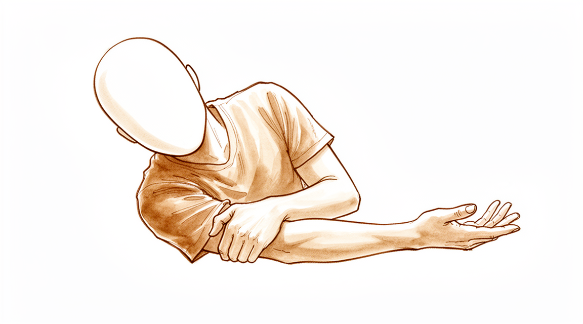
For patients: a plain-language version of this topic is available. See the patient guide.
Overview
Most radial head fractures are undisplaced and managed nonoperatively [4]. For displaced type II and III fractures, optimal treatment remains debated, with insufficient evidence to define a definitive standard of care [19, 25]. Long-term outcomes for operative fixation of displaced but stable isolated partial articular fractures show no appreciable advantage over nonoperative treatment [22]. Consequently, fracture displacement of 2 to 3 mm is not necessarily an indication for surgical fixation in isolated cases [86]. Expert consensus on treatment is challenging to obtain, and the field requires high-level clinical studies, uniform fracture classification, and standardized outcome measures to resolve management of comminuted fractures [1, 6].
Radial head replacement is a reasonable option for unreconstructable fractures, achieving effective radiocapitellar contact and improved stability with satisfactory short- and mid-term results, even in the context of combined elbow injuries [24]. Medium-term data indicate good functional and survival outcomes with a low incidence of complications for complex Mason type III and IV fractures [3, 23]. In patients with Mason type III fractures followed for five years or less, radial head replacement appears to reach better outcomes [5]. Clinical studies of metallic arthroplasty systems confirm that head replacement is a viable option for comminuted fractures and complex elbow trauma [49]. While bipolar-cemented implants show lower revision rates, the overall literature comparing excision to replacement for isolated comminuted fractures remains controversial due to small sample sizes and low evidence levels [50, 81].
The intraoperative decision to fix or replace the radial head is critical to optimize treatment outcomes [144]. In the setting of a terrible triad injury, reconstruction of comminuted radial head fractures is recommended when stable fixation can be achieved, as treatment may independently affect outcome [77]. Although no patient- or injury-related factors were associated with reoperation risk, radial head treatment itself was associated with increased risk, leading to a recommendation for fixation when feasible [91]. Overall reoperation rates are high in patients undergoing operative treatment of radial head and neck fractures [137]. Arthroscopic reduction internal fixation (ARIF) is a safe and viable option for displaced fractures [55], and radial head prosthesis is a viable short-term option for patients with instability after failed open reduction and internal fixation or radial head resection [27]. Adequate knowledge of surgical indications, implant types, and technique is essential for satisfactory outcomes when using a prosthesis for nonreconstructable fractures [41]. Generalizing outcomes after radial head arthroplasty is limited by heterogeneity in indications, injury severity, implant type, and outcome measurements [26]. Acute trauma is the most common indication for radial head arthroplasty, with the Radial Head System being the most commonly used implant [34].
Anatomy & Pathophysiology
Bony Anatomy
The radial head is disk-shaped with a diameter greater than the neck [53]. It is neither completely round nor cylindrical [119], possessing a slightly elliptical cross section that interdigitates precisely with the lesser sigmoid notch and the lateral lip of the trochlea [113]. The head features a shallow cuplike surface articulating proximally with the capitellum and medially with the radial notch of the ulna [53]. It is seated in the lesser sigmoid notch with axial contact against the distal humeral capitellum [32]. The biceps tendon inserts on the tuberosity of the radial head immediately distal to the neck [53]. The radial head has a relatively small nonarticular surface [113], defined as an arc of roughly 90 degrees with its midpoint directly lateral in the neutral position and a slightly greater margin anteriorly [113]. In the normal elbow, the radiographic height difference between the tip of the coronoid and the anterior radial head averages 5 mm [94]. The secondary ossification center of the proximal radius appears as a small sphere between the third and fifth years of life and fuses with the shaft between ages 16 and 18 years [53].
Ligaments and Stability
The primary stabilizers of the elbow are the medial collateral ligament (MCL) and lateral ulnar collateral ligament (LUCL) [119]. The radial head serves as a secondary valgus stabilizer [15, 32] and contributes considerably to neutralizing valgus forces in the axially loaded, fully extended elbow [119]. An intact radial head or its substitute can unload an injured MCL, allowing the ligament to heal without formal repair in most instances [119]. Together with the LUCL, the radial head prevents posterolateral rotatory instability [119]. The buttressing effect of the radial head prevents proximal radial migration when interosseous membrane injury is present, as seen in Essex-Lopresti-type injuries [119]. Ligaments exert the most marked influence on elbow stability, particularly when valgus and varus gravity loads are applied [112]. Small subtle effects of radial head fracture size on elbow kinematics and stability have been observed in both ligament-intact and ligament-deficient elbows [101].
Vascular Supply
The blood supply to the radial head epiphysis is derived through the more distal metaphysis because the entire radial head is covered with articular cartilage [53]. This vascular supply is limited and tenuous [113]. The limited and tenuous vascular supply may be one reason for the occasional nonunions observed after both operative and nonoperative treatment [113].
Mechanisms of Injury
Radial head fractures typically result from a fall on an outstretched hand with the forearm in pronation, resulting in an axial load on the elbow [15]. They are generally caused by longitudinal loading from a fall on an outstretched hand [32]. Most fractures occur from low-energy mechanisms such as a trip and fall on an outstretched hand [66]. Fractures may also occur from a fall onto an outstretched hand with the elbow in extension and valgus [53]. This valgus extension force may produce avulsion of the medial epicondyle, rupture of the medial collateral ligament, and fracture of the olecranon, proximal ulna, or lateral condyle [53]. Specific loading patterns produce distinct injury patterns: * Valgus load: Causes impaction of the radial head into the capitellum, commonly with rupture of the MCL [66]. * Posterolateral rotatory subluxation: Causes a partial articular shear fracture of the anterior radial head, often with rupture of the LCL [66]. * Axial forearm load: Causes impaction of the radial head into the capitellum; more severe trauma produces fracture of the coronoid or rupture of the interosseous membrane and distal radioulnar joint ligaments, termed the Essex-Lopresti injury [66]. * Elbow dislocation: Is another cause of radial head fractures [32]. In trauma involving a fall on the extended arm, the coronoid process is forced under the trochlea, which can cause a shear fracture [7]. The combination of elbow dislocation, radial head fracture, and coronoid fracture is called "the terrible triad of the elbow" [7]. Severe elbow instability and many post-traumatic complications are associated with the terrible triad [7].
Radial neck fractures may occur as a result of elbow dislocation [53]. The radial neck may be fractured by impact against the inferior aspect of the capitellum at the time of posterior dislocation or spontaneous reduction [53]. A radial head fracture may also occur with anterior dislocation of the elbow, producing anterior displacement of the head [53].
Associated Injuries
Radial head fractures can occur in isolation but are often associated with complex injuries, including elbow fractures, dislocations, and soft-tissue injuries [15]. Approximately 30% of patients with radial head fractures have other soft-tissue and skeletal injuries [15]. The incidence of associated injuries with radial head and neck fractures ranges from 11% to 90% [118]. Not all associated injuries are clinically relevant [118]. Increasing patient age, loss of cortical contact, and comminution are related to a higher incidence of associated injuries [118].
Common associated injuries include: * Ligamentous: Tears of the LCLs and/or MCLs are most commonly associated with radial head fractures [66]. Persistent symptoms in LCL injuries are seen in 11% of patients and MCL injuries in 1.5% [118]. * Bony: Dislocations of the elbow and fractures of the coronoid, capitellum, olecranon, and proximal ulna are frequent associations [66]. Ulnar fractures occur in 1.2% to 12% of patients [118]. Capitellum fractures rarely occur in tandem with radial head fractures, with an incidence of 2% [118]. * Distal and Interosseous: Associated injuries include carpal fractures, distal radioulnar joint (DRUJ) and interosseous membrane disruption, coronoid fractures, Monteggia fracture-dislocations, and capitellar fractures [15]. Rupture of the interosseous membrane is uncommon but best diagnosed and treated early, as late reconstruction is challenging and often unsatisfactory [66]. * Scaphoid: Scaphoid fractures are associated with radial head and neck fractures in 3% of trauma patients [172]. There is a 10% incidence of concomitant scaphoid and radial head fractures in men aged 18 to 30 years [172].
Posterolateral dislocation of the elbow accompanies 3% to 14% of radial head fractures [7]. Magnetic resonance imaging (MRI) can detect 60% to 80% of associated ligamentous injuries [118]. The incidence of associated injuries of the elbow with radial head fractures by MR imaging is high [33]. Capitellar osteochondral damage from radial head impaction is seen on MRI 39% to 96% of the time [118], though many findings are clinically insignificant [118]. Concomitant radial head fractures and/or elbow dislocations are associated with significantly worse patient-reported outcomes, greater stiffness, more reoperations, and a higher incidence and grade of heterotopic ossification in anteromedial coronoid facet fractures [154].
Pediatric Anatomy and Pathophysiology
In children, the cartilaginous radial head is resistant to fracture [53]. Isolated radial head fractures are rare because the immature radial head is cartilaginous [78]. Children are more likely to sustain fractures of the radial neck than fractures of the head [53]. Most children sustain fractures of the radial neck, which account for approximately 1% of all children’s fractures and 5% of pediatric elbow fractures [78]. The majority of radial neck injuries occur during a fall onto an outstretched upper extremity with the elbow in a valgus position [78]. Radial neck fractures typically occur in the metaphysis but can extend into the proximal radial physis, producing a Salter-Harris type II pattern [78]. The most common direction of angulation for radial neck fractures is lateral, followed by anterior, then posterior [78].
Approximately 50% of radial neck fractures are associated with other injuries to the elbow [53]. Radial neck fractures can occur in conjunction with an elbow dislocation, either at the time of dislocation or during reduction [78]. The fracture may be completely displaced or intraarticular and may block reduction [78]. Displaced radial neck fractures, particularly in children older than 10 years, may be associated with loss of forearm rotation [53]. In young children, the only sign of a radial head fracture may be a small metaphyseal fragment due to the unossified radial head [78]. True radial head fractures in children are at increased risk of progressive radial head subluxation, osteonecrosis, and radiocapitellar arthrosis [78].
Classification
Speed: The first classification of radial head fractures was described by Speed in 1924, distinguishing between complete and incomplete fractures of the head and neck [135].
Mason: The first widely accepted classification for radial head fractures was developed by Mason in 1954 based on 100 radial head fractures [46]. Mason described Type I fractures as non-displaced marginal fissures or fractures [46]. Type II fractures are defined as displaced marginal fractures with separation or impaction [46]. Type III fractures are displaced comminuted fractures involving the entire radial head [46].
Mason Modifications: Broberg and Morrey added a Type IV to the Mason classification, defined as a radial head fracture combined with elbow dislocation [46]. Johnston added a fourth type in 1962 to signify radial head fractures accompanied by dislocation, irrespective of displacement or fragment comminution [54]. Broberg and Morrey modified the Mason classification in 1987, suggesting that a partial radial head fracture must be at least 30% of the articular surface and displaced at least 2 mm to be considered Mason type II [54]. Hotchkiss modified the Mason classification by adding clinical criteria, defining Type II as a displaced fracture combined with mechanical blocking of joint motion or loss of joint congruity [46]. Hotchkiss defined Type III as comminution that precludes internal fixation and requires resection or prosthetic replacement [46].
Mayo Clinic: The Mayo Clinic classification considers all concomitant lesions and is preferred over Mason's classification for this reason [46]. In the Mayo Clinic classification, letters are added to the Mason type to indicate concomitant lesions, with upper case indicating treated lesions and lower case indicating untreated lesions [46].
PARMa: The Proximal and Articular Radial fractures Management (PARMa) classification is a computed tomography–based algorithm for the management of radial head and neck fractures [87].
Other Considerations: Mason and modified Mason classifications exhibit moderate inter- and intraobserver reliability [54]. These classifications provide inconsistent guidance regarding treatment or prognostic prediction [54]. A limitation of the Mason and Hotchkiss classifications is their poor intra-observer and inter-observer reproducibility [46]. The Mason and Hotchkiss classifications fail to consider concomitant lesions, which are present in nearly 80% of multi-fragment fractures [46]. Contemporary research suggests that Mason's original scheme is limited in its reliability and usefulness [65]. The Hotchkiss modification of the Mason classification may better communicate fracture severity and dictate treatment compared to Mason's original scheme [65]. Radiographs show poor inter- and intra-observer reliability for determining radial head fracture morphology [40]. Assessment of the number of fragments on radiographs is particularly inaccurate [40]. A comprehensive classification of complex fracture-dislocations of the elbow was created that appeared to be reproducible and useful for management [93]. A geometric model developed from three-dimensional computed tomography scans allows classification of partial radial head fractures involving the proximal radial ulnar joint [122]. Ninety-three percent of partial radial head fractures involve the proximal radial ulnar joint [122]. 3DCT and 3D physical modeling provide more accurate fracture classification and characterization of radial head fractures with less proposed variability in treatment [111].
Clinical Presentation
Epidemiology and Mechanism
Radial head fractures are the most common fractures occurring around the elbow [52], accounting for approximately 20% of all elbow fractures [15]. These injuries are frequently accompanied by associated osseous injuries [10]. The typical mechanism involves a fall on an outstretched hand with the forearm in pronation, resulting in an axial load on the elbow [15]. Posterolateral dislocation of the elbow accompanies 3% to 14% of radial head fractures and can occur after a fall on the (nearly) extended arm [7].
Associated Injuries
While radial head fractures can occur in isolation, they are often associated with more complex injuries, including associated elbow fractures, dislocations, and soft-tissue injuries [15]. The incidence of associated osseous injuries of the upper limb is high [37], and these injuries must be considered carefully when treating radial head fractures [30]. Fractures that initially present as uncomplicated displaced or comminuted (Mason type II and III) often have associated ligamentous and/or osteochondral injuries that may have significant implications on prognosis and appropriate treatment protocols [72]. Capitellar cartilage lesions frequently occur concomitantly with higher-grade radial head fractures, with the incidence of these lesions increasing with greater severity of radial head fractures [73].
When a radial head fracture is present, the wrist should be carefully examined for a scaphoid fracture, and vice versa [63]. Five patients have been presented with combined fractures of the carpal scaphoid and the ipsilateral radial head [38]. Most injuries found with MRI in patients with radial head fractures are not symptomatic or of clinical importance in short-term follow-up [12].
Physical Examination
The patient should be questioned carefully about concomitant wrist, forearm, or shoulder pain [15]. Lateral elbow pain and tenderness or limitation in elbow or forearm motion should alert the examiner to the possibility of a radial head fracture [15]. Pain with palpation over the radial head is a clinical finding [15].
The surgeon should examine elbow range of motion (ROM) and assess for a block to pronation/supination or flexion/extension [15]. The surgeon should examine the forearm, wrist, and elbow for: * Tenderness along the course of the interosseous membrane (Essex-Lopresti lesion) [15] * Instability of the distal radioulnar joint (DRUJ) [15] * Pain at the medial side of the elbow (medial collateral ligament [MCL]) [15] * Pain at the lateral side of the elbow (lateral collateral ligament [LCL]) [15]
It is important to determine which structures need to be repaired to avoid complications that could lead to elbow instability [17].
Imaging and Diagnosis
AP and lateral radiographs of the elbow are routinely obtained [15]. Nondisplaced fractures of the radial head may not be visible on standard radiographs [15]. However, nondisplaced fractures may be diagnosed by elevation of the anterior and posterior fat pads (the sail sign) by an intra-articular hemarthrosis [15]. The fat pad sign is usually present on the lateral projection of the elbow radiograph [32]. A positive fat pad sign on a lateral radiograph indicates that fluid is in the elbow joint, which in the acute setting is blood most commonly from a fracture [32].
The radiocapitellar view is accomplished by positioning the patient as for a lateral view but angling the tube 45° toward the shoulder [15]. For comminuted fractures, CT can delineate the location, number, and size of the fragments and is rapidly emerging as a standard imaging method for more complicated radial head fractures [15]. Radiographs show poor inter- and intra-observer reliability for determining radial head fracture morphology, with assessment of the number of fragments being particularly inaccurate [40].
Ultrasound imaging proved to be an effective method for diagnosing occult fractures of the radial head or neck when initial radiograms showed only intraarticular effusion [69]. The absence of the cortical irregularity in the transition zone of the radial head and neck can be used to correctly identify a non-fractured radial head [21]. Because apparently isolated, stable partial fractures of the radial head are infrequently displaced and observers have moderate disagreement regarding the diagnosis of displacement, it is likely that displacement is overdiagnosed [56]. Subsequent radiographs during nonoperative treatment of isolated radial head or neck fractures were unhelpful and might contribute to overtreatment [20].
Classification
The Mason classification categorizes Type I as a minimally displaced fracture, Type II as a displaced fracture, and Type III as a comminuted fracture [15]. Mason proposed a classification scheme where Type I is a nondisplaced fracture, type II is a fracture that is displaced usually involving a single large fragment, type III is a comminuted fracture, and type IV is a fracture associated with an elbow dislocation [32]. Contemporary research suggests that Mason's original scheme is limited in its reliability and usefulness, and that modifications of his classification – in particular that of Hotchkiss – may better communicate fracture severity and dictate treatment of radial head fractures [65]. Broberg and Morrey suggested that to be considered a type 2 fracture, the fragment should constitute ≥ 30% of the articular surface and be displaced by ≥ 2 mm [61].
Investigations
Plain radiography: AP and lateral radiographs of the elbow are routinely obtained for radial head fractures [15]. Nondisplaced radial head fractures may not be visible on radiographs but can be diagnosed by elevation of the anterior and posterior fat pads (the sail sign) caused by intra-articular hemarthrosis [15]. The fat pad sign on lateral radiographs indicates fluid in the elbow joint, which in the acute setting is most commonly blood from a fracture [32]. The absence of cortical irregularity in the transition zone of the radial head and neck can be used to correctly identify a non-fractured radial head [21].
CT: CT scans are useful to rule out subtle, but important, coronoid fractures that may necessitate operative intervention in the setting of radial head fracture with associated ulnar-humeral dislocation [75]. In terrible triad injuries, the imaging appearance of radial head fractures on two-dimensional computed tomography has no measurable influence on treatment recommendations [71].
MRI: It is important to determine which structures need to be repaired to avoid complications that could lead to elbow instability, as assessed by preoperative imaging correlation with intraoperative findings of lateral ulnar collateral ligament tears [17].
Arthroscopy: Elbow arthroscopy has a significant diagnostic value in radial head fractures when compared to standard radiological imaging and revealed concomitant injuries even in patients with uneventful MRI/CT [123].
Aspiration: Aspiration of the intra-articular hematoma and injection of a local anesthetic can be helpful when assessing mechanical blocks to motion [15]. If the patient is unable to tolerate a range of motion examination, aspiration of a hematoma with or without a local anesthetic can provide immediate pain relief and improve the quality of the physical examination [89].
Other Considerations: Approximately 20% of all elbow fractures involve the radial head [15]. Of patients with radial head fractures, 30% have other soft-tissue and skeletal injuries, including carpal fractures, distal radioulnar joint (DRUJ), and interosseous membrane disruption, coronoid fractures, Monteggia fracture-dislocations, capitellar fractures, and medial and lateral collateral ligament injuries [15]. When a radial head fracture is present, the wrist should be carefully examined for a scaphoid fracture [63].
Treatment
Non-Operative
Nonoperative management is indicated for minimally displaced radial head fractures (<3 mm) without a block to elbow flexion or forearm rotation [15, 92, 89]. Long-term patient-reported outcomes are excellent for isolated stable fractures of the radial head or neck [18], and conservative management of isolated Mason II fractures yields favorable therapeutic outcomes with a low incidence of complications [39]. Functional results at one year are similar between nonoperatively and operatively treated adults with isolated Mason type 2 fractures [28], and randomized trials have failed to detect significant differences in functional outcomes between surgical and nonsurgical treatment for this fracture pattern [59]. Initial immobilization in a sling for comfort is recommended for a short period (7 to 10 days or less), followed by early range-of-motion exercises [89, 15]. Immobilization in flexion casts should be avoided, as patients immobilized in flexion demonstrate significantly reduced range of movement compared with those immobilized in extension [114]. Aspiration of the hemarthrosis may be used to encourage motion; one study found that aspiration reduced articular pressure and provided pain relief, though a randomized trial found no significant differences in function or pain between aspiration alone and aspiration with local anesthetic injection [92, 61]. Conservative treatment does not always yield good results [44].
Operative
Indications: Surgical repair is indicated for significantly displaced fractures, those that block motion (especially rotation), or fractures that are part of more complicated injury patterns [15]. Specific indications include displaced fractures with a block to motion, comminuted fragments, associated elbow instability, or retained intra-articular fragments [42, 139]. Clear indications for open reduction and internal fixation (ORIF) include displaced, noncomminuted fractures that impede rotation or are associated with dislocation [42]. Fractures with greater than 2 mm of displacement and greater than 30% of the articular surface involvement (Mason II) are indications for operative fixation, although this remains controversial and unproven [42, 139]. Surgical intervention is specifically indicated when a block to forearm rotation can be attributed to the fracture [136].
Surgical Approach / Technique: ORIF is best suited for young patients with good-quality bone and three or fewer fragments [42, 139]. In young patients, the risks of ORIF must be weighed against the long-term effects of radial head arthroplasty [42]. Attempted fixation with more than three fragments is associated with fragment nonunion, osteonecrosis, fixation failure, and unpredictable forearm motion requiring subsequent hardware removal [42]. Fractures with three or more fragments have a higher incidence of unsatisfactory results with fixation [15], and fixation has a higher failure rate if associated elbow instability is present [15]. However, one series found no difference in clinical outcome or reoperation rate between simple (2 intra-articular pieces) or comminuted (3 or more intra-articular pieces) fractures treated with ORIF [85]. The use of ORIF has fallen out of favor due to technical difficulties, posterior interosseous nerve injury, osteonecrosis, and fixation failure, even with modern implants [92]. A 2002 study showed poor outcomes for fixation of fractures with more than three fragments or those with fragment diastasis or severe impaction, suggesting radial head arthroplasty was preferred in such circumstances [92]. A recent meta-analysis of randomized trials confirmed that radial head arthroplasty is superior to ORIF in these settings [92]. In one nonrandomized comparative study, complication rates were higher with ORIF relative to nonoperative treatment, while clinical outcomes were better in the nonoperative group [139]. Management of partial articular fractures tends to be more successful than complete fractures, likely due to improved stability and compromised vascularity with complete neck fractures [139].
Implant Selection: ORIF with low-profile plates or screws allows stable anatomic reduction while preserving soft tissue attachments [42]. Plates and screw fixation are predominantly used, but malpositioned fixation can impede motion [42]. Low-profile tripod screw fixation provides improved results relative to plate fixation, though screw fixation alone is only indicated for radial neck fractures without comminution [139]. Headless (1.5 to 2.4 mm) or countersunk headed screws in a tripod configuration have less stiffness and less need for implant removal relative to plates, but may be unstable in the presence of comminution [42]. Mini fragment screws (2.7 or 2.0 mm) or headless screws with differential pitch (e.g., Herbert screw, headless compression screw) should be countersunk to prevent screw prominence [15]. Plates should be placed in the “safe zone,” the part of the radial head that does not articulate with the proximal ulna, defined as the arc between lines drawn through the radial styloid and the Lister tubercle [15]. Hardware should be applied to this safe zone [15]. After reduction, plates (precontoured or mini-fragment) should be applied to the anatomic safe zone, though precontoured plates often need adjustment due to anatomical variability [42]. Care must be taken to preserve all soft tissue attachments during dissection [42]. Reduction can be provisionally held with Kirschner wires, and articular impaction or voids can be addressed with bone grafting if needed [42]. Widely displaced fractures devoid of soft tissue attachments can be reconstructed on the back table and secured to the remaining head and neck [42]. Stable fixation with low-profile plates and/or screws angling into the neck is recommended [48]. All other bony and ligamentous pathology must be addressed to avoid postoperative elbow instability [48].
Other Considerations: Radial head excision alone is contraindicated in the presence of extensive damage to primary stabilizers (MCL, coronoid, interosseous membrane, LCL) [15]. The radial head should not be excised with concomitant ligamentous or bony injury, as this leads to loss of radiocapitellar contact forces and precipitates instability [42]. Excision was first proposed over 100 years ago and still has support in the right clinical setting, with good long-term outcomes reported in the presence of a stable elbow [92]. However, if ligament instability is ignored, excision potentiates MCL laxity [92]. Lack of normal radial head-capitellum contact prevents the joint from providing posterolateral rotatory stability in the LUCL-deficient elbow and absorbing longitudinal loads [92]. Without this protective function, interosseous membrane injuries are vulnerable to poor healing, potentially leading to proximal radial migration and ulnar abutment syndrome [92]. Excision should be avoided when ligamentous instability is present [92]. A higher incidence of radiographically demonstrated posttraumatic osteoarthritis in the ulnotrochlear joint has been reported after excision, though these changes do not typically correlate with clinical symptoms [92]. Even with intact collateral ligaments, excision alters elbow kinematics and is infrequently performed [42]. Radial head excision has been documented to alter load transfer and kinematics across the elbow [139]. The benefit of routine replacement versus excision has not been evaluated in randomized clinical trials [139], and there is currently no final answer as to whether excision is superior to replacement for isolated comminuted fractures [81].
Radial Head Arthroplasty: Radial head arthroplasty is an excellent option for restoring radiocapitellar contact and stability in irreconstructable radial head and neck fractures [42]. It is preferred for unreconstructible comminuted fractures due to the high incidence of associated ligamentous and bony injuries [139]. Arthroplasty is a good treatment option for cases with more than three fracture fragments, which have a higher rate of failure with surgical fixation [15]. The most commonly used prosthesis is a modular, metallic, smooth stem noncemented design [15]. Implant options include monoblock or bipolar prostheses, smooth or porous-coated stems, and cemented or noncemented fixation [136]. A prospective study found no difference in functional outcome or range of motion between smooth and porous-coated press-fit stems, but a higher rate of radiographic and symptomatic loosening in press-fit stems [136]. A recent meta-analysis found that rigidly fixed stems (cemented or porous-coated press-fit) had a higher rate of revision and complications [136], while a second meta-analysis found the lowest rates of implant revision with cemented stems compared with porous-coated or smooth stems [136]. Further studies are needed to determine optimal implant design [136]. Prosthetic heads and stems have a wide variety of height, size, and offset to replicate native radial heads [42]. The ideal method of fixation remains elusive, but press-fit prostheses with a rough surface that do not obtain ingrowth or loosen can cause extensive osteolysis [15].
Technical Considerations for Arthroplasty: Measure the size of the radial head diameter and thickness and downsize from the measured size to avoid implant size mismatch or overstuffing [48]. Evaluate radiographically the relationship of the implant to the proximal radioulnar joint (PRUJ) and the coronoid to avoid size mismatch [48]. Fluoroscopic evaluation of the ulnohumeral joint is recommended to avoid gapping [48]. Stability examination should be performed with both trial and final implants [48]. The radial neck should be delivered atraumatically to prevent posterior interosseous nerve palsy [48]. Avoid forced retractor placement behind the neck, maintain the forearm in pronation during approach, avoid aggressive anterior and medial retraction, and do not dissect distal to the biceps tuberosity to prevent posterior interosseous nerve injury [48]. Early motion is recommended to prevent stiffness after radial head arthroplasty [48]. Avoiding overstuffing of the joint is recommended to prevent stiffness [48].
Outcomes and Complications: A report of 10-year follow-up of 16 patients treated with radial head arthroplasty showed promising midterm results, with no development of instability, loss of range of motion, or increased pain compared with the same cohort at 2-year follow-up [136]. However, 2 of the 17 patients developed radiographic osteoarthritis of the ulnohumeral joint [136]. A retrospective study of 55 patients with smooth-stemmed modular metallic radial implants showed that at a mean of 8 years postoperatively, patients had sustained good clinical outcomes with no loss of motion or strength [147]. RHR for complex radial head fractures yielded satisfactory short- to mid-term clinical outcomes, though radiographic complications were relatively high [8]. One study suggests that RHA is the best treatment choice for efficacy and safety in comminuted radial head fractures, while RHR is the safest choice to minimize postoperative complications and enable daily activities [103]. Treatment of radial head fractures may have an independent effect on outcome; reconstruction of comminuted fractures in the context of a terrible triad injury (TTI) is recommended if stable fixation can be achieved [77]. There is a high risk of further surgery after radial head replacement for unstable fractures, as seen in longer-term outcomes at a minimum follow-up of 8 years [31].
Contraindications for Arthroplasty: Radial head arthroplasty should not be performed in the setting of gross wound contamination, if the radial neck cannot be reconstructed to accept an implant, or if the capitellum is deficient or missing from an associated injury [139].
Complications
Associated Injuries
Radial head fractures are frequently accompanied by associated osseous injuries [10]. Concomitant lesions are present in nearly 80% of multi-fragment radial head fractures, particularly Type III fractures [46].
Non-Operative Complications
The complications of radial head fractures are characteristic to their classification [9]. Thirteen percent of patients with radial neck fractures require operative treatment, 21% of which heal with fair or poor outcomes [98].
Operative Complications: ORIF
ORIF of Mason type II and III radial head fractures leads to long-term positive functional outcomes [57].
Operative Complications: Resection
Radial head fractures treated by early resection arthroplasty offer satisfactory functional results in 96% of patients at long-term follow-up, in spite of the radiographic degenerative changes present in the great majority of cases [45]. If impingement symptoms of radial head develop, secondary resection yields good results [70].
Operative Complications: Arthroplasty
Radial head replacement for complex radial head fractures yielded satisfactory short- to mid-term clinical outcomes, though radiographic complications were relatively high [8]. In the short term, radial head prosthesis is a viable option for patients with instability after failed ORIF or radial head resection for radial head fractures [27]. Radial head arthroplasty for fractures has a high potential for reoperation within the first year [62]. Survival rates with uncemented radial head implants remain high at 10 years [62]. Patients report excellent Quick Disability of the Arm, Shoulder, and Hand scores at long-term follow-ups after radial head arthroplasty, despite any need for reoperation [62]. Long-term outcomes for radial head arthroplasty are satisfactory; however, there is a high complication and revision rate [80]. Implant survival for monopolar radial head replacement is 75.1% at 18 years [80]. The highest annual failure rate for monopolar radial head replacement is observed in the first postoperative year [80]. Midterm outcomes of EVOLVE radial head prosthesis are satisfactory, and associated complication rates are low [82]. Longer-term studies will be required to ascertain whether the apparent benefits of radial head arthroplasty are offset by late complications of arthroplasty, such as loosening [121]. Concomitant elbow fractures or dislocations do not affect the longer term outcomes of patients with unreconstructable radial head fractures requiring radial head arthroplasty [131]. Overlengthening is a complication of radial head replacement [74].
Recovery
Light activity (weeks): The provided evidence does not specify a typical week range for the resumption of desk work, driving, or light activities of daily living.
Full activity (months): The provided evidence does not specify a month range for the return to manual work, sport, or full range of motion and strength.
Complete recovery / outcome plateau (months): The provided evidence does not specify a month range for the stabilization of pain, strength, or final functional outcomes.
Rehabilitation protocol: The provided evidence does not detail specific physical therapy phasing, immobilisation duration, weight-bearing or range-of-motion progression schedules, or sling and brace removal timing.
Functional milestones: Radial head arthroplasty patients report excellent Quick Disability of the Arm, Shoulder, and Hand scores at long-term follow-ups, despite any need for reoperation [62].
Other Considerations: Long-term outcomes for radial head arthroplasty are satisfactory; however, there is a high complication and revision rate, resulting in implant survival of 75.1% at 18 years with the highest annual failure rate observed in the first postoperative year [80]. Although radial head arthroplasty for fractures has a high potential for reoperation within the first year, survival rates with uncemented implants remain high at 10 years [62]. Radial head replacement appeared to reach better outcomes in patients with Mason type III radial head fractures followed 5 years or less [5]. The study documents excellent long-term results following resection of the radial head for isolated fractures, with few patients complaining of pain and ranges of motion nearly normal, though pronation and supination were decreased [181]. The outcome of patients undergoing treatment for terrible triad injuries is similar whether the patient's radial head was excised or replaced [180].
Regarding operative management, there is no difference in clinical outcome or reoperation rate between simple (2 intra-articular pieces) or comminuted (3 or more intra-articular pieces) radial head fractures treated with open reduction and internal fixation [85]. The outcomes of the use of biodegradable implants for isolated radial head fractures were comparable to those of metallic implants along with a longer average time to fracture union for biodegradable implants [182].
Prognostic factors include the severity of the radial head fracture, which correlates with longitudinal forearm injury evidenced by the presence of interosseous membrane tearing [178]. Early recognition of longitudinal radioulnar dissociation can aid in timely treatment and improved outcomes, with success rates around 80% for acute cases [177].
Key Evidence
- [L5] The challenge in the coming years will be to perform high-level clinical studies to obtain consensus regarding the most appropriate treatment for comminuted radial head fractures. [1] (10.1007/s00264-018-4082-9)
- [L5] Medium-term data suggest that patients with comminuted radial head fractures do well with radial head replacement. [3] (10.1016/j.jhsa.2012.10.001)
- [L5] The majority of radial head fractures are undisplaced and do not require operative treatment. [4] (10.1016/j.cuor.2006.10.003)
- [L2] Given the available evidence, radial head replacement appeared to reach better outcomes in patients with Mason type III radial head fractures followed 5 years or less. [5] (10.1007/s00590-013-1367-y)
- [L5] Obtaining expert consensus on the treatment of radial head fractures remains challenging. [6] (10.1016/j.jse.2022.10.002)
- [L4] [7] (10.5312/wjo.v6.i11.954)
- [L4] RHR for the treatment of complex radial head fractures yielded satisfactory short- to mid-term clinical outcomes, though radiographic complications were relatively high. [8] (10.5397/cise.2020.00325)
- [L4] The complications of radial head fractures are characteristic to their classification. [9] (10.1016/j.jse.2018.11.047)
- [L4] Radial head fractures are common and frequently accompanied by associated osseous injuries. [10] (10.1016/j.jse.2009.10.015)
- [L2] Most injuries found with MRI in patients with radial head fractures are not symptomatic or of clinical importance in short-term follow-up. [12] (10.1016/j.jse.2011.06.011)
- [L3] It is important to determine which structures need to be repaired to avoid complications that could lead to elbow instability. [17] (10.1016/j.jse.2019.07.006)
- [L4] Long-term patient-reported outcomes were excellent following the nonoperative management of isolated stable fractures of the radial head or neck. [18] (10.2106/jbjs.m.01354)
- [L4] Optimal treatment of type II and III radial head fractures is still the subject of debate, and there is a strong need for randomized clinical trials and uniform fracture classification and outcome measures. [19] (10.1111/j.1758-5740.2010.00101.x)
- [L2] Subsequent radiographs during nonoperative treatment of isolated radial head or neck fractures were unhelpful and might contribute to overtreatment. [20] (10.1016/j.jse.2016.03.007)
- [Paper] The absence of the cortical irregularity can be used to correctly identify a non-fractured radial head. [21] (10.1007/s00402-016-2496-7)
- [Abstract] The long-term results of the operative treatment of displaced but stable isolated partial articular fractures of the radial head demonstrate no appreciable advantage over nonoperative treatment. [22] (10.1016/j.jse.2007.02.052)
- [L4] Radial head implants offer a reliable treatment for complex Mason type III and IV fractures, with good functional and survival outcomes and a low incidence of complications. [23] (10.1016/j.jse.2025.05.038)
- [L5] Radial head replacement is a reasonable option for unreconstructable radial head fractures as it achieves effective radiocapitellar contact and improves stability, with satisfactory short- and mid-term results even with combined elbow injuries. [24] (10.5397/cise.2020.00234)
- [L2] There is insufficient evidence to draw definitive conclusions on optimal treatment of type II-IV radial head fractures. [25] (10.1007/s00402-006-0240-4)
- [L4] The ability to generalize outcomes after radial head arthroplasty is limited by the heterogeneity of indications, injury severity, type of implant, and choice of outcome measurements. [26] (10.1016/j.ocl.2013.03.013)
- [L4] Clinically and radiographically, our results are similar to those reported in the literature, and suggest that in the short term, radial head prosthesis is a viable option for patients with instability after failed ORIF or radial head resection for radial head fractures. [27] (10.1080/17453670510045516)
- [L2] Nonoperatively treated adults with an isolated Mason type 2 radial head fracture have similar functional results after 1 year compared with operatively treated patients. [28] (10.1016/j.jse.2021.02.025)
- [L4] Associated injuries must be considered carefully when treating radial head fractures. [30] (10.1097/01.blo.0000180606.30981.78)
- [L4] [31] (10.1097/corr.0000000000000876)
- [L4] The incidence of associated injuries of the elbow with radial head fractures by MR imaging is high. [33] (10.3109/17453674.2010.483988)
- [L3] For radial head arthroplasties, acute trauma is the most common indication and Radial Head System the most commonly used implant. [34] (10.1177/1758573220987843)
- [L4] The incidence of associated, osseous injuries of the upper limb in radial head fractures is high. [37] (10.1007/s11751-008-0038-8)
- [L4] Five patients are presented with combined fractures of the carpal scaphoid and the ipsilateral radial head. [38] (10.1016/s0020-1383(73)80017-8)
- [L1] Based on the current evidence, conservative management of isolated Mason II radial head fractures yields favorable therapeutic outcomes with a low incidence of complications. [39] (10.1186/s13018-024-05039-6)
- [L3] Radiographs show poor inter- and intra-observer reliability for determining radial head fracture morphology, with assessment of the number of fragments being particularly inaccurate. [40] (10.1016/j.jseint.2021.04.012)
- [L5] Adequate knowledge of the surgical indications, types of implants, and surgical technique are essential for a satisfactory outcome when a radial head prosthesis is used for the treatment of nonreconstructable radial head fractures. [41] (10.5435/jaaos-22-10-633)
- [L1] Recommendations for surgical treatment of radial head and neck fractures according to the Mason classification can now be given with the best available evidence. [43] (10.1016/j.injury.2013.04.003)
- [L4] Conservative treatment of radial head fractures does not always yield good results. [44] (10.1007/s11678-018-0456-2)
- [L4] Radial head fractures treated by early resection arthroplasty offer satisfactory functional results in 96% of patients at long-term follow-up, in spite of the radiographic degenerative changes present in the great majority of cases. [45] (10.1016/j.jse.2010.09.005)
- [L4] [46] (10.1016/j.otsr.2015.06.026)
- [L5] Clinical outcome studies of metallic radial head arthroplasty systems indicate that head replacement is a reasonable option to offer patients with comminuted radial head fractures and complex elbow trauma. [49] (10.1016/j.jhsa.2005.12.005)
- [L4] Radial head replacement is recommended for comminuted fractures with satisfactory medium- and long-term results, though bipolar-cemented implants show lower revision rates. [50] (10.1016/j.injury.2013.09.019)
- [L5] [52] (10.1016/j.jhsa.2008.12.024)
- [L5] [54] (10.1530/eor-24-0035)
- [L4] ARIF is a safe and viable option for treating displaced radial head fractures. [55] (10.1016/j.xrrt.2024.08.001)
- [L4] Because apparently isolated, stable partial fractures of the radial head are infrequently displaced and observers have moderate disagreement regarding the diagnosis of displacement, it is likely that displacement is overdiagnosed. [56] (10.1016/j.jse.2006.10.015)
- [L4] The study results suggest that ORIF of Mason type II and III radial head fractures leads to long-term positive functional outcomes. [57] (10.1016/j.jse.2024.07.022)
- [L1] This trial was unable to detect significant differences in functional outcomes after surgical versus nonsurgical treatment of Mason type-2 radial head fractures in adults. [59] (10.2106/jbjs.21.01483)
- [L5] [61] (10.1302/0301-620x.95b2.29877)
- [L4] Although radial head arthroplasty for fractures has a high potential for reoperation within the first year, survival rates with uncemented implants remain high at 10 years, and patients report excellent Quick Disability of the Arm, Shoulder, and Hand scores at long-term follow-ups, despite any need for reoperation. [62] (10.1016/j.jhsa.2023.04.020)
- [L4] When a radial head fracture is present, the wrist should be carefully examined for a scaphoid fracture, and vice versa. [63] (10.1054/jhsb.2000.0495)
- [L5] Contemporary research suggests that Mason's original scheme is limited in its reliability and usefulness, and that modifications of his classification – in particular that of Hotchkiss – may better communicate fracture severity and dictate treatment of radial head fractures. [65] (10.1007/s11999-012-2319-2)
- [L3] Ultrasound imaging proved to be an effective method for diagnosing occult fractures of the radial head or neck when initial radiograms showed only intraarticular effusion. [69] (10.1016/j.injury.2015.10.050)
- [L3] If impingement symptoms of radial head develop, secondary resection yields good results. [70] (10.1016/j.jse.2011.02.002)
- [L3] The results of this study suggest that in terrible triad injuries, the imaging appearance of radial head fractures has no measurable influence on treatment recommendations. [71] (10.5397/cise.2022.01368)
- [L4] Radial head fractures that initially present as uncomplicated displaced or comminuted (Mason type II and III) often have associated ligamentous and/or osteochondral injuries that may have significant implications on prognosis and appropriate treatment protocols. [72] (10.1016/j.jse.2004.11.003)
- [L4] Capitellar cartilage lesions frequently occurred concomitantly with higher-grade radial head fractures, and the incidence of these lesions increased with greater severity of radial head fractures. [73] (10.1016/j.jhsa.2008.05.016)
- [L4] The review aims to shed light into overlengthening as a complication of radial head replacement and to help identify and treat it. [74] (10.1007/s00402-020-03619-9)
- [L3] Treatment of radial head fractures may have an independent effect on outcome; the authors recommend reconstruction of comminuted radial head fractures in the context of a TTI, providing stable fixation can be achieved. [77] (10.1302/0301-620x.102b12.bjj-2020-2145)
- [L3] Long-term outcomes for radial head arthroplasty are satisfactory; however, there is a high complication and revision rate, resulting in implant survival of 75.1% at 18 years with the highest annual failure rate observed in the first postoperative year. [80] (10.1016/j.jse.2020.11.031)
- [L5] There is currently no final answer to the question if radial head excision is superior to radial head replacement in the treatment of isolated comminuted radial head fractures, as overall literature is marked with controversy and consists of studies with mainly small sample sizes and of low level of evidence. [81] (10.1016/j.injury.2017.01.004)
- [L2] Midterm outcomes of EVOLVE radial head prosthesis are satisfactory, and associated complication rates are low. [82] (10.1177/1758573219850111)
- [L3] In our series, we found no difference in clinical outcome or reoperation rate between simple (2 intra-articular pieces) or comminuted (3 or more intra-articular pieces) radial head fractures treated with ORIF. [85] (10.1016/j.jse.2020.01.031)
- [L2] This retrospective review suggests that fracture displacement of 2 to 3 mm is not necessarily an indication for surgical fixation in isolated fractures of the radial head. [86] (10.1016/j.jse.2013.01.019)
- [L4] The study also provided a treatment algorithm for radial head and neck fractures. [87] (10.1016/j.jseint.2024.09.031)
- [L3] No patient- or injury-related factors were associated with the reoperation risk, but radial head treatment was associated with increased risk, leading to a recommendation for fixation when feasible. [91] (10.1097/corr.0000000000001391)
- [L3] The authors created a comprehensive classification of complex fracture-dislocations of the elbow that appeared to be reproducible and may represent a useful tool for the management of such difficult injuries. [93] (10.1016/j.jse.2011.06.003)
- [L5] This study described the relationship between the coronoid and radial head, noting that the difference in radiographic height between the tip of the coronoid and anterior radial head in the normal elbow averages 5 mm. [94] (10.1016/j.jse.2021.05.025)
- [L4] Thirteen percent of patients with radial neck fractures require operative treatment, 21% of which heal with fair or poor outcomes. [98] (10.1097/bpo.0000000000000387)
- [L4] Small subtle effects of radial head fracture size on elbow kinematics and stability were seen in both the ligament intact and ligament deficient elbows. [101] (10.1016/j.orthres.2004.06.001)
- [L1] This study suggests that RHA is the best treatment of choice for efficacy and safety in the treatment of comminuted radial head fracture, while RHR is the safest choice to minimize postoperative complications and enable patients to perform all daily life activities. [103] (10.1007/s12306-020-00679-3)
- [Paper] 3DCT and 3D physical modeling provide more accurate fracture classification and characterization of fractures of the radial head with less proposed variability in treatment. [111] (10.1007/s12593-013-0107-1)
- [L5] The ligaments have the most marked influence on stability, particularly when the upper limb is positioned such that valgus and varus gravity loads are applied to the elbow. [112] (10.1016/j.jse.2004.09.034)
- [L1] [114] (10.1016/0020-1383(94)90154-6)
- [L3] Longer-term studies will be required to ascertain whether the apparent benefits of radial head arthroplasty are offset by late complications of arthroplasty, such as loosening. [121] (10.1007/s11999-013-3331-x)
- [L3] Ninety-three percent of partial radial head fractures will involve the PRUJ and the geometric model developed allows their classification, potentially helping surgeons decide on optimal treatment. [122] (10.1177/1758573217728492)
- [L4] Elbow arthroscopy has a significant diagnostic value in radial head fractures when compared to standard radiological imaging and revealed concomitant injuries even in patients with uneventful MRI/CT. [123] (10.1186/s12891-019-2726-6)
- [L3] Concomitant elbow fractures or dislocations do not affect the longer term outcomes of patients with unreconstructable radial head fractures requiring radial head arthroplasty. [131] (10.1016/j.jse.2017.06.031)
- [L4] [135] (10.1007/s11999-007-0064-8)
- [L3] Overall reoperation rates are high in patients undergoing operative treatment of radial head and neck fractures. [137] (10.1177/1558944719837691)
- [L5] The intraoperative decision to fix or replace the radial head is critical to optimize treatment outcomes. [144] (10.1016/j.hcl.2004.06.003)
- [L3] While the overall clinical outcomes of surgically managed AMC fractures were good, concomitant radial head fractures and/or elbow dislocations were associated with significantly worse patient-reported outcomes, greater stiffness, more reoperations, and a higher incidence and grade of HO. [154] (10.1016/j.jse.2025.06.025)
- [L4] [172] (10.5435/jaaosglobal-d-19-00055)
- [L5] Early recognition of longitudinal radioulnar dissociation can aid in timely treatment and improved outcomes, with success rates around 80% for acute cases. [177] (10.1016/j.hcl.2007.01.005)
- [L3] The severity of radial head fracture correlates with longitudinal forearm injury evidenced by the presence of interosseous membrane tearing. [178] (10.1007/s11552-013-9561-2)
- [L3] The outcome of patients undergoing treatment for terrible triad injuries is similar whether the patient's radial head was excised or replaced. [180] (10.1302/0301-620x.100b11.bjj-2018-0293.r1)
- [L4] The study documents excellent long-term results following resection of the radial head for isolated fractures, with few patients complaining of pain and ranges of motion nearly normal, though pronation and supination were decreased. [181] (10.2106/00004623-198769030-00010)
- [Paper] The outcomes of the use of biodegradable implants for isolated radial head fractures were comparable to those of metallic implants along with a longer average time to fracture union for biodegradable implants. [182] (10.1016/j.injury.2019.08.005)
See Also
References
[1] Radial head arthroplasty: a historical perspective. International Orthopaedics. 2018. DOI: 10.1007/s00264-018-4082-9
[3] Radial Head Fractures. The Journal of Hand Surgery. 2012. DOI: 10.1016/j.jhsa.2012.10.001
[4] Radial head fractures in adults. Current Orthopaedics. 2007. DOI: 10.1016/j.cuor.2006.10.003
[5] Open reduction and internal-fixation versus radial head replacement in treatment of Mason type III radial head fractures. European Journal of Orthopaedic Surgery & Traumatology. 2013. DOI: 10.1007/s00590-013-1367-y
[6] Decision-making in the treatment of radial head fractures: Delphi methodology. Journal of Shoulder and Elbow Surgery. 2023. DOI: 10.1016/j.jse.2022.10.002
[7] Current concepts in the management of radial head fractures. World Journal of Orthopedics. 2015. DOI: 10.5312/wjo.v6.i11.954
[8] Short- to mid-term outcomes of radial head replacement for complex radial head fractures. Clinics in Shoulder and Elbow. 2020. DOI: 10.5397/cise.2020.00325
[9] Surgical revision of radial head fractures: a multicenter retrospective analysis of 466 cases. Journal of Shoulder and Elbow Surgery. 2019. DOI: 10.1016/j.jse.2018.11.047
[10] The epidemiology of radial head fractures. Journal of Shoulder and Elbow Surgery. 2010. DOI: 10.1016/j.jse.2009.10.015
[12] Magnetic resonance imaging in radial head fractures: most associated injuries are not clinically relevant. Journal of Shoulder and Elbow Surgery. 2011. DOI: 10.1016/j.jse.2011.06.011
[15] Aaos Comprehensive Orthopaedic Review 3. Fractures of the Elbow > I. Radial Head Fractures.
[17] Isolated displaced type II partial articular radial head fracture: correlation of preoperative imaging with intraoperative findings of lateral ulnar collateral ligament tear. Journal of Shoulder and Elbow Surgery. 2020. DOI: 10.1016/j.jse.2019.07.006
[18] Long-Term Outcomes of Isolated Stable Radial Head Fractures. Journal of Bone and Joint Surgery. 2014. DOI: 10.2106/jbjs.m.01354
[19] Management of Radial Head Fractures: Current Concepts. Shoulder & Elbow. 2011. DOI: 10.1111/j.1758-5740.2010.00101.x
[20] The yield of subsequent radiographs during nonoperative treatment of radial head and neck fractures. Journal of Shoulder and Elbow Surgery. 2016. DOI: 10.1016/j.jse.2016.03.007
[21] The cortical irregularity in the transition zone of the radial head and neck: a reliable radiographic sign of an occult radial head fracture. Archives of Orthopaedic and Trauma Surgery. 2016. DOI: 10.1007/s00402-016-2496-7
[22] Long Term Outcome Of Mason 2 Radial Head Fractures Treated With Open Reduction And Internal Fixation. Journal of Shoulder and Elbow Surgery. 2007. DOI: 10.1016/j.jse.2007.02.052
[23] Long-term survival of Acumed anatomical radial head implant for Mason type III-IV fractures: a 15-year follow-up. Journal of Shoulder and Elbow Surgery. 2026. DOI: 10.1016/j.jse.2025.05.038
[24] Radial head resection versus replacement for unreconstructable radial head fractures. Clinics in Shoulder and Elbow. 2020. DOI: 10.5397/cise.2020.00234
[25] Radial head fractures: effectiveness of conservative treatment versus surgical intervention. Archives of Orthopaedic and Trauma Surgery. 2006. DOI: 10.1007/s00402-006-0240-4
[26] Radial Head Fractures. Orthopedic Clinics of North America. 2013. DOI: 10.1016/j.ocl.2013.03.013
[27] Treatment of sequelae of radial head fractures with a bipolar radial head prosthesis: Good outcome after 1–4 years follow-up in 11 patients. Acta Orthopaedica. 2005. DOI: 10.1080/17453670510045516
[28] Operative vs. nonoperative treatment for Mason type 2 radial head fractures: a randomized controlled trial. Journal of Shoulder and Elbow Surgery. 2021. DOI: 10.1016/j.jse.2021.02.025
[30] Associated Injuries Complicating Radial Head Fractures. Clinical Orthopaedics and Related Research. 2005. DOI: 10.1097/01.blo.0000180606.30981.78
[31] High Risk of Further Surgery After Radial Head Replacement for Unstable Fractures: Longer-term Outcomes at a Minimum Follow-up of 8 Years. Clinical Orthopaedics & Related Research. 2019. DOI: 10.1097/corr.0000000000000876
[32] A Lange Medical Book Current Diagnosis Treatment In Orthopedics Fifth Edition. 2Musculoskeletal Trauma Surgery > FRACTURE OF THE RADIAL HEAD (ICD-9:813.05).
[33] Magnetic resonance imaging findings in 46 elbows with a radial head fracture. Acta Orthopaedica. 2010. DOI: 10.3109/17453674.2010.483988
[34] A registry study on radial head arthroplasties in the Netherlands: Indications, types and short-term survival. Shoulder & Elbow. 2021. DOI: 10.1177/1758573220987843
[37] The incidence of associated fractures of the upper limb in fractures of the radial head. Strategies in Trauma and Limb Reconstruction. 2008. DOI: 10.1007/s11751-008-0038-8
[38] Carpal scaphoid fracture associated with fracture of the radial head. Injury. 1973. DOI: 10.1016/s0020-1383(73)80017-8
[39] Comparison of operatively and nonoperatively treated isolated mason type II radial head fractures: a systematic review and meta-analysis. Journal of Orthopaedic Surgery and Research. 2024. DOI: 10.1186/s13018-024-05039-6
[40] Radiographic evaluation of partial articular radial head fractures: assessment of reliability. JSES International. 2021. DOI: 10.1016/j.jseint.2021.04.012
[41] Radial Head Arthroplasty. Journal of the American Academy of Orthopaedic Surgeons. 2014. DOI: 10.5435/jaaos-22-10-633
[42] Rockwood And Green S Fractures In Adults. 40: Fractures of the Proximal Forearm: Olecranon, Proximal Radius, and Radial Head > Operative Treatment of Radial Head and Neck Fractures.
[43] Clinical results after different operative treatment methods of radial head and neck fractures. Injury. 2013. DOI: 10.1016/j.injury.2013.04.003
[44] Reasons for surgical revision after conservatively treated radial head fractures—retrospective study of 70 patients. Obere Extremität. 2018. DOI: 10.1007/s11678-018-0456-2
[45] Resection arthroplasty for radial head fractures: Long-term follow-up. Journal of Shoulder and Elbow Surgery. 2011. DOI: 10.1016/j.jse.2010.09.005
[46] Radial head replacement in adults with recent fractures. Orthopaedics & Traumatology: Surgery & Research. 2016. DOI: 10.1016/j.otsr.2015.06.026
[48] Rockwood And Green S Fractures In Adults. 40: Fractures of the Proximal Forearm: Olecranon, Proximal Radius, and Radial Head > Potential Pitfalls and Preventive Measures.
[49] Radial Head Arthroplasty. The Journal of Hand Surgery. 2006. DOI: 10.1016/j.jhsa.2005.12.005
[50] Radial head, radiocapitellar and total elbow arthroplasties: A review of recent literature. Injury. 2014. DOI: 10.1016/j.injury.2013.09.019
[52] Radial Head Fractures—An Update. The Journal of Hand Surgery. 2009. DOI: 10.1016/j.jhsa.2008.12.024
[53] Tachdjian S Pediatric Orthopaedics From The Texas Scottish Rite Hospital For Children E Book. Pigmented Villonodular Synovitis and Giant Cell Tumor of the Tendon Sheath > Radial Head and Neck Fractures.
[54] Which radial head fractures are best treated surgically?. EFORT Open Reviews. 2024. DOI: 10.1530/eor-24-0035
[55] Arthroscopic reduction internal fixation for displaced radial head fractures: a systematic review of the outcomes and complications. JSES Reviews, Reports, and Techniques. 2024. DOI: 10.1016/j.xrrt.2024.08.001
[56] Apparently isolated partial articular fractures of the radial head: Prevalence and reliability of radiographically diagnosed displacement. Journal of Shoulder and Elbow Surgery. 2007. DOI: 10.1016/j.jse.2006.10.015
[57] Minimum 10-year follow-up after open reduction and internal fixation of radial head fractures Mason type II and III. Journal of Shoulder and Elbow Surgery. 2025. DOI: 10.1016/j.jse.2024.07.022
[59] In Mason Type-2 Radial Head Fracture, Surgical and Nonsurgical Treatment Did Not Differ for Functional Outcomes at 12 Months. Journal of Bone and Joint Surgery. 2021. DOI: 10.2106/jbjs.21.01483
[61] Fractures of the radial head. The Bone & Joint Journal. 2013. DOI: 10.1302/0301-620x.95b2.29877
[62] Radial Head Arthroplasty for Fracture: Implant Survivorship and Outcomes at Mean Follow-Up of 8 Years. The Journal of Hand Surgery. 2025. DOI: 10.1016/j.jhsa.2023.04.020
[63] The Incidence of Simultaneous Fractures of the Scaphoid and Radial Head. Journal of Hand Surgery. 2001. DOI: 10.1054/jhsb.2000.0495
[65] In Brief: The Mason Classification of Radial Head Fractures. Clinical Orthopaedics & Related Research. 2012. DOI: 10.1007/s11999-012-2319-2
[66] Rockwood And Green S Fractures In Adults. 39: Elbow Dislocations and Terrible Triad Injuries > Radial Head Fractures.
[69] Diagnosis of occult radial head and neck fracture in adults. Injury. 2015. DOI: 10.1016/j.injury.2015.10.050
[70] Management of the radial head and linked total elbow arthroplasty. Journal of Shoulder and Elbow Surgery. 2011. DOI: 10.1016/j.jse.2011.02.002
[71] Variation in radial head fracture treatment recommendations in terrible triad injuries is not influenced by viewing two-dimensional computed tomography. Clinics in Shoulder and Elbow. 2023. DOI: 10.5397/cise.2022.01368
[72] Radial head fractures: MRI evaluation of associated injuries. Journal of Shoulder and Elbow Surgery. 2005. DOI: 10.1016/j.jse.2004.11.003
[73] Capitellar Cartilage Injuries Concomitant With Radial Head Fractures. The Journal of Hand Surgery. 2008. DOI: 10.1016/j.jhsa.2008.05.016
[74] Overlengthening of the radial column in radial head replacement: a review of the literature and presentation of a classification system. Archives of Orthopaedic and Trauma Surgery. 2020. DOI: 10.1007/s00402-020-03619-9
[75] Green S Operative Hand Surgery. Dislocation > Fracture-Dislocation (Radial Head).
[77] Infographic: Surgical treatment of the radial head in terrible triad injuries of the elbow. The Bone & Joint Journal. 2020. DOI: 10.1302/0301-620x.102b12.bjj-2020-2145
[78] Campbell S Operative Orthopaedics 4 Volume Set. OPEN REDUCTION AND INTERNAL FIXATION FOR DISPLACED OR ENTRAPPED MEDIAL EPICONDYLE > RADIAL HEAD AND NECK FRACTURES.
[80] Long-term outcome and survival rate of monopolar radial head replacement. Journal of Shoulder and Elbow Surgery. 2021. DOI: 10.1016/j.jse.2020.11.031
[81] Is radial head resection the first choice treatment of comminuted radial head fractures without associated instability?. Injury. 2017. DOI: 10.1016/j.injury.2017.01.004
[82] Midterm outcomes of 146 EVOLVE Proline modular radial head prostheses: a systematic review. Shoulder & Elbow. 2019. DOI: 10.1177/1758573219850111
[85] Open Reduction Internal Fixation of Simple versus Comminuted Radial Head Fractures: Comparison of Clinical Outcomes. Journal of Shoulder and Elbow Surgery. 2020. DOI: 10.1016/j.jse.2020.01.031
[86] A retrospective cohort study of displaced segmental radial head fractures: is 2 mm of articular displacement an indication for surgery?. Journal of Shoulder and Elbow Surgery. 2013. DOI: 10.1016/j.jse.2013.01.019
[87] A proposal for computed tomography–based algorithm for the management of radial head and neck fractures: the Proximal and Articular Radial fractures Management (PARMa) classification. JSES International. 2025. DOI: 10.1016/j.jseint.2024.09.031
[89] Rockwood And Green S Fractures In Adults. 40: Fractures of the Proximal Forearm: Olecranon, Proximal Radius, and Radial Head > Treatment Options for Radial Head and Neck Fractures.
[91] What Factors Are Associated with Reoperation After Operative Treatment of Terrible Triad Injuries?. Clinical Orthopaedics & Related Research. 2020. DOI: 10.1097/corr.0000000000001391
[92] Orthopaedic Knowledge Update Trauma. Fractures of the Proximal Radius and Ulna and Dislocations of the Elbow > Radial Head and Neck Fractures > Treatment.
[93] Complex fracture-dislocations of the proximal ulna and radius in adults: a comprehensive classification. Journal of Shoulder and Elbow Surgery. 2011. DOI: 10.1016/j.jse.2011.06.003
[94] Computed tomography analysis of the relationship between the coronoid and the radial head. Journal of Shoulder and Elbow Surgery. 2021. DOI: 10.1016/j.jse.2021.05.025
[98] Radial Neck Fractures in Children and Adolescents. Journal of Pediatric Orthopaedics. 2016. DOI: 10.1097/bpo.0000000000000387
[101] The effect of radial head fracture size on elbow kinematics and stability. Journal of Orthopaedic Research. 2005. DOI: 10.1016/j.orthres.2004.06.001
[103] Clinical outcomes after internal fixation, arthroplasty and resection for treatment of comminuted radial head fractures: a systematic review and network meta-analysis. MUSCULOSKELETAL SURGERY. 2020. DOI: 10.1007/s12306-020-00679-3
[111] Diagnostic Accuracy of Two-Dimensional and Three-Dimensional Imaging and Modeling of Radial Head Fractures. Journal of Hand and Microsurgery. 2014. DOI: 10.1007/s12593-013-0107-1
[112] Kinematics and stability of the fractured and implant-reconstructed radial head. Journal of Shoulder and Elbow Surgery. 2005. DOI: 10.1016/j.jse.2004.09.034
[113] Green S Operative Hand Surgery. Radial Head.
[114] The non-operative management of radial head fractures: a randomized trial of three treatments. Injury. 1994. DOI: 10.1016/0020-1383(94)90154-6
[118] Rockwood And Green S Fractures In Adults. 40: Fractures of the Proximal Forearm: Olecranon, Proximal Radius, and Radial Head > Radial Head and Neck Fractures.
[119] Orthopaedic Knowledge Update Trauma. Fractures of the Proximal Radius and Ulna and Dislocations of the Elbow > Radial Head and Neck Fractures > Incidence and Classification.
[121] Fixation Versus Replacement of Radial Head in Terrible Triad: Is There a Difference in Elbow Stability and Prognosis?. Clinical Orthopaedics & Related Research. 2014. DOI: 10.1007/s11999-013-3331-x
[122] Involvement of the proximal radial ulnar joint in partial radial head fractures: a novel three-dimensional computed tomography scan evaluation method. Shoulder & Elbow. 2017. DOI: 10.1177/1758573217728492
[123] The value of elbow arthroscopy in diagnosing and treatment of radial head fractures. BMC Musculoskeletal Disorders. 2019. DOI: 10.1186/s12891-019-2726-6
[131] The effect of associated fractures/dislocations on outcomes of unreconstructable radial head fractures managed with radial head arthroplasty: a cohort comparison. Journal of Shoulder and Elbow Surgery. 2017. DOI: 10.1016/j.jse.2017.06.031
[135] Documentation of Associated Injuries Occurring With Radial Head Fracture. Clinical Orthopaedics & Related Research. 2008. DOI: 10.1007/s11999-007-0064-8
[136] Orthopaedic Knowledge Update Sports Medicine 6. Acute/Traumatic Elbow Injuries > Radial Head Fractures.
[137] Reoperation Rates and Costs of Radial Head Arthroplasty Versus Open Reduction and Internal Fixation of Radial Head and Neck Fractures: A Retrospective Database Study. HAND. 2019. DOI: 10.1177/1558944719837691
[139] Rockwood And Green S Fractures In Adults. 39: Elbow Dislocations and Terrible Triad Injuries > Operative Treatment of Radial Head Fractures.
[144] Management of comminuted radial head fractures with replacement arthroplasty. Hand Clinics. 2004. DOI: 10.1016/j.hcl.2004.06.003
[147] Rockwood And Green S Fractures In Adults. 40: Fractures of the Proximal Forearm: Olecranon, Proximal Radius, and Radial Head > Summary, Controversies, and Future Directions Related to Monteggia Fracture–Dislocations and Transolecranon Fracture–Dislocations > Annotated References.
[154] Anteromedial coronoid facet fractures with associated injuries have inferior outcomes to isolated fractures: a clinical outcomes comparison. Journal of Shoulder and Elbow Surgery. 2026. DOI: 10.1016/j.jse.2025.06.025
[172] The Incidence and Risk Factors of Scaphoid Fracture Associated With Radial Head and Neck Fracture in Trauma Patients. JAAOS: Global Research and Reviews. 2019. DOI: 10.5435/jaaosglobal-d-19-00055
[177] Longitudinal Radioulnar Dissociation: Identification and Treatment of Acute and Chronic Injuries. Hand Clinics. 2007. DOI: 10.1016/j.hcl.2007.01.005
[178] MRI Detection of Forearm Soft Tissue Injuries with Radial Head Fractures. HAND. 2013. DOI: 10.1007/s11552-013-9561-2
[180] Radial head resection versus prosthetic arthroplasty in terrible triad injury: a retrospective comparative cohort study. The Bone & Joint Journal. 2018. DOI: 10.1302/0301-620x.100b11.bjj-2018-0293.r1
[181] Resection of the radial head for fracture of the radial head. Long-term follow-up of seventeen cases. The Journal of Bone & Joint Surgery. 1987. DOI: 10.2106/00004623-198769030-00010
[182] WITHDRAWN: Internal fixation of unstable radial head fracture: A comparison of metallic and biodegradable implants. Injury. 2019. DOI: 10.1016/j.injury.2019.08.005