Clinicians › Shoulder
Surgical Materials
Fixation devices, biological augmentations, and irrigation solutions used in shoulder surgery, with a focus on biomechanical stability and healing potential.
Overview
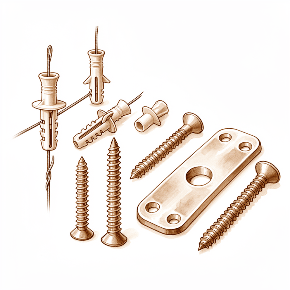
Surgical materials in orthopaedics span a diverse array of implants, grafts, and fixation devices, each governed by specific mechanical and biochemical properties. The selection of these materials is a critical determinant of clinical success, as no single alternative graft material provides all three components for bone regeneration; consequently, clinical applications are dictated by the particular structural and biochemical characteristics of the substitute [26]. While absorbable implants offer the major advantage of negating the need for subsequent removal, the mechanical strength of early-generation devices was often insufficient for certain orthopaedic applications [9]. Similarly, among newer polyblend suture materials, distinct strength and slippage profiles exist, meaning not all polyblend sutures are alike [43]. The 1954 Joint Committee report highlighted the necessity for a permanent organization to test surgical materials, conduct research, encourage standardization, and assist manufacturers, recommending the establishment of the American Surgical Materials Association with funding from three sponsoring medical organizations [5].
Complications associated with these materials are ubiquitous across all methods for managing segmental bone defects but can be limited through careful surgical judgment, patient optimization, and technique [7]. For suture anchors and tacks, complications are categorized as either technique-related or device-related issues, with prevention dependent on the surgeon’s familiarity with the devices and knowledge of their indications and limitations [2]. In soft tissue reconstruction, current evidence suggests that direct repair, autograft reconstruction, or allograft reconstruction are all viable treatment options with similar outcomes for chronic distal biceps tendon ruptures [23]. Objective measures in ACL revision reconstruction using full allograft dowel incorporation have been very good with no failures based on MARS criteria [27]. Although the ultimate fate of allografts is not known, clinical results to date appear to justify continuation of this method of treatment in selected cases in preference to amputation [12].
The utility of specific materials remains subject to ongoing evaluation and debate. The use of artificial bone grafts is expected to increase, but their effectiveness is still a subject of debate due to unclear definitions or limited market surveillance [30]. In a limited case series, a biphasic calcium phosphate bone substitute demonstrated acceptable surgical outcomes [11], while bone substitutes tested generally display a wide range in structural properties and compression strength, indicating suitability for different clinical indications [10]. Biologic reconstructions with viable bone autograft can provide more durable long-term and growing reconstructions in the pediatric population, though this comes at the expense of high short-term complication rates and donor-site morbidity [19]. A longer follow-up is necessary to assess and confirm the superiority of the induced membrane procedure in pediatric reconstruction [28]. Understanding the current evidence and appropriate indications of emerging technologies is of critical importance for their utilization [8].
Anatomy & Pathophysiology
Biomaterials and Host Interface
Material composition, structure, and manufacture determine the mechanical properties and biocompatibility of orthopaedic biomaterials [31]. Atoms and molecules at the surface of a biomaterial possess particular properties that define the interface between the material and the host [81]. Material properties, surface geometry, and local chemistry affect host response and incorporation or failure of the material [81]. Properties of biomaterial surfaces of importance include roughness, morphology, wettability, surface mobility, chemical composition, electrical charge, crystallinity, and modulus [81]. Foreign body lesions are traumatic reactive masses rather than true neoplasms that represent a response to implanted foreign material [80]. The tissue's reaction to foreign material varies with the nature of the foreign substance, the anatomic site involved, the duration of the implant, and the presence or absence of tissue hypersensitivity [80]. Glass, wood, and metal are the most common types of particulate matter associated with a foreign body reaction [80]. Inflammation may destroy the integrity of orthopaedic hardware, which may be evident on physical, radiographic, or intraoperative examination [32]. The type of suture material has a significant effect on the biomechanical behavior of the suture-tendon interface [73]. The mechanical strength of early-generation absorbable implants was insufficient for some orthopaedic applications [9].
Scapular and Clavicular Architecture
The scapula is attached to the axial skeleton by the acromioclavicular and sternoclavicular joints [85]. It is separated from the chest wall by thin gliding fibro-fatty tissue, allowing its smooth excursion over the chest wall [85]. The scapula spans the second through seventh ribs and serves as an attachment for 17 muscles [100]. It is anteverted on the chest wall approximately 30 degrees relative to the body [100]. In humans, the scapula is suspended by muscles alone and has shifted caudally from the cervical position in lower animals [94]. Broadening of the infraspinatus fossa has resulted in a change in the vector of muscle pull from the axillary border of the scapula to the glenoid fossa [94]. This adaptation allows the infraspinatus and teres minor muscles to be more effective in their roles as depressors and external rotators of the humeral head [94]. The acromion has enlarged over time in humans, reflecting the increasing role of the deltoid muscle in shoulder function [94]. The broader attachment of the deltoid on the acromion and its more distal insertion on the humerus have increased its mechanical advantage in shoulder motion [94]. The coracoid process has undergone an increase in size over time [94]. With the shoulder in 90 degrees of abduction, the coracoid extension over the glenohumeral joint can mechanically limit anterior translation of the humerus relative to the glenoid [94].
The glenoid is connected with the flat body of the scapula by the scapular neck [85]. The hook-shaped coracoid process curves forwards from the superior surface of the scapular neck [85]. The distribution of bony mass in the scapula is highly uneven, with areas of thick bone contrasting with areas that are almost translucent [85]. The highest concentration of bony mass in the scapula is located in the glenoid, the scapular neck including the base of the coracoid process, and the lateral border of the scapular body [85]. Two bony pillars extend between the glenoid and the scapular body to transmit compressive forces from the glenoid fossa [85]. The lateral pillar connects the inferior border of the glenoid with the inferior angle of the scapula [85]. The spinal pillar arises from the central part of the glenoid and continues medially to become part of the base of the scapular spine [85]. The weakest bone in the scapula is located primarily in the central part of the biomechanical body, specifically in the infraspinous fossa [85]. The weakest area of the circumference of the biomechanical body of the scapula is the spinomedial angle, which is the connection of the scapular spine and the medial border of the scapula [85].
The clavicle is the only long bone to ossify by intramembranous ossification [86, 93]. It is the first bone in the body to ossify at 5 weeks gestation and the last to fuse with the medial epiphysis at 25 years of age [100]. Fracture of the clavicle is the most common musculoskeletal birth injury [100]. The primary blood supply to the clavicle is periosteal, and no nutrient artery is present [86, 93]. The clavicle serves as the primary stabilizer between the axial skeleton via the sternoclavicular joint and the appendicular skeleton via the acromioclavicular joint [93]. It forms a unique S-shaped curve on the axial view [93]. The distal clavicle is flat in the anteroposterior plane [93]. The clavicle is subcutaneous, and its muscular envelope includes the platysma, pectoralis major, deltoid, and some of the strap muscles of the neck [93].
The superior shoulder suspensory complex provides a stable connection between the scapula and the axial skeleton [86, 93]. It is a bone-soft tissue ring that provides a stable connection of the glenoid and scapula to the clavicle [93]. The superior shoulder suspensory complex is composed of the glenoid, the coracoid process, the coracoclavicular ligaments, the distal clavicle, the acromioclavicular joint, and the acromion [86]. Alternatively, it is described as composed of four bony landmarks: the distal clavicle, acromion, coracoid process, and glenoid neck, and the supporting ligamentous complexes of the acromioclavicular joint and the coracoclavicular ligaments [93]. The coracoclavicular ligaments are the primary stabilizers to superior vertical translation of the distal clavicle [86, 93]. Within the coracoclavicular complex, the conoid ligament is medial and the trapezoid ligament is lateral [93]. The scapula has only one true diarthrodial articulation, the acromioclavicular joint [86]. The sternoclavicular joint is the only true diarthrodial articulation between the upper appendicular and axial skeletons [86]. The posterior sternoclavicular joint capsule and ligaments are the primary stabilizers to anterior and posterior translation of the medial clavicle [86]. The superior and posterior acromioclavicular ligaments are the primary stabilizers to anterior and posterior horizontal translation of the clavicle [86].
The acromion has three ossification centers: the metacromion, the mesoacromion, and the preacromion [86]. Failure of fusion of the acromial ossification centers results in os acromiale [86]. Os acromiale is incomplete fusion of secondary ossification centers, most commonly between the mesoacromion and meta-acromion [100]. The coracobrachialis muscle and the short head of the biceps tendon originate from the coracoid process [86]. The pectoralis minor muscle inserts onto the medial coracoid process [86]. The subchondral bone of the glenoid is relatively flat, and the articular concavity is augmented by cartilage and a circumferential labrum [86]. The glenoid averages 5 degrees of retroversion in relation to the axis of the scapular body [86]. The glenoid is retroverted approximately 5 degrees relative to the scapular body [100]. Normal shoulder motion is approximately two-thirds glenohumeral and one-third scapulothoracic [86]. The coracoacromial ligament contributes to anterosuperior stability in rotator cuff deficiency and should be preserved with irreparable cuff tears to prevent anterosuperior escape [100]. The acromial branch of the thoracoacromial artery runs on the medial aspect of the coracoacromial ligament [100]. The coracoacromial ligament is the arthroscopic landmark for a complete release of the rotator interval for adhesive capsulitis [100].
Proximal Humerus and Glenohumeral Joint
The proximal humerus anatomy comprises four main parts: the humeral head, greater tuberosity, lesser tuberosity, and humeral shaft [82]. The articular head of the humerus is spherical and has a diameter of 37 to 57 mm [82]. The most superior portion of the articular surface of the humeral head averages 8 mm above the greater tuberosity [82]. The humeral version averages 29.8 degrees with a range of 10 to 55 degrees [82]. The humeral head is inclined approximately 130 degrees with respect to the humeral shaft [82]. The humeral head is retroverted an average of 30 degrees [84]. The humeral head averages 19 degrees of retroversion and 41 degrees of inclination [86]. The neck-shaft angle measures an average of 135 degrees [84]. The anatomic neck of the proximal humerus is located at the junction of the articular surface and the tuberosities [82]. The anatomic neck lies at the junction of the head and the tuberosities [84]. The surgical neck represents an indistinct region below the tuberosities but above the humeral shaft [82]. The surgical neck lies below the greater and lesser tuberosities [84]. A fracture involving the anatomic neck is prognostically worse than fractures involving other regions of the proximal humerus with respect to the potential disruption of the vascular supply to the humeral head and subsequent development of avascular necrosis [82]. Fractures of the anatomic neck have a poor prognosis because of complete disruption of the blood supply to the head [84]. Surgical neck fractures are common, and with these, the blood supply to the head is preserved [84].
The bicipital groove lies between the greater and lesser tuberosities and serves as a pathway for the long head of the biceps [82]. The distal aspect of the bicipital groove is internally rotated with respect to the proximal portion [82]. Within the bicipital groove lies the biceps tendon, which is covered by the transverse humeral ligament [84]. The greater tuberosity serves as the attachment site for the supraspinatus, infraspinatus, and teres minor tendons of the rotator cuff [82]. The greater tuberosity provides attachment for the supraspinatus, infraspinatus, and teres minor muscles [84]. The lesser tuberosity serves as the attachment site for the subscapularis tendon [82]. The lesser tuberosity contains the attachment of the subscapularis muscle [84]. The proximal humerus has three centers of ossification: the humeral head, the greater tuberosity, and the lesser tuberosity [86].
The glenoid is a convex structure of shallow depth shaped like an inverted pear [82]. The glenoid cavity is a shallow socket, approximately one third the size of the humeral head [84]. Stability of the glenohumeral joint depends on the capsule, ligament, and muscle [84]. Static stabilizers of the glenohumeral joint include articular congruity, the glenoid labrum, concavity-compression, negative intra-articular pressure, and the glenohumeral capsule and ligaments [86]. The glenoid labrum provides concavity and up to 50% of marginal glenoid socket depth [86]. The rotator cuff stabilizes the glenohumeral joint via joint compression [86]. The rotator cuff consists of four muscles: the subscapularis, supraspinatus, infraspinatus, and teres minor muscles [84]. The teres major is not a rotator cuff muscle [84]. The cuff muscles serve as depressors of the humeral head to allow the deltoid to efficiently abduct the humerus [84]. The infraspinatus and teres minor are external rotators, while the subscapularis is an internal rotator of the humerus [84]. The deltoid and pectoralis major muscles, along with the rotator cuff, cause predictable displacement of fractures around the proximal humerus [84].
The acromion, the coracoacromial ligament, and the coracoid process form the coracoacromial arch, a rigid bony-ligamentous structure that imparts stability to the shoulder girdle [82]. The rotator cuff, subacromial bursa, and subdeltoid bursa pass underneath the coracoacromial arch [82]. Displaced proximal humeral fractures can impede normal movement of structures under the coracoacromial arch, causing impingement and disruption of normal glenohumeral motion [82]. The rotator interval is defined medially by the base of the coracoid, superiorly by the supraspinatus tendon, and inferiorly by the subscapularis tendon [86]. The rotator interval contains the coracohumeral ligament, the superior glenohumeral ligament, and the intra-articular portion of the long head of the biceps tendon [86]. Laxity of the rotator interval results in inferior laxity, known as the sulcus sign [86]. Contracture of the rotator interval is seen with adhesive capsulitis [86]. The coracohumeral ligament restricts external rotation in adduction and is a static restraint to inferior and posterior translation in adduction and external rotation [86].
The proximal humerus receives its blood supply from the anterior and posterior humeral circumflex branches from the third division of the axillary artery [82]. The major blood supply to the humeral head is through the ascending branch of the anterior humeral circumflex artery, which penetrates the head at the bicipital groove and becomes the arcuate artery [84]. The anterolateral ascending branch of the anterior humeral circumflex artery provides the primary blood supply to the humeral head [86]. The anterolateral ascending branch of the anterior humeral circumflex artery travels proximally in the lateral aspect of the intertubercular groove [86]. The terminal intraosseous portion of the anterior humeral circumflex artery enters at the proximal aspect of the intertubercular groove as the arcuate artery [86]. The posterior humeral circumflex artery travels with the axillary nerve, enters the quadrilateral space posteriorly, and anastomoses with a branch of the anterior circumflex to supply the posterior cuff [82]. The anterior humeral circumflex artery arises from the axillary artery at the inferior border of the subscapularis and provides vascular inflow to the humeral head by way of its terminal anterolateral branch known as the artery of Laing or arcuate artery [82]. The ascending branch of the anterior humeral circumflex artery courses parallel to the lateral aspect of the long head biceps tendon and enters the humeral head at the interface of the bicipital groove and greater tuberosity [82]. Injury to the arcuate artery may result in osteonecrosis of the humeral head [82]. Additional extraosseous collateral branches can permit humeral head perfusion despite complete ligation of the arcuate artery [82].
The humeral shaft extends from the level of the insertion of the pectoralis major muscle proximally to the supracondylar ridge distally [84]. The upper portion of the humeral shaft is cylindrical and becomes more flattened in an anteroposterior direction as it proceeds distally [84]. Medial and lateral intermuscular septae divide the arm into anterior and posterior compartments [84]. The anterior compartment of the arm contains the biceps brachii, coracobrachialis, and brachialis muscles, along with the neurovascular bundle coursing along the medial border of the biceps [84]. The posterior compartment of the arm contains the triceps brachii muscle and the radial nerve [84]. Important structures that lie in the vicinity of the shoulder joint include the brachial plexus and axillary artery, which are anterior to the coracoid process of the scapula and humeral head [84]. Nerves innervating muscles around the shoulder include the axillary, suprascapular, subscapular, and musculocutaneous nerves [84]. The acromioclavicular joint is a small diarthrodial joint with an interposed fibrocartilaginous disk [86].
Classification
Suture Anchors and Tacks: Complications associated with suture anchors and tacks are categorized as either technique-related or device-related issues [2].
Acetabular Defects: A classification system for acetabular defects utilizes four radiographic parameters: component location relative to Kohler's line, superior migration of the hip center of rotation above the superior obturator line, degree of ischial osteolysis, and teardrop osteolysis [48]. Type I and II defects possess sufficient host bone support to provide initial stability for a cementless acetabular component, with or without allograft [48]. Type III defects are characterized by the remaining acetabular rim failing to provide adequate initial component stability for reliable biologic fixation and involve more than 3 cm of superior migration [48]. Type IIIA defects present with superolateral migration and less than 15 mm of ischial lysis below the level of the superior obturator line [48]. Type IIIB defects exhibit superomedial migration, with the component extending medial to Kohler line and often greater than 15 mm of ischial lysis below the obturator line; these defects are often associated with severe teardrop osteolysis [48].
Freeze-Dried Allogeneic Segmental Cortical-Bone Grafts: Repairs are classified as Type 1, Type 2, or Type 3 [206]. Type 1 repair involves a fusiform callus forming at the graft-host junctions within four weeks, followed by gradual consolidation between eight and twelve weeks [206]. The callus decreases in size between twelve and sixteen weeks, and graft-host junctions are remodeled to conform to the size of the host bone at twenty-four weeks [206]. Type 2 repair is characterized by resorption of the graft evident by the fourth week, with the graft's original diameter diminished by 50 per cent by the eighth week [206]. A fatigue fracture is seen just distal to the proximal graft-host junction at twelve weeks, and the gross architecture of the fibula is restored by new-bone formation at twenty-four weeks [206]. A second form of Type 2 repair involves an initial resorptive phase occurring between four and eight weeks that is not followed by enough new-bone formation to reconstitute the continuity of the skeletal defect, leaving a thin, tapering remnant of the graft united proximally to the host at twenty-four weeks [206].
FFP Fractures: An FFP type-IIB fracture according to the classification by Rommens is defined as a sacral crush injury with anterior disruption [203]. This injury corresponds to a 61-B3 fracture according to the OTA classification [203].
Other Considerations: The cement substance, rather than collagen fibers, is the important factor differentiating the bone matrix from other calcifying fibrous tissues [139]. The modified Neer classification has not been evaluated in pediatric bone lesions [15]. The MSTS score has not been validated in children [15].
Clinical Presentation
Bone Lesions and Metastatic Disease
A thorough history, physical examination, and appropriate imaging studies often lead to the correct diagnosis, particularly in cases of widespread metastatic bone disease [98]. A solitary bone lesion in a patient with or without a history of cancer should be biopsied to obtain an accurate diagnosis [98]. Presuming a solitary lesion is a bone metastasis in an older patient may lead to the wrong operation, cause extensive contamination, and potentially compromise the life or limb of the patient if the lesion is actually a primary sarcoma of bone [98]. In individuals with recent and known metastatic disease to bone, a new biopsy of bone is not necessary [98]. If the patient has a remote history of cancer or no known metastasis to bone, a biopsy should be performed to confirm that the lesion is not a primary sarcoma [98].
Biopsy Technique: Either a needle or open incisional biopsy is reasonable depending on the availability of expert musculoskeletal radiologists and pathologists [98]. A needle biopsy is usually definitive when differentiating a carcinoma from a sarcoma [98]. Specific immunohistochemical staining may allow determination of the primary site of origin of a carcinoma, most commonly from the lung, breast, thyroid, kidney, or prostate [98]. When there is a pathologic fracture through a lytic lesion, the biopsy can be complicated due to bleeding and early fracture callus [98]. The fracture should be stabilized initially with traction or a cast to allow preliminary staging studies to be completed, which may allow the diagnosis to be made on imaging alone, or there may be a different lesion more amenable to biopsy [98]. Care should be taken to not place traction pins through more distal lesions [98].
If a needle biopsy is nondiagnostic or unable to be done, a careful incisional biopsy should be performed using oncologic principles so as not to preclude subsequent definitive surgical treatment [98]. When possible, the tissue should be obtained from a site near but unaffected by the fracture [98]. The biopsy should be as small as possible, in a longitudinal fashion in line with the extremity, and performed with excellent hemostasis [98]. Tissues contaminated by a postbiopsy hematoma must be considered contaminated by tumor [98]. Cultures should always be sent at the time of biopsy to rule out infection, which can appear radiographically similar to a tumor [98]. If a definitive diagnosis of metastatic disease can be made on an intraoperative frozen section, surgical treatment of the pathologic fracture can be performed at the same operative setting [98]. If the frozen section is nondiagnostic, it is best to wait for the permanent sections before definitively treating the tumor and fracture [98].
Shoulder Instability
The history should define the mechanism of the injury, including the position of the arm, the amount of force applied, and the point of force application [110]. Injury with the arm in extension, abduction, and external rotation favors anterior dislocation [110]. Electoshock, seizures, or a fall on the flexed and adducted arm are commonly associated with posterior dislocation [110]. If the instability is recurrent, the history defines the initial injury, the position or action that results in instability, how long the shoulder stays out, whether radiographs are available with the shoulder out of joint, and what means have been necessary to reduce the shoulder [110]. The history also solicits evidence of neurologic or rotator cuff problems after previous episodes of shoulder instability [110]. Previous treatment of the recurrent instability, as well as the effectiveness of this treatment, should be documented [110].
Physical Examination: An acutely dislocated shoulder is usually very painful, and muscles are in spasm in an attempt to stabilize the joint [110]. The humeral head may be palpable anteriorly [110]. The posterior and lateral aspect of the shoulder shows a hollow beneath the acromion [110]. The arm is held in slight abduction [110]. Passive and active motions are limited by pain [110]. An essential part of the physical examination of an anteriorly dislocated shoulder is assessment of the neurovascular status of the upper extremity and charting of the findings before reduction [110].
Recognition of a posterior dislocation may be impaired by the lack of a striking deformity of the shoulder and by the fact that the shoulder is held in the traditional sling position of adduction and internal rotation [110]. Classic Features of Posterior Dislocation: * Limited external rotation of the shoulder (often to <0 degrees) [110]. * Limited elevation of the arm (often to <90 degrees) [110]. * Posterior prominence and rounding of the shoulder in comparison to the normal side [110]. * Flattening of the anterior aspect of the shoulder [110]. * Prominence of the coracoid process on the dislocated side [110].
Asymmetry of the shoulder contours can often best be visualized by viewing the shoulders from above while standing behind the patient [110]. Motion is limited because the head of the humerus is fixed on the posterior glenoid rim by muscle forces, or the head might actually be impaled on the glenoid rim [110]. With the passage of time, the posterior rim of the glenoid can further impact the fracture of the humeral head and produce a deep hatchet-like defect or a V-shaped compression fracture, which engages the head even more securely [110]. Patients with old, unreduced posterior dislocations of the shoulder can have 30 to 40 degrees of glenohumeral abduction and some humeral rotation as a result of enlargement of the groove [110]. With long-standing disuse of the muscles about the shoulder, atrophy will be present; such atrophy accentuates the flattening of the anterior portion of the shoulder, the prominence of the coracoid, and the fullness of the posterior portion of the shoulder [110]. In the interval before the diagnosis of posterior dislocation of the shoulder is made, the injury may be misdiagnosed as a frozen shoulder for which vigorous therapy may be mistakenly instituted in an attempt to restore range of motion [110].
Post-Surgical Shoulder Pain
The patient will often present with ongoing shoulder pain and weakness [108]. Details should be sought regarding the location of the pain, duration before and after the index surgery, and the current intensity and quality [108]. Radiation of pain past the elbow into the distal extremity, numbness, tingling, and burning symptoms may indicate an extrinsic etiology, such as cervical radiculopathy [108]. Whether there was a period of time after the initial surgery and rehabilitation when the patient was pain-free and regained shoulder function should be determined [108]. History of a new traumatic event versus insidious onset of shoulder pain and dysfunction is also important in determining possible failure mechanisms [108]. The length of physical therapy and rehabilitation after the initial cuff surgery as well as compliance with sling immobilization, the duration of immobilization, and compliance with physical therapy should be assessed [108]. The specifics of the index surgery should be sought from the patient as well as reviewing the reports of the previous surgeries [108]. Original imaging prior to surgery may also help in determining the size of the original tear, the degree of muscle atrophy, and fatty infiltration [108]. The use of tobacco products, history of diabetes, and obesity may have bearing on the likelihood of cuff healing [108]. The patient should be asked about fevers, chills, or malaise, and whether there were any problems with wound healing after the initial surgery as possible indications of postoperative infection [108]. The patient’s work and recreational activity status are important considerations when discussing therapeutic options [108].
Examination and Imaging: The neck should also be examined as cervical spine radiculopathy is a common extrinsic etiology for shoulder pain [108]. Proper evaluation of the cervical spine should include range-of-motion and specific tests for nerve root compression such as Spurling’s test and upper motor neuron signs such as Hoffman’s sign and clonus [108]. A thorough neurovascular examination of both upper extremities will help identify possible brachial plexus lesions, and rare conditions such as thoracic outlet syndrome may be detected [108]. The shoulder exam should progress in the usual systematic fashion, starting with observation of both shoulders and noting asymmetry [108]. Atrophy of the supraspinatus and infraspinatus musculature should be closely evaluated on visualization of both fossae on the posterior shoulder [108]. Palpation of the acromioclavicular joints and the biceps within the intertubercular groove may elucidate possible intrinsic etiologies of pain [108]. Bilateral shoulder motion is checked actively and passively [108]. Particular attention is directed at the examination for stiffness in flexion, internal rotation, and external rotation [108]. Bilateral rotator cuff strength is tested and compared for deficits [108]. Biceps tension tests such as Speed’s, Yergason’s, and O’Brien’s active compression test can be helpful to assess the long head of the biceps [108]. Cross-body adduction may confirm the acromioclavicular joint as an origin of pain [108]. Evaluation of scapulothoracic motion to assess for scapular winging and/or scapular dyskinesis is important, as these conditions may cause chronic shoulder pain [108].
Imaging studies should begin with standard radiographs of the shoulder, including AP, Grashey, scapular Y, and axillary lateral views [108]. Radiographs will help to diagnose other sources of pain such as AC joint arthropathy, glenohumeral joint arthritis, superior humeral head migration, and acromial stress fracture [108]. MRI is helpful in the setting of prior cuff repair surgery as it can detect failed cuff repair-while also providing information about the size of the re-tear, degree of tendon retraction, muscle atrophy, and fatty infiltration [108].
Investigations
Plain radiography: Standardized plain films are almost always sufficient for shoulder evaluation and provide information that cannot be obtained from CT scans [53]. The first key view is the anteroposterior (AP) view in the plane of the scapula, taken so the x-ray beam passes through the glenohumeral joint [53]. This view shows the superoinferior position of the humeral head relative to the glenoid, presence of osteophytes, joint space narrowing, degree of medial displacement, bone quality, loose bodies, and humeral head collapse or deformity [53]. The second key view is the axillary view taken with the arm in the functional position of elevation in the plane of the scapula, referred to as the "truth view" [53]. This view demonstrates glenohumeral relationships in the functional position of elevation, contrasting with CT scans taken with the arm adducted, and enables measurement of posterior subluxation or "functional decentering" not evident with the arm at the side [53]. At least two views are required: an AP in the plane of the glenoid and an axillary projection with the arm in abduction [112]. The standard series should include orthogonal views: a true AP in the scapular plane, an AP view, an axillary view, and a scapular Y view [123]. The true AP in the scapular plane visualizes the anterior greater tuberosity in profile and reveals proximal humeral migration when the arm is in neutral rotation with slight abduction [123]. The AP view with the arm in internal rotation visualizes the posterior aspect of the greater tuberosity and the lesser tuberosity in profile [123]. The axillary view is necessary for evaluating glenohumeral joint instability and determining humeral head position in the glenoid fossa [123]. It may detect occult, locked posterior shoulder dislocation in patients lacking passive external rotation [123]. The scapular Y view visualizes the coracoacromial arch and can reveal coracoacromial spurs associated with rotator cuff pathology [123].
Specific radiographic measurements and signs: The acromiohumeral distance is normally 7 to 14 mm, and the glenohumeral joint space width should be symmetric superiorly and inferiorly [123]. The coracoclavicular distance is normally 1.1 to 1.3 cm [123]. Neer classified acromial morphology as type I (flat), type II (curved), and type III (hooked) [123]. Type III morphology correlates with rotator cuff disease, though no direct causal relationship has been demonstrated, and the classification shows relatively poor interobserver reliability [123]. The Stryker Notch view evaluates Hill–Sachs lesions after dislocation, the West Point view evaluates anterior glenoid bone loss, the Zanca view evaluates the acromioclavicular joint, and the apical oblique view evaluates glenoid rim fracture in instability [123].
Radiography in instability and dislocation: Patients with shoulder instability and dislocations are initially imaged with standard radiographs to assess bony anatomy and humeral head orientation relative to the glenoid [127]. Radiographs provide an initial assessment for bony Bankart and Hill–Sachs lesions among other associated pathologies [127]. In a systematic review of posterior shoulder dislocations, 73% of patients (150) had a missed initial diagnosis due to the lack of an axillary view, Y view, or CT imaging [127]. Of these 150 patients, 98% (147/150) had only AP or lateral views [127]. When axillary or Y-view radiographs were made subsequently, the diagnosis was confirmed in 100% of patients [127]. Silfverskiold et al. found that in 92% of patients (69/75) with suspected shoulder dislocations, both the axillary and scapular "Y" view resulted in the same diagnosis [127]. 81% of patients preferred the scapular "Y" view because of less pain compared to the axillary view [127].
Computed Tomography (CT): CT imaging is frequently used to evaluate fractures, assess bony lesions in recurrent instability, or for preoperative templating for shoulder arthritis [120]. CT with three-dimensional reconstructions is the advanced imaging study of choice for determining the extent of glenoid bone loss in shoulder instability [123]. CT scans may offer a few degrees of increased precision in measuring glenoid version, but this precision does not necessarily improve surgical quality or clinical outcome [53]. Three-dimensional reconstructions based on CT scans of the arthritic shoulder are currently discussed regarding whether they help surgeons achieve better outcomes compared to imaging consisting only of two standardized plain films [115]. CT is helpful for planning fracture surgery and shoulder joint replacement [112]. An algorithm for automated identification of total shoulder arthroplasty implants may provide a clinically meaningful adjunct in assisting with preoperative planning for the failed TSA [156].
Magnetic Resonance Imaging (MRI): MRI is the modality of choice for evaluating the rotator cuff, biceps, and subacromial/subdeltoid bursa [120]. T1-weighted MRI can reveal Hill–Sachs lesions and is often used with magnetic resonance (MR) arthrograms to provide a more detailed picture of joint surfaces [120]. T2-weighted MRI provides better visualization of full-thickness rotator cuff tears [120]. MRI is useful to identify osteonecrosis of the humeral head or a bone tumour [112]. MRI can identify labral tears and rotator cuff tears, although accuracy for these is enhanced by combining the scan with arthrography [112]. Traditional MRI is utilized for evaluation of soft tissues with high contrast and spatial resolution in the management of patients with anterior shoulder instability [117]. MR accuracy in identifying labral and rotator cuff tears in the literature ranges from 70% to 100% [117]. The acquired multi-planar imaging of MRI allows for detailed evaluation of the glenoid, labrum, joint capsule, and rotator cuff in different planes [117]. MRI is often helpful for soft-tissue tumors, bone masses with a soft-tissue component, and any malignant tumor [133]. Plain radiographs are an excellent starting point for most bone tumors [133]. Magnetic resonance imaging indicates that the donor site after autologous osteochondral mosaicplasty is resurfaced with fibrous tissue [134].
MR Arthrography (MRA): MR arthrography refers to MRI of a joint injected with an intra-articular contrast agent such as diluted gadolinium or saline solution [117]. The contrast material is injected prior to MRI by fluoroscopic or ultrasound guidance under strict aseptic technique [117]. By distending the joint capsule, MRA outlines the cartilage, ligaments, and labrum with contrast, increasing sensitivity for detecting tears and other lesions [117]. In the acute dislocation setting, a joint effusion with distension of the joint may outline structures similarly to MRA, making the arthrogram unnecessary [117]. MRA has proven utility by increasing both sensitivity and specificity in detecting injuries to the capsulolabral–ligamentous complex as compared to traditional MRI [117]. A meta-analysis of 6 studies including 4,667 shoulders found greater diagnostic test accuracy for MRA over MRI in the detection of glenoid labral lesions [117]. MRA sensitivity for detecting glenoid labral lesions is 88% and specificity is 93% [117]. MRI sensitivity for detecting glenoid labral lesions is 76% and specificity is 87% [117]. MR arthrography is considered the benchmark for evaluation for labral tears and rarely is indicated for evaluation of rotator cuff pathology [120]. When MRI or MR arthrography is contraindicated (eg, pacemaker, vascular clips), CT arthrography is indicated [120].
MRA positioning and specific findings: Abduction and external rotation (ABER) of the arm is an alternative position utilized to increase the sensitivity and specificity for detecting anteroinferior labroligamentous injury [117]. Limited range of motion or pain may prohibit patients from performing the ABER provocative maneuver [117]. Schreinemachers et al. found that full routine MRI or MRA examination had similar accuracy as the ABER sequence in evaluating the anteroinferior labral–ligamentous complex [117]. Tian et al. found that the sensitivity of MRA with the ABER position for detecting anteroinferior labral lesions was significantly higher than that of the MRA in neutral position [117]. MRA with the ABER position is more effective in identifying Perthes lesions than MRA in neutral position [117]. MRAs can demonstrate a patulous capsule on the coronal, sagittal, and axial imaging in patients with multidirectional instability (MDI) [117]. MRAs can be helpful in evaluating lesions of the rotator interval and other associated findings that may affect the eventual surgical plan [117]. Glenoid dysplasia, increased capsular cross-sectional area, and increased glenoid retroversion have all been found to be associated with increased posterior labral tears and symptomatic instability [117]. Glenoid retroversion was significantly increased in patients with symptomatic posterior labral tears, but there was no significant association between instability and increased humeral head subluxation [117]. The diagnosis of multidirectional instability (MDI) is a clinical one, and the need for expensive and/or invasive imaging should be weighed against the information that will be gained from these studies [117].
Ultrasonography: Ultrasonography is a simple and accurate test for identifying rotator cuff tears and calcific tendinitis [112]. It can be useful in guiding injections or barbotage (aspirating calcific deposits in the rotator cuff) [112]. Ultrasonography is a low-cost alternative to MRI and arthrography for evaluating both skeletal and soft-tissue structures of the shoulder [120]. It can provide immediate, real-time visualization of the rotator cuff, biceps tendon, and calcific deposits [120]. Ultrasonography can be used to measure the subacromial space and detect atrophy of rotator cuff muscles [120]. As a result of providing images in real-time, ultrasonography can evaluate impingement in various positions and motions [120]. Ultrasonography is highly operator dependent and is not as useful for evaluating labral tears or rotator cuff tears that are very small or larger than 3 cm [120]. The most commonly performed joint examination using ultrasonography is the shoulder examination [107]. Accuracy of shoulder ultrasonography depends on the skill of the scanner operator and an awareness of pitfalls that are encountered [107]. Ultrasonography can pick up on partial tears of zone II flexor tendon lacerations that may need treatment but have normal physical examination findings, due to the ability to move the tendon with direct visualization [107]. In a study of 147 patients with rotator cuff calcific tendinitis treated with ultrasonography-guided lavage, 70% of shoulders resulted in significant reduction of symptoms [107]. Calcifications that were softer and middle-sized (12 to 17 mm) had more significant improvement after ultrasonography-guided lavage [107]. Better results after ultrasonography-guided lavage for calcific tendinitis occurred in patients aged 30 to 40 years [107].
Diagnostic Accuracy and Imaging Strategy: The sensitivity of ultrasonography for the detection of full-thickness rotator cuff tears is 98%, specificity is 80%, positive predictive value is 90%, negative predictive value is 95%, and accuracy is 94% [123]. The sensitivity of MRI for the detection of full-thickness rotator cuff tears is 100%, specificity is 68%, positive predictive value is 85%, negative predictive value is 100%, and accuracy is 89% [123]. The purpose of imaging of the shoulder is to help establish the diagnosis, determine the severity of the pathoanatomy, assist in surgical planning, and enable the surgeon to illustrate the condition of the shoulder to the patient [53]. Unless a specific research protocol is in place, the temptation to "overimage" should be resisted, obtaining only the scans or reconstructions that are necessary for the care of the patient [53]. Proper radiographic technique is as important as proper surgical technique to achieve the desired outcome [53]. A robust approach to imaging the shoulder needs to recognize that the shoulder is a three-dimensional structure that cannot be represented by a single planar view [115]. Critical relationships, such as the degree of centering of the humeral head, change with the position of the arm [115]. Shoulder pathology may be found in a large number of different bones and soft tissues [115]. Overlying and superimposed structures as well as metallic implants may complicate imaging the structures of interest [115]. Surgeons need to develop a judicious approach to imaging that yields the information necessary to treat the patient while avoiding the tendency to "over-image" [115]. One can diagnose the vast majority of patients with musculoskeletal tumors without invasive testing with the appropriate use of radiographs and judicious use of advanced imaging modalities [133]. When clinical presentation and imaging fail to provide an answer, biopsy often with molecular testing is an extremely useful tool for musculoskeletal tumors [133]. Performing a poor biopsy for musculoskeletal tumors can lead to devastating consequences for patients [133].
Treatment
Suture Materials and Knots
Suture Selection: Clinical outcome studies comparing suture materials remain elusive, but there is little reason not to use stronger suture material, which is an option within the surgeon's control [1]. Significant differences exist between the ultimate failure loads of various suture types and sizes, with new polyblend materials showing distinct mechanical properties [62].
Knot Configuration: The Zhang knot is a sliding and locking configuration designed to meet the dual arthroscopic demands of high initial stability during tissue tensioning and secure final fixation [25]. Successful deployment requires advancing the sliding knot fully until it is seated directly on the target tissue to minimize the risk of a loose loop that could compromise initial approximation [25]. The key mechanistic step involves sequentially tightening the loops and then maintaining the knot pusher in a stable position while applying tension to the suture to engage the suture-interlocking mechanism [25]. Surgeons must avoid over-tensioning the loop limb while advancing the first half-hitch, as this can impair smooth knot sliding and delivery [25]. Once the first half-hitch is locked and the loop limb is fully tightened, the Zhang knot achieves a definitive and irreversible state of security; any attempt to reverse or untie the knot at this stage risks suture damage or failure [25]. A suture-bridge construct with the non-post limb preserved and the post limb cut at a 4 mm distance from the knot provides the best security [201].
Suture Anchors and Fixation Devices
Complication Prevention: Complications related to suture anchors and tacks can be categorized as technique-related or device-related issues, and prevention depends on the surgeon's familiarity with the devices and knowledge of their indications and limitations [2].
Insertion Technique: Inserting suture anchors at 90° is recommended due to the significantly larger ultimate failure load in both decorticated and non-decorticated bones [182].
Prosthetic Fixation and Contraindications: A modified PHILOS system utilizing a single bone cement calcar screw achieves optimal fixation stability and promotes bone healing, with outstanding clinical outcomes observed in a patient series [16]. The modulus of elasticity varies by a factor of 200,000 between the metal prosthesis (about 200,000 MPa) and the surrounding intact articular cartilage (0.5 to 0.9 MPa), creating a major discontinuity in the deformation of the joint surface under load at the margin of partial resurfacing prostheses [105]. Registry data indicate a 2.5-year revision rate for resurfacing prostheses, which is more than three times the revision rate for stemmed hemiarthroplasty [105]. Severe glenoid cartilage lesions emerged as a contraindication for partial humeral head resurfacing [132].
Bone Grafting and Substitutes
Autografts: Early clinical experience involving reamed autogenous bone graft in the management of nonunion, bone defects, and arthrodesis has been encouraging and has demonstrated the necessary properties to warrant regular consideration of reamed graft for these applications [21]. In the early postoperative period, vascularized bone grafts resemble bones that have had a sham operation and those that have not been operated on, maintaining their strength and stiffness better than non-vascularized grafts during the first three months after the operation [78]. While serious problems persist, the technique of segmental fibular autograft allows for osseous union in the majority of cases [40]. If anatomical and technical considerations are respected, such as preserving the outer table and reconstructing the defect, the patient could be spared the inconvenience of iliac wing fracture following graft harvesting from the anterior iliac crest [4]. This large technique series for preventing autograft-related complications using intercalary frozen autograft augmented with intramedullary cement and bridging plates fixation showed promising functional outcomes and provided durable reconstruction [3].
Allografts and Composites: The combination of an allograft reinforced by a free vascularized fibula promotes initial and long-term mechanical stability with few complications, in particular of mechanical order, for lower limb defects following bone tumour resection in children [13]. The lost intrinsic stability in osseous glenoid defects can be restored by bone grafting, with effectiveness related to both graft height and contour [63]. Biomechanical studies show that glenoid reconstruction with distal tibial allograft (DTA) provides near anatomic reconstruction, leading to increased stability, improved contact area, and decreased loading pressures [35]. Osteochondral allograft (OCA) remains an excellent treatment for osteochondral lesions with durable outcomes at long-term follow-up, though larger cohorts are needed to better identify predictors of success and correct for confounders [65]. Tissue-engineered osteochondral allograft (TE-OCA) is comparable to fresh osteochondral allograft (OCA) in multiple safety and efficacy measures of osteochondral defect repair [131]. Operations involving a nonmassive allograft about the olecranon process may display minimal side effects in comparison to massive allografts, specifically regarding nonunion [173]. A combined graft approach using dermal allograft and biceps tendon autograft may offer a durable and reproducible solution for managing irreparable rotator cuff tears in carefully selected patients [176].
Bone Substitutes: In a limited case series, the studied biphasic bone substitute (BCBS) demonstrated acceptable surgical outcomes for the treatment of benign bone lesions [11]. The combination of demineralized bone matrix (DBM) and platelet-rich plasma (PRP) could serve as a safe bone graft substitute in clinical practice for non-union, achieving low incidence of postoperative complications and satisfactory bony healing rate, though larger and higher quality studies are needed to assess its routine use [170]. The combination therapy of low intensity pulsed ultrasound with increased cortical porosity demonstrated marginal improvement in direct healing over other experimental treatment groups excluding autologous grafts for structural bone allograft incorporation [128].
Ligament Reconstruction and Tendon Repair
Graft Selection: Allografts may be comparable to autografts for crucial effectiveness outcomes in posterior cruciate ligament reconstruction surgery, but insufficient evidence was found to judge crucial safety outcomes due to poor reporting of safety measures and outcomes [60]. Despite relatively low mean subjective outcomes, objective measures were very good with no failures based on MARS criteria and full allograft dowel incorporation in ACL revision reconstruction [27].
Augmentation and Scaffolds: The paper reviews the mechanical and biological characteristics of commercially available extracellular matrix (ECM) materials, delineates indications for their clinical use, and suggests future directions in developing ECM scaffolds for rotator cuff repair [22]. To date, no adverse clinical reactions to the patch have been observed in rotator cuff repair, although no randomized clinical trials have been performed [58]. The technique of arthroscopic incomplete rotator cuff repair with patch augmentation using acellular dermal matrix allograft aims to achieve firm fixation of the patch on the incompletely repaired tendon to enhance biomechanical properties and promote healing [205].
Bone Defect and Osteomyelitis Management
Emerging Technologies: Understanding the current evidence and appropriate indications of emerging technologies in orthopaedic trauma is of critical importance for their utilization [8]. The Masquelet technique for severe open fractures involves understanding the role of the induced membrane, its molecular signature (possessing angiogenic, osteogenic and inductive properties), and capabilities to improve the biological potency of graft materials, with good results expected when tips on membrane preservation and graft optimization are applied [17]. From a health economic perspective, the Masquelet technique is associated with less costs compared to bone transport [17].
Grafting Strategies: Primary bone grafting offers advantages including a single-stage procedure, superior osteoconductive, osteoinductive, and osteogenic properties of the graft, and the ability to add antibiotics for local release, but has disadvantages including donor site morbidity, limited availability, risk of early resorption, risk of relapse of infection, and slow and unreliable graft incorporation [55]. Bone graft substitutes offer advantages including no donor site morbidity and the ability to add antibiotics for local release, but have disadvantages including association with persistent drainage from wounds and seromas and usually uncontrolled release of antibiotics [55]. The induced membrane (Masquelet) technique combines the advantages of antibiotic-impregnated cement spacers with those of delayed bone grafting, features a highly vascularized induced membrane rich in growth and osteoinductive factors, and provides a confined space for the application of the bone graft, but has disadvantages including being a two-stage procedure, increased risk of antibiotic resistance, limited availability of bone graft, and potential for prolonged healing and recovery time [55].
Antibiotic Delivery and Soft Tissue: Biodegradable antibiotic delivery devices release most of their incorporated antibiotic content after degradation, can carry a wider range of antibiotic agents, leave no substratum for bacterial colonization, and are easy to use, but have disadvantages including increased risk of antibiotic resistance, association with persistent drainage from wounds and seromas, and some release acidic degradation products limiting the volume that can be used [50]. Bioactive glass has antimicrobial, osteoconductive, and angiogenic properties and integrates with bone and soft tissue, but good soft tissue coverage is required [50]. Local flaps provide well-vascularized, healthy tissue that covers the defect improving bone and wound healing, but have disadvantages including pedicle length limiting the distance the flap can be transferred to and donor site morbidity [50]. Vascularized free flaps provide well-vascularized, healthy tissue that covers the defect improving bone and wound healing, but have disadvantages including the need for microsurgical anastomoses with high risk of complications and failure of graft, prolonged operating time, and contraindication in peripheral arterial disease [50]. Megaprosthesis offers early restoration of function and single-stage surgery, but has disadvantages including availability only at specialized centers, high associated costs, risk of dislocation, risk of recurrence, and no options for revision surgery [55].
Chronic Osteomyelitis Principles: Chronic osteomyelitis is very difficult if not impossible to completely eradicate, with recurrences occurring years after the original diagnosis, and the aim of treatment is resolution of symptoms and restoration of function [101]. The most important step for a successful outcome in chronic osteomyelitis is prompt diagnosis, which is often challenging especially on the background of vascular disease, diabetes, and immunodeficiency where systemic symptoms and features are subtle or absent [101]. Management of chronic osteomyelitis can be very complex and requires a multidisciplinary approach [101]. "Orthoplastic" care refers to the collaboration of orthopedic trauma surgeons and plastic surgeons for the optimal management of patients requiring complex reconstructive procedures following trauma, osteomyelitis, or tumors [101]. In the case of chronic infections, the orthoplastic team must be supported by musculoskeletal radiologists, infectious disease physicians, microbiologists, vascular surgeons, prosthetists, clinical psychologists, and nursing staff experienced in treating patients following limb reconstructions [101]. The main goals of treatment for chronic osteomyelitis are removal of all the devitalized tissue and pathogens and elimination of the inflammatory process, dead space management, stimulation of bone repair when an aseptic environment has been established, restoration of the mechanical axis of the limb and previous functional capacity, and reduction of the risk of recurrence [101]. These goals in chronic osteomyelitis treatment can be achieved with a combination of aggressive surgical debridement and management of the dead space, along with targeted, effective antibiotic treatment [101]. Chronic osteomyelitis remains a serious health problem worldwide and represents an economic burden to any health system, because of its high incidence of hospitalization, need for multiple procedures, and high risk of recurrence [102]. Before the initiation of treatment for chronic osteomyelitis, causal host disorders such as diabetes and peripheral vascular disease should be addressed [102]. Prevention of osteomyelitis is of paramount importance, as in the case of open fractures, the diabetic foot, and the implantation of orthopedic devices [102]. The gold standard for the diagnosis of chronic osteomyelitis is positive bone cultures and histopathologic examination of the bone [102]. FDG-PET combined with CT appears to be the most accurate imaging technique for detecting osteomyelitis, especially in the axial skeleton, but if it is unavailable, leukocyte scintigraphy can be used with satisfactory diagnostic accuracy in the peripheral skeleton [102]. The management of chronic osteomyelitis is challenging and complicated by the presence of sequestra, biofilms, impaired local vascularity, compromised tissue envelopes, and multiple comorbidities [102]. For optimal treatment of chronic osteomyelitis, a multidisciplinary approach is advocated [102]. Treatment for chronic osteomyelitis should be individualized according to severity of disease, chronicity, clinical, and radiologic response to treatment [102]. Treatment for chronic osteomyelitis should ideally include a combined antimicrobial and surgical treatment, with effective management of the dead space and soft tissue reconstruction where required [102]. Regardless of all the advances in this field, the risk of recurrence and risk of amputation remain high in chronic osteomyelitis [102]. The length of follow-up for chronic osteomyelitis remains a subject of debate, but should be at least 5 years to ensure the absence of subclinical infection [102]. "Cure" of chronic osteomyelitis cannot be safely declared as once osteomyelitis has been established, complete eradication of the infection is extremely difficult if not impossible [102]. Future directions in the management of osteomyelitis include the development of species-specific antibiotics, exploring new pathways of binding sites of microorganisms, exploiting biofilms, and improving the understanding of mechanisms of genome mutations leading to resistance to specific antibiotics [102]. The use of novel biocompatible, biodegradable antibiotic carriers, combining osteoinductive and osteoconductive properties, and compatible with a wider range of antibiotics could be a solution in the management of bone defects in osteomyelitis [102]. Antimicrobial implant coatings may be an option preventing implant colonization and biofilm formation [102].
Septic Nonunion Management: Septic nonunions are one of the most challenging reconstructive procedures facing the orthopedic traumatologist and often infected nonunions are limb-threatening conditions [103]. The approach to septic nonunions is to maximize the treatment decisions toward eradicating infection, utilizing hardware removal, wide debridement, soft tissue coverage as needed, culture-specific local and IV antibiotics, and bony stabilization that minimized foreign material at the site of infection in the initial phase of a multistage management protocol [103]. Local antibiotic delivery for septic nonunions is typically with antibiotic beads between serial debridements and with antibiotic PMMA spacers or antibiotic rods after definitive wound closure at the conclusion of the initial stage [103]. Stabilization for septic nonunions is with an antibiotic rod when applicable (e.g., diaphyseal tibia nonunion), otherwise with external fixation [103]. The second phase of management for septic nonunions is dedicated to culture-specific antibiotic treatment, and an infectious disease team experienced in the management of osteomyelitis associated with fractures is critical to direct the selection and duration of antibiotic therapy and manage any encountered side effects [103]. Close communication between orthopedic and ID teams is important as unexpected events often necessitate changes in management plans for septic nonunions [103]. The second phase of management for septic nonunions ends when clinical, laboratory, and radiographic signs of infection are absent, usually after 6 weeks of therapy [103]. The third phase in the management of infected nonunions typically mimics the management of atrophic aseptic nonunions [103]. One important decision in the third phase of septic nonunion management is whether to discontinue antibiotic therapy prior to nonunion repair or to continue therapy through and after nonunion repair [103]. Surgeons generally lean toward continuation of oral therapy until union has occurred whenever there is any doubt regarding the success in eradication of infection in septic nonunions [103].
General Skeletal Infection: Skeletal infection may be treated with pharmacologic and/or surgical intervention, and treatment should be initiated with broad-spectrum antimicrobial coverage and narrowed down once cultures and sensitivities of specimens from the affected site are obtained [32]. With regard to surgical intervention for skeletal infection, adequate débridement remains a crucial step in the treatment process [32]. The decision to replace or retain hardware in skeletal infection may also be contingent on the integrity of both native and
Complications
Suture and Anchor Complications
Suture mechanical parameters are significantly influenced by suture type, size, passage of time, and incubation media [74]. Narrow tape sutures provide reliable tissue apposition and fixation while decreasing potential adverse effects from knot prominence [141]. With the exception of the Roeder knot, stress relaxation was similar provided that a secure knot was formed at the time of original tying [174].
Bone Graft and Allograft Complications
The harvest of autologous bone graft is frequently associated with complications [24]. Biologic reconstructions with viable bone autograft are associated with high short-term complication rates and donor-site morbidity [19]. Primary bone grafting carries disadvantages including donor site morbidity, risk of early resorption, risk of relapse of infection, and slow, unreliable graft incorporation [55]. Persistent drainage from wounds and seromas is associated with bone graft substitutes, and the release of antibiotics from these substitutes is usually uncontrolled [55]. The induced membrane (Masquelet) technique is associated with an increased risk of antibiotic resistance, limited availability of bone graft, and prolonged healing and recovery time [55]. Fixation devices and bone transport are associated with frequent pin-site complications and a high incidence of re-interventions [55]. Megaprosthesis carries a risk of dislocation and recurrence, with no options for revision surgery existing [55]. Fresh tissue-antigen-matched grafts were the least likely to be associated with long-term complications, as antigen-mismatching and freezing had cumulative deleterious effects on cartilage [144]. The average duration in situ for allografts retrieved because of complications associated with the graft was twenty-eight months, while the average duration for those retrieved because of complications associated with a tumor was thirty months [70].
Donor Site Complications
If anatomical and technical considerations are respected, such as preserving the outer table and reconstructing the defect, the patient could be spared iliac wing fracture following graft harvesting [4]. To prevent hernias through donor sites for iliac-bone grafts, full-thickness iliac-crest bone grafts should be taken from the anterior or posterior portion of the crest rather than the middle [76]. A method of taking iliac-bone graft was used more than 200 times with no hematomas requiring drainage, no wound infections, no muscle herniation, and no need for later removal of the wire [161].
Implant and Hardware Complications
About one in five patients had a complication after reverse total shoulder arthroplasty, and about one in ten patients required further surgery [61]. Reoperation rates for clavicle fracture exceed 20%, with the vast majority of reoperations being performed for device removal [64]. Hardware failure in the setting of flail chest surgical fixation is rare but occurs, with a reported 1.2% fixation failure rate in a study of 650 operative cases [126]. Plate removal due to discomfort and chest stiffness occurred in 1.4% of cases in a study of 650 operative flail chest fixations [126]. Plates and screws can become loose or break, causing local discomfort in slim individuals or leading to a nonunion [126]. The use of smooth intramedullary wires should be avoided due to the risk of migration into adjacent organs [126]. The use of absorbable plates may be associated with foreign body reaction and fluid accumulation and is generally not recommended [126]. Methyl methacrylate caused no gross reaction that could be recognized clinically or roentgenographically in thirty-three patients after being in contact with bone for periods as long as fourteen years and four months [18]. Despite major primary complications and high incidence of radiographic signs of degenerative changes after 8.8 years, mainly good clinical results were achieved with Judet's bipolar prosthesis [38].
Infection and Soft Tissue Complications
The literature suggests a 1.2% risk of superficial wound infections in flail chest injuries and a 3% risk of serious infections with surgical fixation of flail chest injuries [126]. Pneumonia and sepsis are the two most common causes of mortality in patients with flail chest injuries [126]. The risk of empyema is higher in the presence of a retained hemothorax, retained chest tubes for an extended length of time, chest tubes placed under semi-sterile conditions, or deep infection adjacent to orthopedic implants [126]. Inflammation may destroy the integrity of orthopaedic hardware [32]. Biodegradable antibiotic delivery devices are associated with persistent drainage from wounds and seromas and carry an increased risk of antibiotic resistance [50]. Some biodegradable antibiotic delivery devices release acidic degradation products limiting the volume that can be used [50]. Vascularized free flaps carry a high risk of complications and failure of graft due to the need for microsurgical anastomoses and are contraindicated in peripheral arterial disease [50]. Local flaps are associated with donor site morbidity [50].
Other Considerations
Complications are ubiquitous with all methods of managing segmental bone defects but can be limited with careful surgical judgment, patient optimization, and technique [7]. The combination of an allograft reinforced by a free vascularized fibula promotes initial and long-term mechanical stability with few complications, in particular of mechanical order [13]. Fusion construct or type of bone graft does not affect time to union or complication rates in scapulothoracic fusion for facioscapulohumeral dystrophy [67]. Insufficient evidence was found to judge crucial safety outcomes for allografts versus autografts in posterior cruciate ligament reconstruction due to poor reporting of safety measures and outcomes [60].
Recovery
Sutures and Anchors: Biocomposite anchors are not uniform; they differ in chemical composition, resorption patterns, timelines, and capacity for bone replacement [45]. In a sheep model utilizing an interposition bioresorbable scaffold with a vented anchor for primary rotator cuff repair, improved histology correlated with improved final construct strength at the 12-week time point [75].
Bone Grafting and Reconstruction: Harvest of autologous bone graft is frequently associated with complications [24]. Autologous ICBGT biomechanically improves anterior shoulder stability in long-term follow-up, whereas allografts did not demonstrate any bone-mediated biomechanical effect at follow-up due to resorption [68]. Patients undergoing MPFLR with either autograft or allograft can expect improvement in clinical outcomes [213]. In a dog model, the subsequent repair of fresh allografts is not adversely affected by immunosuppressive therapy [212]. Bone grafting restores lost intrinsic stability, with effectiveness related to both graft height and contour [63]. A local vascularized scapula bone graft serves as an innovative technique supplementing the broad spectrum of surgical techniques to treat posterior glenohumeral instability [42].
Implants and Prostheses: The 5-year clinical glenoid survival rate for hybrid all-polyethylene glenoid anatomic total shoulder arthroplasty implants was 99.2% [20]. The 5-year radiographic glenoid survival rate for these implants was 93.5% [20]. The trabecular metal monoblock acetabular cup system showed excellent early clinical and radiographic behavior [77]. Surgeons must understand factors affecting implant survivorship, including patient-specific variables, modifiable lifestyle factors, and surgical technique, to ensure optimal outcomes [69]. Clinical outcomes of reverse total shoulder arthroplasty at a minimum follow-up of 1 year were similar in high- and lower-risk groups for iatrogenic suprascapular neuropathy by screw violation [71]. New implants must withstand vigorous challenges, and future extensive and long-term studies can enable improved implants using human tissue [66].
Long-Term Outcomes and Durability: A large technique series for preventing autograft-related complications showed promising functional outcomes and provided durable reconstruction [3]. In a limited case series, the studied biphasic bone substitute demonstrated acceptable surgical outcomes [11]. Given its outstanding clinical outcomes observed in the current patient series, advocating for the modified PHILOS system utilizing a single bone cement calcar screw holds considerable clinical significance in enhancing patients' prognoses [16]. At follow-up ranging from 8 to 18 years, 37 patients were clinically assessed as good, 9 as fair, and none as poor for ankle-joint injuries [37]. Roentgenograms revealed apparent complete regeneration of both articular surfaces of the transplanted joint while preserving original contours in a ten-year follow-up case report [47]. Osteochondral allografts remain an excellent treatment for osteochondral lesions with durable outcomes at long-term follow-up, though larger cohorts are needed to better identify predictors of success and correct for confounders [65]. Optimizing treatment for osteochondral allografts in shoulder surgical procedures relies on long-term results, careful patient selection, and realistic expectations [160]. Evidence supporting the association between the size and number of grafts used and the time to osseous healing and return to sport after osteochondral autograft transplantation of the capitellum is currently limited [162].
Key Evidence
- [L5] Clinical outcome studies comparing suture materials remain elusive, but there is little reason not to use stronger suture material, and this is an option within the surgeon's control. [1] (10.1016/j.arthro.2019.12.009)
- [L4] Complications related to suture anchors and tacks can be categorized as technique-related or device-related issues, and prevention depends on the surgeon's familiarity with the devices and knowledge of their indications and limitations. [2] (10.1177/0363546505284240)
- [L4] This large technique series for preventing autograft-related complications showed promising functional outcomes and provided durable reconstruction. [3] (10.1186/s13018-024-05240-7)
- [Case_report] If anatomical and technical considerations are respected, such as preserving the outer table and reconstructing the defect, the patient could be spared this inconvenience. [4] (10.1016/j.otsr.2011.03.026)
- [L5] The report outlines the need for a permanent organization to test surgical materials, conduct research, encourage standardization, and assist manufacturers, recommending the establishment of the American Surgical Materials Association with funding from three sponsoring medical organizations. [5] (10.2106/00004623-195436020-00026)
- [L5] The application of this technique clinically is minimum. [6] (10.2106/00004623-198466090-00016)
- [L5] Complications are ubiquitous with all methods but can be limited with careful surgical judgment, patient optimization, and technique. [7] (10.5435/jaaos-d-14-00018r1)
- [L5] The bone substitutes tested display a wide range in structural properties and compression strength, indicating that they will be suitable for different clinical indications. [10] (10.1186/1471-2474-12-34)
- [L4] In a limited case series, the studied BCBS demonstrated acceptable surgical outcomes. [11] (10.1186/s12891-022-05843-3)
- [L4] Although the ultimate fate of the allografts is not known, the clinical results to date appear to justify continuation of this method of treatment in selected cases in preference to amputation. [12] (10.2106/00004623-197355010-00001)
- [Paper] The combination of an allograft reinforced by a free vascularized fibula promotes initial and long-term mechanical stability with few complications, in particular of mechanical order. [13] (10.1016/j.otsr.2010.02.003)
- [L1] [15] (10.2106/jbjs.22.00716)
- [L4] Given its outstanding clinical outcomes observed in the current patient series, advocating for this fixation device holds considerable clinical significance in enhancing patients' prognoses. [16] (10.1186/s12891-025-08854-y)
- [L5] [17] (10.1186/s13018-026-06698-3)
- [L4] Methyl methacrylate caused no gross reaction that could be recognized clinically or roentgenographically in thirty-three patients, after being in contact with bone for periods as long as fourteen years and four months. [18] (10.2106/00004623-197254020-00005)
- [L5] Biologic reconstructions with viable bone autograft can provide more durable long-term reconstructions and growing reconstructions in the pediatric population at the expense of high short-term complication rates and donor-site morbidity. [19] (10.5435/jaaos-d-25-00228)
- [L3] We observed a 5-year clinical and radiographic glenoid survival rate of 99.2% and 93.5%, respectively. [20] (10.1177/2325967126s00537)
- [L5] Early clinical experience involving reamed autogenous bone graft in the management of nonunion, bone defects, and arthrodesis has been encouraging and has demonstrated the necessary properties to warrant regular consideration of reamed graft for these applications. [21] (10.5435/jaaos-d-16-00512)
- [L5] The paper reviews the mechanical and biological characteristics of commercially available ECM materials, delineates indications for their clinical use, and suggests future directions in developing ECM scaffolds for rotator cuff repair. [22] (10.1016/j.jse.2007.03.008)
- [L1] Currently, available evidence suggests that direct repair, autograft reconstruction, or allograft reconstruction are all viable treatment options with similar outcomes. [23] (10.1016/j.xrrt.2022.02.007)
- [L5] [25] (10.1002/atn2.70108)
- [L5] No single alternative graft material provides all three components for bone regeneration, and clinical applications for substitutes are dictated by their particular structural and biochemical properties. [26] (10.5435/00124635-199501000-00001)
- [L4] Despite relatively low mean subjective outcomes, objective measures were very good with no failures based on MARS criteria and full allograft dowel incorporation. [27] (10.1177/2325967113s00072)
- [L4] A longer follow-up is necessary to assess and confirm the superiority of this pediatric reconstruction technique. [28] (10.1016/j.otsr.2015.06.027)
- [L5] The use of artificial bone grafts is expected to increase, but their effectiveness is still a subject of debate due to unclear definitions or limited market surveillance. [30] (10.1016/j.injury.2011.06.010)
- [L1] Biomechanical studies show that glenoid reconstruction with DTA provides near anatomic reconstruction, leading to increased stability, improved contact area, and decreased loading pressures. [35] (10.1016/j.arthro.2025.05.007)
- [L4] At follow-up ranging from 8 to 18 years, 37 patients were clinically assessed as good, 9 as fair, and none as poor. [37] (10.1016/s0020-1383(69)80036-7)
- [L4] Despite major primary complications and high incidence of radiographic signs of degenerative changes after 8.8 years, mainly good clinical results were achieved with Judet's bipolar prosthesis. [38] (10.1016/j.jse.2010.05.022)
- [L4] While serious problems persist, the technique allows for osseous union in the majority of cases. [40] (10.2106/00004623-198567010-00013)
- [L5] These results suggest that some suture materials may provide a superior alternative to wire for cerclage fixation techniques with select clinical application. [41] (10.1177/1758573217735323)
- [L5] It is an innovative technique supplementing the broad spectrum of surgical techniques to treat PGHI. [42] (10.1016/j.xrrt.2026.100772)
- [L5] Among newer polyblend suture materials, there are different strength and slippage profiles, and not all polyblend sutures are alike. [43] (10.1177/0363546509332816)
- [L5] Not all biocomposite anchors are the same; they have different chemical compositions, resorption patterns, timelines, and ability to be replaced by bone. [45] (10.1016/j.arthro.2019.08.023)
- [L5] [48] (10.1016/j.arth.2007.01.018)
- [L5] To date, no adverse clinical reactions to the patch have been observed, although no randomized clinical trials have been performed. [58] (10.1016/j.arthro.2019.02.006)
- [L1] Allografts may be comparable to autografts for crucial effectiveness outcomes, but insufficient evidence was found to judge crucial safety outcomes due to poor reporting of safety measures and outcomes. [60] (10.1016/j.asmr.2020.07.017)
- [L3] About one in five patients had a complication and one in ten required further surgery. [61] (10.1302/0301-620x.104b3.bjj-2021-0856.r2)
- [L5] Significant differences were found between ultimate failure loads of various suture types and sizes, with new polyblend materials showing distinct mechanical properties. [62] (10.1007/s00167-010-1186-1)
- [Abstract] The lost intrinsic stability can be restored by bone grafting, with effectiveness related to both graft height and contour. [63] (10.1016/j.jse.2007.02.045)
- [L3] However, reoperation rates exceed 20%, the vast majority of reoperations being performed for device removal. [64] (10.1186/s12891-022-05075-5)
- [L5] OCA remains an excellent treatment for osteochondral lesions with durable outcomes at long-term follow-up, though larger cohorts are needed to better identify predictors of success and correct for confounders. [65] (10.1016/j.arthro.2025.05.020)
- [L5] New implants must withstand vigorous challenges, and future extensive and long-term studies can enable improved implants using human tissue. [66] (10.1016/j.arthro.2019.05.001)
- [L3] Fusion construct or type of bone graft does not affect time to union or complication rates. [67] (10.1016/j.jse.2023.01.025)
- [L4] The autologous ICBGT procedure biomechanically improves anterior shoulder stability in long-term follow-up whereas the usage of allografts did not show any bone-mediated biomechanical effect at follow-up due to resorption. [68] (10.1016/j.jse.2021.03.029)
- [L4] Surgeons must understand factors affecting implant survivorship, including patient-specific variables, modifiable lifestyle factors, and surgical technique, to ensure optimal outcomes. [69] (10.5435/jaaos-d-21-00302)
- [L4] [70] (10.2106/00004623-200107000-00001)
- [L3] However, the clinical outcomes of RTSA at a minimum follow-up of 1 year were similar in the high- and lower-risk groups. [71] (10.1016/j.jse.2021.10.024)
- [L5] The type of suture material has a significant effect on the biomechanical behavior of the suture-tendon interface. [73] (10.1177/0363546508314793)
- [Paper] Suture type and size, the passage of time and the incubation media have all had a significant (P < 0.01) effect on the mechanical parameters measured. [74] (10.1016/s0020-1383(03)00089-5)
- [L5] Improved histology was correlated with improved final construct strength at the 12-week time point. [75] (10.1016/j.jse.2019.05.024)
- [L4] To prevent this rare complication, full-thickness iliac-crest bone grafts should be taken from the anterior or posterior portion of the crest rather than the middle. [76] (10.2106/00004623-198365070-00022)
- [L4] The implant showed excellent early clinical and radiographic behavior. [77] (10.1016/j.arth.2008.09.027)
- [L5] In the early postoperative period, vascularized bone grafts resemble bones that have had a sham operation and those that have not been operated on, maintaining their strength and stiffness better than non-vascularized grafts during the first three months after the operation. [78] (10.2106/00004623-199072060-00002)
- [L5] The combination therapy demonstrated marginal improvement in direct healing over other experimental treatment groups excluding autologous grafts. [128] (10.1186/1749-799x-3-20)
- [L5] TE-OCA is comparable to OCA in multiple safety and efficacy measures of osteochondral defect repair. [131] (10.1177/03635465251409083)
- [L3] Severe glenoid cartilage lesions emerged as a contraindication for partial humeral head resurfacing. [132] (10.1016/j.arthro.2019.11.057)
- [L4] However, magnetic resonance imaging indicates that the donor site is resurfaced with fibrous tissue. [134] (10.1177/0363546507306465)
- [L5] The use of this material may be beneficial in total knee arthroplasty where its improved fatigue properties may be an advantage under high stresses. [138] (10.1016/j.arth.2007.09.027)
- [L5] The cement substance, rather than collagen fibers, is the important factor differentiating the bone matrix from other calcifying fibrous tissues. [139] (10.2106/00004623-195234020-00013)
- [L5] Narrow tape sutures provide reliable tissue apposition and fixation while decreasing potential adverse effects from knot prominence. [141] (10.1016/j.asmr.2021.07.014)
- [L5] Fresh tissue-antigen-matched grafts were the least likely to be associated with long-term complications, as antigen-mismatching and freezing had cumulative deleterious effects on cartilage. [144] (10.2106/00004623-198971090-00004)
- [L5] This algorithm may provide a clinically meaningful adjunct in assisting with preoperative planning for the failed TSA and allows for scalable expansion with additional radiographic data and validation efforts. [156] (10.1016/j.jse.2023.03.028)
- [L4] Optimizing treatment relies on long-term results, careful patient selection, and realistic expectations. [160] (10.2106/jbjs.rvw.16.00001)
- [L4] The authors used this method more than 200 times in the past two years with no hematomas requiring drainage, no wound infections, no muscle herniation, and no need for later removal of the wire. [161] (10.2106/00004623-197860030-00030)
- [L1] Evidence supporting the association between the size and number of grafts used and the time to osseous healing and return to sport is currently limited. [162] (10.1016/j.arthro.2017.01.046)
- [L3] The combination of DBM and PRP could serve as a safe bone graft substitute in clinical practice for non-union, achieving low incidence of postoperative complications and satisfactory bony healing rate, though larger and higher quality studies are needed to assess its routine use. [170] (10.1186/s12891-021-04840-2)
- [Case_report] This report demonstrates that operations involving a nonmassive allograft about the olecranon process may display minimal side effects in comparison to massive allografts, specifically regarding nonunion. [173] (10.1016/j.xrrt.2021.12.006)
- [L5] With the exception of the Roeder knot, relaxation was similar provided that a secure knot was formed at the time of original tying. [174] (10.1016/j.arthro.2011.01.010)
- [L5] This combined graft approach may offer a durable and reproducible solution for managing irreparable rotator cuff tears in carefully selected patients. [176] (10.1016/j.eats.2025.103824)
- [L5] The clinical relevance is that inserting suture anchors at 90° is recommended due to the significantly larger ultimate failure load in both decorticated and non-decorticated bones. [182] (10.1186/s13018-019-1209-7)
- [L5] A suture-bridge construct with the non-post limb preserved and the post limb cut at a 4 mm distance from the knot provides the best security. [201] (10.1186/s13018-022-03180-8)
- [L5] [203] (10.1186/s12891-021-04933-y)
- [L5] The technique aims to achieve firm fixation of the patch on the incompletely repaired tendon to enhance biomechanical properties and promote healing. [205] (10.1016/j.eats.2023.07.050)
- [L5] [206] (10.2106/00004623-197860080-00011)
- [L5] The subsequent repair of fresh allografts is not adversely affected by immunosuppressive therapy. [212] (10.2106/00004623-198163030-00015)
- [L4] Patients undergoing MPFLR with either autograft or allograft can expect to experience improvement in clinical outcomes. [213] (10.1177/23259671211046639)
See Also
- Os Acromiale
- Rotator Cuff
- Fractures
- Shoulder Instability
- Frozen Shoulder
- Thoracic Outlet Syndrome
- Shoulder Arthritis
- Total shoulder arthroplasty
- Calcific Tendinitis
- Rotator cuff repair
- Clavicle Fracture
- Suprascapular neuropathy
References
[1] Editorial Commentary : Living in the Material World: Could Viscoelastic Properties of Sutures Impact Patient Outcomes?. Arthroscopy. 2020. DOI: 10.1016/j.arthro.2019.12.009
[2] Suture Anchors and Tacks for Shoulder Surgery, Part II. The American Journal of Sports Medicine. 2006. DOI: 10.1177/0363546505284240
[3] Is intercalary frozen autograft augmented with intramedullary cement and bridging plates fixation a durable reconstruction?. Journal of Orthopaedic Surgery and Research. 2024. DOI: 10.1186/s13018-024-05240-7
[4] Iliac wing fracture following graft harvesting from the anterior iliac crest: Literature review based on a case report. Orthopaedics & Traumatology: Surgery & Research. 2012. DOI: 10.1016/j.otsr.2011.03.026
[5] REPORT OF THE JOINT COMMITTEE FOR THE STUDY OF SURGICAL MATERIALS. The Journal of Bone & Joint Surgery. 1954. DOI: 10.2106/00004623-195436020-00026
[6] Free microvascular epiphyseal-plate transplantation. An experimental study in dogs.. The Journal of Bone & Joint Surgery. 1984. DOI: 10.2106/00004623-198466090-00016
[7] Management of Segmental Bone Defects. Journal of the American Academy of Orthopaedic Surgeons. 2015. DOI: 10.5435/jaaos-d-14-00018r1
[8] Chapter 3 Emerging Technologies in Orthopaedic Trauma. 2021.
[9] Initially, the implants to be studied were prepared by casting the polymers into sheets or films, which permitted basic investigations on the biological behavior of the corn pounds in bone tissue but were not suitable for the fixation of fractures.. 1991.
[10] Microstructure and biomechanical characteristics of bone substitutes for trauma and orthopaedic surgery. BMC Musculoskeletal Disorders. 2011. DOI: 10.1186/1471-2474-12-34
[11] Treatment of benign bone lesions with an injectable biphasic bone substitute. BMC Musculoskeletal Disorders. 2022. DOI: 10.1186/s12891-022-05843-3
[12] Allograft Replacement of All or Part of the End of a Long Bone Following Excision of a Tumor. The Journal of Bone & Joint Surgery. 1973. DOI: 10.2106/00004623-197355010-00001
[13] Is combining massive bone allograft with free vascularized fibular flap the children's reconstruction answer to lower limb defects following bone tumour resection?. Orthopaedics & Traumatology: Surgery & Research. 2010. DOI: 10.1016/j.otsr.2010.02.003
[15] Allograft Versus Bioactive Glass (BG-S53P4) in Pediatric Benign Bone Lesions. Journal of Bone and Joint Surgery. 2023. DOI: 10.2106/jbjs.22.00716
[16] Modified PHILOS system utilizing a single bone cement calcar screw achieves optimal fixation stability and promotes bone healing: a biomechanical study and corresponding case series. BMC Musculoskeletal Disorders. 2025. DOI: 10.1186/s12891-025-08854-y
[17] The masquelet technique for severe open fractures: current insights. Journal of Orthopaedic Surgery and Research. 2026. DOI: 10.1186/s13018-026-06698-3
[18] The Fate of Patients with Methyl Methacrylate Cranioplasty. The Journal of Bone & Joint Surgery. 1972. DOI: 10.2106/00004623-197254020-00005
[19] Autograft and Biologic Living Bone Reconstructions in Orthopaedic Oncology. Journal of the American Academy of Orthopaedic Surgeons. 2025. DOI: 10.5435/jaaos-d-25-00228
[20] Poster 243. Long-Term Survival Rate of Hybrid All-Polyethylene Glenoid Anatomic Total Shoulder Arthroplasty Implants at 2- and 5-Years Postoperatively. Orthopaedic Journal of Sports Medicine. 2026. DOI: 10.1177/2325967126s00537
[21] Basic Science and Clinical Application of Reamed Sources for Autogenous Bone Graft Harvest. Journal of the American Academy of Orthopaedic Surgeons. 2018. DOI: 10.5435/jaaos-d-16-00512
[22] Commercially available extracellular matrix materials for rotator cuff repairs: State of the art and future trends. Journal of Shoulder and Elbow Surgery. 2007. DOI: 10.1016/j.jse.2007.03.008
[23] Outcomes and complications after different surgical techniques for the treatment of chronic distal biceps tendon ruptures: a systematic review and quantitative synthesis. JSES Reviews, Reports, and Techniques. 2022. DOI: 10.1016/j.xrrt.2022.02.007
[24] Plate Fixation of Ununited Humeral Shaft Fractures: Effect of Type of Bone Graft on Healing. 2007.
[25] The Zhang Knot: A Locking Slip‐Knot for Arthroscopy. Arthroscopy Techniques. 2026. DOI: 10.1002/atn2.70108
[26] Alternatives to Autogenous Bone Graft: Efficacy and Indications. Journal of the American Academy of Orthopaedic Surgeons. 1995. DOI: 10.5435/00124635-199501000-00001
[27] ACL Revision Reconstruction. Orthopaedic Journal of Sports Medicine. 2013. DOI: 10.1177/2325967113s00072
[28] Surgical technique and indications of the induced membrane procedure in children. Orthopaedics & Traumatology: Surgery & Research. 2016. DOI: 10.1016/j.otsr.2015.06.027
[30] Bioactive and osteoinductive bone graft substitutes: Definitions, facts and myths. Injury. 2011. DOI: 10.1016/j.injury.2011.06.010
[31] Orthopaedic Basic Science Fifth Edition Print Ebook. Biology and Mechanics of the Skeletal Extracellular Matrix > Summary.
[32] Orthopaedic Basic Science Fifth Edition Print Ebook. Venous Thromboembolic Disease and Fat Embolism Syndrome > Summary.
[35] Distal Tibial Allograft for the Treatment of Shoulder Instability Provides Excellent Results With Low Rates of Recurrent Instability: A Systematic Review of Clinical and Biomechanical Studies. Arthroscopy. 2025. DOI: 10.1016/j.arthro.2025.05.007
[37] Ankle-joint injuries. Injury. 1969. DOI: 10.1016/s0020-1383(69)80036-7
[38] Mid- to long-term results after bipolar radial head arthroplasty. Journal of Shoulder and Elbow Surgery. 2010. DOI: 10.1016/j.jse.2010.05.022
[40] A roentgenographic, biomechanical, and histological evaluation of vascularized and non-vascularized segmental fibular canine autografts.. The Journal of Bone & Joint Surgery. 1985. DOI: 10.2106/00004623-198567010-00013
[41] Is suture comparable to wire for cerclage fixation? A biomechanical analysis. Shoulder & Elbow. 2017. DOI: 10.1177/1758573217735323
[42] A local vascularized scapula bone graft to treat posterior glenohumeral instability—an innovative surgical technique. JSES Reviews, Reports, and Techniques. 2026. DOI: 10.1016/j.xrrt.2026.100772
[43] Arthroscopic Suture Material and Knot Type. The American Journal of Sports Medicine. 2009. DOI: 10.1177/0363546509332816
[44] Cruciate ligament prosthesis: strength, creep, and fatigue properties.. The Journal of Bone and Joint Surgery. American Volume. 1976.
[45] Editorial Commentary: Fill It up: The Fate of “Absorbable” Implants. Arthroscopy. 2019. DOI: 10.1016/j.arthro.2019.08.023
[47] CASE REPORT AND TEN-YEAR FOLLOW-UP. 1966.
[48] Addressing Severe Bone Deficiency. The Journal of Arthroplasty. 2007. DOI: 10.1016/j.arth.2007.01.018
[50] Rockwood And Green S Fractures In Adults. 21: Psychosocial Aspects of Recovery After Trauma > Dead Space Management.
[53] Rockwood And Matsen S The Shoulder. Arthroscopic Management of Prearthritic and Arthritic Conditions of the Shoulder and the Postarthroplasty Shoulder > Radiographic Evaluation.
[55] Rockwood And Green S Fractures In Adults. 21: Psychosocial Aspects of Recovery After Trauma > Management of Bone Defects.
[58] Rotator Cuff Repair: Patch the Shoulder. Arthroscopy: The Journal of Arthroscopic & Related Surgery. 2019. DOI: 10.1016/j.arthro.2019.02.006
[60] Comparative Effectiveness and Safety of Allografts and Autografts in Posterior Cruciate Ligament Reconstruction Surgery: A Systematic Review. Arthroscopy, Sports Medicine, and Rehabilitation. 2020. DOI: 10.1016/j.asmr.2020.07.017
[61] Complications and further surgery after reverse total shoulder arthroplasty. The Bone & Joint Journal. 2022. DOI: 10.1302/0301-620x.104b3.bjj-2021-0856.r2
[62] Loop security and tensile properties of polyblend and traditional suture materials. Knee Surgery, Sports Traumatology, Arthroscopy. 2010. DOI: 10.1007/s00167-010-1186-1
[63] Anterior Inferior Bone Grafting Can Restore Stability In Osseous Glenoid Defects. Journal of Shoulder and Elbow Surgery. 2007. DOI: 10.1016/j.jse.2007.02.045
[64] Surgical treatment, complications, reoperations, and healthcare costs among patients with clavicle fracture in England. BMC Musculoskeletal Disorders. 2022. DOI: 10.1186/s12891-022-05075-5
[65] Editorial Commentary : Osteochondral Allograft Outcomes Are Durable at Longer Follow‐Up. Arthroscopy. 2025. DOI: 10.1016/j.arthro.2025.05.020
[66] Editorial Commentary: New Kid on the Block: Rotator Cuff Patch Augmentation Requires Rigorous Scrutiny. Arthroscopy. 2019. DOI: 10.1016/j.arthro.2019.05.001
[67] Outcomes of scapulothoracic fusion in patients with facioscapulohumeral dystrophy: a comparison of allograft versus autograft bone grafting. Journal of Shoulder and Elbow Surgery. 2023. DOI: 10.1016/j.jse.2023.01.025
[68] The Biomechanical Effect of Bone Grafting and Bone Graft Remodeling in Shoulder Instability Patients. Journal of Shoulder and Elbow Surgery. 2021. DOI: 10.1016/j.jse.2021.03.029
[69] Survivorship of Anatomic Total Shoulder Arthroplasty. Journal of the American Academy of Orthopaedic Surgeons. 2022. DOI: 10.5435/jaaos-d-21-00302
[70] Retrieved Human Allografts. The Journal of Bone and Joint Surgery-American Volume. 2001. DOI: 10.2106/00004623-200107000-00001
[71] Three-dimensional analysis of baseplate screw penetration in reverse total shoulder arthroplasty: risk of iatrogenic suprascapular neuropathy by screw violation. Journal of Shoulder and Elbow Surgery. 2022. DOI: 10.1016/j.jse.2021.10.024
[73] Influence of Suture Material on the Biomechanical Behavior of Suture-Tendon Specimens. The American Journal of Sports Medicine. 2008. DOI: 10.1177/0363546508314793
[74] Suture characteristics following incubation in synovial fluid or phosphate buffered saline. Injury. 2004. DOI: 10.1016/s0020-1383(03)00089-5
[75] A prospective study comparing tendon-to-bone interface healing using an interposition bioresorbable scaffold with a vented anchor for primary rotator cuff repair in sheep. Journal of Shoulder and Elbow Surgery. 2020. DOI: 10.1016/j.jse.2019.05.024
[76] Hernias through donor sites for iliac-bone grafts.. The Journal of Bone & Joint Surgery. 1983. DOI: 10.2106/00004623-198365070-00022
[77] Migration of the Trabecular Metal Monoblock Acetabular Cup System. The Journal of Arthroplasty. 2010. DOI: 10.1016/j.arth.2008.09.027
[78] Biological and physical properties of autogenous vascularized fibular grafts in dogs.. The Journal of Bone & Joint Surgery. 1990. DOI: 10.2106/00004623-199072060-00002
[80] Green S Operative Hand Surgery. BOX 59.1 Ganglions of the Hand and Wrist > Foreign Body Lesions.
[81] Orthopaedic Basic Science Fifth Edition Print Ebook. Biology and Mechanics of the Skeletal Extracellular Matrix > Surface Properties.
[82] Rockwood And Matsen S The Shoulder. Shoulder and Elbow Specialty Clinic Workers’ Survey > ANATOMY.
[84] A Lange Medical Book Current Diagnosis Treatment In Orthopedics Fifth Edition. 2Musculoskeletal Trauma Surgery > SHOULDER AND ARM INJURIES.
[85] Rockwood And Green S Fractures In Adults. 29: Principles of Nonunion and Bone Defect Treatment > Applied Anatomy Related to Scapular Fractures.
[86] Aaos Comprehensive Orthopaedic Review 3. Anatomy of the Shoulder, Arm, and Elbow > I. Shoulder.
[93] Aaos Comprehensive Orthopaedic Review 3. Fractures of the Clavicle, Scapula, and Glenoid > I. Clavicular Fractures.
[94] Rockwood And Matsen S The Shoulder. Developmental Anatomy of the Shoulder and Anatomy of the Glenohumeral Joint > Development of Individual Regions.
[98] Rockwood And Green S Fractures In Adults. 21: Psychosocial Aspects of Recovery After Trauma > Biopsy.
[100] Miller S Review Of Orthopaedics. Genetics of musculoskeletal conditions and abnormalities are summarized in Table 1.27 > UPPER EXTREMITY > SHOULDER.
[101] Rockwood And Green S Fractures In Adults. 21: Psychosocial Aspects of Recovery After Trauma > Principles of Treatment of Osteomyelitis.
[102] Rockwood And Green S Fractures In Adults. 21: Psychosocial Aspects of Recovery After Trauma > Summary, Controversies, and Future Directions Related to Osteomyelitis.
[103] Rockwood And Green S Fractures In Adults. 29: Principles of Nonunion and Bone Defect Treatment > Operative Treatment of Nonunion.
[105] Rockwood And Matsen S The Shoulder. Arthroscopic Management of Prearthritic and Arthritic Conditions of the Shoulder and the Postarthroplasty Shoulder > Techniques for Anatomic Arthroplasty > Prosthesis Selection for Anatomic Arthroplasty.
[107] Orthopaedic Knowledge Update Sports Medicine 6. Diagnostic Ultrasonography and Ultrasonography-Guided Procedures > Annotated References.
[108] Rockwood And Matsen S The Shoulder. Fractures, Dislocations, and Acquired Problems of the Shoulder in Children > APPROACH TO FAILED CUFF SURGERY.
[110] Rockwood And Matsen S The Shoulder. Fractures, Dislocations, and Acquired Problems of the Shoulder in Children > Clinical Findings.
[112] Apley And Solomon S Concise System Of Orthopaedics And Trauma. INVESTIGATION.
[115] Rockwood And Matsen S The Shoulder. Developmental Anatomy of the Shoulder and Anatomy of the Glenohumeral Joint > SENIOR EDITOR COMMENTARY.
[117] Rockwood And Green S Fractures In Adults. 29: Principles of Nonunion and Bone Defect Treatment > Magnetic Resonance Imaging and Arthrography.
[120] Orthopaedic Knowledge Update 13 Ebook Without Multimedia. Shoulder Anatomy and Biomechanics, Clinical Evaluation, Imaging > Clinical Evaluation > Imaging.
[123] Aaos Comprehensive Orthopaedic Review 3. Imaging of the Shoulder and Elbow > I. Shoulder.
[126] Rockwood And Green S Fractures In Adults. Signs and Symptoms of Chest Wall Injuries > Management of Expected Adverse Outcomes and Unexpected Complications Related to Flail Chest Injuries.
[127] Rockwood And Green S Fractures In Adults. 29: Principles of Nonunion and Bone Defect Treatment > Radiography.
[128] Effects of low intensity pulsed ultrasound with and without increased cortical porosity on structural bone allograft incorporation. Journal of Orthopaedic Surgery and Research. 2008. DOI: 10.1186/1749-799x-3-20
[131] Tissue-Engineered Osteochondral Allograft Versus Fresh Osteochondral Allograft: Comparable Cartilage and Subchondral Bone Repair in a 14-Month Equine Osteochondral Defect Model. The American Journal of Sports Medicine. 2026. DOI: 10.1177/03635465251409083
[132] Five‐year Outcome of Arthroscopic Partial Humeral Head Resurfacing.. Arthroscopy. 2019. DOI: 10.1016/j.arthro.2019.11.057
[133] Orthopaedic Knowledge Update 13 Ebook Without Multimedia. Evaluation and Treatment of Musculoskeletal Tumors > Summary.
[134] Donor Site Evaluation after Autologous Osteochondral Mosaicplasty for Cartilaginous Lesions of the Elbow Joint. The American Journal of Sports Medicine. 2007. DOI: 10.1177/0363546507306465
[138] Highly Cross-Linked Ultrahigh Molecular Weight Polyethylene With Improved Fatigue Resistance for Total Joint Arthroplasty. The Journal of Arthroplasty. 2008. DOI: 10.1016/j.arth.2007.09.027
[139] AN ELECTRON-MICROSCOPIC STUDY OF THE CRYSTALLINE INORGANIC COMPONENT OF BONE AND ITS RELATIONSHIP TO THE ORGANIC MATRIX. The Journal of Bone & Joint Surgery. 1952. DOI: 10.2106/00004623-195234020-00013
[141] Mechanical Comparison of High‐Strength Tape Suture Versus High‐Strength Round Suture. Arthroscopy, Sports Medicine, and Rehabilitation. 2021. DOI: 10.1016/j.asmr.2021.07.014
[144] The fate of articular cartilage after transplantation of fresh and cryopreserved tissue-antigen-matched and mismatched osteochondral allografts in dogs.. The Journal of Bone & Joint Surgery. 1989. DOI: 10.2106/00004623-198971090-00004
[156] Artificial intelligence for automated identification of total shoulder arthroplasty implants. Journal of Shoulder and Elbow Surgery. 2023. DOI: 10.1016/j.jse.2023.03.028
[160] Osteochondral Allografts in Shoulder Surgical Procedures. JBJS Reviews. 2016. DOI: 10.2106/jbjs.rvw.16.00001
[161] Taking the iliac-bone graft.. The Journal of Bone & Joint Surgery. 1978. DOI: 10.2106/00004623-197860030-00030
[162] Return to Play After Osteochondral Autograft Transplantation of the Capitellum: A Systematic Review. Arthroscopy. 2017. DOI: 10.1016/j.arthro.2017.01.046
[170] Preliminary outcomes of the combination of demineralized bone matrix and platelet Rich plasma in the treatment of long bone non-unions. BMC Musculoskeletal Disorders. 2021. DOI: 10.1186/s12891-021-04840-2
[173] Allograft reconstruction of olecranon after traumatic bone loss: a case report. JSES Reviews, Reports, and Techniques. 2022. DOI: 10.1016/j.xrrt.2021.12.006
[174] The Effect on Loop Elongation and Stress Relaxation During Longitudinal Loading of FiberWire in Shoulder Arthroscopic Knots. Arthroscopy. 2011. DOI: 10.1016/j.arthro.2011.01.010
[176] Combined Superior Capsular Reconstruction Using Dermal Allograft and Biceps Tendon Autograft. Arthroscopy Techniques. 2025. DOI: 10.1016/j.eats.2025.103824
[182] When deadman theory meets footprint decortication: a suture anchor biomechanical study. Journal of Orthopaedic Surgery and Research. 2019. DOI: 10.1186/s13018-019-1209-7
[201] A biomechanical study of the medial row tightening on stability of single stitch suture-bridge construct. Journal of Orthopaedic Surgery and Research. 2022. DOI: 10.1186/s13018-022-03180-8
[203] Is cement-augmented sacroiliac screw fixation with partially threaded screws superior to that with fully threaded screws concerning compression and pull-out force in fragility fractures of the sacrum? – a biomechanical analysis. BMC Musculoskeletal Disorders. 2021. DOI: 10.1186/s12891-021-04933-y
[205] Arthroscopic Incomplete Rotator Cuff Repair With Patch Augmentation Using Acellular Dermal Matrix Allograft. Arthroscopy Techniques. 2023. DOI: 10.1016/j.eats.2023.07.050
[206] Freeze-dried allogeneic segmental cortical-bone grafts in dogs.. The Journal of Bone & Joint Surgery. 1978. DOI: 10.2106/00004623-197860080-00011
[212] Short-term immunosuppression with fresh segmental fibular allografts in dogs.. The Journal of Bone & Joint Surgery. 1981. DOI: 10.2106/00004623-198163030-00015
[213] Autograft Versus Allograft for Medial Patellofemoral Ligament Reconstruction: A Systematic Review. Orthopaedic Journal of Sports Medicine. 2021. DOI: 10.1177/23259671211046639