Patients › Wrist
Proximal Row Carpectomy
A wrist salvage operation for an arthritic wrist (SLAC/SNAC) that keeps useful movement — how it works, recovery, and how it compares with fusion.
Why this operation has been suggested
Dr Kieran Hirpara, an upper-limb surgeon at Mater Private Hospital Rockhampton, starts with the least invasive options that suit your condition. Patients are generally referred to our clinic by their GP; if a physiotherapist has suggested you see us, you will still need a referral from your GP in order to be eligible for the Medicare rebate. At your appointment we take a history, examine your wrist, and arrange imaging where it is needed to work out what is wrong.
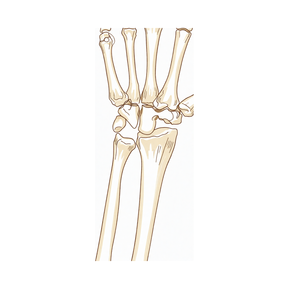
A proximal row carpectomy removes two or three of the small bones on the little-finger side of the back of your wrist. We usually suggest it when wear-and-tear arthritis or a bone that has lost its blood supply (Kienböck disease) has damaged those bones and other treatments have not given you enough relief. Activity change, physiotherapy or hand therapy, and splinting come first for long-standing problems. For some acute injuries, surgery may be the right choice straight away.
The operation keeps your wrist moving rather than stiffening it. It aims to relieve pain and let you use your wrist for everyday tasks. With careful selection, including people who do heavy work with their hands, results have held up over many years. We will talk through whether it suits you and decide together.
Before the operation
Your surgeon will plan the operation using scans of your wrist, such as X-rays or an MRI, which show the bones and cartilage in detail. On the day, you will need to stop eating and drinking seven hours before surgery. We ask for seven hours rather than six so you can be brought forward if the theatre list runs early. Your surgeon will tell you which of your usual medicines to skip and which to take. Bring a written list of everything you take. Arrange for someone to drive you home afterwards, and wear loose, comfortable clothing with sleeves that are easy to get on and off. If you have other medical conditions, you may need blood tests or a review with the anaesthetist.
On the day
You will arrive at the hospital's surgical admissions unit, where you are checked in and prepared for theatre. You will then meet the anaesthetist. This operation is done under general anaesthetic. A regional nerve block is sometimes added for post-operative pain relief; the anaesthetist will discuss this with you on the day. You are then taken into the operating theatre, where the operation is performed.
You will wake up in the recovery area, where nurses monitor you while the anaesthetic wears off. Once you are stable, you will either go to the ward or go home, depending on the procedure and your recovery.
What the operation involves
Your surgeon makes a single cut over the back of your wrist. Through this cut, they remove the two or three small wrist bones that are damaged. The bones next in line, the round end of one bone and its matching socket in your forearm, are left in place. These surfaces have healthy cartilage, the smooth gliding tissue that lets joints move without pain. Your surgeon checks this cartilage carefully during the operation, because it is what the wrist will glide on afterwards.
One structure matters a great deal: a strong strap of tissue on the palm side of the wrist that helps hold the remaining bones steady. Your surgeon takes care to preserve it, as it supports the wrist's movement once the damaged bones are gone.
The aim is a simpler wrist. Instead of two rows of small bones grinding on worn surfaces, you have one smooth joint doing the work. Nothing metal or plastic is put in, and no bone is wired or screwed together, so there is nothing to remove later.
The cut is closed with stitches. A dressing goes over the top, and you keep that dressing on for about 10 days.
Because no bones need to knit together, your wrist does not need to be held still in a cast afterwards. Your hand can start moving early, which is one of the reasons this operation suits people who want to keep using their wrist.
Your surgeon will talk you through the plan before the day, and you can ask questions at any point before the operation goes ahead.
After the operation
You will wake up in the recovery area, then move to the ward once you are stable. Most patients stay one night in hospital after this operation, though some are able to go home the same day. Pain relief is given as you need it, and the nurses will check on you regularly. Your wrist will be wrapped in a soft dressing. A sling or a half plaster along the front of your forearm is sometimes used, and sometimes not; you will be told what applies to you before you leave. Because no bones need to knit together, your hand can start moving early. We leave the dressing on for about 10 days; please do not take it off before then unless we tell you to. We change or remove it when we see you. Someone should stay with you for the first 24 hours after you get home.
Recovery
Recovery from this operation is often quicker than people expect. Because no bones need to knit together, your wrist is not held stiff in a cast. Your hand can start moving early, and gentle movement is usually encouraged from the first days.
Expect some pain and swelling around the back of your wrist at first. This settles gradually as the weeks pass. Keeping your hand raised when you are resting helps, and taking your pain relief as prescribed keeps you comfortable while the soreness eases. Some people find the swelling is worse in the mornings and improves as they use the hand during the day.
Your rehabilitation is with Ruby Doolan, our hand therapist at Extend Rehabilitation. She will guide your exercises and make any splint you need, usually within about a week of surgery. Early on, you will work on gentle bending and straightening of the wrist and fingers. As movement returns and the swelling settles, the exercises build up to gripping, lifting and using your hand for everyday tasks like dressing, eating and writing. Once you can grip without pain, heavier tasks come back into the picture.
Driving is not something to rush. You will need to be out of any sling or splint, able to hold the wheel with both hands and react in an emergency stop, and off strong pain medication. See our guide on driving after upper-limb surgery for the full detail.
Everyone heals at their own pace, so your timeline may differ. Your surgeon and your hand therapist will guide you along the way.
What can go wrong
Most patients do well, but problems can occasionally happen. Your surgeon and the team monitor you closely to spot any issue early.
Some wrists develop more wear-and-tear arthritis over the years after this operation. You might notice pain or grinding that slowly builds again, or the wrist feeling stiffer than it was. If this happens, bring it up at your next review so it can be checked with new scans.
A small number of people need a bigger operation later, where the whole wrist is stiffened by joining the remaining bones together. This is usually because pain has returned and other treatments have not helped enough. If your wrist pain keeps getting worse rather than better, tell your surgeon, who will talk you through the options.
Because this operation removes bone rather than fixing bone together, the usual risks of bones failing to join do not apply here. There is also nothing metal or plastic left in the wrist, so no further surgery is needed to remove hardware.
If you notice a deep, throbbing pain that does not ease with simple painkillers, or swelling and redness that spreads out from the wound, contact the clinic straight away. These signs need checking promptly rather than waiting for your next appointment.
Some people are left with ongoing wrist pain, or find they cannot return to the work or activities they did before. If pain or weakness is still limiting you well after recovery, raise it at your review. Your surgeon and hand therapist can look at what else might help, from further therapy to other treatments.
The complications table on this page lists typical rates if you want the specifics.
When to call us
Call us if you have a fever, or if redness, swelling or discharge from the wound gets worse. Call us if you have deep, throbbing pain that simple painkillers do not ease. Go to emergency if you have sudden severe pain, calf swelling or pain, or shortness of breath. Go to emergency if you lose feeling in your hand or cannot move your fingers or wrist. These signs need checking straight away.
Where to read more about the condition
This page is about the operation itself. The condition it treats, including what the evidence shows about when surgery helps and when it does not, is covered in more detail on the SLAC and SNAC Wrist page.
Evidence & references
This is the clinical evidence summary written for health professionals. It is technical, and it lists the research this page was built from. You do not need to read it to understand your treatment or to make a decision about it.
Overview
- At long-term followup, all patients older than thirty-five years of age at the time of proximal row carpectomy maintained a satisfactory range of motion, grip strength, and pain relief and were satisfied with the result [1].
- Proximal row carpectomy with interposition arthroplasty is an effective motion-sparing procedure for patients with proximal capitate and/or lunate fossa arthritis, improving pain and function [6].
- Short- to mid-term outcomes in patients with end-stage wrist arthritis affecting the capitate who undergo proximal row carpectomy and meniscus interposition arthroplasty are comparable with those receiving proximal row carpectomy alone [7].
Anatomy & Pathophysiology
Bony Anatomy
- The wrist includes the distal radioulnar, radiocarpal, and ulnocarpal joints and the eight carpal bones and their proximal and distal articulations and attached ligaments [23].
- The proximal row of carpal bones consists of the scaphoid, lunate, triquetrum, and pisiform [23].
- The distal row of carpal bones consists of the trapezium, trapezoid, capitate, and hamate [23].
- The capitate is the largest carpal bone [23].
- The pisiform and trapezoid are the smallest carpal bones [23].
- The capitate articulates with seven other bones [23].
- The pisiform articulates with one other bone, the triquetrum [23].
- The radiocarpal joints are formed by the articulation of the distal radius with the scaphoid and lunate through their respective concave facets on the distal radius and the triquetrum on the triangular fibrocartilage [23].
- The distal concave articular surfaces of the proximal carpal row form the midcarpal articulations with the distal row [23].
- The distal radius has three articular components: the scaphoid fossa, the lunate fossa, and the sigmoid notch [27].
- The scaphoid and lunate fossae allow articulation with the scaphoid and lunate bones, respectively [27].
- The sigmoid notch allows articulation with the ulna medially [27].
- A ridge between the scaphoid and lunate fossa corresponds with the scapholunate interval [27].
- The concave elliptical distal radius is oriented in the sagittal plane with an average of 11 degrees of volar tilt [27].
- In the frontal plane, the average radial inclination of the distal radius is 23 degrees [27].
- Radial length, measured from the tip of the radial styloid to the ulnar articular surface, averages 13 mm [27].
- The distal ulnar convexity articulates at the lesser sigmoid notch of the distal radius [23].
- The sigmoid notch articular surface accommodates the ulnar head through two thirds of its arc [23].
- There is about a 20-degree inclination of the distal ulna at its articulation with the radius [23].
Ligaments
- Extrinsic carpal ligaments connect the radius or the ulna to the carpus [28].
- In general, volar ligaments are stronger than dorsal ligaments [28].
- The radioscaphocapitate ligament connects to the waist of the scaphoid, around which the scaphoid rotates, and limits ulnar translation of the carpus [28].
- The long radiolunate ligament helps to limit ulnar translocation of the carpus [28].
- The short radiolunate ligament helps control lunate position [28].
- The radioscapholunate ligament is a vascular conduit, not a true ligament, also known as the ligament of Testut [28].
- The ulnocapitate ligament attaches to the ulnar head and originates from the volar margin of the ulnar fovea [28].
- The ulnocapitate ligament is the most superficial or palmar of the palmar ulnocarpal ligaments [28].
- The dorsal radiocarpal ligament has a trapezoidal shape and passes from the dorsal rim of the distal radius to the lunate and the triquetrum [28].
- Fibers of the dorsal radiocarpal ligament insert onto the dorsal lunotriquetral interosseous ligament [28].
- The scapholunate interosseous ligament is a major stabilizer of the wrist and the most commonly injured wrist ligament [28].
- The scapholunate interosseous ligament is C-shaped, consisting of dorsal, palmar, and interosseous portions, with the dorsal portion being the strongest and thickest [28].
- The scapholunate interosseous ligament provides a flexion force on the lunate given its attachment to the scaphoid [28].
- The lunotriquetral interosseous ligament is C-shaped, where the volar portion is the thickest and strongest [28].
- The lunotriquetral interosseous ligament provides an extension moment on the lunate given its attachment to the triquetrum [28].
- The capitohamate ligament is a thick ligament, 5 × 5 mm in cross section, with extensions to the third or fourth metacarpals [28].
- The dorsal intercarpal ligament passes from the dorsal tubercle of the triquetrum to the distal pole of the scaphoid [28].
- The dorsal intercarpal ligament reinforces the elastic dorsal wrist capsule and helps stabilize the scapholunate articulation with a contribution to the dorsal scapholunate interosseous ligament from its deep fibers [28].
- The space of Poirier is an area adjacent to the proximal capitate without ligamentous attachment, situated ulnar to the radioscaphocapitate ligament and radial to the long radiolunate in the floor of the carpal tunnel [28].
- The space of Poirier is a weak area that is vulnerable to instability [28].
- The distal carpal row separates from the lunate through the space of Poirier during a perilunate dislocation [28].
- The triangular fibrocartilage complex includes the ulnar collateral ligament, the dorsal and volar radioulnar ligaments, the articular disc, the meniscal homologue, the extensor carpi ulnaris sheath, and the ulnolunate and ulnotriquetral ligament [23].
- The triangular fibrocartilage attaches to the base of the ulnar styloid and separates the hyaline cartilage–covered ulnar head from the styloid [23].
Biomechanics and Kinematics
- The wrist joint’s motion planes include flexion, extension, radial deviation, ulnar deviation, and circumduction [28].
- There is minimal carpal motion with pronosupination [28].
- Approximately 62° of wrist extension occurs through the radiocarpal joint [28].
- 62% of wrist flexion occurs through the midcarpal joint [28].
- The midcarpal joint is mostly responsible for 20° and 40° of radial and ulnar deviation, respectively [28].
- The midcarpal joint is responsible for the “dart thrower’s motion,” which involves moving from radial extension into ulnar flexion positioning of the wrist [28].
- The radius bears 80% of the axial load transmitted through the radiocarpal joint in neutral ulnar variance [28].
- The ulna bears 20% of the axial load transmitted through the radiocarpal joint in neutral ulnar variance [28].
- The proximal row of carpal bones forms an intercalated segment between the distal carpal row and the distal radius and is bound into a functional unit by the scapholunate interosseous ligament and lunotriquetral interosseous ligament [28].
- The distal row is rigid, with little motion between its bones due to stout intercarpal ligaments, and acts as a functional unit with the scaphoid bridging both rows [28].
- During wrist flexion from neutral, the distal row flexes and ulnarly deviates slightly while the scaphoid also pronates [28].
- During wrist flexion from neutral, the proximal row flexes differentially, with more rotation through the scaphoid, followed by the triquetrum and the lunate [28].
- The proximal row translates dorsally during wrist flexion [28].
- During wrist extension from neutral, the distal row extends and radially deviates slightly while the scaphoid also supinates [28].
- During wrist extension from neutral, the proximal row extends differentially, with more motion in the scaphoid, followed by the triquetrum and then the lunate [28].
- The proximal row translates palmarly during wrist extension [28].
- Hysteresis can be quantified using 4DCT wrist kinematics and is greater in wrists with scapholunate ligament injury than in healthy wrists [31].
- A normative range of median radiolunate interosseous proximities during wrist motion has been quantified [20].
Classification
- Triquetro-lunate fusions are the commonest type of fusions in the carpal region [22].
Clinical Presentation
- In patients with continued pain over the radial side of the carpus, attention should be given to the other carpal bones and the distal radius and not just the scaphoid [14].
- Os styloideum must be suspected in patients with persistent pain in the dorsal aspect of the hand or wrist [33].
Investigations
Imaging Modalities
- MRI is the modality of choice for imaging radiographically occult fractures of the hand and wrist [29].
- The primary advantages of MRI compared with CT and radiography are improved tissue characterization, especially of soft tissues such as ligamentous structures in the wrist and synovium in the hand, and the lack of ionizing radiation [29].
- Modern MRI is generally at 1.5T or 3T, with 3T being much preferred for hand and wrist imaging, especially for imaging small fields of view [29].
- 7T MRI has recently become approved for clinical use and has the potential to become a powerful tool for hand and wrist imaging as applications are developed [29].
- MRI with contrast enhancement is most commonly used to determine whether soft-tissue lesions are solid or cystic or, in the case of rheumatologic imaging, to better visualize erosions and synovial burden [29].
- Dynamic contrast enhancement has been used with inconsistent results to assess for the presence of avascular necrosis in the lunate or scaphoid after injury [29].
- MR arthrography can be performed for evaluation of the triangular fibrocartilage and intercarpal ligament tears, but this is generally unnecessary with the increasing availability of high field MRI [29].
Arthroscopy
- To be successful with wrist arthroscopy and mitigate iatrogenic injury, a clear understanding of the topographical and 3-dimensional spatial anatomic relationships in the wrist as well as a patient’s unique anatomic variances is critical [12].
- A good experience in wrist arthroscopy is necessary for the realization of arthroscopic radioscapholunate fusion [8].
- In a comparative study of needle arthroscopy versus conventional arthroscopy, there was no difference between radiocarpal or midcarpal visualization and surgeon-rated ease of use, while diagnostic confidence was the same between the two groups [17].
Treatment
- At long-term followup, all patients older than thirty-five years of age at the time of proximal row carpectomy maintained satisfactory range of motion, grip strength, and pain relief and were satisfied with the result [1].
- Regardless of the construct used, if the distal radius articular surface is well reduced and other principles of fracture fixation are applied, most patients treated with a dorsal spanning plate can expect to regain functional wrist range of motion [2].
- The use of a wrist fixator allows open wound care and permits free access to the wrist for early secondary operations [10].
- No complications occurred due to the arm and elbow supports or fingertraps in the context of horizontal fingertrap traction in distal radial fractures [9].
Complications
- Postoperatively, extension of the fingers remained poor for over 3 months following an unusual carpometacarpal fracture-dislocation [4].
- No complications occurred due to arm and elbow supports or fingertraps during horizontal fingertrap traction in distal radial fractures [9].
Recovery
- At long-term follow-up, all patients older than thirty-five years of age at the time of proximal row carpectomy maintained a satisfactory range of motion [1].
- At long-term follow-up, all patients older than thirty-five years of age at the time of proximal row carpectomy maintained satisfactory grip strength [1].
- At long-term follow-up, all patients older than thirty-five years of age at the time of proximal row carpectomy maintained satisfactory pain relief [1].
- At long-term follow-up, all patients older than thirty-five years of age at the time of proximal row carpectomy were satisfied with the result [1].
- Proximal row carpectomy with interposition arthroplasty improves pain in patients with proximal capitate and/or lunate fossa arthritis [6].
- Proximal row carpectomy with interposition arthroplasty improves function in patients with proximal capitate and/or lunate fossa arthritis [6].
- Short- to mid-term outcomes for patients with end-stage wrist arthritis affecting the capitate who undergo proximal row carpectomy and meniscus interposition arthroplasty are comparable with those receiving proximal row carpectomy alone [7].
- Postoperatively, recovery of the wrist was rapid in a case of unusual carpometacarpal fracture-dislocation [4].
- In a case of unusual carpometacarpal fracture-dislocation, extension of the fingers remained poor for over 3 months postoperatively [4].
Key Evidence
- [L4] At the time of long-term followup, all patients older than thirty-five years of age at the time of a proximal row carpectomy had maintained a satisfactory range of motion, grip strength, and pain relief and were satisfied with the result. [1] (10.2106/jbjs.e.00261)
- [L4] Regardless of the construct used, if the distal radius articular surface is well reduced and other principles of fracture fixation are applied, most patients treated with a DSP can expect to regain functional wrist ROM. [2] (10.1177/15589447241247335)
- [L5] Postoperatively recovery of the wrist was rapid, though extension of the fingers remained poor for over 3 months. [4] (10.1016/0020-1383(94)90161-9)
- [L4] Proximal row carpectomy with interposition arthroplasty is an effective motion-sparing procedure for patients with proximal capitate and/or lunate fossa arthritis, improving pain and function. [6] (10.1177/15589447241298721)
- [L3] Our short- to mid-term outcomes in patients with end-stage wrist arthritis affecting the capitate who undergo PRC and meniscus interposition arthroplasty are comparable with those receiving PRC alone. [7] (10.1177/15589447241262052)
- [L5] The authors present their technique and tips for performing this arthroscopic intervention, noting that a good experience in wrist arthroscopy is necessary for its realization. [8] (10.1016/j.eats.2022.02.015)
- [L4] No complications occurred due to the arm and elbow supports or fingertraps. [9] (10.1016/s0020-1383(99)00161-8)
- [L4] The use of a wrist fixator allows open wound care and permits free access to the wrist for early secondary operations. [10] (10.1016/s0020-1383(99)00267-3)
- [L5] To be successful with wrist arthroscopy and mitigate iatrogenic injury, a clear understanding of the topographical and 3-dimensional spatial anatomic relationships in the wrist as well as a patient’s unique anatomic variances is critical. [12] (10.1016/j.eats.2024.103223)
- [L4] In patients with continued pain over the radial side of the carpus, attention should be given to the other carpal bones and the distal radius and not just the scaphoid. [14] (10.1016/0020-1383(95)00081-j)
- [L2] In this study, there was no difference between radiocarpal or midcarpal visualization and surgeon-rated ease of use, while diagnostic confidence was the same between two groups. [17] (10.1177/15589447241265982)
- [Paper] This study quantifies a normative range of median radiolunate interosseous proximities during wrist motion. [20] (10.1177/15589447251352124)
- [L5] Triquetro-lunate fusions are the commonest type of fusions in the carpal region. [22] (10.1016/0020-1383(95)00192-1)
- [L3] This study demonstrated that hysteresis can be quantified using 4DCT wrist kinematics and is greater in wrists with scapholunate ligament injury than in healthy wrists. [31] (10.1177/17531934261468199)
- [L4] Os Styloideum is an infrequent pathology and must be suspected in patients with persistent pain in the dorsal aspect of the hand or wrist. [33] (10.1177/15589447251317232)
References
[1] Proximal Row Carpectomy. Journal of Bone and Joint Surgery. 2005. DOI: 10.2106/jbjs.e.00261
[2] What to Expect? Use of Supplemental Fixation With a Concomitant Dorsal Spanning Plate for Complex Intraarticular Distal Radius Fractures. HAND. 2024. DOI: 10.1177/15589447241247335
[4] An unusual carpometacarpal fracture—dislocation. Injury. 1994. DOI: 10.1016/0020-1383(94)90161-9
[6] Outcomes of Proximal Row Carpectomy With Interposition Arthroplasty for Advanced Wrist Arthritis. HAND. 2024. DOI: 10.1177/15589447241298721
[7] Outcomes of Patients Receiving Proximal Row Carpectomy and Meniscus Interposition Allografts for the Treatment of End-Stage Wrist Arthritis: A Comparative Study. HAND. 2024. DOI: 10.1177/15589447241262052
[8] Arthroscopic Radioscapholunate Fusion: Surgical Technique. Arthroscopy Techniques. 2022. DOI: 10.1016/j.eats.2022.02.015
[9] Horizontal fingertrap traction in distal radial fractures. Injury. 1999. DOI: 10.1016/s0020-1383(99)00161-8
[10] The treatment of complex carpal dislocations by external fixation. Injury. 2000. DOI: 10.1016/s0020-1383(99)00267-3
[12] Wrist Arthroscopy: Positioning, Portal Placement, and Diagnostic Evaluation. Arthroscopy Techniques. 2024. DOI: 10.1016/j.eats.2024.103223
[14] The suspected scaphoid fracture and isotope bone imaging. Injury. 1995. DOI: 10.1016/0020-1383(95)00081-j
[17] Needle Arthroscopy Versus Conventional Arthroscopy in the Evaluation of Carpal Pathology: A Comparative Study. HAND. 2024. DOI: 10.1177/15589447241265982
[20] Four-Dimensional Computed Tomography-Derived Radiolunate Arthrokinematics With a Case Study in Four-Corner Arthrodesis. HAND. 2025. DOI: 10.1177/15589447251352124
[22] An atypical fracture of a triquetral-lunate synostosis. Injury. 1996. DOI: 10.1016/0020-1383(95)00192-1
[23] Campbell S Operative Orthopaedics 4 Volume Set. NERVE INJURIES AT THE LEVEL OF THE HAND AND WRIST > ANATOMY.
[27] A Lange Medical Book Current Diagnosis Treatment In Orthopedics Fifth Edition. 2Musculoskeletal Trauma Surgery > FRACTURES AND DISLOCATIONS OF THE DISTAL AND MID-FOREARM.
[28] Aaos Comprehensive Orthopaedic Review 3. Carpal Instability* > II. Anatomy and Biomechanics (See Chapter 92).
[29] Orthopaedic Knowledge Update 13 Ebook Without Multimedia. Anatomy, Evaluation, Clinical Examination, and Imaging > Imaging: Advances in Imaging of the Hand and Upper Extremity > Magnetic Resonance Imaging.
[31] Hysteresis in dynamic wrist kinematics: a 4DCT comparison between healthy and scapholunate ligament-injured wrists. Journal of Hand Surgery (European Volume). 2026. DOI: 10.1177/17531934261468199
[33] Os Styloideum and Third Metacarpal Partial Coalition Nonunion After Traumatic Fracture: A Report of Three Cases. HAND. 2025. DOI: 10.1177/15589447251317232