Clinicians › Elbow
Anatomy & Biomechanics
Elbow anatomy & biomechanics: ulnohumeral, radiocapitellar joints, valgus stress, and implications for UCL injury/OCD in throwers.
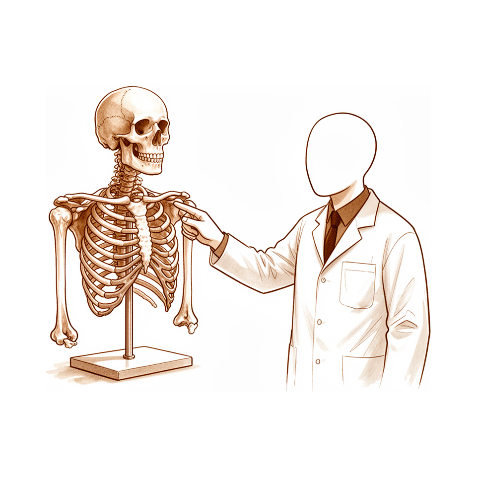
For patients: a plain-language version of this topic is available. See the patient guide.
Overview
Elbow stability and complex kinematics depend on a combination of bony articulation and soft-tissue stabilizers [12]. The anterior bundle of the medial ulnar collateral ligament serves as the primary restraint to valgus loads [20]. Consequently, tendon reconstructions for elbow soft tissue stabilizers should be tightened at 30 degrees of flexion rather than 90 degrees [20]. Understanding elbow biomechanics and injury mechanisms provides valuable insight into the variations of pathology observed in complex elbow dislocations [5]. The evaluation of the elbow requires an understanding of anatomy, biomechanics, and diagnostic tests, with particular attention paid to functional biomechanics necessary when treating complex pathology [10]. Additionally, the surgical management of proximal ulna fractures must take the complex biomechanics of the elbow joint into account [19]. Thorough knowledge of distal biceps insertional and footprint anatomy is essential for understanding the biomechanics of rupture and reconstruction and to avoid nerve injuries [2].
Longitudinal stability of the forearm relies on the radial head, interosseous membrane, and distal radioulnar joint working together [13]. In the shoulder, understanding normal and degenerative anatomy is necessary to restore joint function and prevent complications such as glenoid loosening or cuff failure in shoulder arthroplasty [9].
General upper extremity principles dictate that a thorough understanding of biomechanical principles and neurological pathways is necessary for the hand surgeon in the management of upper limb spasticity [4]. Detailed knowledge about the anatomical situation in areas of pin implantation is of great importance for unilateral external fixation in the upper limb [24]. Preparation and detailed anatomic knowledge are important in surgical procedures for upper extremity trauma [7]. Eight studies followed International Society of Biomechanics standards for quantifying upper body kinematics during functional upper limb tasks [3]. Furthermore, biomechanical and clinical studies are needed to understand the true clinical relevance of anatomic variations in the ulnar greater sigmoid notch coverage angle [1].
Osseous Anatomy
Distal Humerus
The helical path of the distal humeral groove serves as the anatomical origin for the vortical movement, or screw motion, of the elbow joint [27]. Cartilage thickness on the distal humerus is not uniform [11]. This non-uniformity modifies the morphologic shape and diameters of the humeral articular surface [11]. CT-based measurements of distal humerus morphology in healthy adults provide a basis for the design of distal humeral orthopaedic implants [30].
Ulna
The ulnar greater sigmoid notch possesses a "coverage angle" that involves both bone and cartilage contributions [1].
Radiocapitellar Joint
Restoration of the anatomic radial head height is critical when performing radial head arthroplasty to maintain normal joint biomechanics [32].
Proximal Radioulnar Joint
A significant negative relationship exists between the Alpha Angle and the Beta Angle in the proximal radioulnar joint [26]. These angles emphasize the biomechanical impact of joint congruence on bony coverage in the proximal radioulnar joint [26].
Ligaments and Joint Capsule
Medial Collateral Ligament
The anterior bundle of the medial collateral ligament contains isometric fibers originating from a broader area extending more medially in the coronal plane than previously described [67]. This broader medial origin allows the ligament to reconcile isometricity with robustness [67]. When performing tendon reconstructions for the medial ulnar collateral ligament, the graft should be tightened at 30 degrees of flexion rather than 90 degrees [20]. Repair of the ulnar collateral ligament using an internal brace restores resistance to valgus stress to near-intact levels without causing joint overconstraint [60].
Lateral Collateral Ligament and Other Ligaments
In 40% of human subjects, the distal oblique bundle within the distal membranous portion of the interosseous membrane stabilizes the distal radioulnar joint [66]. Regarding the anterior oblique ligament, stress is equalized at 60 degrees of flexion [72]. This specific angle represents the position where the anterior oblique ligament is unlikely to sustain damage [72].
Joint Capsule and General Biomechanics
Ligaments may function as a 'static-dynamic' stabilizer rather than acting solely as static structures [59]. A thorough understanding of relevant neurovascular and ligamentous anatomy is critical for safely performing surgical procedures about the elbow [8]. Similarly, comprehensive anatomical knowledge is essential for surgeons to understand the biomechanics of rupture and reconstruction of the distal biceps tendon [2]. This anatomical mastery is also required to avoid nerve injuries during distal biceps reconstruction [2].
Surgical Considerations
Approaches and Visualization: A combination of two ligament-preserving approaches enables viewing the entirety of the joint surface [18]. These ligament-preserving techniques may serve as an alternative to ligament-releasing approaches for capitellum surgery [18].
Arthroscopic Management: Arthroscopic treatment for coronoid process nonunion facilitates the diagnosis and management of associated lesions, including lateral collateral ligament imbrication [65]. This approach also allows for the removal of loose bodies [65]. The Zhang knot offers a technically feasible and mechanically promising addition to the arthroscopic armamentarium [69]. A mechanical forearm holder provides an effective means to temporarily or permanently maintain the elbow joint in any desired degree of extension or flexion during elbow arthroscopy without the need for an assistant [71].
Fixation and Hardware: In distal biceps tendon repair, two suture anchors demonstrated the least displacement after loading [70]. Hardware removal may be required in approximately 25 percent of patients with anterior chondral buttress plating due to impingement in deep flexion [64].
Muscles and Tendons
Elbow Ligaments and Tendons
The anterior bundle insertion of the medial ulnar collateral ligament (MCL) complex is more elongated and distally tapered than previously described [15]. Geometric centroids of the MCL complex footprints are predictable within 0.8 to 1.3 mm for most footprints [15]. Biometric mapping has defined the specific common flexor tendon origins and their sizes on the medial epicondyle, including their position relative to the MCL [90]. Regarding the ulnar collateral ligament, the middle and distal thirds of the insertional footprint on the ulna did not significantly contribute to resistance at 5 N·m of valgus load [84]. Placing a graft within the footprint of the native ulnar collateral ligament may better approximate normal joint mechanics [82].
Neurovascular structures in the cubital fossa are in close proximity to the distal biceps tendon and its tuberosity insertion [25]. Dynamic rotation significantly changes the positional relationship between these neurovascular structures and the distal biceps tendon insertion [25]. The footprints of the posterolateral ligament of the elbow have been described, with clinical significance demonstrated in cases of elbow instability caused by acute ligament tears and elbow pain due to ligament enthesopathy [91]. There is no truly isometric location for lateral ulnar collateral ligament tendon graft reconstruction tunnels [87].
For the triceps tendon, gross measurement of the insertion overestimates and inaccurately represents the true insertional footprint compared with histologic measurement [63]. Anatomic coverage of the triceps footprint allows more surface area for healing in double-row repair techniques [75].
Distal Biceps Tendon
The distal biceps tendon insertion footprint is located on the posterior/ulnar aspect, centered at approximately 30 degrees anterior to the lateral/coronal plane with the forearm fully supinated [88]. An anatomic Footprint repair restores superior biomechanical supination strength and endurance compared to a conventional Endobutton technique in a clinical setting [83]. The single-incision arthrotunneling technique recreates the anatomic footprint and biomechanics of the native biceps [86]. This single-incision arthrotunneling technique has a reduced complication profile compared to a two-incision approach [86].
Other Muscles and Tendons
Endoscopic proximal hamstring repair is supported by cadaveric mapping of the anatomic landscape encountered endoscopically [80]. Biometric data for the pronator quadratus muscle provides valuable information for surgical procedures involving the distal forearm [89]. Margin convergence techniques with single stitch and subsequent footprint repair may have adverse effects on muscle properties and tensile loading on repair, increasing the risk of retear of repairs [85].
Neurovascular Anatomy
General Principles
Thorough anatomical knowledge is essential for understanding the biomechanics of rupture and reconstruction of the distal biceps tendon and for avoiding nerve injuries [2]. A comprehensive grasp of biomechanical principles and neurological pathways is necessary for the hand surgeon [4]. Appreciation of relevant neurovascular and ligamentous structures is critical to safely performing surgical procedures about the elbow [8]. In the upper limb, the anatomical base of pin placement is of paramount importance [23]. Furthermore, a thorough understanding of forearm anatomy, including the locations of cutaneous nerves, muscular intervals, and deep neurovascular structures, is essential to adequately perform surgical exposure of the radius and ulna [76].
Nerve Anatomy and Dynamics
Dynamic rotation significantly changes the positional relation of neurovascular structures with the distal biceps tendon insertion [25]. The relative position of the median nerve with respect to the ulnar insertion of the brachialis muscle changes with elbow and forearm movements [81]. Understanding the anatomy of radial nerve branches at the elbow is of utmost importance when devising a reconstructive strategy for upper limb paralysis [74]. Magnetic resonance neurography provides anatomical correlation and pinpoints nerve pathology, enhancing diagnostic confidence and guiding treatment planning [73].
Vascular Anatomy
The vascular anatomy for vascularized olecranon bone grafts is consistent, and flap harvest is simple and straightforward in all cadaveric specimens [61]. The arterial supply of the proximal ulna is relatively consistent and flows in opposite directions from two separate areas [62]. Understanding the extraosseous and intraosseous arterial anatomy of the adult elbow may help avoid iatrogenic injury to the intraosseous circulation during trauma or extensive dissection [79].
Surgical Safety and Injury Prevention
In a cadaveric model, penetration of the posterior humeral cortex at the suprapectoral location results in proximity to the axillary nerve and should be avoided [22]. A high index of suspicion for neurovascular entrapment led to prompt surgical treatment, resulting in an optimal outcome with full recovery of motor, sensory, and sympathetic functions at 1-year follow-up [78].
Biomechanics and Function
General Principles and Methodology
Deformable mechanics examines the effect of forces and motions on internal body stresses [35]. In musculoskeletal biomechanics, this field focuses on understanding the natural behavior of joint tissues, including both soft and hard tissue types [35]. When measuring tissue response to loading, it is essential to distinguish intrinsic material properties from extrinsic structural properties [35]. The term "Stiffness" requires an exact description of the load configuration and the precise localization and kind of deformation measured [49]. Graphical statics remains a useful tool for visualizing, understanding, and checking the actions of force groups in modern biomechanical problems [54]. Eight studies followed International Society of Biomechanics standards for quantifying upper body kinematics [3].
Elbow Biomechanics and Stability
The ulnar greater sigmoid notch coverage angle involves both bone and cartilage contribution, with anatomic variations requiring further biomechanical and clinical studies to determine clinical relevance [1]. The load sharing ratio for the radiocapitellar and ulnotrochlear joints is 58%:42%, respectively [57]. This study is the first to comprehensively evaluate loading mechanics across both native elbow joints simultaneously throughout the entire functional range of elbow flexion and forearm rotation [57]. Axial loading increases the contact area of the radiocapitellar joint [52]. Radial head excision causes altered elbow kinematics and increased laxity [58]. Recent changes in elbow arthroplasty device design and implantation methods are driven by biomechanical and clinical outcome-based research to better reproduce elbow kinematics [50].
The lateral ulnar collateral ligament origin center is 10.7 mm from the lateral epicondyle and insertion is 3.3 mm from the apex of the supinator crest [34]. The MUCL anterior bundle is the strongest component of the ligamentous complex and the primary restraint to valgus stress [34]. It is subdivided into anterior and posterior bands which provide reciprocal function, with the anterior band tight in extension and the posterior band tight in flexion [34]. Detailed analysis of the fibers of the medial ulnar collateral ligament allows for further understanding of its kinematic function [21]. The MUCL provides group coverage area and kinematic function for each degree of motion arc, allowing selective reconstruction according to mechanism of injury and dominant fibers affected [34]. Both TightRope and traditional docking ulnar collateral ligament reconstruction techniques restored native joint kinematics from 15 to 75 degrees of flexion under low loading conditions [43]. Further biomechanical and clinical studies are needed to confirm long-term outcomes for humeral ligament reconstruction using Shark Screw tendon in medial and posterolateral elbow instability [14].
Shoulder Biomechanics
Patient-specific musculoskeletal modeling of shoulder joint biomechanics can contribute significantly to predicting pathology and optimizing postoperative function [38].
Upper Extremity Kinematics and Throwing Biomechanics
The biomechanics of throwing in baseball and football differ significantly, leading to distinct injury patterns between sports [16]. Professional pitchers had greater biomechanical efficiency than collegiate pitchers [55]. Clinicians should assess for altered mechanics or fatigue-related compensation during return-to-play for MLB pitchers with lower-body injuries [56].
Common Sites of Injury
Elbow
Understanding elbow biomechanics and injury mechanisms provides insight into the variations of pathology observed in complex elbow dislocations [5]. Anatomic variations in the ulnar greater sigmoid notch coverage angle require biomechanical and clinical studies to determine their true clinical relevance [1]. Detailed analysis of the medial ulnar collateral ligament fibers allows for further understanding of its kinematic function [21]. The heterogeneity of anterior bundle injuries in patients undergoing UCL reconstruction includes a variety of injury configurations and chronic attritional damage [45]. Regarding posteromedial rotatory incongruity fractures, ligamentous repair alone may not reliably restore kinematics for small subtype 2 fractures [33]. Larger posteromedial rotatory incongruity fractures should ideally have fragments fixed or reconstructed [33].
Forearm
The forearm operates as a series of interconnected parts where damage to one component can affect stability in multiple areas [28]. Injury to the interosseous membrane contributes more to the disruption of the normal distribution of axial loads across the elbow than injury to the distal radioulnar joint [42].
Distal Biceps
Thorough knowledge of distal biceps insertional and footprint anatomy is essential for understanding the biomechanics of rupture and reconstruction [2]. This anatomical knowledge is also essential to avoid injuries of the nerves during reconstruction [2].
Shoulder
Understanding normal and degenerative shoulder anatomy is necessary to restore joint function and prevent complications such as glenoid loosening or cuff failure [9].
Pediatric Elbow
Five distinct fracture patterns of the humeral medial epicondyle are identifiable on plain radiographs and are associated with specific injury mechanisms [36, 37]. Identification of humeral medial epicondyle injury patterns is a key first step in understanding the variability in clinical outcomes with different management strategies [41]. Treatment rationale for humeral medial epicondyle injuries is often predicated on restoring elbow biomechanics through anatomical restoration of the UCL [41].
Wrist
Open anatomical reduction with stabilization of all injured structures, followed by six to eight weeks of immobilization, is probably optimum treatment for fracture-dislocation of the radiocarpal joint [44].
Surgical Anatomy
Bony Anatomy and Articular Surface
An MRI anatomic study of 78 elbows examined the bone and cartilage contribution to the ulnar greater sigmoid notch “coverage angle” [1]. Biomechanical and clinical studies are needed to understand the true clinical relevance of anatomic variations in the ulnar greater sigmoid notch [1]. Cartilage thickness of the distal humerus is not uniform [11]. This non-uniform cartilage thickness modifies the morphologic shape and diameters of the humeral articular surface [11].
Neurovascular and Ligamentous Structures
Thorough knowledge of distal biceps insertional and footprint anatomy is essential for surgeons to understand the biomechanics of rupture and reconstruction [2]. In a cadaveric model, penetration of the posterior humeral cortex at the suprapectoral location results in proximity to the axillary nerve [22]. Consequently, penetration of the posterior humeral cortex at the suprapectoral location should be avoided due to proximity to the axillary nerve [22].
Surgical Approaches and Exposure
A combination of two ligament-preserving approaches enables viewing the entirety of the capitellum joint surface [18]. Ligament-preserving approaches to the capitellum may represent an alternative to ligament-releasing approaches [18]. A limited exposure technique for fractured capitellum uses guide wires and cannulated screws inserted from the posterior to the anterior aspects [47]. This limited exposure technique is designed to limit soft tissue dissection and avoid damage to the articular cartilage [47].
Minimally Invasive Lateral Ulnar Collateral Ligament Techniques: The minimally invasive nature of arthroscopic lateral ulnar collateral ligament plication/reconstruction preserves tissue [51]. Preservation of tissue in these minimally invasive techniques leads to a shorter rehabilitation phase [51] and reduces the risk of postoperative mobility restrictions [51]. Arthroscopic-assisted lateral ulnar collateral ligament reconstruction necessitates a long learning curve and experience to work at the posterolateral compartment [48].
Other Arthroscopic and Open Techniques: Arthroscopic extensor carpi radialis brevis muscle resection for chronic elbow lateral epicondylitis is operator dependent due to a lack of clear definition of resection landmarks [46]. Anatomic direct repair of chronic distal biceps brachii tendon rupture without interposition graft optimizes the approach using the most secure fixation method [53]. This anatomic direct repair allows repair without loss of range movement or function [53].
General Principles and Biomechanics
A thorough understanding of biomechanical principles and neurological pathways is necessary for the hand surgeon in upper limb spasticity management [4]. The proper surgical management of proximal ulna fractures requires taking the complex biomechanics of the elbow joint into account [19]. The anatomical base of pin placement is of paramount importance in the upper limb [23].
Key Evidence
- [L5] Biomechanical and clinical studies are needed to understand the true clinical relevance of these anatomic variations. [1] (10.1016/j.jse.2015.06.006)
- [L4] Thorough knowledge of the anatomy is essential for the surgeon in order to understand the biomechanics of rupture and reconstruction of the distal biceps tendon and to avoid injuries of the nerves. [2] (10.1007/s00167-014-3322-9)
- [L4] Eight studies followed International Society of Biomechanics standards for quantifying upper body kinematics. [3] (10.1016/j.jelekin.2018.02.011)
- [L5] A thorough understanding of biomechanical principles and neurological pathways is necessary for the hand surgeon. [4] (10.1177/17531934261434453)
- [L4] Understanding elbow biomechanics and the injury mechanism provides valuable insight into the variations of pathology that may be observed. [5] (10.5435/jaaos-d-14-00023)
- [L5] The issue emphasizes the importance of preparation and detailed anatomic knowledge in surgical procedures for upper extremity trauma, providing comprehensive descriptions of exposures and coverage with clinical examples. [7] (10.1016/j.hcl.2014.08.004)
- [L5] An appreciation of the relevant neurovascular and ligamentous structures is critical to safely performing surgical procedures about the elbow. [8] (10.5435/00124635-200905000-00007)
- [L5] Understanding normal and degenerative anatomy is necessary to restore joint function and prevent complications such as glenoid loosening or cuff failure. [9] (10.1302/2058-5241.6.210014)
- [L4] Cartilage thickness is not uniform and modifies the morphologic shape and diameters of the humeral articular surface. [11] (10.1016/j.jse.2016.10.012)
- [L5] This article discusses the basic anatomy of the elbow and the biomechanics of this joint, noting that a combination of bony articulation and soft-tissue stabilizers accounts for the elbow's stability and complex kinematics. [12] (10.1016/j.csm.2004.06.008)
- [L4] The review outlines the integrated anatomy and biomechanics of the forearm unit, emphasizing that longitudinal stability relies on the radial head, interosseous membrane, and distal radioulnar joint working together, and discusses current diagnostic and treatment options. [13] (10.1111/j.1758-5740.2012.00207.x)
- [L5] Further biomechanical and clinical studies are needed to confirm long-term outcomes. [14] (10.1016/j.jseint.2026.101786)
- [L5] The study quantified the 3D anatomy of the medial ulnar collateral ligament complex, revealing that the anterior bundle insertion is more elongated and distally tapered than previously described, with geometric centroids predictable within 0.8 to 1.3 mm for most footprints. [15] (10.1177/2325967118762751)
- [L5] The biomechanics of throwing in baseball and football differ significantly, leading to distinct injury patterns between sports. [16] (10.1177/23259671251407244)
- [L5] A combination of these two ligament-preserving approaches enables viewing the entirety of the joint surface and may represent an alternative to ligament-releasing approaches. [18] (10.1016/j.jse.2022.01.013)
- [L5] The aim of this review is to illustrate the proper surgical management of proximal ulna fractures using modern osteosynthetic implants and novel techniques while taking the complex biomechanics of the elbow joint into account. [19] (10.1302/2058-5241.4.180022)
- [L5] The article reviews the anatomy and biomechanics of soft tissue stabilizers, emphasizing that the anterior bundle of the medial ulnar collateral ligament is the primary restraint to valgus loads and that tendon reconstructions should be tightened at 30 degrees of flexion rather than 90 degrees. [20] (10.1016/j.jhsa.2019.10.034)
- [L5] Detailed analysis of the fibers of the MUCL allows for further understanding of its kinematic function. [21] (10.1177/2325967118762750)
- [L5] In a cadaveric model, penetration of the posterior humeral cortex at the suprapectoral location results in proximity to the axillary nerve and should be avoided. [22] (10.1016/j.jse.2014.06.038)
- [Paper] In the upper limb the anatomical base of pin placement is of paramount importance. [23] (10.1016/s0020-1383(99)00255-7)
- [L5] Detailed knowledge about the anatomical situation in the areas of pin implantation is of great importance. [24] (10.1016/s0020-1383(99)00258-2)
- [L5] Neurovascular structures in the cubital fossa are in close proximity to the distal biceps tendon and its tuberosity insertion, and dynamic rotation significantly changes their positional relation with the tendon insertion. [25] (10.1136/jisakos-2019-000326)
- [L4] This study describes anatomical structures of the PRUJ and highlights a significant negative relationship between the Alpha Angle and the Beta Angle, emphasizing the biomechanical impact of joint congruence on bony coverage. [26] (10.1016/j.xrrt.2026.100695)
- [L5] This is the first anatomical study clarifying the origin of the vortical movement of the elbow due to the helical path of the distal humeral groove based on a statistical shape model of the humerus. [27] (10.1016/j.jse.2025.08.018)
- [Paper] The forearm operates as a series of interconnected parts where damage to one can affect stability in multiple areas, and physicians need a holistic understanding of these components to quickly diagnose the proper injury patterns before the condition worsens. [28] (10.1016/j.hcl.2020.06.001)
- [L4] The findings provide a basis for the design of distal humeral orthopaedic implants, ensuring greater alignment with the anatomical structure of the distal humerus and improved surgical outcomes. [30] (10.1186/s12891-024-07858-4)
- [L5] Restoration of the anatomic radial head height is critical when performing radial head arthroplasty to maintain normal joint biomechanics. [32] (10.1016/j.jhsa.2021.11.006)
- [L5] For small subtype 2 fractures, ligamentous repair alone may not reliably restore kinematics, and larger fractures should ideally have fragments fixed or reconstructed. [33] (10.1016/j.jse.2018.02.018)
- [L4] This large cohort confirmed five distinct fracture patterns that are identifiable on plain radiographs and are associated with specific injury mechanisms. [36] (10.1177/2325967126s00493)
- [L4] This large cohort confirmed five distinct fracture patterns that are identifiable on plain radiographs and are associated with specific injury mechanisms. [37] (10.1177/2325967126s00189)
- [L5] Patient-specific musculoskeletal modeling of shoulder joint biomechanics can contribute significantly to predicting pathology and optimizing postoperative function. [38] (10.1016/j.jseint.2024.04.006)
- [L4] As the treatment rationale for ME injuries is often predicated on restoring elbow biomechanics through anatomical restoration of the UCL, identification of these injury patterns is potentially a key first step in understanding the variability in clinical outcomes with different management strategies for medial elbow injuries. [41] (10.1177/2325967126s00151)
- [L5] Injury to the interosseous membrane contributes more to the disruption of the normal distribution of axial loads across the elbow than injury to the distal radioulnar joint. [42] (10.1016/j.jse.2018.07.016)
- [L5] Both the TR and DO techniques restored native joint kinematics from 15 to 75 degrees of flexion under low loading conditions. [43] (10.1177/0363546513482567)
- [L4] Open anatomical reduction with stabilization of all injured structures, followed by six to eight weeks of immobilization, is probably optimum treatment. [44] (10.2106/00004623-197759020-00011)
- [L4] This study shows the heterogeneity of anterior bundle injuries in patients undergoing UCL reconstruction, with a variety of injury configurations and chronic attritional damage observed. [45] (10.1016/j.jse.2019.01.017)
- [L5] One limitation of our technique is that there is no clear definition of the resection landmarks, which makes it operator dependent. [46] (10.1016/j.eats.2023.07.017)
- [L4] The described technique uses a limited exposure with guide wires and cannulated screws inserted from the posterior to the anterior aspects to limit soft tissue dissection and avoid damage to the articular cartilage. [47] (10.1016/s0020-1383(99)00254-5)
- [L5] However, the demanding technique necessitates a long learning curve and experience to work at the posterolateral compartment. [48] (10.1016/j.eats.2024.103101)
- [Paper] The term "Stiffness" of a structure always requires an exact description of the load configuration and the exact localization and kind of deformation measured. [49] (10.1016/s0020-1383(00)80040-6)
- [L5] Recent changes in device design and implantation methods are driven by biomechanical and clinical outcome-based research to better reproduce elbow kinematics, resulting in more durable and long-lasting joint replacement procedures. [50] (10.1302/2058-5241.2.160064)
- [L5] The minimally invasive nature of this technique preserves tissue, leading to a shorter rehabilitation phase and reducing the risk of postoperative mobility restrictions. [51] (10.1016/j.eats.2025.103529)
- [L5] Axial loading increases the contact area of the radiocapitellar joint. [52] (10.1016/j.jse.2020.08.042)
- [L4] Our method optimizes the approach to direct repair using the most secure fixation method and allows anatomic repair without loss of range movement or function. [53] (10.1016/j.jse.2012.01.012)
- [Paper] Graphical statics is an almost forgotten, intuitive drawing method for solving plane mechanical problems that remains a useful tool for visualizing, understanding and checking the actions of force groups occurring in modern biomechanical problems. [54] (10.1016/s0020-1383(00)80041-8)
- [L4] Professional pitchers had greater biomechanical efficiency than collegiate pitchers. [55] (10.1177/03635465221119194)
- [L3] Clinicians should assess for altered mechanics or fatigue-related compensation during return-to-play. [56] (10.1177/2325967126s00471)
- [L5] The study establishes a load sharing ratio of 58%:42% for the radiocapitellar and ulnotrochlear joints, respectively, and is the first to comprehensively evaluate loading mechanics across both native joints simultaneously throughout the entire functional range of elbow flexion and forearm rotation. [57] (10.1177/1758573220961025)
- [L5] Radial head excision causes altered elbow kinematics and increased laxity. [58] (10.2106/00004623-200408000-00018)
- [L5] Based on these perspectives, ligaments could function as a 'static-dynamic' stabilizer rather than a simple static one. [59] (10.1016/j.jseint.2024.01.006)
- [L5] The procedure did not lead to joint overconstraint while also returning the ligament to near-intact levels of resisting valgus stress. [60] (10.1177/23259671221134829)
- [L5] Vascular anatomy is consistent and flap harvest is simple and straightforward in all cadaveric specimens. [61] (10.1016/j.jhsa.2019.05.014)
- [L5] The arterial supply of the proximal ulna is relatively consistent and flows in opposite directions from the 2 separate areas. [62] (10.1016/j.jhsa.2011.02.011)
- [L5] Gross measurement of the triceps tendon insertion overestimates and inaccurately represents the true insertional footprint compared with histologic measurement. [63] (10.1016/j.jhsa.2021.05.003)
- [L4] The technique is mechanically sound in accordance with the buttress principles; however, considering its articular location, hardware removal may be required in approximately 25 percent of patients due to impingement in deep flexion. [64] (10.1016/j.jseint.2026.101717)
- [L5] The technique allows for both the diagnosis and management of associated lesions, including lateral collateral ligament imbrication or the removal of loose bodies. [65] (10.1016/j.eats.2025.103668)
- [L5] The distal oblique bundle in the distal membranous portion may stabilize the distal radioulnar joint in 40% of human subjects who have this ligament. [66] (10.1016/j.jhsa.2009.01.015)
- [L5] Isometric ligaments within the anterior bundle of the medial collateral ligament originate from a broader area extending more medially in the coronal plane than previously thought, which explains how the ligament reconciles isometricity and robustness. [67] (10.1016/j.jse.2011.07.018)
- [L5] The Zhang knot presents a technically feasible and mechanically promising addition to the arthroscopic armamentarium. [69] (10.1002/atn2.70108)
- [L1] Two suture anchors demonstrated the least displacement after loading. [70] (10.1177/2325967121s00747)
- [L5] This technique simply and reproducibly provides an effective means to temporarily or permanently maintain the elbow joint in any desired degree of extension or flexion during elbow arthroscopy without the need for an assistant. [71] (10.1016/j.eats.2024.102991)
- [L5] The angle at which the anterior oblique ligament stress was equalized was 60 degrees, suggesting that 60 degrees is the angle at which the ligament is unlikely to be damaged. [72] (10.1016/j.jse.2020.05.033)
- [Paper] Its main benefit is providing anatomical correlation and pinpointing nerve pathology, thereby enhancing diagnostic confidence and guiding appropriate treatment planning. [73] (10.1177/23259671251400763)
- [L5] Understanding the anatomy of the radial nerve branches at the elbow is of utmost importance when devising a reconstructive strategy for upper limb paralysis. [74] (10.1016/j.jhsa.2023.11.021)
- [L5] The technique provides anatomic coverage of the triceps footprint, allowing more surface area for healing. [75] (10.1016/j.eats.2024.103129)
- [L5] A thorough understanding of forearm anatomy, including the locations of cutaneous nerves, muscular intervals, and deep neurovascular structures, is essential to adequately perform surgical exposure of the radius and ulna. [76] (10.5435/00124635-201107000-00006)
- [L4] A high index of suspicion for neurovascular entrapment led to prompt surgical treatment, resulting in an optimal outcome with full recovery of motor, sensory, and sympathetic functions at 1-year follow-up. [78] (10.1016/j.jse.2017.09.034)
- [L5] Understanding this anatomy may help avoid iatrogenic injury to the intraosseous circulation during trauma or extensive dissection. [79] (10.2106/00004623-199711000-00007)
- [L5] This cadaveric study maps the anatomic landscape encountered endoscopically and supports the efficacy and safety of endoscopic proximal hamstring repair. [80] (10.1177/2325967118s00147)
- [L5] This study confirms that the relative position of the median nerve with respect to the ulnar insertion of the brachialis muscle changes with elbow and forearm movements. [81] (10.1016/j.jse.2021.03.022)
- [L5] By placing the graft within the footprint of the native UCL, this reconstruction may better approximate normal joint mechanics. [82] (10.1016/j.jse.2012.12.023)
- [L3] This study is the first to demonstrate that an anatomic Footprint repair restores superior biomechanical supination strength and endurance compared to a conventional Endobutton technique in a clinical setting. [83] (10.1177/1758573218815312)
- [L5] The middle and distal thirds of the insertional footprint of the UCL on the ulna did not significantly contribute to resistance at 5 N·m of valgus load. [84] (10.1177/2325967118825294)
- [L5] MC techniques with single stitch and subsequent footprint repair may have adverse effects on muscle properties and tensile loading on repair, increasing the risk of retear of repairs. [85] (10.1371/journal.pone.0162110)
- [L5] The single-incision arthrotunneling technique is a safe and effective repair that recreates the anatomic footprint and biomechanics of the native biceps and has a reduced complication profile compared to a two-incision approach. [86] (10.1177/17585732211034818)
- [L5] There is no truly isometric location for LUCL tendon graft reconstruction tunnels. [87] (10.1177/0363546509346049)
- [L5] This study is the first to quantitatively describe the angular location of the radial tuberosity and the relationship of the distal biceps tendon on the tuberosity, finding the insertion footprint is on the posterior/ulnar aspect centered at approximately 30 degrees anterior to the lateral/coronal plane with the forearm fully supinated. [88] (10.1016/j.jse.2007.05.005)
- [L5] This study provides valuable biometric data for surgical procedures involving the distal forearm by detailing the anatomy of the pronator quadratus muscle. [89] (10.1186/s12891-025-08914-3)
- [L5] The measurements found have allowed the creation of a map of the specific common flexor tendon origins and their sizes on the medial epicondyle, as well as their position relative to the MCL. [90] (10.1016/j.jhsa.2024.04.008)
- [L4] Its footprints were described, and its clinical significance was demonstrated in cases of elbow instability caused by acute ligament tears and elbow pain due to ligament enthesopathy. [91] (10.1016/j.jse.2023.08.033)
See Also
References
[1] The ulnar greater sigmoid notch “coverage angle”: bone and cartilage contribution. Magnetic resonance imaging anatomic study on 78 elbows. Journal of Shoulder and Elbow Surgery. 2015. DOI: 10.1016/j.jse.2015.06.006
[2] Clinical relevance of distal biceps insertional and footprint anatomy. Knee Surgery, Sports Traumatology, Arthroscopy. 2014. DOI: 10.1007/s00167-014-3322-9
[3] Use of optical motion capture for the analysis of normative upper body kinematics during functional upper limb tasks: A systematic review. Journal of Electromyography and Kinesiology. 2018. DOI: 10.1016/j.jelekin.2018.02.011
[4] Achieving balance in upper limb spasticity management. Journal of Hand Surgery (European Volume). 2026. DOI: 10.1177/17531934261434453
[5] Management of Complex Elbow Dislocations. Journal of the American Academy of Orthopaedic Surgeons. 2015. DOI: 10.5435/jaaos-d-14-00023
[7] Options for Surgical Exposure and Soft Tissue Coverage in Upper Extremity Trauma. Hand Clinics. 2014. DOI: 10.1016/j.hcl.2014.08.004
[8] Surgical Approaches to the Elbow. Journal of the American Academy of Orthopaedic Surgeons. 2009. DOI: 10.5435/00124635-200905000-00007
[9] Biomechanics of anatomic and reverse shoulder arthroplasty. EFORT Open Reviews. 2021. DOI: 10.1302/2058-5241.6.210014
[10] Chapter 26 Anatomy, Biomechanics, Physical Examination, and Imaging of the Elbow. 2020.
[11] Cartilage thickness of distal humerus and its relationships with bone dimensions: magnetic resonance imaging bilateral study in healthy elbows. Journal of Shoulder and Elbow Surgery. 2017. DOI: 10.1016/j.jse.2016.10.012
[12] Elbow anatomy and structural biomechanics. Clinics in Sports Medicine. 2004. DOI: 10.1016/j.csm.2004.06.008
[13] Re: ‘Longitudinal instability of the forearm: anatomy, biomechanics, and treatment considerations’. Shoulder & Elbow. 2012. DOI: 10.1111/j.1758-5740.2012.00207.x
[14] Anatomical Feasibility of Using Shark Screw® Tendon for Humeral Ligament Reconstruction in Medial and Posterolateral Elbow Instability. JSES International. 2026. DOI: 10.1016/j.jseint.2026.101786
[15] Quantitative Anatomic Analysis of the Medial Ulnar Collateral Ligament Complex of the Elbow. Orthopaedic Journal of Sports Medicine. 2018. DOI: 10.1177/2325967118762751
[16] A Review of Research on Throwing Biomechanics, Upper Extremity Injuries, and Treatment of Throwing Injuries in Professional Baseball and Football. Orthopaedic Journal of Sports Medicine. 2026. DOI: 10.1177/23259671251407244
[18] Surgical Approaches To The Capitellum: A Comparative Anatomic Study. Journal of Shoulder and Elbow Surgery. 2022. DOI: 10.1016/j.jse.2022.01.013
[19] Fractures of the proximal ulna: current concepts in surgical management. EFORT Open Reviews. 2019. DOI: 10.1302/2058-5241.4.180022
[20] Elbow Biomechanics: Soft Tissue Stabilizers. The Journal of Hand Surgery. 2020. DOI: 10.1016/j.jhsa.2019.10.034
[21] Functional Classification of the Medial Ulnar Collateral Ligament: An In Vivo Kinematic Study With Computer-Aided Design. Orthopaedic Journal of Sports Medicine. 2018. DOI: 10.1177/2325967118762750
[22] Safety of open suprapectoral and subpectoral biceps tenodesis: an anatomic assessment of risk for neurologic injury. Journal of Shoulder and Elbow Surgery. 2015. DOI: 10.1016/j.jse.2014.06.038
[23] Unknown Title. Injury. 2000. DOI: 10.1016/s0020-1383(99)00255-7
[24] The anatomical base of unilateral external fixation in the upper limb. Injury. 2000. DOI: 10.1016/s0020-1383(99)00258-2
[25] Dynamic rotational alteration in positional relationship of neurovascular structures and distal biceps tendon insertion: a cadaveric study. Journal of ISAKOS. 2019. DOI: 10.1136/jisakos-2019-000326
[26] Functional shape of the proximal radioulnar joint: anatomical characterization through Alpha and Beta Angles. JSES Reviews, Reports, and Techniques. 2026. DOI: 10.1016/j.xrrt.2026.100695
[27] Anatomical explanation of the screw motion of the elbow joint. Journal of Shoulder and Elbow Surgery. 2026. DOI: 10.1016/j.jse.2025.08.018
[28] Biomechanical Factors in Stability of the Forearm. Hand Clinics. 2020. DOI: 10.1016/j.hcl.2020.06.001
[30] CT-based measurement and analysis of distal humerus morphology in healthy adults from Northern China. BMC Musculoskeletal Disorders. 2024. DOI: 10.1186/s12891-024-07858-4
[32] Effect of Radiocapitellar Joint Over/Under Stuffing on Elbow Joint Contact Pressure. The Journal of Hand Surgery. 2023. DOI: 10.1016/j.jhsa.2021.11.006
[33] Posteromedial Rotatory Incongruity of the Elbow: A Computational Kinematics Study. Journal of Shoulder and Elbow Surgery. 2018. DOI: 10.1016/j.jse.2018.02.018
[34] Orthopaedic Knowledge Update 13 Ebook Without Multimedia. Anatomy, Biomechanics, Physical Examination, and Imaging of the Elbow > Annotated References.
[35] Orthopaedic Knowledge Update 13 Ebook Without Multimedia. Musculoskeletal Biomechanics > Deformable Mechanics.
[36] Poster 196. Fracture of the Humeral Medial Epicondyle in Children and Adolescents: Injury Mechanism Differs by Fracture Pattern. Orthopaedic Journal of Sports Medicine. 2026. DOI: 10.1177/2325967126s00493
[37] Fracture of the Humeral Medial Epicondyle in Children and Adolescents: Injury Mechanism Differs by Fracture Pattern. Orthopaedic Journal of Sports Medicine. 2026. DOI: 10.1177/2325967126s00189
[38] Modeling the future of shoulder arthroplasty. JSES International. 2025. DOI: 10.1016/j.jseint.2024.04.006
[41] Fracture Patterns in the Pediatric Humeral Medial Epicondyle: An MRI-Based Investigation. Orthopaedic Journal of Sports Medicine. 2026. DOI: 10.1177/2325967126s00151
[42] Effects of axial forearm instability on force transmission across the elbow. Journal of Shoulder and Elbow Surgery. 2019. DOI: 10.1016/j.jse.2018.07.016
[43] Biomechanical Evaluation of the TightRope Versus Traditional Docking Ulnar Collateral Ligament Reconstruction Technique. The American Journal of Sports Medicine. 2013. DOI: 10.1177/0363546513482567
[44] Fracture-dislocation of the radiocarpal joint. The Journal of Bone & Joint Surgery. 1977. DOI: 10.2106/00004623-197759020-00011
[45] The pathoanatomy of the anterior bundle of the medial ulnar collateral ligament. Journal of Shoulder and Elbow Surgery. 2019. DOI: 10.1016/j.jse.2019.01.017
[46] Arthroscopic Extensor Carpi Radialis Brevis Muscle Resection for Chronic Elbow Lateral Epicondylitis. Arthroscopy Techniques. 2023. DOI: 10.1016/j.eats.2023.07.017
[47] Cannulated screw fixation of fractured capitellum: surgical technique through a limited approach. Injury. 2000. DOI: 10.1016/s0020-1383(99)00254-5
[48] Arthroscopic‐Assisted Lateral Ulnar Collateral Ligament Reconstruction for Posterolateral Rotatory Instability of the Elbow: A Technical Note. Arthroscopy Techniques. 2024. DOI: 10.1016/j.eats.2024.103101
[49] Stiffness — an unknown world of mechanical science?. Injury. 2000. DOI: 10.1016/s0020-1383(00)80040-6
[50] Current concepts in elbow arthroplasty. EFORT Open Reviews. 2017. DOI: 10.1302/2058-5241.2.160064
[51] Arthroscopic Lateral Ulnar Collateral Ligament Plication/Reconstruction With Augmented Lateral Collateral Ligament Imbrication. Arthroscopy Techniques. 2025. DOI: 10.1016/j.eats.2025.103529
[52] The contact area of the radiocapitellar joint under pronation and supination with axial load using a 3-dimensional computed tomography: an in vivo study. Journal of Shoulder and Elbow Surgery. 2021. DOI: 10.1016/j.jse.2020.08.042
[53] Anatomic direct repair of chronic distal biceps brachii tendon rupture without interposition graft. Journal of Shoulder and Elbow Surgery. 2012. DOI: 10.1016/j.jse.2012.01.012
[54] Graphical statics a forgotten tool for solving plane mechanical problems. Injury. 2000. DOI: 10.1016/s0020-1383(00)80041-8
[55] Determinants of Biomechanical Efficiency in Collegiate and Professional Baseball Pitchers. The American Journal of Sports Medicine. 2022. DOI: 10.1177/03635465221119194
[56] Poster 173. Leg Up or Letdown? Exploring the Link Between Lower-Body Injury and Upper Extremity Injury in MLB Pitchers. Orthopaedic Journal of Sports Medicine. 2026. DOI: 10.1177/2325967126s00471
[57] Biomechanics of axial load transmission across the native human elbow. Shoulder & Elbow. 2020. DOI: 10.1177/1758573220961025
[58] The Effect of Radial Head Excision and Arthroplasty on Elbow Kinematics and Stability. The Journal of Bone and Joint Surgery-American Volume. 2004. DOI: 10.2106/00004623-200408000-00018
[59] Elbow anatomy in perspective of joint capsule and surrounding aponeuroses: a narrative review. JSES International. 2024. DOI: 10.1016/j.jseint.2024.01.006
[60] Intersurgeon Consistency of Ulnar Collateral Ligament Repair With Internal Brace: A Biomechanical Analysis. Orthopaedic Journal of Sports Medicine. 2022. DOI: 10.1177/23259671221134829
[61] Vascularized Olecranon Bone Graft: An Anatomical Study and Novel Technique. The Journal of Hand Surgery. 2020. DOI: 10.1016/j.jhsa.2019.05.014
[62] Vascular Anatomy of the Proximal Ulna. The Journal of Hand Surgery. 2011. DOI: 10.1016/j.jhsa.2011.02.011
[63] Histologic Evaluation of the Triceps Brachii Tendon Insertion: Implications for Triceps-Sparing Surgery. The Journal of Hand Surgery. 2022. DOI: 10.1016/j.jhsa.2021.05.003
[64] Anterior chondral buttress plating for complex coronal shear fractures of the distal humerus. A retrospective case series. JSES International. 2026. DOI: 10.1016/j.jseint.2026.101717
[65] Arthroscopic Treatment for Coronoid Process Nonunion of the Elbow. Arthroscopy Techniques. 2025. DOI: 10.1016/j.eats.2025.103668
[66] Interosseous Membrane of the Forearm: Length Change of Ligaments During Forearm Rotation. The Journal of Hand Surgery. 2009. DOI: 10.1016/j.jhsa.2009.01.015
[67] In vivo and 3-dimensional functional anatomy of the anterior bundle of the medial collateral ligament of the elbow. Journal of Shoulder and Elbow Surgery. 2012. DOI: 10.1016/j.jse.2011.07.018
[69] The Zhang Knot: A Locking Slip‐Knot for Arthroscopy. Arthroscopy Techniques. 2026. DOI: 10.1002/atn2.70108
[70] Poster 186: Repair of the Distal Biceps Tendon: A Systematic Review & Network Meta-Analysis of Biomechanical Performance. Orthopaedic Journal of Sports Medicine. 2022. DOI: 10.1177/2325967121s00747
[71] Use of a Mechanical Forearm Holder During Elbow Arthroscopy in Lateral Decubitus Position. Arthroscopy Techniques. 2024. DOI: 10.1016/j.eats.2024.102991
[72] Evaluation of anterior oblique ligament tension at the elbow joint angle—a cadaver study. Journal of Shoulder and Elbow Surgery. 2021. DOI: 10.1016/j.jse.2020.05.033
[73] Magnetic Resonance Neurography Findings in Clinically Suspected Posterior Interosseous Neuropathy: Response. Orthopaedic Journal of Sports Medicine. 2026. DOI: 10.1177/23259671251400763
[74] A Fresh Cadaver Study on the Innervation of Brachioradialis and Extensor Carpi Radialis Longus Muscles. The Journal of Hand Surgery. 2024. DOI: 10.1016/j.jhsa.2023.11.021
[75] Arthroscopic Repair of the Triceps Tendon Avulsion—Double‐Row Repair. Arthroscopy Techniques. 2024. DOI: 10.1016/j.eats.2024.103129
[76] Surgical Exposures of the Radius and Ulna. Journal of the American Academy of Orthopaedic Surgeons. 2011. DOI: 10.5435/00124635-201107000-00006
[78] An important lesson in assessing neurovascular involvement in proximal humeral fractures: the presence of neuropathic pain in a dysvascular limb. Journal of Shoulder and Elbow Surgery. 2018. DOI: 10.1016/j.jse.2017.09.034
[79] The Extraosseous and Intraosseous Arterial Anatomy of the Adult Elbow (dagger). The Journal of Bone and Joint Surgery (American Volume)*. 1997. DOI: 10.2106/00004623-199711000-00007
[80] Endoscopic Proximal Hamstring Repair: Portal Site Anatomy - A Cadaveric Study. Orthopaedic Journal of Sports Medicine. 2018. DOI: 10.1177/2325967118s00147
[81] The Distance of the Median Nerve from the Brachialis Muscle Changes Significantly with Elbow and Forearm Movements. Journal of Shoulder and Elbow Surgery. 2021. DOI: 10.1016/j.jse.2021.03.022
[82] Biomechanical Evaluation of the Tight Rope and Traditional Docking Ulnar Collateral Ligament Reconstruction Techniques: Kinematic and Failure Testing. Journal of Shoulder and Elbow Surgery. 2013. DOI: 10.1016/j.jse.2012.12.023
[83] Biomechanical comparison of transosseous cortical button and Footprint repair techniques for acute distal biceps tendon ruptures. Shoulder & Elbow. 2019. DOI: 10.1177/1758573218815312
[84] The Middle and Distal Aspects of the Ulnar Footprint of the Medial Ulnar Collateral Ligament of the Elbow Do Not Provide Significant Resistance to Valgus Stress: A Biomechanical Study. Orthopaedic Journal of Sports Medicine. 2019. DOI: 10.1177/2325967118825294
[85] Biomechanical Effect of Margin Convergence Techniques: Quantitative Assessment of Supraspinatus Muscle Stiffness. PLOS ONE. 2016. DOI: 10.1371/journal.pone.0162110
[86] Biomechanical analysis of single-incision anatomic repair technique for distal biceps tendon rupture using tunneling device. Shoulder & Elbow. 2021. DOI: 10.1177/17585732211034818
[87] Isometric Placement of Lateral Ulnar Collateral Ligament Reconstructions: A Biomechanical Study. The American Journal of Sports Medicine. 2009. DOI: 10.1177/0363546509346049
[88] Distal biceps tendon insertion: An anatomic study. Journal of Shoulder and Elbow Surgery. 2008. DOI: 10.1016/j.jse.2007.05.005
[89] Detailed morphometric analysis of the pronator quadratus muscle: a cadaveric study. BMC Musculoskeletal Disorders. 2025. DOI: 10.1186/s12891-025-08914-3
[90] Mapping Origins of Tendons on the Medial Epicondyle to Improve Treatment of Medial Epicondylitis: Anatomical Study. The Journal of Hand Surgery. 2025. DOI: 10.1016/j.jhsa.2024.04.008
[91] The posterolateral ligament of the elbow: anatomy and clinical relevance. Journal of Shoulder and Elbow Surgery. 2024. DOI: 10.1016/j.jse.2023.08.033