Clinicians › Hand
Finger Fractures
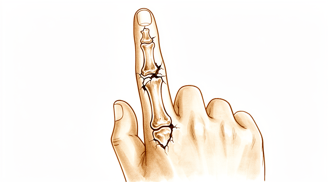
Phalangeal and metacarpal fractures of the hand — non-operative care and indications for fixation.
For patients: a plain-language version of this topic is available. See the patient guide.
Overview
The majority of hand fractures are managed successfully without operation [1, 2]. Conservative functional techniques represent the optimum treatment for most patients with single metacarpal fractures [2]. Surgical intervention offers distinct advantages in properly selected cases [1], and indications for operative treatment are now more clearly defined as understanding of finger fractures improves and osteosynthesis techniques continue to evolve [12]. The primary goals of phalangeal fracture treatment are to restore anatomy and preserve function while enabling early mobilization [16].
In the pediatric population, most hand and phalangeal fractures are treated nonoperatively with good results [5, 15]. However, a subset of pediatric phalangeal fractures requires prompt recognition and surgical intervention to minimize complications [5, 15]. Articular fractures of the fingers in children often result in sequelae, with rates as high as 50% when the fracture was displaced initially [7].
Outcomes for finger fractures vary according to fracture type, surgeon experience, and patient compliance [12]. Specific injury patterns carry higher risks of adverse events or reoperation. Injuries sustained in the thumb and index finger were more likely to undergo unplanned reoperation [4]. A quarter of open finger fractures will likely need more than one surgical procedure, a requirement especially common in more severely injured fingers due to crush or with vascular impairment [10]. Postoperative finger stiffness occurred in 43% of fractures treated with titanium plates and/or screws for unstable proximal phalangeal fractures [3].
Anatomy & Pathophysiology
Bony Anatomy
All phalanges consist of a proximal base, a central diaphysis, and a distal head [53]. Unlike metacarpals, the bases of all phalanges develop as metaphyses rather than heads [53]. The distal portion of the distal phalanx is referred to as the tuft [53]. Fingers follow a typical pattern of relative lengths: the tip of the index finger extends to the base of the nail of the middle finger, the tip of the ring finger to the mid-aspect of the middle finger nail, and the tip of the small finger to a corresponding position [53]. A midline volar longitudinal groove is consistently present in the middle phalanges [126]. This groove is most pronounced at the mid-phalangeal shaft and is deepest in the middle and ring fingers [126].
Joint Anatomy & Kinematics
The articulations of the fingers form a triarticular chain that flexes toward the thumb and the palm to allow grasp [72]. These joints function in the direction of flexion and possess two firm collateral ligaments and a thick reinforced anterior capsule, known as the volar plate [72]. In contrast, the fibrous dorsal capsule is thin and lax [72]. The interphalangeal articulations function uniquely in flexion–extension, with trochlear-shaped articulations that remain closely congruent throughout excursion [72]. Flexion ranges are approximately 85 degrees at the metacarpophalangeal joint, 115 degrees at the proximal interphalangeal joint, and 80 degrees at the distal interphalangeal joint [72].
Intraarticular fractures that disrupt joint congruency can occur at either the distal (condylar fracture) or proximal (pilon or proximal condylar fracture) articular surface [53]. Most commonly, these intraarticular phalangeal fractures are produced by an axial loading injury [53]. The normal ulnar inclination of the fingers occurs at the metacarpophalangeal joints [77]. This inclination is most marked in the index finger, less in the middle and little fingers, and almost non-existent in the ring finger [77]. It results from anatomical factors including asymmetry of the metacarpal heads and collateral ligaments, tendon crossing on the ulnar side of the longitudinal axis, intrinsic muscle predominance, and forward displacement of the ulnar metacarpals [77].
Soft Tissue & Tendon Anatomy
Intrinsic and extrinsic tendon insertions act as deforming forces to create typical angulation patterns in phalangeal fractures [53]. Proximal and middle phalangeal shaft fractures typically collapse into apex volar angulation due to the proximal flexion moment of the intrinsics and the distal extensor moment of the extensor mechanism [53]. The extensor tendons pass from the forearm onto the dorsum of the hand through six compartments beneath the extensor retinaculum [76]: * First compartment: Contains the extensor pollicis brevis and abductor pollicis longus [76]. * Second compartment: Contains the extensors carpi radialis longus and brevis [76]. * Third compartment: Contains the extensor pollicis longus [76]. * Fourth compartment: Contains the four tendons of the extensor digitorum communis plus the extensor indicis proprius [76]. * Fifth compartment: Contains the extensor digiti quinti [76]. * Sixth compartment: Contains the extensor carpi ulnaris [76].
The interosseous muscles produce lateral movements of fingers through their insertions on the lateral aspect of the base of the proximal phalanges [82]. When the metacarpophalangeal joint is in extension, the interosseous muscles extend the distal phalanges [82]. When the metacarpophalangeal joint is in flexion, the interosseous muscles reinforce flexion of the proximal phalanx and lose their extensor action on the distal phalanges [82]. The lumbrical muscles are able to extend the two distal phalanges whether the metacarpophalangeal joint is in extension or flexion [82]. They participate in extension of the distal phalanges by pulling distally on the flexor profundus tendon when this muscle is at rest [82].
The palmar skin is anchored to the underlying fascial planes by a system of fibrous tracts [73]. It is separated from the superficial palmar fascia by a layer of fatty tissue divided into compartments by fibrous septa, forming fat pads [73]. The metacarpophalangeal pad sits transversely over the base of the fingers from the ulnar to the radial border of the hand [73]. The pulp has a lobulated palmar pad where fibrous septa join the periosteum of the distal phalanx to the deep aspect of the dermis [73]. The soft tissue of the fingertips is tightly anchored [74]. Conversely, the soft tissue of the fingers between the fingertips and the area of the aponeurosis is mobile and flexible [74]. This mobile soft tissue overlies the fibrous tendon sheaths and extends from the distal phalanges to the metacarpophalangeal joints [74].
Cutaneous striations that make up fingerprints reflect the arrangement of the papillary ridges of the underlying dermis [73]. The specific distribution of cutaneous striations over the pulps forms a typical concentric pattern [73]. Cutaneous striae play an important part in the retention of an object during gripping by preventing sliding [73]. The concentric arrangement of striae at the pulp ensures the presence of a group of striae perpendicular to the force exerted, whatever its direction [73]. The palmar creases do not overlie the joints whose flexion induces them to fold [73]. Only the middle digit creases coincide with the proximal interphalangeal joints [73]. The distal palmar crease lies just distal to the distal interphalangeal joint [73]. The proximal palmar crease lies almost halfway down the proximal phalanx [73].
Pathophysiology & Deformity Mechanics
Apex volar angulation of 10° or more involving the proximal phalangeal diaphysis will affect the balance of the extensor mechanism, potentially producing a pseudoclaw deformity [70]. Lateral angular deformities of the phalanges are associated with more complex injuries involving bone loss [70]. Phalangeal deformity primarily involving both proximal and middle phalanges can comprise rotation, lateral, volar, or apex dorsal angulation, shortening, and often a combination of these [70]. Malrotation of phalangeal fractures does not remodel and can result in problems with grip formation [29]. Prolonged immobilization of simple finger injuries can result in significant joint stiffness [29].
The mallet finger deformity is characterized by a loss of active distal interphalangeal joint extension with full passive range of motion evident [55]. It reflects the loss of normal extensor force transmission via the terminal tendon insertion onto the distal phalanx [55]. In mallet finger, the unopposed flexor digitorum profundus pulls the distal joint into flexion [55]. The usual mechanism of injury involves sudden passive flexion of the actively extended distal interphalangeal joint [55]. Disruption of the terminal tendon may be entirely confined to the tendon or may involve an avulsed fracture fragment from the dorsal lip of the distal phalanx proximal articular surface [55]. The clinical appearance of soft tissue and bony mallet fingers is similar, with the distal joint resting in a flexion posture that cannot be actively changed [55]. Full passive extension of the distal interphalangeal joint is possible in mallet finger [55].
In the "lumbrical plus" finger, shortening injury that acutely violates the flexor digitorum profundus insertion leads to tendon retraction [33]. The retracted flexor digitorum profundus tendon creates tension on the extensor mechanism through the lumbrical, causing paradoxical interphalangeal joint extension with active digit flexion [33]. The "lumbrical plus" deformity is treated with release of the radial lateral band [33].
Small amounts of metacarpal shortening or dorsal angulation cause minimal functional impairment [48]. Early motion of adjacent joints in closed simple metacarpal fractures expedites recovery of motion and strength without adversely affecting fracture alignment [48]. The absence of the proximal phalanx of the middle or ring finger creates a hole through which small objects can pass and impairs scooping maneuvers [24]. This absence causes remaining fingers to tend to deviate toward the midline of the hand [24]. The third and fourth metacarpal heads help stabilize the metacarpal arch by providing attachments for the transverse metacarpal ligament [24]. Excising the third metacarpal shaft removes the origin of the adductor pollicis and weakens pinch [24].
The small finger plays an important role in palmar grip because of the mobility of its carpometacarpal joint and the action of the hypothenar muscles [35]. It increases the span of the hand for grasp owing to its abduction moment [35]. The ring finger forms the keystone of the palmar arch and participates in power grip [52]. It plays a minimal role in precision pinch [52]. Ring finger ray resection can have negative effects on hand function, including decreased key and chuck pinch strengths compared to amputation through the proximal phalanx [52]. Central ray deletion can have a negative impact on manual dexterity [52]. When the index finger is amputated at or more proximal to its proximal interphalangeal joint level, the remaining stump is useless and can hinder pinch between the thumb and middle finger [75].
Classification
Pediatric Phalangeal Neck Fractures
Pediatric phalangeal neck fractures are classified by displacement and fragment contact. Type I fractures are defined as undisplaced injuries [138]. Type II fractures are displaced but retain some bone-to-bone contact between the distal and proximal fragments [36]. Type III fractures are displaced with complete loss of bone-to-bone contact at the fracture site [36]. A distinct Type II D pattern tends to occur in young children, with the majority involving the middle phalanx [43]. Additionally, Salter-Harris type II fractures of the proximal phalanx represent the most frequent phalangeal fractures in children [42].
General Phalangeal Fracture Patterns
Phalangeal deformity can comprise rotation, lateral, volar, or apex dorsal angulation, shortening, and often a combination of these [70]. Lateral angular deformities are associated with more complex injuries involving bone loss [70]. In adults, phalangeal neck fractures of the proximal phalanx are rare and present limited options for unstable fractures [21].
Clinical Presentation
Epidemiology and Demographics
Finger phalangeal fractures constitute approximately 10% of all fractures encountered [40] and represent the second most common fracture in males [40]. The annual incidence of hand fractures is 3.7 per 1000 for men and 1.3 per 1000 for women [22]. Approximately 70% of phalangeal and metacarpal fractures occur in patients aged 11 to 45 years [53], with phalangeal fractures being more common in men than women [53]. The little finger is the most frequently affected digit, accounting for 32.5% of finger phalangeal fractures, followed by the ring finger at 25.8% [40]. In a study of 1569 fractures, little finger metacarpal fractures represented 27% of the total [22].
In the pediatric population, hand and finger fractures are the second most common fracture presenting to emergency departments [29]. These injuries exhibit a bimodal age distribution with peaks at 0 to 2 years and 12 to 16 years [29]. The most commonly injured locations are the base of the proximal phalanx (67%) of the border rays, specifically the little finger (52.2%) and thumb (23.5%) [29]. Salter-Harris type II fractures are extremely common in children, with the little finger proximal phalanx being the most frequently injured site [29]. Phalangeal fractures are the most common hand fractures in children, with Salter-Harris type II fractures of the proximal phalanx being the most frequent [42].
Open phalangeal fractures are relatively common, with the highest prevalence in males aged 36 to 64 years [40]. The distal phalanges are the commonest site for open phalangeal fractures, where 25.3% of fractures were open [40]. About 55% of patients with multiple phalangeal fractures have other phalangeal fractures as associated injuries, with an average patient age of 55.4 years [40].
Mechanism of Injury
Direct blow or assault is the most common mode of injury, accounting for 39.1% of cases, followed by falls from standing height at 29.5% and sports injuries at 23.8% [40]. Patients injured by direct blows or sports injuries are younger and more likely to be male than those injured by standing falls [40]. Fractures caused by direct blows or assaults are more prevalent on the radial side, with 47.4%, 50.0%, and 43.4% of middle finger, index finger, and thumb fractures respectively resulting from these mechanisms [40]. Conversely, more fractures of the little and ring fingers are caused by falls and sports injuries [40].
In children, toddlers and preschoolers usually sustain hand injuries at home as crush injuries, while adolescents are most often injured outside the home during sporting activities [29]. Salter-Harris II fractures typically result from jamming or hyperextension injuries causing abduction deformity [29]. Spiral fractures of the proximal phalanx can result from finger wrestling [20].
Clinical Examination and Diagnosis
Appropriate evaluation includes clinical examination and radiographs [29]. Clinical examination must assess for open injuries and angular and rotational malalignment of the injured ray [29]. Rotational alignment is confirmed by ensuring all fingers point to the scaphoid tubercle when flexed [29]. Radiographs should include PA, lateral, and oblique views of the injured location [29].
For Salter-Harris II fractures, coronal plane deformity is easy to assess, but extra care is required to detect rotational deformity, which is not obvious on radiographs [29]. Malrotation does not remodel and can result in problems with grip formation [29]. The classic “jammed” finger with a swollen, painful PIP joint usually involves a volar plate injury or small nondisplaced avulsion fracture off the volar base of the middle finger epiphysis [29]. Phalangeal neck and condyle fractures present similarly to a simple “jammed” finger and are often missed [29]. Seymour fractures are open fractures that are often missed; diagnosis relies on nail plate/cuticle disruption and a displaced distal phalanx fracture on radiographs [29]. A meticulous examination of the patient’s hand is required for injury diagnosis [53].
Injuries to the thumb and index finger are more likely to undergo unplanned reoperation, which may guide initial treatment decision-making and postoperative follow-up [4]. Common preoperative comorbidities, including smoking status and diabetes mellitus, increase the likelihood of postoperative complications in patients undergoing surgical treatment [60]. Social deprivation influences both the pattern and management of hand fractures [22]. Little finger metacarpal fractures were associated with social deprivation in men [22]. For women, fractures with an unclear mechanism or where the patient was intoxicated and could not recall the mechanism showed a clear association with deprivation [22]. Affluent patients were more likely to receive operative treatment [22].
Investigations
Plain radiography: A true lateral radiograph is essential for the accurate diagnosis of phalangeal neck fractures in children [149]. In the context of carpometacarpal fracture-dislocations, a true lateral radiograph is required because swelling can obscure the deformity [99]. On posteroanterior views, the loss of parallel joint surfaces at the carpometacarpal articulations is indicative of carpometacarpal fracture-dislocation [99].
Ultrasonography: Ultrasonography may provide greater accuracy in fracture diagnostics by revealing small avulsed bony fragments that are missed on radiographs [144]. This modality can be beneficial in diagnosing occult fractures, especially in children [144].
CT: A CT scan is sometimes beneficial to determine the extent of joint surface involvement and to guide appropriate intervention for carpometacarpal fracture-dislocations [99]. Quantitative 3DCT analysis of fracture fragments provides useful information that could facilitate surgery and analysis of complex fractures of the base of the middle phalanx [145].
Treatment
Non-Operative
The majority of hand fractures are managed without surgery, although operative intervention offers distinct advantages in properly selected cases [1]. Indications for surgical treatment of finger fractures are clearly defined, and while results vary based on fracture type, surgeon experience, and patient compliance, operative techniques and implants continue to evolve [12]. Well-reduced, minimally angulated, or nonangulated proximal phalangeal fractures can be effectively treated with functional casts that do not immobilize the wrist [107]. Traction splinting is a successful method for closed proximal phalangeal fractures [11], and a non-invasive thermoplastic traction platform provides a safe and effective alternative [116]. A hand-based non-invasive skin traction orthosis is a simple, inexpensive option that yields promising results, with a preference for shorter traction duration compared to forearm-based orthoses [125]. For stable proximal phalangeal fractures, a splint keeping the MCP joint flexed at 90° and the IP joints in extension is recommended, with occasional active PIP motion encouraged to compress and stabilize the fracture [104]. Non-operative treatment of transverse metacarpal fractures typically results in excellent functional outcomes, apart from mild cosmetic abnormality [95]. There is no good evidence that more marked malunion of metacarpal neck fractures causes reduced hand function or unacceptable deformity [95], and no single non-operative technique has been shown to be superior in published studies [95].
In pediatric patients, most hand fractures are treated nonoperatively with good results [5, 15]. The majority can be managed with closed reduction, appropriate immobilization, and early motion [29]. Buddy taping is a non-inferior treatment modality for most paediatric finger fractures compared to splint immobilization [96]. Taping displaced extra-articular phalangeal finger fractures in children can be recommended irrespective of the degree of displacement or the need for reduction [30]. Nonsurgical management is supported for most Salter-Harris Type II fractures of the proximal phalanx of the small finger in children [130]. Displaced phalangeal neck fractures in children do not necessarily displace with nonsurgical treatment [69].
For professional athletes, treatment is dictated by nonbiological factors such as season timing, player position, and hand dominance, often requiring a team approach to balance return-to-play risks against financial and professional consequences [132]. In baseball, treatment should be immediate, with nonoperative splinting for at least 3 weeks for nondisplaced fractures and surgery for displaced fractures [110].
Operative
Indications: A subset of pediatric phalangeal fractures requires prompt recognition and surgical intervention to minimize complications [5, 15]. Displaced phalangeal neck fractures in children require reduction and pin fixation, which can usually be achieved through a closed fashion [29]. Seymour fractures are open fractures that require removal of the nail plate, débridement of the fracture site, extrication of interposed nail bed, and reduction of the fracture [29]. If a Seymour fracture is unstable, it may require Kirschner wire placement in addition to immobilization in a splint or cast [29].
Surgical Approach / Technique: Open reduction and internal fixation (ORIF) of proximal phalangeal fractures has achieved excellent outcomes, providing a benchmark for recovery [13]. Most fractures treated with closed reduction and periarticular pinning of the base and shaft of the proximal phalanx healed within 4 weeks, with the majority of patients having excellent or good results [46]. Open reduction and interfragmentary screw fixation is an effective treatment modality for symptomatic non-union of distal phalangeal fractures with minimal morbidity, resulting in union and normal function in all patients [41]. Ligamentotaxis provides excellent functional results for closed phalangeal fractures at the PIP joint, with a low incidence of postoperative complications similar to other commonly used surgical techniques [14]. All six fingers treated with intramedullary fixation through a volar extra-tendon sheath approach obtained satisfactory union, and no patient complained of pain at the final follow-up [8]. Percutaneous compressive bone tie was used to treat two intraarticular proximal phalangeal fractures of the thumb with satisfactory outcomes, including full range of movements within 3 weeks, no complications, and no need for implant removal [23]. Condyle fractures in children can often be treated with closed reduction and pinning, as open procedures increase the risk for osteonecrosis [29].
Implant Selection: No differences in the outcome of finger proximal phalangeal fractures were observed between dorsal and lateral plate fixation [9]. Non-locking plates are appropriate for most metacarpal and phalangeal fractures necessitating plate fixation [47]. Intramedullary compression screw fixation for proximal phalangeal fractures results in good functional outcomes and a low complication rate [31]. Dual antegrade intramedullary headless screw fixation of proximal phalanx fractures resulted in excellent postoperative motion, near-normal grip strength, positive self-reported patient outcomes, and no complications with follow-up of at least 1 year [44]. The Joshi's External Stabilization System (JESS) has had extensive use in the management of hand injuries [28].
Other Considerations: A finger undergoing any type of surgery is likely to be stiffer than one that was treated nonoperatively [104]. Injuries sustained in the thumb and index finger were more likely to undergo unplanned reoperation after vascular reconstruction, which may guide initial treatment decision-making and postoperative follow-up [4]. A quarter of open finger fractures will likely need more than one surgical procedure, especially in more severely injured fingers due to crush or with vascular impairment [10].
Outcomes and Complications: Extension lags of the PIP joints were found in 67% of all fractured fingers treated with plate fixation for extra-articular fractures of the proximal phalanx [38]. At final follow-up or plate removal, an improved average total active motion (TAM) of 213° was found in patients treated with plate fixation for extra-articular proximal phalangeal fractures [38]. In a retrospective review of 105 pediatric patients treated with closed reduction pin fixation of a displaced proximal phalanx fracture, the complication rate was 4.8%, including infection, pin site complication, and malunion [29]. Thirty-six of 105 pediatric patients treated with closed reduction pin fixation for displaced proximal phalanx fractures had postoperative stiffness, with 31 requiring therapy [29]. Phalangeal neck fractures had the highest rate of postoperative stiffness in pediatric patients treated with closed reduction pin fixation [29]. Thirty-one pediatric patients available for follow-up at 1 year or greater after closed reduction pin fixation reported return of full motion, no pain, and happiness with function and appearance, despite 22% having a measurable coronal plane deformity on radiograph [29]. Missed Seymour fractures have a high rate of complication including infection and nail or physeal growth disturbance [29]. Residual stiffness of the injured finger was observed in cases of nonunion without avascular necrosis of finger phalangeal neck fractures in children [63]. Articular fractures of the fingers in children often result in sequelae, with a rate as high as 50% when the fracture was displaced initially [7]. Recommended treatment for Seymour fractures includes a dose of IV antibiotic in the emergency department followed by a 7- to 10-day course of oral antibiotic, with a first-generation cephalosporin being the preferred antibiotic [29].
Complications
Postoperative Stiffness and Range of Motion
Phalangeal fractures treated with a titanium plate tend to deteriorate in total active motion compared to metacarpal fractures [26]. Percutaneous pinning of proximal phalangeal base fractures results in a permanent reduction in range of motion at the MCP and PIP joints [68]. Uncorrected angulation of 25 degrees or more in the adult or older child usually results in loss of both flexion and extension of the proximal interphalangeal joint [115]. In children over age ten with impacted fractures in the proximal third of the proximal phalanx, two of six patients had malunion with loss of significant flexion and extension of the proximal interphalangeal joint [115]. Among seventeen adults with impacted fractures in the proximal third of the proximal phalanx, nine had malunion [115]. Seven adults first seen more than five weeks after injury had angulation of 25 to 70 degrees with significant loss of both flexion and extension at the proximal interphalangeal joint [115].
Reoperation and Surgical Failure
A quarter of open finger fractures will likely need more than one surgical procedure, especially in more severely injured fingers due to crush or vascular impairment [10]. Injuries sustained in the thumb and index finger were more likely to undergo unplanned reoperation after vascular reconstruction [4]. The risk for postoperative complications and reoperations after closed reduction and percutaneous pinning of closed proximal phalanx fractures is considerable [108]. The remaining patients in a cohort of phalangeal fractures had either suffered an unusual type of injury or developed an arthrosis associated with a long delay between injury and operation [32].
Pediatric Complications
Complications of distal phalanx fractures in children are frequent [57].
Other Considerations
Bone resorption occurred in the digit reconstructed using the circumferential method seven months after surgery for pulley reconstruction [50]. The true incidence of bone resorption following pulley reconstruction remains unclear as radiographs are not commonly performed after such procedures [50].
Recovery
General Outcomes and Prognosis: Results vary according to fracture type, surgeon experience, and patient compliance [12].
Non-Operative Recovery: In pediatric populations, most phalangeal neck fractures in children less than 3 years old obtained satisfactory results [19]. Complete remodelling of a malunited phalangeal neck fracture occurred over a 1-year period in a 3-year-old boy treated conservatively [49].
Operative Recovery: Excellent outcomes were achieved after open reduction and internal fixation of proximal phalangeal fractures, providing a benchmark for recovery [13]. Dynamic external fixation (DEF) provides excellent functional results for closed phalangeal fractures at the PIP joint, with a low incidence of postoperative complications similar to other commonly used surgical techniques [14]. All six fingers obtained satisfactory union of the fractures, and no patient complained of pain at the final follow-up after intramedullary fixation through a volar extra-tendon sheath approach [8]. No differences in the outcome of finger proximal phalangeal fractures treated by dorsal and lateral plate fixation were observed [9]. Intramedullary compression screw (IMS) fixation for proximal phalangeal fractures is associated with good functional outcomes and a low complication rate [31]. Dual antegrade intramedullary headless screw (IMHS) fixation of proximal phalanx fractures resulted in excellent postoperative motion, near-normal grip strength, positive self-reported patient outcomes, and no complications with follow-up of at least 1 year [44]. Most fractures healed within 4 weeks after closed reduction and periarticular pinning of base and shaft fractures of the proximal phalanx [46]. The majority of patients had excellent or good results after closed reduction and periarticular pinning of base and shaft fractures of the proximal phalanx [46]. The stepwise surgical algorithm for displaced pediatric phalangeal neck fractures produces 92% good-excellent results even in late-presenting fractures [152].
Complications and Adverse Outcomes: Bone resorption occurred in the digit reconstructed using the circumferential method seven months after surgery [50]. Patients who suffered an unusual type of injury or developed an arthrosis associated with a long delay between injury and operation had less favorable outcomes [32].
Key Evidence
- [L5] The majority of hand fractures can be treated without surgery, though surgery offers distinct advantages in properly selected cases. [1] (10.1016/j.jhsa.2013.02.017)
- [L5] Most hand fractures can be managed successfully without operation, and conservative functional techniques are the optimum treatment for the majority of patients with single metacarpal fractures. [2] (10.1177/1753193420928820)
- [L2] Postoperative finger stiffness occurred in 43% of fractures. [3] (10.1016/j.jhsa.2014.06.107)
- [L3] Injuries sustained in the thumb and index finger were more likely to undergo unplanned reoperation, which may guide initial treatment decision-making and postoperative follow-up. [4] (10.1177/15589447221109635)
- [L5] Most pediatric hand fractures are treated nonoperatively with good results, but a subset of phalangeal fractures requires prompt recognition and surgical intervention to minimize complications. [5] (10.1097/01.blo.0000205890.88952.97)
- [L4] Articular fractures of the fingers in children often result in sequelae, with a rate as high as 50% when the fracture was displaced initially. [7] (10.1016/s0749-0712(21)00213-4)
- [L4] All six fingers obtained satisfactory union of the fractures, and no patient complained of pain at the final follow-up. [8] (10.1142/s0218810411005230)
- [L3] No differences in the outcome of finger proximal phalangeal fractures treated by dorsal and lateral plate fixation were observed. [9] (10.1007/s00402-017-2650-x)
- [L3] A quarter of open finger fractures will likely need more than one surgical procedure, especially in more severely injured fingers, due to crush or with vascular impairment. [10] (10.1177/15589447211043191)
- [L4] We believe we have shown its success in the treatment of closed proximal phalangeal fractures. [11] (10.1016/s0020-1383(01)00138-3)
- [L5] Fractures of the fingers are better understood, indications for surgical treatment are more clearly defined, and operative techniques and implants for osteosynthesis are continuing to evolve and improve, though results vary according to fracture type, surgeon experience, and patient compliance. [12] (10.1054/jhsb.2002.0889)
- [L3] Excellent outcomes were achieved, providing a benchmark for recovery after ORIF of proximal phalangeal fractures. [13] (10.1177/1753193416670591)
- [L2] DEF provides excellent functional results for closed phalangeal fractures at the PIP joint, with a low incidence of postoperative complications similar to other commonly used surgical techniques. [14] (10.1177/17531934251350453)
- [Paper] Most pediatric phalangeal fractures can be treated nonsurgically, but a small subset benefits from surgical intervention. [15] (10.1016/j.jhsa.2025.08.015)
- [L5] The primary goals of phalangeal fracture treatment are to restore anatomy and preserve function while enabling early mobilization. [16] (10.1016/j.hcl.2013.08.006)
- [L4] Most phalangeal neck fractures in children less than 3 years old obtained satisfactory results. [19] (10.1186/s13018-025-05849-2)
- [L4] These two cases present an uncommon mechanism resulting in a spiral fracture of the proximal phalanx of the index finger, which to our knowledge has not been reported previously in the English literature. [20] (10.1016/0020-1383(92)90162-l)
- [L4] Phalangeal neck fractures of the proximal phalanx in adults are rare with limited options for unstable fractures. [21] (10.1016/j.injury.2010.06.017)
- [L3] [22] (10.1177/1753193410381823)
- [L4] The technique was used to treat two intraarticular proximal phalangeal fractures of the thumb with satisfactory outcomes, including full range of movements within 3 weeks, no complications, and no need for implant removal. [23] (10.1177/1753193415615032)
- [L2] The phalangeal fractures tend to deteriorate %TAM than metacarpal fractures. [26] (10.1016/s0363-5023(11)60047-6)
- [Paper] The system has had extensive use in the management of hand injuries. [28] (10.1016/s0020-1383(97)88363-5)
- [L1] With the current data, we can conclude that taping these finger fractures can be recommended irrespective of the degree of displacement or the need for reduction. [30] (10.1177/17531934241293338)
- [L4] This updated systematic review finds good functional outcomes and a low complication rate following the use of IMS fixation for proximal phalangeal fractures. [31] (10.1177/15589447251329597)
- [L4] The remaining patients had either suffered an unusual type of injury, or had developed an arthrosis, associated with a long delay between injury and operation. [32] (10.1016/0020-1383(81)90217-5)
- [L4] [36] (10.1177/1753193408091430)
- [L4] [38] (10.1007/s00402-015-2155-4)
- [L4] Open reduction and interfragmentary screw fixation is an effective treatment modality for symptomatic non-union of distal phalangeal fractures with minimal morbidity, resulting in union and normal function in all patients. [41] (10.1177/1753193407087866)
- [L5] Phalangeal fractures are the most common hand fractures in children, with Salter-Harris type II fractures of the proximal phalanx being the most frequent. [42] (10.5435/jaaos-d-16-00199)
- [Paper] Type II D phalangeal neck fractures tend to occur in young children and the majority involve the middle phalanx. [43] (10.1055/s-0040-1703097)
- [L4] Dual antegrade IMHS fixation of proximal phalanx fractures resulted in excellent postoperative motion, near-normal grip strength, positive self-reported patient outcomes, and no complications with follow-up of at least 1 year. [44] (10.1177/1558944717750919)
- [L4] Most fractures healed within 4 weeks, and the majority of patients had excellent or good results. [46] (10.1016/j.jhsa.2014.05.008)
- [L5] Non-locking plates are appropriate for most metacarpal and phalangeal fractures necessitating plate fixation. [47] (10.1016/j.jhsa.2011.09.023)
- [L5] Recent reports confirm that small amounts of metacarpal shortening or dorsal angulation cause minimal functional impairment, and early motion of adjacent joints in closed simple metacarpal fractures expedites recovery of motion and strength without adversely affecting fracture alignment. [48] (10.1097/01.blo.0000205888.04200.c5)
- [L5] Complete remodelling of a malunited phalangeal neck fracture occurred over a 1-year period in a 3-year-old boy treated conservatively, representing the second such occurrence in the literature. [49] (10.1016/j.injury.2003.09.042)
- [L4] Bone resorption occurred in the digit reconstructed using the circumferential method seven months after surgery, though the true incidence remains unclear as radiographs are not commonly performed after such procedures. [50] (10.1177/1753193412453413)
- [L5] [53] (10.1016/j.hcl.2012.05.032)
- [L4] Complications of distal phalanx fractures in children are frequent. [57] (10.1016/j.jhsa.2017.03.042)
- [L3] Common preoperative comorbidities, including smoking status and diabetes mellitus, increase the likelihood of postoperative complication in patients with hand and finger fractures and/or dislocations undergoing surgical treatment. [60] (10.1177/15589447221120847)
- [L4] However, there was residual stiffness of the injured finger. [63] (10.1016/j.jhsa.2014.05.017)
- [L3] Both techniques resulted in permanent reduction in range of motion at the MCP and PIP joints. [68] (10.1016/s0363-5023(12)60053-7)
- [L4] Our findings suggest that displaced phalangeal neck fractures do not necessarily displace with nonsurgical treatment. [69] (10.1016/j.jhsa.2020.02.019)
- [L5] [70] (10.1016/j.jhsa.2013.07.014)
- [L4] [95] (10.1177/1753193414548170)
- [L1] Buddy taping is a non-inferior treatment modality for most paediatric finger fractures compared to splint immobilization. [96] (10.1177/1753193418822692)
- [L4] [104] (10.1177/1558944717735947)
- [L2] Well-reduced, minimally angulated, or nonangulated fractures of the proximal phalanges of the fingers can be effectively treated using functional casts without immobilizing the wrist. [107] (10.1016/j.jhsa.2012.02.017)
- [L3] The risk for postoperative complications and reoperations after CRPP of closed proximal phalanx fractures is considerable, and surgeons should counsel patients before surgery about these risks. [108] (10.1016/j.jhsg.2023.11.004)
- [L5] Treatment should be immediate, with nonoperative splinting for at least 3 weeks for nondisplaced fractures and surgery for displaced fractures. [110] (10.1016/j.hcl.2012.05.033)
- [L4] [115] (10.2106/00004623-196951070-00006)
- [L4] This non-invasive technique using a thermoplastic traction platform is safe & effective in the management of proximal phalangeal fractures. [116] (10.1016/j.jht.2021.02.001)
- [L4] The study recommends the use of hand-based non-invasive skin traction orthosis as a simple, inexpensive, and non-invasive option for managing phalangeal fractures, noting that it yields promising results with a preference for shorter traction duration compared to forearm-based orthoses. [125] (10.1016/j.jht.2023.12.012)
- [L5] A midline volar longitudinal groove is consistently present in the middle phalanges, is most pronounced at the mid-phalangeal shaft, and is deepest in the middle and ring fingers. [126] (10.1016/j.jhsa.2022.10.004)
- [L4] Our findings support nonsurgical management of most FBPP of the small finger in children. [130] (10.1016/j.jhsa.2024.04.002)
- [L5] Management of phalangeal fractures in professional athletes is dictated by nonbiological factors such as season timing, player position, and hand dominance, often requiring a team approach to balance return-to-play risks against financial and professional consequences. [132] (10.1016/j.hcl.2012.05.035)
- [L4] [138] (10.1054/jhsb.2000.0506)
- [L4] Ultrasonography may provide greater accuracy in fracture diagnostics by revealing small avulsed bony fragments missed on radiographs and can be beneficial in diagnosing occult fractures, especially in children. [144] (10.1016/j.jhsa.2015.02.022)
- [L4] Quantitative 3DCT analysis of fracture fragments provides useful information that could facilitate surgery and analysis of complex fractures of the base of the middle phalanx. [145] (10.1007/s11552-014-9665-3)
- [Paper] K-wire fixation is required even for minimally displaced fractures, and a true lateral view is essential for accurate diagnosis. [149] (10.1016/j.injury.2015.02.018)
- [L4] The stepwise surgical algorithm produces 92% good-excellent results even in late-presenting fractures. [152] (10.1016/s0363-5023(09)60118-0)
See Also
- Mallet Finger
- Dislocations
References
[1] Hand Fractures: A Review of Current Treatment Strategies. The Journal of Hand Surgery. 2013. DOI: 10.1016/j.jhsa.2013.02.017
[2] Current methods, outcomes and challenges for the treatment of hand fractures. Journal of Hand Surgery (European Volume). 2020. DOI: 10.1177/1753193420928820
[3] Predictors of Postoperative Finger Stiffness After Treatment of Unstable Proximal Phalangeal Fractures With Titanium Plates And/Or Screws. The Journal of Hand Surgery. 2014. DOI: 10.1016/j.jhsa.2014.06.107
[4] Phalangeal Fractures Requiring Vascular Reconstruction: Epidemiology and Factors Predictive of Reoperation. HAND. 2022. DOI: 10.1177/15589447221109635
[5] Pediatric Phalanx Fractures. Clinical Orthopaedics and Related Research. 2006. DOI: 10.1097/01.blo.0000205890.88952.97
[7] ARTICULAR FRACTURES OF THE FINGERS IN CHILDREN. Hand Clinics. 2000. DOI: 10.1016/s0749-0712(21)00213-4
[8] INTRAMEDULLARY FIXATION OF PROXIMAL PHALANGEAL FRACTURES THROUGH A VOLAR EXTRA-TENDON SHEATH APPROACH. Hand Surgery. 2011. DOI: 10.1142/s0218810411005230
[9] Dorsal versus lateral plate fixation of finger proximal phalangeal fractures: a retrospective study. Archives of Orthopaedic and Trauma Surgery. 2017. DOI: 10.1007/s00402-017-2650-x
[10] Reoperation After Operative Treatment of Open Finger Fractures. HAND. 2022. DOI: 10.1177/15589447211043191
[11] Old principles revisited—traction splinting for closed proximal phalangeal fractures. Injury. 2002. DOI: 10.1016/s0020-1383(01)00138-3
[12] Treatment of Fractures of the Fingers. What’s New?. Journal of Hand Surgery. 2003. DOI: 10.1054/jhsb.2002.0889
[13] Pattern of recovery after open reduction and internal fixation of proximal phalangeal fractures in the finger: a prospective longitudinal study. Journal of Hand Surgery (European Volume). 2016. DOI: 10.1177/1753193416670591
[14] Clinical outcomes of ligamentotaxis in closed phalangeal fractures: a systematic review. Journal of Hand Surgery (European Volume). 2025. DOI: 10.1177/17531934251350453
[15] Pediatric Finger Fractures: Preventing Big Problems After Small Fractures. The Journal of Hand Surgery. 2026. DOI: 10.1016/j.jhsa.2025.08.015
[16] Treatment of Phalangeal Fractures. Hand Clinics. 2013. DOI: 10.1016/j.hcl.2013.08.006
[19] Clinical results of phalangeal neck fractures in children aged 3 years or younger: a case series of 37 patients. Journal of Orthopaedic Surgery and Research. 2025. DOI: 10.1186/s13018-025-05849-2
[20] Spiral fracture of the proximal phalanx of the index finger by finger wrestling. Injury. 1992. DOI: 10.1016/0020-1383(92)90162-l
[21] Phalangeal neck fractures of the proximal phalanx of the fingers in adults. Injury. 2010. DOI: 10.1016/j.injury.2010.06.017
[22] The epidemiology of fractures of the hand and the influence of social deprivation. Journal of Hand Surgery (European Volume). 2010. DOI: 10.1177/1753193410381823
[23] Percutaneous compressive bone tie for reduction of condylar phalangeal fractures. Journal of Hand Surgery (European Volume). 2016. DOI: 10.1177/1753193415615032
[24] Campbell S Operative Orthopaedics 4 Volume Set. AMPUTATION THROUGH THE SURGICAL NECK OF THE HUMERUS > MIDDLE OR RING FINGER RAY AMPUTATIONS.
[26] Predictors of the Postoperative Range of Finger Motion for Comminuted Hand and Finger Fractures Treated with a Titanium Plate. The Journal of Hand Surgery. 2011. DOI: 10.1016/s0363-5023(11)60047-6
[28] Joshi's External Stabilization System (JESS): a simple mini external fixator for the management of hand trauma and its sequels. Injury. 1997. DOI: 10.1016/s0020-1383(97)88363-5
[29] Orthopaedic Knowledge Update 13 Ebook Without Multimedia. Pediatric Forearm, Wrist, and Hand Trauma > Hand and Finger Fractures.
[30] Buddy taping after reduction of displaced extra-articular phalangeal finger fractures in children: a randomized controlled trial. Journal of Hand Surgery (European Volume). 2024. DOI: 10.1177/17531934241293338
[31] Intramedullary Compression Screw Fixation of Proximal Phalanx Fractures: An Updated Systematic Review. HAND. 2025. DOI: 10.1177/15589447251329597
[32] Fractures of phalanges. Injury. 1981. DOI: 10.1016/0020-1383(81)90217-5
[33] Miller S Review Of Orthopaedics. 4. Fingertip injuries with tissue loss.
[35] Green S Operative Hand Surgery. SMALL FINGER RAY AMPUTATION.
[36] Phalangeal Neck Fractures with Concurrent Vascular Injury. Journal of Hand Surgery (European Volume). 2008. DOI: 10.1177/1753193408091430
[38] Plate fixation of extra-articular fractures of the proximal phalanx: do new implants cause less problems?. Archives of Orthopaedic and Trauma Surgery. 2015. DOI: 10.1007/s00402-015-2155-4
[40] Rockwood And Green S Fractures In Adults. 3: Biologic and Biophysical Technologies for the Enhancement of Fracture Repair > Finger Phalanges.
[41] OPEN REDUCTION AND INTERFRAGMENTARY SCREW FIXATION FOR SYMPTOMATIC NONUNION OF DISTAL PHALANGEAL FRACTURES. Journal of Hand Surgery (European Volume). 2008. DOI: 10.1177/1753193407087866
[42] Pediatric Phalanx Fractures. Journal of the American Academy of Orthopaedic Surgeons. 2016. DOI: 10.5435/jaaos-d-16-00199
[43] Type II D Phalangeal Neck Fractures in Children: A Series of 20 Cases Treated According to a Preset Stepwise Algorithm. Journal of Hand and Microsurgery. 2021. DOI: 10.1055/s-0040-1703097
[44] Dual Antegrade Intramedullary Headless Screw Fixation for Treatment of Unstable Proximal Phalanx Fractures. HAND. 2018. DOI: 10.1177/1558944717750919
[46] Outcomes of Closed Reduction and Periarticular Pinning of Base and Shaft Fractures of the Proximal Phalanx. The Journal of Hand Surgery. 2014. DOI: 10.1016/j.jhsa.2014.05.008
[47] Non-Locked and Locked Plating Technology for Hand Fractures. The Journal of Hand Surgery. 2011. DOI: 10.1016/j.jhsa.2011.09.023
[48] Extraarticular Hand Fractures in Adults. Clinical Orthopaedics and Related Research. 2006. DOI: 10.1097/01.blo.0000205888.04200.c5
[49] Remodelling in a malunited phalangeal neck fracture. Injury. 2004. DOI: 10.1016/j.injury.2003.09.042
[50] Bone resorption of the proximal phalanx secondary to pulley reconstruction. Journal of Hand Surgery (European Volume). 2012. DOI: 10.1177/1753193412453413
[52] Green S Operative Hand Surgery. RING FINGER RAY AMPUTATION.
[53] Phalangeal Fractures. Hand Clinics. 2012. DOI: 10.1016/j.hcl.2012.05.032
[55] A Lange Medical Book Current Diagnosis Treatment In Orthopedics Fifth Edition. 9Hand Surgery > 3. Mallet Finger.
[57] Complications of Distal Phalanx Fractures in Children. The Journal of Hand Surgery. 2017. DOI: 10.1016/j.jhsa.2017.03.042
[60] Comorbidities Associated With Increased Likelihood of Postoperative Surgical Site Infection in Patients Treated for Hand or Finger Fracture and/or Dislocations. HAND. 2022. DOI: 10.1177/15589447221120847
[63] Nonunion Without Avascular Necrosis of Finger Phalangeal Neck Fractures in Children: Report of 4 Cases. The Journal of Hand Surgery. 2014. DOI: 10.1016/j.jhsa.2014.05.017
[68] Percutaneous Pinning of Proximal Phalangeal Base Fractures: Unacceptable Outcomes. The Journal of Hand Surgery. 2012. DOI: 10.1016/s0363-5023(12)60053-7
[69] Outcomes of Phalangeal Neck Fractures in a Pediatric Population. The Journal of Hand Surgery. 2020. DOI: 10.1016/j.jhsa.2020.02.019
[70] Malunited Fractures in the Hand. The Journal of Hand Surgery. 2014. DOI: 10.1016/j.jhsa.2013.07.014
[72] Exam Of The Hand Wrist 2Ed. Movements of the fingers > Articulations of the fingers.
[73] Exam Of The Hand Wrist 2Ed. Macroscopic appearance of palmar skin > Fingerprints.
[74] Dupuytren S Disease And Related Hyperproliferative Disorders. 2. Palmar Fibromatosis or the Loss of Flexibility of the Palmar Finger Tissue: A New Insight into the Disease Process of Dupuytren Contracture > 2.2 The Skin Anchoring Fibers in the Normal and Diseased Hand > 2.2.1 The External View of the Soft Tissue of the Fingers.
[75] Campbell S Operative Orthopaedics 4 Volume Set. AMPUTATION THROUGH THE SURGICAL NECK OF THE HUMERUS > AMPUTATIONS OF SINGLE FINGERS.
[76] Campbell S Operative Orthopaedics 4 Volume Set. RECONSTRUCTION OF FINGER FLEXORS: SINGLE-STAGE TENDON GRAFT > EXTENSOR TENDONS.
[77] Exam Of The Hand Wrist 2Ed. Movements of the fingers > Normal ulnar inclination of the fingers.
[82] Exam Of The Hand Wrist 2Ed. Mechanisms of finger flexion and extension > Actions of the intrinsic muscles.
[95] The non-operative management of hand fractures. Journal of Hand Surgery (European Volume). 2014. DOI: 10.1177/1753193414548170
[96] Buddy taping versus splint immobilization for paediatric finger fractures: a randomized controlled trial. Journal of Hand Surgery (European Volume). 2019. DOI: 10.1177/1753193418822692
[99] Campbell S Operative Orthopaedics 4 Volume Set. FRACTURES, DISLOCATIONS, AND LIGAMENTOUS INJURIES OF THE HAND AND WRIST > FINGER METACARPALS.
[104] Proximal Phalanx Fracture Management. HAND. 2017. DOI: 10.1177/1558944717735947
[107] Extra-Articular Fractures of the Proximal Phalanges of the Fingers: A Comparison of 2 Methods of Functional, Conservative Treatment. The Journal of Hand Surgery. 2012. DOI: 10.1016/j.jhsa.2012.02.017
[108] Risks of Reoperation in Surgically Treated Fractures of the Proximal Phalanx. Journal of Hand Surgery Global Online. 2024. DOI: 10.1016/j.jhsg.2023.11.004
[110] Phalangeal Fractures in Baseball: Commentary. Hand Clinics. 2012. DOI: 10.1016/j.hcl.2012.05.033
[115] Impacted Fractures in the Proximal Portion of the Proximal Phalanx of the Finger. The Journal of Bone & Joint Surgery. 1969. DOI: 10.2106/00004623-196951070-00006
[116] A traction orthosis for the treatment of proximal phalangeal fractures. Journal of Hand Therapy. 2022. DOI: 10.1016/j.jht.2021.02.001
[125] The use of non-invasive skin traction orthosis in managing phalangeal fractures. Journal of Hand Therapy. 2024. DOI: 10.1016/j.jht.2023.12.012
[126] The Volar Midline Longitudinal Groove of the Middle Phalanx: An Anatomic Study and Clinical Implications. The Journal of Hand Surgery. 2024. DOI: 10.1016/j.jhsa.2022.10.004
[130] Long-Term Clinical Outcomes Following Nonsurgical Management of Salter-Harris Type II Fractures of the Proximal Phalanx of the Small Finger: A Prospective Cohort Study. The Journal of Hand Surgery. 2025. DOI: 10.1016/j.jhsa.2024.04.002
[132] Football Commentary: Phalangeal Fractures—Displaced/Nondisplaced. Hand Clinics. 2012. DOI: 10.1016/j.hcl.2012.05.035
[138] PHALANGEAL NECK FRACTURES IN CHILDREN: CLASSIFICATION AND OUTCOME IN 66 CASES. Journal of Hand Surgery. 2001. DOI: 10.1054/jhsb.2000.0506
[144] Ultrasonography Can Be Used to Assess Phalanx Fractures. The Journal of Hand Surgery. 2015. DOI: 10.1016/j.jhsa.2015.02.022
[145] Quantitative 3-Dimensional CT Analyses of Fractures of the Middle Phalanx Base. HAND. 2014. DOI: 10.1007/s11552-014-9665-3
[149] A review of phalangeal neck fractures in children. Injury. 2015. DOI: 10.1016/j.injury.2015.02.018
[152] A Minimally Invasive Stepwise Surgical Algorithm for Displaced Pediatric Phalangeal Neck Fractures: The Importance of Percutaneous Reduction. The Journal of Hand Surgery. 2009. DOI: 10.1016/s0363-5023(09)60118-0