Clinicians › Hand
Soft Tissue Trauma
Hand soft tissue trauma: assessment, infection risk, and management principles for crush, injection, and compartment syndrome injuries.
Overview
Appropriate management of the soft-tissue envelope is essential to a good outcome in high-energy extremity trauma [1]. Orthopaedic surgeons must adhere to strict principles of soft-tissue management to ensure appropriate decision making and accurate prediction of outcome [1]. Management of major and extensive soft-tissue injuries associated with skeletal fractures requires a planned approach based on broad training and knowledge rather than one or two past experiences [3]. Open fractures are serious injuries requiring prompt antibiotic administration, timely surgical débridement, skeletal stabilization, and soft-tissue coverage [5]. Soft tissue coverage is required to salvage traumatized limbs and restore adequate function and form [6]. The decision on how to approach soft tissue coverage is complex and requires consideration of the wound character, patient factors, and anticipated functional results [8].
Specific injury patterns dictate distinct management priorities. Emergent reduction, early soft tissue cover, and orderly surgical fixation are essential for managing complex open carpal dissociations [9]. Soft tissue defects of the upper extremity require durable coverage that will allow early postoperative mobilization and rehabilitation to maximize functional outcomes [27]. Prevention remains the ideal treatment for iatrogenic extravasation injuries, and when they occur, they must be diagnosed and treated promptly to minimize the amount of soft tissue injury [4]. Patients with firework-related hand injuries often require reconstruction rather than repair, and many will require secondary procedures [66]. Prevention is the best treatment for firework-related hand injuries [66].
Further experience, as well as clinical and basic research, is required to define optimal indications and benefits of vacuum-assisted wound closure compared with traditional methods of wound management [29]. Knowledge of modern repair techniques and rehabilitation protocols may improve patient outcomes for open extensor tendon injuries [56]. There is insufficient evidence to determine the best splint type or specific indications for surgery for mallet finger injuries [57].
Anatomy & Pathophysiology
Bony Anatomy
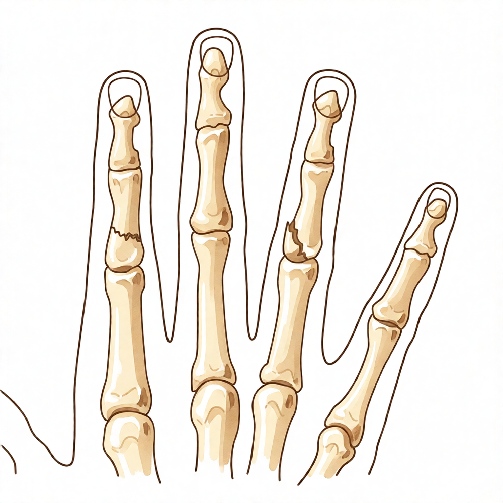
The skeleton of the hand and wrist comprises 27 bones, 19 of which are long bones [47]. The hand itself consists of 19 bones, 17 articulations, and 19 muscles situated entirely within the hand [43]. The skeletal structure is divided into five rays, each forming a polyarticulated chain of metacarpals and phalanges [47]. The thumb ray is the shortest, comprising only three bones: a metacarpal and two phalanges [47]. The remaining four digital rays consist of four skeletal segments each: a metacarpal and three phalanges [47]. In the sagittal plane, the thumb metacarpal makes an angle of approximately 45 degrees with the second metacarpal [47]. The transverse axis of the palm forms an acute angle of approximately 75 degrees with the longitudinal axis [47]. Epiphyseal plates are located at the proximal ends of the phalanges and the first metacarpal, whereas they are located at the distal ends of the other metacarpals [47]. The index metacarpal is the most firmly fixed, while the fifth metacarpal is semi-independent with a range of flexion–extension of approximately 20 degrees [79]. The ring metacarpal has about 10 degrees of mobility in flexion and extension [79].
Soft Tissue & Vascular Anatomy
The blood and nerve supplies of the hand are continuous with those of the rest of the limb [43]. Extrinsic extensors traverse six distinct fibroosseous retinacular compartments at the wrist level [46]. Compartment contents: * First compartment: Abductor pollicis longus and extensor pollicis brevis [46]. * Second compartment: Extensor carpi radialis longus and extensor carpi radialis brevis [46]. * Third compartment: Extensor pollicis longus, which turns abruptly radialward about Lister tubercle [46]. * Fourth compartment: Extensor indicis proprius, lying deep to the four tendons of the extensor digitorum communis [46]. * Fifth compartment: Extensor digiti quinti [46]. * Sixth compartment: Extensor carpi ulnaris tendon [46].
Sagittal band fibers insert onto the volar proximal phalanx and the lateral borders of the volar plate [46]. Rupture or attenuation of these fibers allows the extrinsic extensor tendon to sublux to the ulnar side of the metacarpal head, causing ulnar deviation of the finger [46]. The flexor digitorum profundus inserts on the proximal volar aspect of the distal phalanx [46]. The flexor digitorum superficialis inserts via radial and ulnar slips into the proximal metaphysis of the middle phalanx [83]. The flexor pollicis longus inserts into the proximal base of the thumb distal phalanx [83]. The A2 and A4 pulleys are the most essential in maintaining the mechanical advantage of the flexor tendons [83]. The tenosynovium lining the fibroosseous tunnel supplies nutrition and lubrication to the poorly vascularized flexor tendons [83]. Within the sheath, tendon vascularity is supplied via the vincula system, comprising the vinculum longus and brevis [83].
The dorsal interossei are abductors located on the radial side of the index and middle fingers and the ulnar side of the middle and ring fingers [77]. The volar interossei are adductors located on the ulnar side of the index finger and the radial side of the ring and little fingers [77]. The middle finger possesses two dorsal interossei (abductors) and no volar interossei (adductors) because the central axis of the hand lies within it [77]. The superficial head of each dorsal interosseous muscle abducts and weakly flexes the proximal phalanx [77]. The deep head of each dorsal interosseous muscle flexes and weakly abducts the proximal phalanx while extending the middle and distal phalanges [77]. The volar interossei form the ulnar lateral band of the index finger and the radial lateral band of the ring and little fingers [77]. The abductor digiti quinti inserts onto the ulnar lateral tubercle at the base of the proximal phalanx of the little finger [77]. The opponens digiti quinti arises from the pisohamate ligament and the hook of the hamate, inserting onto the ulnar side of the diaphysis of the fifth metacarpal [77].
The princeps pollicis artery crosses the first intermetacarpal space and runs along the ulnar side of the first metacarpal bone [80]. It divides into two terminal rami, the collateral palmar arteries of the thumb, at the level of the cutaneous flexion crease of the metacarpophalangeal joint [80]. Only 15% of anatomical dissections fall into the classical category of palmar artery layout in the thumb [80]. The superficial palmar fascia covers a triangular area of the central palm, with the proximal corner facing directly proximal [86]. When present, the palmaris longus tendon terminates in continuity with the fibers of the proximal corner of the superficial palmar fascia [86]. Fibers dorsal to the digital neurovascular bundle are collectively called Cleland ligament, while those palmar to the neurovascular bundle are called Grayson ligament [86]. The natatory ligament spans the web spaces between the fingers [86].
Cutaneous Anatomy & Functional Units
The dorsal skin possesses a normal pilosebaceous system, unlike the palm [85]. Dorsal skin has loose connections with deeper planes, allowing free gliding and full flexion at the digital joints [85]. Finger flexion produces significant lengthening of the dorsal skin, with an average increase of 3 cm in the middle finger from extension to full flexion [85]. Flexion at the metacarpophalangeal joint alone requires an average skin lengthening of 1.25 cm [85]. The dorsal and palmar skin areas are independent due to a system of adhesions anchoring their common boundary to the underlying plane [85].
One cutaneous unit on the dorsum extends from the wrist to the proximal interphalangeal joints of the fingers and the interphalangeal joint of the thumb [76]. The dorsal covering of the interphalangeal articulations of the digits forms a unique cutaneous unit characterized by considerable excess of skin when the digits are in extension [76]. The palm forms a cutaneous unit extending from the distal transverse crease of the wrist up to the transverse crease at the base of the digits [76]. The skin of the radial portion of the palm covers the thenar eminence and is the mobile portion [76]. The skin of the ulnar and distal portion of the palm covers the hypothenar eminence, where mobility is poor [76]. The central triangular part of the palm has fixed, poorly vascularized skin covering almost directly the superficial palmar aponeurosis [76].
Skin dimensions: * Thumb distal to the metacarpophalangeal joint: approximately 9 cm wide and 8 cm long [78]. * Thumb and first metacarpal skin loss: 13 cm wide and 12 cm long [78]. * Palmar and dorsal surfaces of the hand: 12 cm by 10 cm [78]. * Each finger (palmar and dorsal aspects): 7 cm by 10 cm [78].
Pathophysiology & Injury Mechanisms
The nature of injury causes specific anatomic disruptions that lead to functional deficits in delayed presentation cases [18]. Inadequate fixation results in malunion and nonunion, secondarily affecting other joints and movement [18]. Patients with delayed presentation of mutilating hand injuries present with chronic pain, stiffness, malpositioning, scissoring, and sometimes severe deformities [18].
Projectile composition and shape determine a bullet's tendency to deform, fragment, or spin [31]. Low-velocity bullets usually travel at 1000 ft/sec or less, whereas high-velocity bullets travel at 2000 ft/sec or more [31]. Most military weapons range from 2400 ft/sec to 2900 ft/sec [31]. The severity and character of the wound are directly dependent on the kinetic energy imparted on the tissue [31]. Tissue damage is determined by missile velocity and mass, as well as bullet shape, type, and interactions within the tissues [31]. Bullet fragmentation, deformation, and production of secondary missiles contribute significantly to wounding potential [31]. Laceration or crushing of tissues are the predominant mechanisms of tissue injury from low-velocity missiles [31]. Shock waves are not considered a significant determinant of tissue injury [31].
Fragments do not always travel in straight lines in blast or penetrating missile injuries [14]. Small entry wounds can be associated with extensive injury in blast or penetrating missile injuries [14]. Intra-articular air or foreign bodies seen radiographically indicate joint penetration in blast injuries [14]. Closed degloving of the thumb is a rare condition caused by crushing and elongation forces that detach soft tissues without skin disruption [118]. Hand trauma, particularly surgical trauma and repetitive mechanical stress, is associated with the onset of Dupuytren's disease [113]. A bite wound located over a joint or tendon is a risk factor for more severe infection, which could require hospitalization [71]. Intrinsic muscle attachments to the lateral bands can allow proximal interphalangeal (PIP) joint extension despite complete disruption of the central slip over the PIP joint or the extensor tendon more proximally [71].
The tenosynovial sheath of the flexor pollicis longus is continuous with the radial bursa, and the tenosynovial sheath to the little finger is continuous with the ulnar digital bursa [83]. In some patients, the radial and ulnar bursae communicate, allowing a horseshoe abscess to spread between the thumb and little finger if infection occurs in the flexor tendon sheath of either digit [83]. Following injury, flexor tendon healing occurs through both extrinsic and intrinsic mechanisms [83]. Extrinsic tendon healing occurs via cells brought to the site of repair by ingrowth of capillaries and fibroblasts, with formation of adhesions following at the repair site [83]. Intrinsic healing occurs from tenocytes within the tendon [83]. The goal of flexor tendon repair and postoperative care is to encourage both intrinsic and extrinsic healing without the formation of thick adhesions, which would limit tendon excursion and ultimately result in restricted motion of the finger [83]. Current evidence regarding the diagnosis of hand compartment syndrome is based mostly on case reports and small case series with varying etiology, lacking a consensus reference standard or reliable diagnostic criteria [33].
Classification
Burn Depth: Burns are categorized according to the degree of damage to epithelium, dermis, subcutaneous tissue, and other underlying structures [128]. First-degree burn injuries involve damage to the epidermis only and do not have any open wounds or blistering [128]. Second-degree burns are defined as partial-thickness burns, meaning that the zone of injury encompasses the epidermis and varying degrees of dermis [128]. These are subcategorized as superficial and deep [128]. Superficial second-degree burns exhibit blister formation and are painful as a result of the involvement of nerve endings in the dermis [128]. They will often heal via re-epithelialization with local wound care over the course of 10 to 14 days [128].
Gustilo: A spectrum of injury severity for open fractures has been described by different classification systems, with the most severe injuries represented by the Gustilo classification [65].
SATT: The SATT classification system for open hand injuries is based first on identifying the severity of the initial injury and determining whether the involved parts are viable [112]. It takes into account anatomical localisation (isolated vs. extended) and topography (volar vs. dorsal) to direct the correct management of the injury [112]. The system also considers the type of injury (sharp vs. crush-avulsed) to ensure that the hand will be functional to the full extent possible [112].
Wong: Wong et al proposed a clinical classification system for high-pressure injection injuries consisting of mild, moderate, and severe categories [52]. This clinical classification system for high-pressure injection injuries is used to guide treatment [52].
Clinical Presentation
General Principles and Evaluation
Management of major and extensive soft-tissue injuries associated with skeletal fractures requires a planned approach based on broad training and knowledge rather than limited past experiences [3]. On presentation with a severe upper limb injury, the patient should be managed according to advanced trauma life support principles because there may be associated significant injuries [22]. The neurovascular status of the limb should be carefully evaluated and documented before surgical intervention [22]. During initial management, the wound should be handled only for removal of gross contaminants, for photographic documentation, and to seal it from the environment [22]. Assessment of the pathophysiologic condition, careful inventory of the injured structures, and early aggressive wound excision and reconstruction should be followed by expertly directed rehabilitation to return optimal hand function [24].
Evaluation of the injured hand starts with obtaining a proper history and knowing the hand dominance and nature of job performed by the individual [18]. In delayed presentation of hand injuries, the assessment of loss in various tissue components can only be made at the time of debridement [18]. Basic investigations include wound swabs for culture and antibiotic sensitivity, and radiographs are done for all patients [18]. Soft tissue ultrasound imaging, computed tomography or MRI, and assessment of vascular status by Doppler ultrasonography and angiography are done if necessary [18]. A clear understanding of acetabular osseous anatomy and surrounding soft tissues is essential for evaluation and management [13].
Specific Injury Mechanisms and Presentations
Radiographs are invaluable in the assessment of musculoskeletal blast injuries, and the presence and severity of osseous injury will have a major bearing on the overall treatment plan [14]. In blast injuries, metallic foreign material indicates the type of weapon and the depth of penetration, while the absence of metallic fragments usually indicates a through-and-through wound and the need to search for an inconspicuous entry point [14]. Foreign material such as shoe leather, dirt, and plastic casing fragments may not be radiopaque but still may be associated with severe injury in blast trauma [14]. Fragments do not always travel in straight lines, and small entry wounds can be associated with extensive injury in blast or penetrating missile trauma [14].
Surgical treatment by tendon transfer yields excellent restoration of extensor function to the thumb in cases of sequential traumatic bilateral extensor pollicis brevis rupture [11]. The clinical importance of closed rupture of the extensor indicis tendon lies in its presentation as a swelling over the dorsum of the hand after trauma, causing a diagnostic dilemma, as the rupture does not cause any motor deficit [104]. Sword assaults can cause hand injuries with devastating loss of function for the victims [20]. Individuals presenting with self-inflicted gunshot wounds to the hand are more likely to be older, to require multiple operations, to develop infections, and to present with acute carpal tunnel syndrome requiring urgent surgical decompression [95].
The COVID-19 pandemic and consequent restrictions of activity have had substantial impacts on the patterns of hand trauma and its management, resulting in a decrease in overall presentations, particularly elective cases, and alterations in injury type and mechanism [19]. Necrotizing Sweet syndrome may be easily mistaken for necrotizing fasciitis [105]. Major complications following trigger finger release are unlikely; however, minor complications are prominent [106]. Infections after acute repairs of upper-extremity lacerations were rare [107]. Accurate diagnosis and treatment of acute burn-related hand injuries require a multidisciplinary team [108].
Compartment Syndrome
Acute compartment syndrome is a clinical emergency requiring vigilance, prompt diagnosis, and treatment [32]. Risk factors for acute compartment syndrome include high-energy injuries, specific fracture patterns, and younger age [32]. Treating providers should recognize crush injury, prolonged decubitus, and infection as the most common causes of acute hand compartment syndrome [99]. Early recognition and surgical intervention of compartment syndrome of the hand after infiltration injury in medically complex patients will reduce morbidity in this patient population [103]. A neoplasm should be considered in the differential diagnosis of the cause of a compartment syndrome, as a soft-tissue sarcoma can cause necrosis and edema leading to acute compartment syndrome [51]. This case highlights the importance of considering rare causes, like soft tissue sarcoma, in cases of compartment syndrome without clear trauma [16].
Infections and Necrotizing Conditions
The key to effective treatment of necrotizing fasciitis is prompt diagnosis and immediate and thorough debridement of infected necrotic tissue [17]. When signs of rapidly progressive soft-tissue infection develop, Aeromonas hydrophila should be considered as a causative pathogen [35]. Deterioration in the clinical condition or a poor response to conservative treatment for upper extremity infections following common carp fish handling requires a meticulous surgical drainage and excision of both infected and necrotic tissues [39]. Clinical findings suggestive of the need for hospitalization for cat bite infections of the hand include location of the bite over a joint or tendon sheath, erythema, pain, and swelling [97]. Patients presenting with acute fight bites within 24 hours generally have excellent outcomes regardless of treatment, whereas those presenting with infection or delayed presentation require operative debridement [100]. The clinician should diagnose osteomyelitis based on certain clinical manifestations and on laboratory and imaging findings [50].
Tumors and Masses
A thorough history, physical examination, and baseline plain x-rays are the foundation of accurate diagnosis for benign bony and soft tissue tumors of the hand [23]. Many benign tumors have characteristics that enable the clinician to arrive at a diagnosis without biopsy [23]. Certain lesions with a characteristic appearance and benign features may be managed nonsurgically with observation alone [23]. Vigilance in monitoring for signs of malignancy is encouraged, even for seemingly benign tumors [23]. Signs of malignancy, such as rapid growth, increasing pain, or other atypical features, mandate a thorough workup, and surgical management should be considered [23]. Due to the complexity of treatment, any soft-tissue swelling suspected of malignancy is best referred directly to a sarcoma centre [34]. Early diagnosis and surgery are critical for treating intramuscular epithelioid sarcoma presenting as extrinsic flexor tightness in the forearm [49].
High-Pressure Injection Injuries
An accurate history for high-pressure injection injuries includes pressure of the injection gun, exact material injected, estimate of volume injected, and distance from gun to extremity [52]. A thorough physical examination includes the entire upper extremity, documenting entry and exit wounds, assessing the neurovascular examination of the digit or hand, tendon involvement, and crepitus [52]. The entry wound is often just a small puncture, and injected material can sometimes exude or is expressible [52]. Pain is often not present initially [52]. The small size of the entry site might lead inexperienced patients to underestimate the true extent of the injury, resulting in devastating delay or inappropriate treatment [52]. Radiographs are helpful; in cases of air or radiodense materials, they can assess the extent of spread and deep tissue injury [52]. Most high-pressure injection injuries are work related, and accurate documentation is imperative, including whether safety training was provided for the employee [52].
Delayed Presentation and Sequelae
Various factors affect the course of events, modalities of treatment, progress, results of treatment, and prognosis in patients who present late at the appropriate facility [18]. Inadequate fixation results in malunion and nonunion secondarily affecting other joints and movement in delayed presentation cases [18]. The onset of infection and secondary problems lead to certain sequelae in delayed presentation cases [18].
Investigations
Clinical Evaluation: A systematic approach to the physical examination of the hand is essential due to the high density of structures within a small space [44]. Clinical evaluation of the injured or dysfunctional hand and wrist requires combining patient history with a careful physical examination to pinpoint or narrow the scope of possible pathologic processes [44]. Signs of malignancy, such as rapid growth, increasing pain, or other atypical features, mandate a thorough workup for soft tissue lesions [23].
Plain Radiography: Augmenting plain radiographs with additional imaging modalities like ultrasound or dark-field imaging may aid in the diagnosis of septic arthritis of the proximal interphalangeal joint [127]. The diagnosis of Bizarre Parosteal Osteochondromatous Proliferation (Nora Lesion) in pediatric phalanges can be challenging due to presenting symptoms and radiographic findings [153].
MRI: MRI is the imaging modality of choice for the majority of soft-tissue tumors [122]. A thorough evaluation including quality imaging is essential to identify sarcomas before unplanned resection [122]. In Dupuytren's disease, MRI is probably most useful in identifying additional pathology such as flexor tendon bowstringing [89]. MRI may also provide a quantitative noninvasive measure of cellularity in affected areas, serving as an index of biologic activity [89]. However, the potential staging tool of MRI for cellularity in Dupuytren's disease has not been investigated yet on a large scale [89]. MR assessment of Dupuytren's disease is hindered by the resolution of current equipment, orientation issues due to multiplanar deformities of the fingers, and lack of intraoperative availability [89].
Arthrography: Arthrography is recommended to assist in deciding whether to proceed with surgical or conservative treatment for soft tissue injuries to the dorsum of the metacarpophalangeal joint [156].
Other Considerations: Diagnostic tests such as imaging and serum laboratory studies are useful in determining hand and wrist pathology but can be expensive, time consuming, and often nonspecific [44]. An 8-MHz Doppler tone assessment may be used to identify superficially displaced neurovascular bundles when Dupuytren cords lie beneath soft fleshy prominences, though false-negatives are possible with this method [89].
Treatment
General Principles and Initial Management
Management of major and extensive soft-tissue injuries associated with skeletal fractures requires a planned approach based on broad training and knowledge, not one or two past experiences [3]. Open fractures require prompt antibiotic administration, timely surgical débridement, skeletal stabilization, and soft-tissue coverage [5]. The decision on how to approach soft tissue coverage requires consideration of the wound character, patient factors, and anticipated functional results [8]. Treatment goals for hand fractures include restoration of length, alignment, and articular congruity, along with stabilization and soft tissue repair [54]. Minimal, accurate débridement and decompression with early stability are crucial in the early management of ballistic hand trauma [63]. Clinical decision-making for open fractures of the hand is influenced by injury mechanism, timing, environment, and patient factors [42]. Antibiotic guidelines remain less definitive for hand fractures compared to other areas [42]. The surgical plan for acute burned upper extremities should prioritize key functional recovery over range of motion of all individual joints [60]. Goals in treating upper extremity mangling injuries include preserving life, preserving tissue, preserving function, and reconstructing and restoring function of both the extremity and the patient [90]. General principles for managing mangling injuries include complete debridement of devitalized tissue, restoration of good vascularity, rigid skeletal fixation while minimizing additional soft tissue injury, stable vascularized soft tissue coverage, timely intervention for repair and reconstruction, comprehensive rehabilitation, and secondary procedures as needed [90].
Mangled extremity injuries require routine trauma protocols that address the patient as a whole because 10% to 17% of these patients will have an associated life-threatening injury [91]. Prophylactic antibiotics should be administered as soon as possible and tetanus prophylaxis should be administered as indicated for mangled extremity injuries [91]. The injured extremity should be evaluated for adequate vascular perfusion, and vascular surgery consultation should be obtained if a vascular injury is suspected [91]. A sterile dressing should be applied to open wounds and the limb immobilized as soon as possible to minimize ongoing soft tissue damage until the patient is in the operating room [91]. The presence of an acute compartment syndrome, which occurs at a rate of 1% to 10% in open fractures, should be sought out during evaluation [91]. Radiographic evaluation for mangled extremity injuries should include two orthogonal views of any involved joints or long bones, as well as the joint above and below any confirmed fractures [91]. Photographs of the extremity should be obtained whenever possible to provide documentation of the extent of the initial injury and serve as a visual record of progress [91]. A comprehensive musculoskeletal examination should be performed to rule out any concomitant musculoskeletal injuries in polytrauma patients [91].
The outcome of the open fracture is largely dependent on the quality of the soft-tissue envelope [92]. Planning of the soft-tissue coverage is just as vital as the orthopaedic fracture fixation techniques [92]. Orthoplastic teams have demonstrated improved adherence to fracture treatment guidelines and have demonstrated improved patient outcomes [92]. Orthopaedic and plastic surgeons working in isolation often produce less-than-optimal outcomes compared to a collaborative orthoplastic strategy [140]. Orthoplastic teams comprise orthopaedic surgeons trained in microsurgery, orthopaedic trauma surgeons, and plastic surgeons with microsurgical expertise dedicated to limb salvage [140]. Débridement and temporary fixation can be achieved in a single surgical procedure, with the promise of early soft-tissue coverage enabling a comprehensive, excisional approach to débridement [140].
Debridement and Wound Management
Early debridement, antibiotic treatment, reconstruction, and rehabilitation offer patients the best chance for full functional recovery for gunshot wounds to the hand [130]. Early treatment with debridement and stabilization is recommended for ballistic fractures of the hand and wrist [132]. Primary bone grafting is considered for ballistic fractures of the hand and wrist due to lack of follow-up and patient noncompliance [132]. Secondary free flap reconstruction delays rehabilitation, prolongs hospital stay, and increases overall costs and number of operations [22]. Granulation tissue develops in response to wound colonization, is poorly penetrated by antibiotics, and is a precursor to fibrosis [22]. Tissue edema peaks within 3 days of injury and contributes to joint stiffness, fibrosis, inadequate mobilization, and extension of the physiologic zone of injury [22]. Serial debridement may result in overall greater tissue loss caused by desiccation, extension of the zone of injury, and edema and granulation tissue compromising delineation between viable and nonviable tissue [22]. Larger wounds resulting from serial debridement compared with primary debridement and reconstruction make free tissue transfer more likely and longer pedicles necessary [22]. A serial approach to debridement may be indicated when conversion of a contaminated wound to a cleaner wound is impossible, such as after severe crush injuries, severe multiplanar degloving, massive wound contamination, electrical burn injuries, or in patients with other life-threatening injuries [22].
The time elapsed between injury and surgery is not an important risk factor for a good outcome in Zone 2 flexor tendon injuries; rather, outcomes depend on proper surgical methods, the surgeon's experience, and early mobilization [40]. Conservative management of tendinous mallet finger injuries that have been neglected for 2 to 4 weeks can be treated as well as those injuries in patients presenting within the first 2 weeks of injury with low long-term complication rates [114]. The lowest rate of sequelae for neonatal limb compartment syndrome was observed when decompression was performed within the first 24 hours [55]. Metallic foreign material in blast injuries indicates the type of weapon and the depth of penetration [14]. The absence of metallic fragments in blast injuries usually indicates a through-and-through wound and the need to search for an inconspicuous entry point [14]. Fragments in blast injuries do not always travel in straight lines, and small entry wounds can be associated with extensive injury [14].
Soft Tissue Coverage and Reconstruction
Free flap soft tissue reconstruction should be selected early in the treatment algorithm if a better end result can be anticipated, as it allows for early coverage with composite reconstruction of all damaged or missing tissues and early mobilization to restore function [2]. Local and regional flaps are used for soft tissue reconstruction within the hand to protect against contractures and facilitate mobility [10]. The PIA flap, either alone or in combination with local flaps, provides reliable soft-tissue coverage and favorable functional outcomes in patients with severe hand injuries [15]. A treatment strategy based on the location of soft tissue defect and the perforator anatomy allows for expedient upper limb wound coverage in one stage using perforator based propeller flaps [26]. An individualized design of thoracodorsal artery perforator chimeric flap provides a flexible design for customized coverage of complex soft tissue defects with limited donor site morbidity [41]. Pelnac® artificial dermis assisted by vacuum sealing drainage can be considered as a viable alternative for addressing severe injuries or complex wound conditions in severe avulsion injuries of the fingers [36].
Optimal timing of wound coverage for open fractures is within 7 to 10 days of the initial injury [92]. The aesthetic appearance is often improved with the use of a fasciocutaneous flap compared to other tissue types [92]. Soft-tissue surgeons and orthopaedic trauma surgeons should collaborate for optimal patient outcomes in extremity soft-tissue reconstruction associated with fracture [92]. Free flap reconstruction has been used simultaneously with delayed replantation to provide coverage of vital structures such that the replantation need not be performed as a subsequent staged procedure [134]. Negative pressure wound therapy (NPWT) maintains wound homeostasis and reduces wound exudate and soft tissue edema in extremity salvage [134]. NPWT with instillation further enhances wound healing and expedites the debridement of acute and chronic wounds [134]. Use of NPWT on the amputation stump may shorten the delay from initial ectopic banking to subsequent delayed replantation [134]. Twelve evidence-based recommendations were developed for the use of NPWT in traumatic wounds and reconstructive surgery, with the strongest evidence base for skin grafts and the weakest for primary treatment of burns [37]. Further experience is required, as well as clinical and basic research, to define optimal indications and benefits of vacuum-assisted wound closure compared with traditional methods of wound management [29]. Treatment of Morel-Lavallée lesions is predominantly conservative or minimally invasive with good functional outcomes [38].
Special Considerations and Referral
Any soft-tissue swelling suspected of malignancy is best referred directly to a sarcoma centre due to the complexity of treatment [34]. Improved survival rates on the battlefield have resulted in new and ongoing challenges in the care of U.S. military personnel, necessitating the application of new technology and treatment alternatives [111]. The technique of concomitant use of antibiotic-loaded calcium sulphate and bone marrow aspirate proved safety and efficacy in eradicating the infection and bony healing of the defects after debridement in adult chronic osteomyelitis [110].
Complications
Infection and Wound Complications
In agricultural upper extremity injuries, the incidence of clinical infection at 6 months correlates with injury severity: 2 patients developed infection for type 1 injuries, 14 for type 2 injuries, and 24 for type 3 injuries [61]. Among type 2 injuries, clinical infections comprised 9 superficial infections, 4 deep soft tissue infections, and 1 case of osteomyelitis [61]. Type 3 injuries presented with a higher burden of deep pathology, including 6 superficial infections, 12 deep soft tissue infections, and 6 cases of osteomyelitis [61]. In type 3 wounds, infections diagnosed after 1 month from injury included 3 deep tissue infections and 2 cases of osteomyelitis [61]. The larger wounds resulting from serial debridement, compared with primary debridement and reconstruction, make free tissue transfer more likely and longer pedicles necessary [22].
Functional Deficits and Deformity
Inadequate fixation of delayed-presenting hand injuries results in malunion and nonunion, secondarily affecting other joints and movement [18]. The onset of infection and secondary problems in these delayed-presenting injuries leads to certain sequelae [18]. Deep injury to the first web space may result in adduction contracture and require secondary reconstruction if not prevented [73].
Nonunion and Bone Healing
Associated soft tissue injury is a risk factor for nonunion in phalangeal fractures in severely injured hands [126]. Neurovascular injury appears to be responsible for the effect of associated soft tissue injury on nonunion in these fractures [126]. Segmental bone loss also predisposed to nonunion in phalangeal fractures in severely injured hands [126]. Insufficient contact between fracture fragments, caused by segmental bone loss, precludes proper bone healing [126]. Nonunion occurred in ten fractures with segmental bone loss, and cancellous bone graft was used in only one of these fractures [126].
Vascular and Nerve Complications
In gunshot-related upper extremity injuries, one patient underwent transradial amputation for complications following a vascular injury [12]. In electrical burns of the upper extremities, apparently healthy tissue surrounding a wound may become necrotic [135]. The walls of vessels through which current traverses are friable and brittle, with endothelial changes and parietal thrombi attached to the intima [135]. Extensive necrosis of the media in electrical burns causes vessels to become brittle to the point that ligation is impossible [135]. Massive gangrene of an extremity is an occasional cause of electrical burns due to severe damage to the media and intima of vessels [135].
Rehabilitation and Outcome Factors
Tearing types of injury, such as those caused by saws, led to poorer outcomes for Zone II flexor tendon injuries compared with sharp injuries at an average follow-up of 4 years [70]. Service members with more severe initial peripheral nerve injuries had worse final outcomes [150].
Recovery
Light activity (weeks): The evidence base does not specify a typical week range for the resumption of desk work, driving, or light activities of daily living.
Full activity (months): The evidence base does not specify a typical month range for the return to manual work, sport, or full range of motion and strength.
Complete recovery / outcome plateau (months): Most patients achieve maximal recovery by 2 to 3 years following surgical resection for soft-tissue sarcoma of the extremities [152].
Rehabilitation protocol: Appropriate management of the soft-tissue envelope is essential to a good outcome [1]. Free flap soft tissue reconstruction allows for early coverage with composite reconstruction of all damaged or missing tissues and early mobilization to restore function [2]. Management of major and extensive soft-tissue injuries associated with skeletal fractures requires a planned approach and action based on broad training and knowledge, and not one or two past experiences [3]. Local and regional flaps for hand coverage focus on a systematic approach to treating challenging conditions with optimal soft tissue reconstruction that protects against contractures and facilitates mobility [10]. For patients presenting more than a month after soft tissue mallet finger injury, continuous extension splinting for 6 weeks, extending to 12-14 weeks if necessary, is preferred as residual extensor lag is largely an aesthetic concern [72]. Surgical therapy for frostbite of the hand is postponed until there is clear demarcation between healthy and necrotic tissue [74].
Functional milestones: At a long-term follow-up, the patient-reported outcome measures and objective outcomes of the homodigital unipedicle neurovascular island flap are satisfactory and it is a safe and reliable flap [58]. At 6-month follow-up, all three patients in the homodigital subcutaneous flap case series retained robust soft tissue coverage with no functional restrictions or cold intolerance [64]. Soft tissue reconstruction seeks to restore both the aesthetic appearance and the function of the hand [67]. Although associated with a relatively high rate of complications, vascularised fibular grafts for reconstruction of extremity bone defects have a high rate of successful limb salvage and a good long-term functional outcome [69]. The results of staged management of severe open fractures with bone defects are reproducible and good [75]. Forty-seven (96 per cent) of the forty-nine shoulders had a good clinical result after distal release of the contracture [163].
Other Considerations: Prevention remains the ideal treatment for iatrogenic extravasation injuries [4]. When extravasation injuries occur, they must be diagnosed and treated promptly to minimize the amount of soft tissue injury [4]. Deterioration in the clinical condition or a poor response to conservative treatment for upper extremity infections following fish handling requires a meticulous surgical drainage and excision of both infected and necrotic tissues [39]. Injuries to the upper extremities seem to have limited effect on long-term outcome in patients with polytrauma, as long as no injury was caused to the brachial plexus [53]. The lowest rate of sequelae in neonatal limb compartment syndrome was observed when decompression was performed within the first 24 hours [55]. Despite advances in understanding and declining amputation rates, the risk of long-term morbidity with diminished function and chronic symptoms remains high for high-pressure injection injuries to the hand [154]. The role of systemic steroids in the treatment of high-pressure injection injuries to the hand is uncertain [154]. Initial cultures of the wound on presentation were helpful in predicting the development of wound sepsis in one prospective analysis, although not all authors have found initial cultures to be helpful [161].
Key Evidence
- [L5] Appropriate management of the soft-tissue envelope is essential to a good outcome, and orthopaedic surgeons must adhere to strict principles of soft-tissue management to ensure appropriate decision making and accurate prediction of outcome. [1] (10.5435/00124635-199701000-00005)
- [L5] Free flap soft tissue reconstruction should be selected early in the treatment algorithm if a better end result can be anticipated, as it allows for early coverage with composite reconstruction of all damaged or missing tissues and early mobilization to restore function. [2] (10.1016/j.hcl.2007.02.007)
- [L5] Management of major and extensive soft-tissue injuries associated with skeletal fractures requires a planned approach and action based on broad training and knowledge, and not one or two past experiences. [3] (10.2106/00004623-196244040-00018)
- [L5] Prevention remains the ideal treatment for these iatrogenic injuries, and when they do occur, they must be diagnosed and treated promptly to minimize the amount of soft tissue injury. [4] (10.1016/j.jhsa.2011.10.001)
- [L5] Soft tissue coverage is required to salvage traumatized limbs and restore adequate function and form. [6] (10.1016/j.hcl.2017.09.007)
- [L5] The decision on how to approach soft tissue coverage is complex and requires consideration of the wound character, patient factors, and anticipated functional results. [8] (10.1016/j.hcl.2020.03.011)
- [Case_report] The authors emphasize that emergent reduction, early soft tissue cover, and orderly surgical fixation are essential for managing complex open carpal dissociations. [9] (10.1016/j.jhsa.2007.07.025)
- [L4] The purpose of this review was to present an overview of local and regional flaps commonly used for soft tissue reconstruction within the hand, focusing on a systematic approach to treating challenging conditions with optimal soft tissue reconstruction that protects against contractures and facilitates mobility. [10] (10.1016/j.jhsa.2013.09.027)
- [Case_report] Surgical treatment by tendon transfer yields excellent restoration of extensor function to the thumb. [11] (10.1016/j.jhsa.2007.02.007)
- [L4] [12] (10.1016/j.jhsa.2021.03.020)
- [L4] [14] (10.2106/00004623-200207000-00022)
- [L4] The PIA flap, either alone or in combination with local flaps, provides reliable soft-tissue coverage and favorable functional outcomes in patients with severe hand injuries. [15] (10.1016/j.injury.2026.113151)
- [L4] This case highlights the importance of considering rare causes, like soft tissue sarcoma, in cases of compartment syndrome without clear trauma. [16] (10.1016/j.jhsg.2025.01.003)
- [L2] The key to effective treatment is prompt diagnosis and immediate and thorough debridement of infected necrotic tissue. [17] (10.1016/j.jhsa.2014.04.010)
- [L5] [18] (10.1016/j.hcl.2016.07.008)
- [L4] The COVID-19 pandemic and consequent restrictions of activity have had substantial impacts on the patterns of hand trauma and its management, resulting in a decrease in overall presentations, particularly elective cases, and alterations in injury type and mechanism. [19] (10.1177/15589447211028918)
- [L4] This case series demonstrates the extent and severity of hand injuries that can be caused by sword assaults with devastating loss of function for the victims. [20] (10.1177/1753193410381576)
- [L5] [22] (10.1016/j.hcl.2014.01.001)
- [L5] [23] (10.1016/j.jhsa.2010.08.015)
- [L5] Assessment of the pathophysiologic condition, careful inventory of the injured structures, and early aggressive wound excision and reconstruction should be followed by expertly directed rehabilitation to return optimal hand function. [24] (10.5435/00124635-199507000-00005)
- [L4] Primary closure is a viable option for Type III open injuries if specific criteria are met, including debridement within 12 hours, no skin loss, and approximation without tension. [25] (10.1016/j.injury.2007.01.013)
- [L4] A treatment strategy based on the location of soft tissue defect and the perforator anatomy allows for expedient upper limb wound coverage in one stage. [26] (10.1016/s0363-5023(11)60028-2)
- [L5] Soft tissue defects of the upper extremity require durable coverage that will allow early postoperative mobilization and rehabilitation to maximize functional outcomes. [27] (10.1016/j.hcl.2014.08.002)
- [L5] Further experience is required, as well as clinical and basic research, to define optimal indications and benefits compared with traditional methods of wound management. [29] (10.5435/00124635-200209000-00002)
- [L5] Delayed wound closure is the rule and emergency free tissue transfer is the exception in major trauma centres due to practical constraints like team availability and patient condition. [30] (10.1016/j.injury.2007.03.011)
- [L5] [31] (10.1016/s0749-0712(21)00457-1)
- [L5] Current evidence regarding the diagnosis of hand compartment syndrome is based mostly on case reports and small case series with varying etiology, lacking a consensus reference standard or reliable diagnostic criteria. [33] (10.1016/j.jhsa.2015.01.034)
- [L5] Due to the complexity of treatment, any soft-tissue swelling suspected of malignancy is best referred directly to a sarcoma centre. [34] (10.1302/2058-5241.2.170005)
- [L5] When signs of rapidly progressive soft-tissue infection develop in such a patient, Aeromonas hydrophila should be considered as a causative pathogen. [35] (10.2106/jbjs.c.00923)
- [L4] This approach can be considered as a viable alternative for addressing severe injuries or complex wound conditions. [36] (10.1186/s13018-025-05547-z)
- [L1] Twelve evidence-based recommendations were developed for the use of NPWT in traumatic wounds and reconstructive surgery, with the strongest evidence base for skin grafts and the weakest for primary treatment of burns. [37] (10.1016/s0020-1383(11)00041-6)
- [L4] Treatment is predominantly conservative or minimally invasive with good functional outcomes. [38] (10.1177/2325967121s00025)
- [L4] Deterioration in the clinical condition or a poor response to conservative treatment requires a meticulous surgical drainage and excision of both infected and necrotic tissues. [39] (10.1054/jhsb.2001.0660)
- [L4] The time elapsed between injury and surgery is not an important risk factor for a good outcome; rather, outcomes depend on proper surgical methods, the surgeon's experience, and early mobilization. [40] (10.1177/17531934211024435)
- [L4] It provided a flexible design for customized coverage of complex soft tissue defects with limited donor site morbidity. [41] (10.1186/s13018-023-03852-z)
- [L5] Clinical decision-making is influenced by injury mechanism, timing, environment, and patient factors, while antibiotic guidelines remain less definitive for hand fractures compared to other areas. [42] (10.1016/j.hcl.2013.08.008)
- [L4] Early diagnosis and surgery are critical for treating this sarcoma. [49] (10.1016/j.jhsa.2018.02.020)
- [Paper] The clinician should diagnose osteomyelitis based on certain clinical manifestations and on laboratory and imaging findings. [50] (10.1016/s0020-1383(11)70128-0)
- [L4] A neoplasm should be considered in the differential diagnosis of the cause of a compartment syndrome, as a soft-tissue sarcoma can cause necrosis and edema leading to acute compartment syndrome. [51] (10.2106/00004623-199508000-00017)
- [L4] [52] (10.1016/j.jhsa.2012.07.005)
- [L3] Injuries to the upper extremities seem to have limited effect on long-term outcome in patients with polytrauma, as long as no injury was caused to the brachial plexus. [53] (10.1302/0301-620x.99b2.37999)
- [L4] Despite nonsurgical management and late fasciotomy (> 24 hours) being the most described treatments, the lowest rate of sequelae was observed when decompression was performed within the first 24 hours. [55] (10.1016/j.jhsa.2023.08.013)
- [L4] Knowledge of modern repair techniques and rehabilitation protocols may improve patient outcomes. [56] (10.1016/j.jhsa.2014.06.136)
- [L4] There is insufficient evidence to determine the best splint type or specific indications for surgery. [57] (10.1016/j.jhsa.2009.06.018)
- [L4] At a long-term follow-up, the patient-reported outcome measures and objective outcomes of this flap are satisfactory and it is a safe and reliable flap. [58] (10.1177/17531934231172081)
- [L5] The surgical plan should prioritize key functional recovery over range of motion of all individual joints. [60] (10.1016/j.jhsa.2025.02.020)
- [L2] [61] (10.1016/j.jhsa.2007.09.003)
- [L5] Minimal, accurate débridement and decompression with early stability are crucial. [63] (10.5435/00124635-201002000-00006)
- [L4] At 6-month follow-up, all three patients retained robust soft tissue coverage with no functional restrictions or cold intolerance. [64] (10.1007/s11552-010-9279-3)
- [L5] [65] (10.1186/s13018-026-06698-3)
- [L4] Patients often require reconstruction rather than repair, and many will require secondary procedures, making prevention the best treatment. [66] (10.1016/j.jhsa.2014.08.041)
- [L5] Soft tissue reconstruction seeks to restore both the aesthetic appearance and the function of the hand. [67] (10.1016/j.jhsa.2009.04.035)
- [L4] Although associated with a relatively high rate of complications, each reconstruction method is useful, with a high rate of successful limb salvage and a good long-term functional outcome. [69] (10.1302/0301-620x.99b9.bjj-2017-0219.r1)
- [L3] Tearing types of injury, such as those caused by saws, led to poorer outcomes for Zone II flexor tendon injuries compared with sharp injuries at an average follow-up of 4 years. [70] (10.1016/j.jhsa.2012.09.021)
- [L5] [71] (10.5435/jaaos-23-01-47)
- [L5] For patients presenting more than a month after injury, the authors prefer continuous extension splinting for 6 weeks, extending to 12-14 weeks if necessary, as residual extensor lag is largely an aesthetic concern. [72] (10.1016/j.jhsa.2013.03.050)
- [L4] The first web space requires particular consideration because deep injury may result in adduction contracture and require secondary reconstruction if not prevented. [73] (10.1016/j.jhsa.2017.01.028)
- [L5] Surgical therapy is postponed until there is clear demarcation between healthy and necrotic tissue. [74] (10.1016/j.jhsa.2014.01.035)
- [L5] The results of staged management of severe open fractures with bone defects are reproducible and good. [75] (10.2106/jbjs.24.00647)
- [L4] Individuals presenting with SI GSWs are more likely to be older, to require multiple operations, to develop infections, and to present with acute carpal tunnel syndrome requiring urgent surgical decompression. [95] (10.1177/15589447211014603)
- [L3] Clinical findings suggestive of the need for hospitalization include location of the bite over a joint or tendon sheath, erythema, pain, and swelling. [97] (10.1016/j.jhsa.2013.11.003)
- [L4] Treating providers should recognize crush injury, prolonged decubitus, and infection as the most common causes of acute hand compartment syndrome. [99] (10.1177/15589447221084012)
- [L4] Patients presenting within 24 hours generally have excellent outcomes regardless of treatment, whereas those presenting with infection or delayed presentation require operative debridement. [100] (10.1016/j.jhsa.2013.03.002)
- [L4] Early recognition and surgical intervention of compartment syndrome of the hand after infiltration injury in medically complex patients will reduce morbidity in this patient population. [103] (10.1016/j.jhsg.2021.12.002)
- [L4] The clinical importance of this case lies in its presentation as a swelling over the dorsum of the hand after trauma, causing a diagnostic dilemma, as the rupture of the extensor indicis tendon does not cause any motor deficit. [104] (10.1177/1753193412439271)
- [L4] Necrotizing Sweet syndrome may be easily mistaken for necrotizing fasciitis; early diagnosis and treatment with systemic steroids can alter the disease course and potentially the patient's outcome. [105] (10.1016/j.jhsa.2017.08.019)
- [L3] Major complications following trigger finger release are unlikely; however, minor complications are prominent. [106] (10.1177/15589447221081869)
- [L2] Infections after acute repairs of upper-extremity lacerations were rare. [107] (10.1016/j.jhsa.2025.01.030)
- [L5] Accurate diagnosis and treatment of acute burn-related hand injuries require a multidisciplinary team. [108] (10.1016/j.jhsa.2015.02.033)
- [L4] The technique proved safety and efficacy in eradicating the infection and bony healing of the defects after debridement. [110] (10.1007/s00264-018-4063-z)
- [L5] Improved survival rates on the battlefield have resulted in new and ongoing challenges in the care of U.S. military personnel, necessitating the application of new technology and treatment alternatives to provide the best care to those wounded in war. [111] (10.1016/j.jhsa.2007.07.007)
- [L4] [112] (10.1016/j.injury.2008.06.007)
- [L1] Hand trauma, particularly surgical trauma and repetitive mechanical stress, is associated with the onset of Dupuytren's disease. [113] (10.1177/17531934251360545)
- [L3] Conservative management of tendinous mallet finger injuries that have been neglected for 2 to 4 weeks can be treated as well as those injuries in patients presenting within the first 2 weeks of injury with low long-term complication rates. [114] (10.1016/j.jhsa.2014.06.140)
- [L4] Closed degloving of the thumb is a rare condition caused by crushing and elongation forces that detaches soft tissues without skin disruption. [118] (10.1016/j.jhsa.2007.06.016)
- [L4] [126] (10.1054/jhsb.2000.0486)
- [L5] Augmenting plain radiographs with additional imaging modalities like ultrasound or dark-field imaging may aid in diagnosis. [127] (10.1016/j.jhsa.2021.04.004)
- [L5] [128] (10.1016/j.jhsa.2010.03.019)
- [L4] Given the current evidence related to management of gunshot wounds to the hand, early debridement, antibiotic treatment, reconstruction, and rehabilitation offer patients the best chance for full functional recovery. [130] (10.1016/j.jhsa.2013.02.011)
- [L4] Early treatment with debridement and stabilization is recommended, with primary bone grafting considered due to lack of follow-up and patient noncompliance. [132] (10.1177/1558944717697432)
- [L4] [134] (10.1016/j.hcl.2019.01.002)
- [L4] [135] (10.2106/00004623-195840010-00003)
- [L5] Fasciocutaneous flaps are the optimal choice for initial soft tissue reconstruction when secondary procedures are foreseen due to their pliable nature and ease of re-elevation. [138] (10.1016/j.hcl.2014.01.004)
- [L4] The technique of two simultaneous pedicled flaps is a simple and successful alternative to free tissue transfer for coverage of complex soft tissue defects in the hand and forearm, particularly in critically ill, multi-extremity injured patients where free tissue transfer is not indicated. [141] (10.1177/1753193409347428)
- [L5] Local hand flaps offer excellent coverage of soft tissue defects by replacing like with like, frequently resulting in optimum functional and aesthetic outcomes while sparing patients from more complicated repair methods. [145] (10.1016/j.hcl.2013.12.004)
- [L4] Service members with more severe initial injuries had worse final outcomes. [150] (10.1016/j.jhsa.2020.08.004)
- [L4] Most patients will achieve maximal recovery by 2 to 3 years following surgical resection for soft-tissue sarcoma of the extremities. [152] (10.2106/jbjs.23.01007)
- [L4] The diagnosis in pediatric cases can be challenging due to presenting symptoms and radiographic findings. [153] (10.1016/j.jhsa.2020.05.002)
- [L5] Despite advances in understanding and declining amputation rates, the risk of long-term morbidity with diminished function and chronic symptoms remains high, and the role of systemic steroids in treatment is uncertain. [154] (10.5435/jaaos-22-01-38)
- [L4] The authors recommend arthrography to assist in deciding whether to proceed with surgical or conservative treatment. [156] (10.1054/jhsb.2001.0656)
- [L4] Initial cultures of the wound on presentation were helpful in predicting the development of wound sepsis in one prospective analysis, although not all authors have found initial cultures to be helpful. [161] (10.1016/s0749-0712(02)00055-0)
- [L3] Forty-seven (96 per cent) of the forty-nine shoulders had a good clinical result after distal release of the contracture. [163] (10.2106/00004623-199802000-00010)
See Also
- Mallet Finger
- Dupuytren's Disease
- Flexor tendon repair
- Trigger finger release
- Tumors
- Reconstructive Surgery
References
[1] Soft-Tissue Injuries Associated With High-Energy Extremity Trauma: Principles of Management. Journal of the American Academy of Orthopaedic Surgeons. 1997. DOI: 10.5435/00124635-199701000-00005
[2] Indications and Selection of Free Flaps for Soft Tissue Coverage of the Upper Extremity. Hand Clinics. 2007. DOI: 10.1016/j.hcl.2007.02.007
[3] Major and Extensive Soft-Tissue Injuries Complicating Skeletal Fractures. The Journal of Bone & Joint Surgery. 1962. DOI: 10.2106/00004623-196244040-00018
[4] Extravasation Injuries. The Journal of Hand Surgery. 2011. DOI: 10.1016/j.jhsa.2011.10.001
[5] Chapter 14 Evaluation and Management of Soft-Tissue Injury and Open Fractures. 2021.
[6] Traumatic Wounds of the Upper Extremity. Hand Clinics. 2018. DOI: 10.1016/j.hcl.2017.09.007
[8] Soft Tissue Coverage for Severe Infections. Hand Clinics. 2020. DOI: 10.1016/j.hcl.2020.03.011
[9] Complex Carpal Dissociation With Open, Complete, and Divergent Trapezium, Capitate, and Hamate Dislocation: A Case Report. The Journal of Hand Surgery. 2007. DOI: 10.1016/j.jhsa.2007.07.025
[10] Local and Regional Flaps for Hand Coverage. The Journal of Hand Surgery. 2014. DOI: 10.1016/j.jhsa.2013.09.027
[11] Sequential Traumatic Bilateral Extensor Pollicis Brevis Rupture: A Case Report. The Journal of Hand Surgery. 2007. DOI: 10.1016/j.jhsa.2007.02.007
[12] Gunshot-Related Upper Extremity Nerve Injuries at a Level 1 Trauma Center. The Journal of Hand Surgery. 2022. DOI: 10.1016/j.jhsa.2021.03.020
[13] Chapter 33 Evaluation and Management of Acetabular Fractures. 2021.
[14] Blast and Fragment Injuries of the Musculoskeletal System. The Journal of Bone & Joint Surgery. 2002. DOI: 10.2106/00004623-200207000-00022
[15] Posterior interosseous artery flap for severe hand injuries: Outcomes of reconstruction combined with local and regional flaps. Injury. 2026. DOI: 10.1016/j.injury.2026.113151
[16] Forearm Compartment Release with Incidental Soft Tissue Sarcoma. Journal of Hand Surgery Global Online. 2025. DOI: 10.1016/j.jhsg.2025.01.003
[17] Necrotizing Fasciitis. The Journal of Hand Surgery. 2014. DOI: 10.1016/j.jhsa.2014.04.010
[18] Challenges Posed by Delayed Presentation of Mutilating Hand Injuries. Hand Clinics. 2016. DOI: 10.1016/j.hcl.2016.07.008
[19] The Impact of COVID-19 on Hand Trauma. HAND. 2021. DOI: 10.1177/15589447211028918
[20] Severe hand injuries resulting from Samurai sword assaults: a Dublin case series. Journal of Hand Surgery (European Volume). 2010. DOI: 10.1177/1753193410381576
[22] Free Muscle Flaps for Reconstruction of Upper Limb Defects. Hand Clinics. 2014. DOI: 10.1016/j.hcl.2014.01.001
[23] Benign Bony and Soft Tissue Tumors of the Hand. The Journal of Hand Surgery. 2010. DOI: 10.1016/j.jhsa.2010.08.015
[24] Management of the Mangled Hand and Forearm. Journal of the American Academy of Orthopaedic Surgeons. 1995. DOI: 10.5435/00124635-199507000-00005
[25] Early versus delayed closure of open fractures. Injury. 2007. DOI: 10.1016/j.injury.2007.01.013
[26] Clinical Application of Perforator Based Propeller Flaps in Upper Limb Soft Tissue Reconstruction. The Journal of Hand Surgery. 2011. DOI: 10.1016/s0363-5023(11)60028-2
[27] Soft Tissue Coverage of the Upper Extremity. Hand Clinics. 2014. DOI: 10.1016/j.hcl.2014.08.002
[29] New Techniques in Wound Management: Vacuum-Assisted Wound Closure. Journal of the American Academy of Orthopaedic Surgeons. 2002. DOI: 10.5435/00124635-200209000-00002
[30] Early versus delayed closure of open fractures. Injury. 2007. DOI: 10.1016/j.injury.2007.03.011
[31] THE MANAGEMENT OF PENETRATING TRAUMA TO THE HAND. Hand Clinics. 1999. DOI: 10.1016/s0749-0712(21)00457-1
[32] Chapter 8 Acute Compartment Syndrome. 2021.
[33] Acute Compartment Syndrome of the Hand. The Journal of Hand Surgery. 2015. DOI: 10.1016/j.jhsa.2015.01.034
[34] Diagnosis and treatment of soft-tissue sarcomas of the extremities and trunk. EFORT Open Reviews. 2017. DOI: 10.1302/2058-5241.2.170005
[35] Aeromonas Hydrophila Necrotizing Fasciitis. The Journal of Bone & Joint Surgery. 2006. DOI: 10.2106/jbjs.c.00923
[36] Pelnac® artificial dermis assisted by vacuum sealing drainage for treatment of severe avulsion injuries of the fingers. Journal of Orthopaedic Surgery and Research. 2025. DOI: 10.1186/s13018-025-05547-z
[37] Evidence-based recommendations for the use of Negative Pressure Wound Therapy in traumatic wounds and reconstructive surgery: Steps towards an international consensus. Injury. 2011. DOI: 10.1016/s0020-1383(11)00041-6
[38] ARE SPORTS INJURIES THE MOST COMMON CAUSE OF MOREL-LAVALLEE LESIONS IN THE PEDIATRIC POPULATION?. Orthopaedic Journal of Sports Medicine. 2021. DOI: 10.1177/2325967121s00025
[39] Upper Extremity Infections Following Common Carp Fish (Cyprinus Carpio) Handling. Journal of Hand Surgery. 2002. DOI: 10.1054/jhsb.2001.0660
[40] Up to five-week delay in primary repair of Zone 2 flexor tendon injuries: outcomes and complications. Journal of Hand Surgery (European Volume). 2021. DOI: 10.1177/17531934211024435
[41] Individualized design of thoracodorsal artery perforator chimeric flap for customized reconstruction of complex three-dimensional defects in the extremities. Journal of Orthopaedic Surgery and Research. 2023. DOI: 10.1186/s13018-023-03852-z
[42] Open Fractures of the Hand with Soft Tissue Loss. Hand Clinics. 2013. DOI: 10.1016/j.hcl.2013.08.008
[43] Exam Of The Hand Wrist 2Ed. INTRODUCTION.
[44] Orthopaedic Knowledge Update 13 Ebook Without Multimedia. Anatomy, Evaluation, Clinical Examination, and Imaging > Evaluation and Clinical Examination: Current Concepts.
[46] A Lange Medical Book Current Diagnosis Treatment In Orthopedics Fifth Edition. 9Hand Surgery > Image DISORDERS OF THE MUSCULATURE OF THE HAND.
[47] Exam Of The Hand Wrist 2Ed. 1.1 SKELETON OF THE HAND > The osseous skeleton.
[49] Intramuscular Epithelioid Sarcoma Presenting as Extrinsic Flexor Tightness in the Forearm. The Journal of Hand Surgery. 2018. DOI: 10.1016/j.jhsa.2018.02.020
[50] Management of bone infections in adults: the surgeon's and microbiologist's perspectives. Injury. 2011. DOI: 10.1016/s0020-1383(11)70128-0
[51] Synovial sarcoma presenting as an acute compartment syndrome.. The Journal of Bone & Joint Surgery. 1995. DOI: 10.2106/00004623-199508000-00017
[52] High-Pressure Injection Injuries. The Journal of Hand Surgery. 2012. DOI: 10.1016/j.jhsa.2012.07.005
[53] Injuries to the upper extremities in polytrauma. The Bone & Joint Journal. 2017. DOI: 10.1302/0301-620x.99b2.37999
[54] Chapter 95 Hand Trauma. 2019.
[55] Neonatal Limb Compartment Syndrome: A Comprehensive Review. The Journal of Hand Surgery. 2025. DOI: 10.1016/j.jhsa.2023.08.013
[56] Open Extensor Tendon Injuries. The Journal of Hand Surgery. 2015. DOI: 10.1016/j.jhsa.2014.06.136
[57] Mallet Finger Injuries. The Journal of Hand Surgery. 2009. DOI: 10.1016/j.jhsa.2009.06.018
[58] Long-term patient-reported outcome measures of fingertip coverage with a homodigital unipedicle neurovascular island flap. Journal of Hand Surgery (European Volume). 2023. DOI: 10.1177/17531934231172081
[60] The Acute Burned Upper Extremity: Evolution of Practices and Management. The Journal of Hand Surgery. 2025. DOI: 10.1016/j.jhsa.2025.02.020
[61] The Microbiology and Risk of Infection Following Open, Agricultural Upper Extremity Injuries. The Journal of Hand Surgery. 2008. DOI: 10.1016/j.jhsa.2007.09.003
[63] Early Management of Ballistic Hand Trauma. American Academy of Orthopaedic Surgeon. 2010. DOI: 10.5435/00124635-201002000-00006
[64] The Homodigital Subcutaneous Flap for Soft Tissue Coverage of the Dorsum of the Finger: A Case Series. HAND. 2010. DOI: 10.1007/s11552-010-9279-3
[65] The masquelet technique for severe open fractures: current insights. Journal of Orthopaedic Surgery and Research. 2026. DOI: 10.1186/s13018-026-06698-3
[66] Firework-Related Injuries of the Hand. The Journal of Hand Surgery. 2015. DOI: 10.1016/j.jhsa.2014.08.041
[67] Soft Tissue Reconstruction of the Hand. The Journal of Hand Surgery. 2009. DOI: 10.1016/j.jhsa.2009.04.035
[69] Vascularised fibular grafts for reconstruction of extremity bone defects after resection of bone and soft-tissue tumours. The Bone & Joint Journal. 2017. DOI: 10.1302/0301-620x.99b9.bjj-2017-0219.r1
[70] Clinical Outcomes of Zone II Flexor Tendon Repair Depending on Mechanism of Injury. The Journal of Hand Surgery. 2012. DOI: 10.1016/j.jhsa.2012.09.021
[71] Human and Other Mammalian Bite Injuries of the Hand. Journal of the American Academy of Orthopaedic Surgeons. 2015. DOI: 10.5435/jaaos-23-01-47
[72] Soft Tissue Mallet Finger Injuries With Delayed Treatment. The Journal of Hand Surgery. 2013. DOI: 10.1016/j.jhsa.2013.03.050
[73] Severe Hand Injuries From Fireworks: Injury Patterns, Outcomes, and Fireworks Types. The Journal of Hand Surgery. 2017. DOI: 10.1016/j.jhsa.2017.01.028
[74] Frostbite of the Hand. The Journal of Hand Surgery. 2014. DOI: 10.1016/j.jhsa.2014.01.035
[75] Modern Management of Severe Open Fractures of the Extremities. Journal of Bone and Joint Surgery. 2025. DOI: 10.2106/jbjs.24.00647
[76] Exam Of The Hand Wrist 2Ed. Functional cutaneous units.
[77] Green S Operative Hand Surgery. Interosseous and Hypothenar Muscles.
[78] Exam Of The Hand Wrist 2Ed. Planning skin cover of the hand and forearm.
[79] Exam Of The Hand Wrist 2Ed. The arches of the hand > The metacarpal arch.
[80] Exam Of The Hand Wrist 2Ed. Techniques of investigation of the arterial supply by J P Melki > Vascularization of the thumb > Palmar aspect.
[83] A Lange Medical Book Current Diagnosis Treatment In Orthopedics Fifth Edition. 9Hand Surgery > FLEXOR TENDON INJURY.
[85] Exam Of The Hand Wrist 2Ed. The dorsal skin.
[86] Green S Operative Hand Surgery. PERTINENT ANATOMY.
[89] Dupuytren S Disease And Related Hyperproliferative Disorders. 54. The Future of Dupuytren’s Research and Treatment > 54.4 Mechanical Measurements and Procedures > 54.4.4 Imaging.
[90] Green S Operative Hand Surgery. EVOLUTION IN THE TREATMENT OF MANGLING INJURIES > PRINCIPLES.
[91] Rockwood And Green S Fractures In Adults. Effect of Blast on the Musculoskeletal System > Principles of Management.
[92] Orthopaedic Knowledge Update Trauma. Extremity Soft-Tissue Reconstruction Associated With Fracture > Summary.
[95] Point Blank: A Retrospective Review of Self-inflicted Gunshot Wounds to the Hand. HAND. 2021. DOI: 10.1177/15589447211014603
[97] Cat Bite Infections of the Hand: Assessment of Morbidity and Predictors of Severe Infection. The Journal of Hand Surgery. 2014. DOI: 10.1016/j.jhsa.2013.11.003
[99] Surgical Demographics of Acute Hand Compartment Syndrome. HAND. 2022. DOI: 10.1177/15589447221084012
[100] Acute Fight Bite. The Journal of Hand Surgery. 2013. DOI: 10.1016/j.jhsa.2013.03.002
[103] Isolated Compartment Syndrome of the Hand After Intravenous Doxycycline Infiltration Injury. Journal of Hand Surgery Global Online. 2022. DOI: 10.1016/j.jhsg.2021.12.002
[104] Closed rupture of the extensor indicis tendon: An unusual cause of swelling over the dorsum of the hand. Journal of Hand Surgery (European Volume). 2012. DOI: 10.1177/1753193412439271
[105] Necrotizing Sweet Syndrome of the Upper Extremity After Elective Hand Surgery. The Journal of Hand Surgery. 2018. DOI: 10.1016/j.jhsa.2017.08.019
[106] Complications Following Endoscopic and Open Trigger Finger Release: A Retrospective Comparative Study. HAND. 2022. DOI: 10.1177/15589447221081869
[107] Prophylactic Antibiotics for Uncomplicated Upper-Extremity Lacerations: A Retrospective Cohort Study of Private Insurance Claims. The Journal of Hand Surgery. 2025. DOI: 10.1016/j.jhsa.2025.01.030
[108] Management of the Acutely Burned Hand. The Journal of Hand Surgery. 2015. DOI: 10.1016/j.jhsa.2015.02.033
[110] One-stage surgery for adult chronic osteomyelitis: concomitant use of antibiotic-loaded calcium sulphate and bone marrow aspirate. International Orthopaedics. 2018. DOI: 10.1007/s00264-018-4063-z
[111] Injuries Sustained to the Upper Extremity Due to Modern Warfare and the Evolution of Care. The Journal of Hand Surgery. 2007. DOI: 10.1016/j.jhsa.2007.07.007
[112] Versatility of the island forearm flap in the management of extensive skin defects of the hand. Injury. 2008. DOI: 10.1016/j.injury.2008.06.007
[113] Incidence of Dupuytren’s disease following hand trauma: a systematic review. Journal of Hand Surgery (European Volume). 2025. DOI: 10.1177/17531934251360545
[114] Soft-Tissue Mallet Injuries: A Comparison of Early and Delayed Treatment. The Journal of Hand Surgery. 2014. DOI: 10.1016/j.jhsa.2014.06.140
[118] Closed Degloving of the Thumb. The Journal of Hand Surgery. 2007. DOI: 10.1016/j.jhsa.2007.06.016
[122] Chapter 68 Soft-Tissue Tumors: Evaluation and Diagnosis. 2020.
[126] Treatment of Phalangeal Fractures in Severely Injured Hands. Journal of Hand Surgery. 2001. DOI: 10.1054/jhsb.2000.0486
[127] Septic Arthritis of the Proximal Interphalangeal Joint After Rattlesnake Bite. The Journal of Hand Surgery. 2022. DOI: 10.1016/j.jhsa.2021.04.004
[128] Acute Management of Burn Injuries to the Hand and Upper Extremity. The Journal of Hand Surgery. 2010. DOI: 10.1016/j.jhsa.2010.03.019
[130] Management of Gunshot Wounds to the Hand: A Literature Review. The Journal of Hand Surgery. 2013. DOI: 10.1016/j.jhsa.2013.02.011
[132] Current Trends in the Management of Ballistic Fractures of the Hand and Wrist: Experiences of a High-Volume Level I Trauma Center. HAND. 2017. DOI: 10.1177/1558944717697432
[134] Revascularization and Replantation in the Hand. Hand Clinics. 2019. DOI: 10.1016/j.hcl.2019.01.002
[135] Electrical Burns of the Upper Extremities. The Journal of Bone & Joint Surgery. 1958. DOI: 10.2106/00004623-195840010-00003
[138] Refinements and Secondary Surgery After Flap Reconstruction of the Traumatized Hand. Hand Clinics. 2014. DOI: 10.1016/j.hcl.2014.01.004
[140] Chapter 16 Extremity Soft-Tissue Reconstruction Associated With Fracture. 2021.
[141] Simultaneous pedicled flaps for coverage of complex blast injuries to the forearm and hand (with supplemental external fixation to the iliac crest for immobilization). Journal of Hand Surgery (European Volume). 2009. DOI: 10.1177/1753193409347428
[145] Local Flaps of the Hand. Hand Clinics. 2014. DOI: 10.1016/j.hcl.2013.12.004
[150] Combat-Sustained Peripheral Nerve Injuries in the United States Military. The Journal of Hand Surgery. 2021. DOI: 10.1016/j.jhsa.2020.08.004
[152] Mapping the Course of Recovery Following Limb-Salvage Surgery for Soft-Tissue Sarcoma of the Extremities. Journal of Bone and Joint Surgery. 2024. DOI: 10.2106/jbjs.23.01007
[153] Bizarre Parosteal Osteochondromatous Proliferation (Nora Lesion) in Pediatric Phalanges. The Journal of Hand Surgery. 2021. DOI: 10.1016/j.jhsa.2020.05.002
[154] High-pressure Injection Injuries to the Hand. Journal of the American Academy of Orthopaedic Surgeons. 2014. DOI: 10.5435/jaaos-22-01-38
[156] Treatment of Soft Tissue Injuries to the Dorsum of the Metacarpophalangeal Joint (Boxer’s Knuckle). Journal of Hand Surgery. 2002. DOI: 10.1054/jhsb.2001.0656
[161] Antimicrobial management of mutilating hand injuries. Hand Clinics. 2003. DOI: 10.1016/s0749-0712(02)00055-0
[163] Contracture of the Deltoid Muscle. The Journal of Bone and Joint Surgery (American Volume). 1998. DOI: 10.2106/00004623-199802000-00010