Clinicians › Shoulder
Post-Surgical Residual Symptom Injections
Diagnostic and therapeutic post-operative injections for residual symptoms. Timing, indications, prognostic value of response. Distinguishing scar-related inflammation from structural failure.
Overview
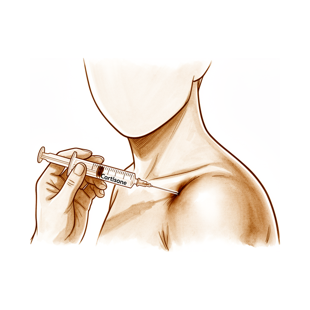
Post-surgical residual symptom injections serve as a targeted intervention for persistent pain and functional limitation following shoulder procedures. A single image-guided corticosteroid injection yields statistically and clinically relevant improvements in shoulder function and pain for glenohumeral arthritis, with benefits sustained up to 4 months [1]. For adhesive capsulitis, multiple-site corticosteroid injections demonstrate clinical advantage over placebo for short- and intermediate-term composite outcomes, although these benefits dissipate over time [3]. In the context of refractory frozen shoulder, intra-articular steroid injection improves pain prior to arthroscopic pan-capsular release and at final follow-up [54], while repeat manipulation under anesthesia with corticosteroid and local anaesthetic injection remains a valuable option before surgical intervention for recurrence [22].
The timing of injections relative to surgery is a critical determinant of postoperative risk and outcome. The risk of reoperation for rotator cuff repair significantly declines if more than 6 months elapse between a preoperative injection and the surgery [4]. Conversely, preoperative injections may increase the risk of revision rotator cuff repair and subacromial decompression by up to 150% in patients 6 to 12 months after index surgery compared with those who did not receive an injection [7]. Consequently, treatment should be withheld if a rotator cuff repair is planned within the following 6 months [40]. For shoulder arthroplasty, corticosteroid injections within 12 months prior to anatomic and reverse procedures do not compromise patient-reported outcomes during a minimum of 2-year follow-up [8], though the timing of injection remains a critical factor in postoperative outcomes [39]. Postoperatively, intra-articular corticosteroid injection in the early period after arthroscopic rotator cuff repair provides satisfactory pain relief and range of motion improvement without increasing re-tear rates or deteriorating clinical outcomes at 2-year follow-up [16]. A single-dose injection at 6 weeks postoperatively for stiffness significantly improves pain, function, and return to activities of daily living without increasing retear risk [18].
Image-guided techniques, particularly ultrasound, are associated with statistically significant greater improvement in shoulder pain and function at 6 weeks compared to blind injections [13]. Ultrasound-guided subscapular cortisone injection prior to arthroscopic superior medial scapuloplasty for snapping scapula syndrome results in significantly better recovery in patients who gain a good transient response [10]. While the effect of subacromial triamcinolone acetonide, hyaluronic acid, and saline injections is best seen after the first and second injections, the necessity of repeating subacromial injections more than two times is questioned due to diminishing effects [5]. Clinicians must remain aware of rare but significant complications, such as central serous retinopathy causing loss of visual acuity following steroid injection into the shoulder bursa, which requires prompt ophthalmic referral [11]. Expansion of indications for periarticular and intra-articular injections should be approached with caution due to potential negative effects on articular cartilage [36]. Delayed application of leukocyte-rich platelet-rich plasma following rotator cuff repair did not improve function as measured by patient-reported outcome measures and Constant score at 1 year postoperatively [14].
Anatomy & Pathophysiology
Bony Anatomy
The proximal humerus comprises the humeral head, greater tuberosity, lesser tuberosity, and humeral shaft [66]. The articular head is spherical with a diameter of 37 to 57 mm [66], an arc of approximately 160 degrees covered by articular cartilage [78], and a radius of curvature of approximately 25 mm, which is slightly larger in men than in women [78]. The most superior portion of the articular surface averages 8 mm above the greater tuberosity [66], or 8 to 10 mm superior to the top of the greater tuberosity [78]. Head height is approximately 5.6 cm above the superior border of the pectoralis major tendon [80]. The humeral head is inclined approximately 130 degrees with respect to the humeral shaft [66], with inclination ranging from 30 to 55 degrees [78]. Humeral version averages 29.8 degrees, with a range of 10 to 55 degrees [66], and the head is retroverted 30 degrees relative to the transepicondylar axis of the humerus [80]. Mean humeral retroversion is around 26 degrees in healthy adults [73], though proximal humeral retroversion is highly variable, ranging from 0 to 55 degrees depending on the measurement method [78]. The anatomic neck lies at the junction of the head and the tuberosities [67] and serves as an attachment for the shoulder capsule [80]. The surgical neck is more distal than the anatomic neck and is more often involved in fractures [80]. The bicipital groove lies between the greater and lesser tuberosities and serves as a pathway for the long head of the biceps [66].
The glenoid is a convex structure of shallow depth shaped like an inverted pear [66], approximately one-third the size of the humeral head [67]. The subchondral bone of the glenoid is relatively flat, with articular concavity augmented by cartilage and a circumferential labrum [69]. The glenoid articular surface radius of curvature is 2 to 3 mm larger than that of the humeral head [78], with a glenoid radius of curvature ranging from 22 to 28 mm [78]. The glenoid diameter ranges from 18 to 30 mm superior anteroposteriorly and 21 to 35 mm inferior anteroposteriorly [78], with a superoinferior height ranging from 30 to 48 mm [78]. Glenoid inclination averages 4.2 degrees, with a range of -7 to 20 degrees [78]. The glenoid is retroverted approximately 5 degrees relative to the scapular body [80], with version averaging 1.5 degrees of retroversion and a range of 10.5 degrees anteversion to 9.5 degrees retroversion [78]. The normal position of the glenoid surface in relation to the axis of the scapular body ranges from 2 degrees of anteversion to 7 degrees of retroversion [78]. Glenoid surface area ranges from 4 to 6 mm [78], and glenoid cartilage thickness is 2.16 mm [78].
The scapula is attached to the axial skeleton by the acromioclavicular and sternoclavicular joints [68] and spans the second through seventh ribs, serving as an attachment for 17 muscles [80]. It is separated from the chest wall by thin gliding fibro-fatty tissue, allowing smooth excursion over the chest wall [68]. The scapula is anteverted on the chest wall approximately 30 degrees relative to the body [80]. The glenoid is connected with the flat body of the scapula by the scapular neck [68]. The coracoid process curves forwards from the superior surface of the scapular neck [68], and the scapular spine ends in a flattened bony process, the acromion, which curves forwards [68]. The highest concentration of bony mass in the scapula is located in the glenoid, the scapular neck, and the lateral border of the scapular body [68]. Two bony pillars transmit compressive forces from the glenoid fossa: the lateral pillar connects the inferior border of the glenoid with the inferior angle, and the spinal pillar arises from the central part of the glenoid and continues medially to become part of the base of the scapular spine [68]. The weakest bone in the scapula is located primarily in the central part of the biomechanical body, specifically in the infraspinous fossa, while the weakest area of the circumference of the biomechanical body is the spinomedial angle [68].
The acromion, coracoacromial ligament, and coracoid process form the coracoacromial arch, a rigid bony-ligamentous structure that imparts stability to the shoulder girdle [66]. The rotator cuff, subacromial bursa, and subdeltoid bursa pass underneath the coracoacromial arch [66]. The acromion has three ossification centers: the metacromion, mesoacromion, and preacromion [69]. Failure of fusion of the acromial ossification centers results in os acromiale, which is incomplete fusion of secondary ossification centers, most commonly between the mesoacromion and meta-acromion [80]. The clavicle is the first bone to ossify, occurring in the fifth week of gestation, and is the only long bone to ossify by intramembranous ossification [69]. It is also the last to fuse, with the medial epiphysis fusing at age 20 to 25 years [69]. Fracture of the clavicle is the most common musculoskeletal birth injury [80]. The primary blood supply to the clavicle is periosteal, with no nutrient artery present [69].
The humeral shaft extends from the level of the insertion of the pectoralis major muscle proximally to the supracondylar ridge distally [67]. The upper portion of the humeral shaft is cylindrical and becomes more flattened in an anteroposterior direction as it proceeds distally [67]. Medial and lateral intermuscular septae divide the arm into anterior and posterior compartments [67]. The anterior compartment contains the biceps brachii, coracobrachialis, and brachialis muscles, along with the neurovascular bundle, while the posterior compartment contains the triceps brachii muscle and the radial nerve [67]. The average neck-shaft angle is 45 degrees, with a range of 30 to 50 degrees [78], though the neck-shaft angle measures an average of 135 degrees [67]. Arthritic shoulders have a flatter neck-shaft angle close to 50 degrees [78]. The distance from the lateral base of the coracoid process to the lateral margin of the greater tuberosity is called the lateral humeral offset [78]. A significant decrease in lateral humeral offset reduces the lever arms for the deltoid and supraspinatus muscles, weakening abduction and impairing function, whereas a significant increase causes excessive tension on soft tissues, resulting in loss of motion and likely accelerating polyethylene wear [78]. Medial humeral offset in the coronal plane ranges from 4 to 14 mm, and posterior humeral offset in the transverse plane ranges from -2 to 10 mm [78]. Humeral articular malposition of more than 4 mm led to increased subacromial contact in biomechanical cadaver studies, and an offset of 8 mm in any direction significantly decreased passive range of motion [78].
Ossification of the scapular body begins at the eighth week of gestation [69]. The formation of the humerus begins with the appearance of the cartilage anlage, which is present by the fifth week of gestation, and the primary ossification center for the humerus appears at about the sixth week [73]. By the time of birth, the entire humeral diaphysis is completely ossified, while the proximal humerus is primarily cartilaginous [73]. Ossification centers of the proximal humerus can be detected with ultrasonography as early as the 38th week of gestation [73]. The proximal humerus has three centers of ossification: the humeral head, greater tuberosity, and lesser tuberosity [69]. The ossification center for the humeral head is usually present at birth, the greater tuberosity ossification center appears by 1 to 3 years of age, and the lesser tuberosity ossification center appears by 5 years of age [73]. Proximal humeral ossification centers fuse by 5 to 7 years of age to form the humeral head [73]. The proximal humeral physis closes by 14 to 17 years of age in girls and by 16 to 18 years in boys [73]. Humeral retroversion averages 65 degrees in infants and young children and gradually decreases, approaching adult values by 11 years of age [73]. Eighty percent of subsequent humeral growth comes from the proximal humeral physis, which accounts for approximately 40% of the growth of the entire upper extremity [73]. Less than 75% of humeral growth occurs before 2 years of age, and more than 85% of humeral growth occurs by 8 years of age [73].
Soft Tissue Anatomy
The rotator cuff is a sheet of conjoined tendons closely applied over the shoulder capsule and inserting mainly into the greater tuberosity of the humerus [74]. It consists of the subscapularis in front, supraspinatus above, and infraspinatus and teres minor behind [74]. The subscapularis inserts into the lesser tuberosity [74], while the greater tuberosity provides attachment for the supraspinatus, infraspinatus, and teres minor muscles [67]. The teres major is not a rotator cuff muscle [67]. The rotator cuff stabilizes the head of the humerus by pulling it firmly into the glenoid whenever the deltoid lifts the arm forwards or sideways [74]. The cuff muscles serve as depressors of the humeral head to allow the deltoid to efficiently abduct the humerus [67]. The infraspinatus and teres minor are external rotators, while the subscapularis is an internal rotator of the humerus [67]. The deltoid and pectoralis major muscles, along with the rotator cuff, cause predictable displacement of fractures around the proximal humerus [67].
The coracoacromial arch is formed by the acromion process posterosuperiorly, the coracoid process anteriorly, and the coracoacromial ligament joining them [74]. The subacromial bursa separates the tendons from the coracoacromial arch and allows them to glide [74]. The biceps tendon lies within the bicipital groove and is covered by the transverse humeral ligament, which is an important stabilizer of the biceps tendon [67]. The coracoacromial ligament contributes to anterosuperior stability in rotator cuff deficiency and should be preserved with irreparable cuff tears to prevent anterosuperior escape [80]. The acromial branch of the thoracoacromial artery runs on the medial aspect of the coracoacromial ligament [80]. The coracoacromial ligament is the arthroscopic landmark for a complete release of the rotator interval for adhesive capsulitis [80].
The subscapular bursa lies between the subscapularis tendon and the neck of the scapula [70]. It communicates with the joint cavity between the superior and middle glenohumeral ligaments [70]. The subscapular bursa protects the tendon of the subscapularis at the point where it passes under the base of the coracoid process and over the neck of the scapula [70]. It is linked to the coracoid process by a suspensory ligament [70]. In 28% of dissected specimens, the subscapular bursae merged with the subcoracoid bursae, forming a unique wide bursa [70]. The subscapular bursa often houses loose bodies in the shoulder [70]. Synovitis of the shoulder may be most intense in the subscapular bursa region, where small fringes or villi can project into the joint cavity [70]. A bursa may be present between the infraspinatus muscle and the capsule, though it is uncommon and not in communication with the joint cavity [70]. Other synovial recesses are usually located in the anterior portion of the capsule [70]. DePalma described six common variations or types of recesses in the anterior capsule, with Type 1 recesses, present in 30.2% of cases, having one synovial recess [70].
Vascular & Neural
The primary blood supply to the humeral head is through the ascending branch of the anterior humeral circumflex artery, which penetrates the head at the bicipital groove and becomes the arcuate artery [67]. The anterolateral ascending branch of the anterior humeral circumflex artery provides the primary blood supply to the humeral head [69]. The terminal intraosseous portion of the anterior humeral circumflex artery enters the humeral head at the proximal aspect of the intertubercular groove as the arcuate artery [69]. Fractures of the anatomic neck have a poor prognosis because of complete disruption of the blood supply to the head, whereas surgical neck fractures are common and preserve the blood supply to the head [67]. The brachial plexus and axillary artery lie anterior to the coracoid process of the scapula and humeral head [67]. Nerves innervating muscles around the shoulder include the axillary, suprascapular, subscapular, and musculocutaneous nerves [67].
Kinematics
Normal shoulder motion is approximately two-thirds glenohumeral and one-third scapulothoracic [69]. The scapula has only one true diarthrodial articulation, the acromioclavicular joint [69]. The sternoclavicular joint is the only true diarthrodial articulation between the upper appendicular and axial skeletons [69]. The posterior sternoclavicular joint capsule and ligaments are the primary stabilizers to anterior and posterior translation of the medial clavicle [69]. The acromioclavicular joint is a small diarthrodial joint with an interposed fibrocartilaginous disk [69]. The superior and posterior acromioclavicular ligaments are the primary stabilizers to anterior and posterior translation of the clavicle, while the coracoclavicular ligaments are the primary stabilizers to superior translation of the distal clavicle [69].
Classification
French Arthroscopic Society: This system classifies calcific deposit morphology in the shoulder [143].
Gärtner: This classification categorizes calcifications based on initial radiological evaluation, with type I, type II, and type III designations [151].
Sugaya: This system assesses the structural integrity of surgical repairs via magnetic resonance imaging (MRI) [154].
Other Considerations: Current classifications exhibit poor reliability in categorizing glenoid defects post-reverse shoulder arthroplasty removal [134]. In the context of lateral epicondylitis meta-analyses, heterogeneity impacts vary by time point; VAS scores at 1-month (I2 = 25%) and DASH scores at 6-month (I2 = 31%) show low impact, while VAS scores at 3-month (I2 = 75%) and 6-month (I2 = 78%) show high impact, likely due to clinical diversity [147]. VAS scores at 12-month follow-up demonstrate moderate heterogeneity (I2 = 49%) [147]. Risk of bias assessments for these models note that selective reporting is a high risk in 3-month VAS studies due to the absence of standardized orthopaedic PROMs [147]. The study by Wolf et al. received an overall unclear risk of bias rating from one rater, suggesting higher risk than other studies in the 6-month VAS model, while a second rater assigned a low risk of bias across all domains [147].
Evidence quality is formally assessed using the Grading of Recommendations, Assessment, Development, and Evaluations (GRADE) framework [156]. High-quality evidence indicates that further research is unlikely to change confidence in the estimate of effect, with no known or suspected reporting biases and all domains fulfilled [156]. Moderate-quality evidence suggests further research likely to have an important impact on confidence and might change the estimate, with one domain not fulfilled [156]. Low-quality evidence indicates further research likely to have an important impact and is likely to change the estimate, with two domains not fulfilled [156]. Very low-quality evidence reflects uncertainty about the estimate, with three domains not fulfilled [156]. Where only single trials are available, evidence from studies with less than 200 participants is downgraded for inconsistency and imprecision and rated as low-quality [156]. Studies may be further downgraded to very low quality if limitations of study design or risk of bias are found [156].
Clinical Presentation
The clinical presentation of residual shoulder symptoms is defined by the underlying pathology and the response to prior interventions. In glenohumeral arthritis, patients experience statistically and clinically relevant improvements in function and pain for up to 4 months following a single, image-guided corticosteroid injection [1]. For adhesive capsulitis, multiple-site corticosteroid injections demonstrate a clinical advantage over placebo for short- and intermediate-term composite outcomes [3]. In primary frozen shoulder, functional scores are significantly higher in the multisite-injection group compared to the single-injection group [19]. The average UCLA score for the multisite group is "good," whereas the single-injection group achieves "moderate" results [19]. Specifically, 94.7% of patients in the multisite group meet the threshold UCLA score, compared to 84.2% in the single-injection group [19]. All patients in this frozen shoulder trial achieve the minimal clinically important difference (MCID) in the ASES score at the end of follow-up [19].
Subacromial Impingement and Rotator Cuff Pathology: Subacromial corticosteroid injection is an effective short-term therapy for symptomatic subacromial impingement syndrome [21]. Blind injections into the subacromial bursa are as effective as ultrasound-guided injections for improving pain and function after short-term follow-up [25]. Subacromial injections of human placenta hydrolysate produce significant improvements in pain, functional level, and quality of life in shoulder impingement syndrome [26]. However, subacromial corticosteroid injection provides no long-term benefit in patients with rotator cuff disorders [27]. Corticosteroid injections provide at best minimal transient pain relief in a small number of patients with rotator cuff tendinosis and cannot modify the natural course of the disease [28]. A subacromial hyaluronate injection produces similar pain and functional improvement to corticosteroid at short-term follow-up for impingement syndrome [29].
Post-Surgical and Refractory Cases: Intra-articular corticosteroid injection in the early postoperative period after arthroscopic rotator cuff repair provides satisfactory pain relief and range of motion improvement without increasing the re-tear rate or deteriorating clinical outcomes at 2-year follow-up [16]. Delayed application of platelet-rich plasma post-rotator cuff repair does not improve function as measured by patient-reported outcome measures and Constant score at 1 year postoperatively [14]. Conversely, subacromial injection of platelet-rich plasma provides greater improvement in pain and functional outcomes compared to corticosteroids at 1-year follow-up [47]. The combination of hyaluronate acid and platelet-rich plasma is superior to steroids for pain relief less than 6 months using injection therapy for partial rotator cuff tears [48]. Intra-articular steroid injection improves pain just before an arthroscopic pan-capsular release and at final follow-up in all patients with refractory frozen shoulder [54]. A repeat manipulation under anaesthesia with corticosteroid and local anaesthetic injection is a valuable option before proceeding to surgery for recurrence of idiopathic frozen shoulder [22]. Patients who gain a good transient response to a preoperative ultrasound-guided subscapular cortisone injection obtain significantly better recovery than those who do not [10].
Arthroscopic Capsular Release Outcomes: In patients undergoing arthroscopic capsular release for idiopathic frozen shoulder, 90% state that their pain is relieved significantly by the surgery [32]. The mean preoperative visual analogue scale (VAS) pain score is 6.6 (SD: 2.4), and the postoperative pain score is 1.0 (SD: 2.0) [32]. Ten percent of patients continue to have some pain despite the intervention [32]. Ninety-nine percent of the total cohort state they would recommend this treatment to a friend with proven stage II idiopathic frozen shoulder [32]. Ninety percent of patients state that the surgery allows them to have an uninterrupted night's sleep [32]. All 136 patients were unable to sleep through the night preoperatively [32]. Sixty-two percent of patients have an uninterrupted night's sleep by week 1 following intervention, and 84% achieve this by week 6 [32]. Residual MRI findings 6 months after manipulation under ultrasound-guided cervical nerve root block for frozen shoulder have no significant correlation with clinical symptoms [50].
Injection Technique and Complications: The accuracy of intra-articular glenohumeral injections does not appear to depend on the experience of the physician and may be irrelevant in treating shoulder pain of multiple origins [23]. A significant number of blind subacromial injections may miss the subacromial bursa or infiltrate regional structures [20]. A study comparing MRI findings with surgeon confidence at having successfully injected the subacromial space demonstrated only a 66% positive correlation [20]. In a randomized prospective study evaluating short-term response to subacromial or biceps tendon sheath steroid injections, significantly improved Shoulder Function Assessment and visual analogue scale scores are observed in the ultrasound-guided group at 6 weeks [20]. In the blind injection group, the injected steroid is inaccurately placed in most patients, whereas in the ultrasound-guided group, only 2 of 21 injections involve anatomic areas other than the intended target [20]. Subacromial injections with corticosteroids can cause a transient MRI signal change that mimics a rotator cuff tear [33]. Clinicians should be aware of the rare but significant complication of central serous retinopathy causing loss of visual acuity after steroid injection into the shoulder bursa to allow prompt referral to an ophthalmic specialist if visual disturbances are noticed [11].
Neurological and Other Residual Symptoms: Two-thirds of graft failures after Latarjet procedure require reoperations, and half of the nerve injuries in the study lead to residual symptoms [2]. Most axillary nerve lesions in rotator cuff arthropathy and after reverse shoulder arthroplasty are transient with almost complete recovery at 6 months and scarce functional impact [139]. Suprascapular nerve lesions in rotator cuff arthropathy and after reverse shoulder arthroplasty appear to behave differently with poor functional results and lower potential for complete recovery [139].
Perioperative Pain Management: Patients who undergo multimodal shoulder injections during arthroscopic rotator cuff repair under interscalene brachial plexus block anesthesia have significantly lower VAS pain scores at 12 hours postoperatively and a reduced incidence of rebound pain compared with the control group [56]. Liposomal bupivacaine provides similar overall pain relief as interscalene nerve block after shoulder arthroplasty, with no significant differences found in any variables after the day of surgery [53].
Investigations
Injection Accuracy and Guidance
Ultrasound guidance significantly improves the accuracy and clinical success of shoulder injections compared to blind techniques. In a randomized prospective study of subacromial or biceps tendon sheath steroid injections, the ultrasound-guided group demonstrated significantly improved Shoulder Function Assessment and visual analogue scale scores at 6 weeks, whereas the blind injection group had inaccurate steroid placement in most patients [20]. Only 2 of 21 injections in the ultrasound-guided group involved anatomic areas other than the intended target [20]. With appropriate training, ultrasound represents an inexpensive, noninvasive, safe, and readily available modality for ensuring accurate subacromial placement [20]. It may play a role in patients with clinically evident outlet impingement who remain symptomatic despite prior blind injections [20]. For acromioclavicular joint injection, ultrasound guidance significantly improves the success rate [166], as a high level of clinical injection success substantiated with arthrography has not been previously demonstrated [6]. In primary frozen shoulder, a prospective double-blind randomized trial found no significant differences in pain and functional outcomes between ultrasound-guided and blind intra-articular corticosteroid injections, although ultrasound guidance was associated with greater accuracy [41].
Imaging Interpretation and Artifacts
Caution is required when interpreting magnetic resonance imaging scans of the shoulder soon after corticosteroid injection. Subacromial injections with corticosteroids can cause transient MRI signal changes that mimic a full-thickness rotator cuff tear [33].
Standard Imaging Protocols
The purpose of shoulder imaging is to help establish the diagnosis, determine the severity of the pathoanatomy, assist in surgical planning, and enable the surgeon to illustrate the condition to the patient [43]. Unless a specific research protocol is in place, the temptation to “overimage” should be resisted, obtaining only the scans or reconstructions necessary for patient care [43].
Plain radiography: Standardized plain films are almost always sufficient for shoulder arthroplasty planning [43]. The standard shoulder series should include orthogonal views: a true AP view in the scapular plane, an AP view, an axillary view, and a scapular Y view [92]. At least two views are required: an anteroposterior in the plane of the glenoid and an axillary projection with the arm in abduction to show the relationship of the humeral head to the glenoid [84]. The first key radiographic view is the anteroposterior (AP) in the plane of the scapula, taken so that the x-ray beam passes through the glenohumeral joint [43]. The true AP view in the scapular plane visualizes the anterior greater tuberosity in profile and can reveal proximal humeral migration [92]. The AP view with the arm in internal rotation visualizes the posterior aspect of the greater tuberosity and the lesser tuberosity in profile [92]. The second key radiographic view is the axillary view, taken with the arm in the functional position of elevation in the plane of the scapula, referred to as the “truth view” [43]. This standardized axillary truth view demonstrates glenohumeral relationships in the functional position of elevation, unlike CT scans which are taken with the arm in the adducted position [43]. It enables the measurement of posterior subluxation or “functional decentering” that is not evident in images taken with the arm at the side [43]. The axillary view is necessary in evaluating glenohumeral joint instability and enables determination of the humeral head position in the glenoid fossa [92]. It may detect occult, locked posterior shoulder dislocation in a patient who exhibits a lack of passive external rotation [92]. The scapular Y view provides visualization of the coracoacromial arch and can reveal coracoacromial spurs associated with rotator cuff pathology [92]. Normal measurements include an acromiohumeral distance of 7 to 14 mm, a symmetric superior and inferior glenohumeral joint space width, and a coracoclavicular distance of 1.1 to 1.3 cm [92].
For patients presenting with shoulder instability and dislocations, initial imaging with standard radiographs provides an overview of bony anatomy and initial assessment for bony Bankart and Hill–Sachs lesions [95]. If the patient can abduct the arm, an axillary view must be obtained to evaluate for anterior or posterior humeral head subluxation or dislocation [95]. If the patient is unable to abduct due to the acuity of injury, a scapular “Y” view must be obtained to evaluate the relationship of the humeral head to the glenoid [95]. In a systematic review of posterior shoulder dislocations, 73% of patients had a missed initial diagnosis due to the lack of an axillary view, Y view, or CT imaging [95]. When axillary or Y-view radiographs were made subsequently, the diagnosis of posterior dislocation was confirmed in 100% of patients [95]. In a comparison of axillary and scapular “Y” views in 75 consecutive patients with suspected shoulder dislocations, both views resulted in the same diagnosis in 69 patients (92%) [95]. 81% of patients preferred the scapular “Y” view because of less pain [95]. The radiology technician preferred the “Y” view due to the ease of obtaining the image compared to the axillary view [95]. A Velpeau view can be obtained in patients who are guarding, done with the patient in the sling and the radiographic plate positioned posteriorly and under the shoulder [95]. Special radiographic views that assist in identifying pathology related to shoulder instability include the Stryker Notch, West Point, and Bernageau profile views [95]. The Stryker Notch view is indicated to evaluate Hill-Sachs lesion after dislocation [92]. The West Point view is indicated for anterior glenoid bone loss [92]. The Zanca view is indicated for the AC joint [92]. The Apical oblique view is indicated to evaluate for glenoid rim fracture in instability [92]. The Serendipity view is indicated for the sternoclavicular joint [92].
MRI: Magnetic resonance imaging (MRI) is useful to identify osteonecrosis of the humeral head, or a bone tumour [84]. MRI can identify labral tears and rotator cuff tears, although the accuracy for these is enhanced by combining the scan with arthrography [84]. MRI is the modality of choice for evaluating the rotator cuff, biceps, and subacromial/subdeltoid bursa [91]. T1-weighted MRI can reveal Hill-Sachs lesions and is often used with magnetic resonance (MR) arthrograms to provide a more detailed picture of the joint surfaces [91]. T2-weighted MRI provides better visualization of full thickness rotator cuff tears [91]. MR accuracy in identifying labral and rotator cuff tears in the literature ranges from 70% to 100% [88]. The sensitivity of MRI for the detection of full-thickness rotator cuff tears is 100% [92]. The specificity of MRI for the detection of full-thickness rotator cuff tears is 68% [92]. The positive predictive value of MRI for the detection of full-thickness rotator cuff tears is 85% [92]. The negative predictive value of MRI for the detection of full-thickness rotator cuff tears is 100% [92]. The accuracy of MRI for the detection of full-thickness rotator cuff tears is 89% [92].
CT: Computed tomography (CT) is helpful for planning fracture surgery and shoulder joint replacement [84]. CT with three-dimensional reconstructions is the advanced imaging study of choice for determining the extent of glenoid bone loss in the setting of shoulder instability [92].
Ultrasound: Ultrasound is a simple and accurate test for identifying rotator cuff tears and calcific tendinitis [84]. Ultrasound can be useful in guiding injections or barbotage (aspirating calcific deposits in the rotator cuff) [84]. Ultrasonography is a low-cost alternative to MRI and arthrography for evaluating both skeletal and soft-tissue structures of the shoulder [91]. Ultrasonography can provide immediate, real-time visualization of the rotator cuff, biceps tendon, and calcific deposits [91]. Ultrasonography can be used to measure the subacromial space and detect atrophy of rotator cuff muscles [91]. The most commonly performed joint examination using ultrasonography is the shoulder examination [81]. Accuracy of rotator cuff ultrasonography depends on the skill of the scanner operator and an awareness of pitfalls that are encountered [81]. Ultrasonography can pick up on partial tears that may need treatment but have normal physical examination findings due to the ability to move the tendon [81]. Ultrasonography is a dynamic tool for usual and unusual disorders [81]. In a study of 147 patients with rotator cuff calcific tendinitis treated with ultrasonography-guided lavage, 70% of shoulders resulted in significant reduction of symptoms [81]. Calcifications that were softer and middle-sized (12 to 17 mm) had more significant improvement in ultrasonography-guided lavage for calcific tendinitis [81]. Better results occurred in patients aged 30 to 40 years in ultrasonography-guided lavage for calcific tendinitis [81]. Ultrasonography is highly operator dependent and is not as useful for evaluating labral tears or rotator cuff tears that are very small or larger than 3 cm [91]. The sensitivity of ultrasonography for the detection of full-thickness rotator cuff tears is 98% [92]. The specificity of ultrasonography for the detection of full-thickness rotator cuff tears is 80% [92]. The positive predictive value of ultrasonography for the detection of full-thickness rotator cuff tears is 90% [92]. The negative predictive value of ultrasonography for the detection of full-thickness rotator cuff tears is 95% [92]. The accuracy of ultrasonography for the detection of full-thickness rotator cuff tears is 94% [92].
Arthroscopy: Arthroscopy is useful for diagnosing and treating subacromial impingement, intra-articular lesions, detachment of the glenoid labrum and rotator cuff tears [84].
MR Arthrography Specifics
MR arthrography (MRA) refers to MRI of a joint that has been injected with an intra-articular contrast agent such as diluted gadolinium or saline solution [88]. The contrast material is injected prior to MRI by fluoroscopic or ultrasound guidance under strict aseptic technique [88]. By distending the joint capsule, the cartilage, ligaments, and labrum are outlined with contrast, increasing the sensitivity for detecting tears and other lesions [88]. In the acute dislocation setting, a joint effusion with distension of the joint may outline structures similarly, making the arthrogram unnecessary [88]. MRA has proven utility by increasing both sensitivity and specificity in detecting injuries to the capsulolabral–ligamentous complex as compared to traditional MRI [88]. In a meta-analysis of 6 studies including 4,667 shoulders, MRA had greater diagnostic test accuracy for glenoid labral lesions than MRI (MRA sensitivity 88% and specificity 93% vs. MRI sensitivity 76% and specificity 87%) [88]. MR arthrography is considered the benchmark for evaluation for labral tears and rarely is indicated for evaluation of rotator cuff pathology [91]. When MRI or MR arthrography is contraindicated, CT arthrography is indicated [91].
Abduction and external rotation (ABER) of the arm is an alternative position utilized to increase the sensitivity and specificity for detecting anteroinferior labroligamentous injury [88]. Limited ROM or pain may prohibit patients from performing the ABER provocative maneuver [88]. In a retrospective study, full routine MRI or MRA examination had similar accuracy as the ABER sequence in evaluating the anteroinferior labral–ligamentous complex [88]. In a similar study, the sensitivity of MRA with the ABER position for detecting anteroinferior labral lesions was significantly higher than that of the MRA in neutral position and more effective in identifying Perthes lesions [88]. MRAs can demonstrate a patulous capsule on the coronal, sagittal, and axial imaging in patients with multidirectional instability [88]. MRAs can be helpful in evaluating lesions of the rotator interval and other associated findings that may affect the eventual surgical plan [88]. The presence of glenoid dysplasia, increased capsular cross-sectional area, and increased glenoid retroversion have been found to be associated with increased posterior labral tears and symptomatic instability [88]. Glenoid retroversion was significantly increased in patients with symptomatic posterior labral tears, but there was no significant association between instability and increased humeral head subluxation [88]. The diagnosis of multidirectional instability is a clinical one, and the need for expensive and/or invasive imaging should be weighed against the information that will be gained from these studies [88].
Preoperative Imaging Recommendations
The authors recommend strong consideration of performing arthroscopy prior to open Latarjet if a preoperative MRI is not obtained or if a preoperative MRI identifies additional intra-articular pathology [164]. We recommend routine radiographic follow-up after use of metallic anchors to ensure identification of early failure by anchor pullout [165].
Treatment
Non-Operative
Corticosteroid Injections: Efficacy and Duration A single image-guided corticosteroid injection for glenohumeral arthritis yields statistically and clinically relevant improvements in shoulder function and pain for up to 4 months [1]. In adhesive capsulitis, multiple-site corticosteroid injections demonstrate a clinical advantage over placebo for short- and intermediate-term composite outcomes, although these short-term benefits dissipate over time [3]. For sustained pain relief exceeding 3 months, prolotherapy and PRP rank highest, while HA and prolotherapy are associated with better long-term functional outcomes for rotator cuff injuries [61].
Corticosteroid Injections: Technique and Guidance Blind subacromial injections frequently miss the subacromial bursa or infiltrate regional structures, with only a 66% positive correlation between MRI findings and surgeon confidence in successful injection [20]. In a randomized prospective study, the ultrasound-guided group showed significantly improved Shoulder Function Assessment and visual analogue scale scores at 6 weeks, whereas the blind injection group had inaccurate steroid placement in most patients [20]. Ultrasound imaging is an inexpensive, noninvasive, safe, and readily available modality for ensuring accurate subacromial placement [20]. Successful ultrasound-guided injection of the acromioclavicular joint has been substantiated with arthrography [6]. Variations in corticosteroid and anesthetic doses and types among orthopaedic surgeons, rheumatologists, and physical medicine and primary-care physicians highlight the need for additional investigations to establish uniform injection guidelines [60].
Corticosteroid Injections: Perioperative Timing and Risks The risk of reoperation significantly declines if more than 6 months elapse between a preoperative shoulder injection and rotator cuff repair [4]. Consequently, caution is required when deciding to inject a patient, and treatment should be withheld if a rotator cuff repair is planned within the following 6 months [40]. Shoulder arthroscopy should be avoided within 4 weeks of a pre-operative steroid injection unless strong justification exists [138]. Timing and frequency are critical for preoperative shoulder corticosteroid injections; clinicians should minimize preoperative injections or delay primary rotator cuff repair for 6 months following injection [30]. Expansion of indications for periarticular and intra-articular injections requires caution, as they may alleviate pain but potentially harm articular cartilage [36].
Multisite and Specific Joint Injections For primary frozen shoulder, functional scores are significantly higher in the multisite-injection group than in the single-injection group, with 94.7% of patients in the multisite group meeting the threshold UCLA score compared to 84.2% in the single-injection group [19]. All patients achieved the minimal clinically important difference in the ASES score at the end of the follow-up period in a trial comparing multisite and single glenohumeral injections for primary frozen shoulder [19]. Patients who gained a good transient response to a preoperative ultrasound-guided subscapular cortisone injection obtained significantly better recovery than those who did not after arthroscopic superior medial scapuloplasty for snapping scapula syndrome [10].
Other Injection Agents and Techniques Peritendinous leucocyte-poor platelet-rich plasma injections improve symptomatic chronic rotator cuff tendinopathies and partial-thickness rotator cuff tears [101]. Patients undergoing multimodal shoulder injections during arthroscopic subacromial rotator cuff repair under interscalene brachial plexus block anesthesia had significantly lower VAS pain scores at 12 hours postoperatively and a reduced incidence of rebound pain compared with the control group [56]. The beneficial effect of treating frozen shoulder with manipulation under anaesthetic and injection is not related to the duration of presenting symptoms [136]. In a prospective trial for frozen shoulder, intra-articular lidocaine injections combined with manual therapy were administered, with participants advised to rest and refrain from vigorous activities for one week regardless of pain relief [110]. A consensus study provided information from expert NHS clinicians to help determine the method of suprascapular nerve block injection delivery in the conduct of a future clinical trial for rotator cuff disorders [35]. A patient experienced significant pain relief and improvement of range of motion after the procedure for suprascapular neuropathy secondary to reverse shoulder arthroplasty [63].
Complications
Infection Risk: Preoperative shoulder injections are associated with an increased risk of revision rotator cuff repair and subacromial decompression by up to 150% in patients 6 to 12 months after index surgery compared with patients who did not receive a preoperative injection [7]. This association demonstrates frequency and time dependence, with increased rotator cuff revision rates observed [37]. The risk of reoperation significantly declines if there is more than 6 months between injection and rotator cuff repair [4]. Regarding postoperative infection, procedures performed within 2 weeks of an injection may increase the risk [55], and preoperative corticosteroid joint injections within 4 weeks of arthroscopic shoulder procedures are associated with increased postoperative infection rates [38]. Conversely, the risk of postoperative infection is not significantly increased when injections are given more than 1 month before surgery [38], and the association between injection and increased postoperative infection risk was not noted when shoulder arthroscopy or arthroplasty occurred >3 months after injection [24].
Neurological and Systemic Complications: Loss of visual acuity due to central serous retinopathy has been reported after steroid injection into the shoulder bursa [11]. Clinicians should be aware of central serous retinopathy as a rare but significant complication to allow prompt referral to an ophthalmic specialist if visual disturbances are noticed after injection [11].
Post-Arthroscopic Complications: Postarthroscopic glenohumeral chondrolysis is characteristically marked by unexpected progressive pain and loss of joint motion in the weeks to months following arthroscopic surgery [144]. Patients often present with pain out of proportion to that expected in the normal postoperative period [144]. The mean time from surgery to documentation of initial symptoms in postarthroscopic glenohumeral chondrolysis was 254 days [144]. Radiographic changes include joint-space narrowing, periarticular bone erosion, subchondral cysts, and lack of osteophyte formation [144]. MRI findings include diffuse loss of the articular cartilage on both the glenoid and humeral head surfaces, with cortical irregularity and patchy areas showing a change in signal intensity in subchondral marrow consistent with subchondral sclerosis and marrow edema [144]. Shoulder arthroscopy findings include complete loss of articular cartilage from both the glenoid and humerus, destruction of the normal osseous anatomy, and a profound red villous synovitis [144].
Other Considerations: Serious complications can occur following treatment with biologic injections, including infections requiring multiple surgical procedures and inflammatory reactions [49]. In a study of the Latarjet procedure, two-thirds of the graft failures required reoperations [2], and half of the nerve injuries led to residual symptoms [2].
Recovery
Efficacy and Duration of Symptom Relief: Multiple-site corticosteroid injections demonstrate a clinical advantage over placebo for short- and intermediate-term composite outcome assessments in adhesive capsulitis [3]. In the early postoperative period following arthroscopic rotator cuff repair, intra-articular corticosteroid injection provides satisfactory pain relief and range of motion improvement without increasing the re-tear rate or deteriorating clinical outcomes at 2-year follow-up [16]. For sustained pain relief exceeding 3 months in rotator cuff injuries, prolotherapy and PRP rank highest, while HA and prolotherapy are associated with better long-term functional outcomes [61]. Both ultrasound-guided needling combined with subacromial corticosteroid injection and high-energy extracorporeal shockwave therapy successfully improve function and pain, with high satisfaction rates at 1-year follow-up for calcific tendinitis of the rotator cuff [146]. Good pain relief and improved shoulder function are reported at a mean of 5 years postoperatively for arthroscopic capsular release in patients with Type I Diabetes Mellitus [149].
Impact on Surgical Outcomes and Complications: Preoperative shoulder injections may increase the risk of revision rotator cuff repair and subacromial decompression by up to 150% in patients 6 to 12 months after index surgery compared with patients who did not receive a preoperative injection [7]. The risk of reoperation significantly declines if there is more than 6 months between injection and rotator cuff repair [4]. Timing and frequency are critical; consideration should be given to minimizing preoperative injections in patients requiring rotator cuff repair or delaying primary rotator cuff repair for 6 months following injection [30]. The timing of the injection is a critical factor in postoperative outcomes for shoulder arthroplasty [39]. The association between shoulder injection and postoperative infection risk was not noted when shoulder arthroscopy or arthroplasty occurred >3 months after injection [24]. Evidence suggests caution when administering injections in the immediate postoperative period after shoulder arthroscopy due to infection risk [9]. The delayed application of PRP postrotator cuff repair did not improve function as measured by patient-reported outcome measures and Constant score at 1 year postoperatively [14]. Two-thirds of the graft failures after Latarjet procedure required reoperations, and half of the nerve injuries led to residual symptoms [2].
Technique and Protocol Considerations: The effect of subacromial injections was best seen after the first and second injections, questioning the necessity of repeating injections more than two times [5]. High levels of clinical injection success for the acromioclavicular joint, substantiated with arthrography, have been demonstrated [6]. Four injections with corticosteroid with or without distension were better than treatment-as-usual in the short term for adhesive capsulitis, but no difference was found between any of the groups in the long run [58]. The effect of corticosteroid injections compared with physiotherapeutic interventions for adhesive capsulitis decreased over time, with only a small effect in favour of injections in the longer term [51]. Repeated later intraarticular injections under pressure may gradually restore free mobility in rigid shoulders [64].
Key Evidence
- [L4] Patients experienced statistically and clinically relevant improvements in shoulder function and pain up to 4 months after injection. [1] (10.1016/j.jse.2020.08.008)
- [L4] Two-thirds of the graft failures required reoperations, and half of the nerve injuries in this study led to residual symptoms. [2] (10.1016/j.jse.2020.09.002)
- [L1] Multiple-site corticosteroid injections showed clinical advantage over placebo for short- and intermediate-term composite outcome assessments, though short-term benefits of steroids dissipated over time. [3] (10.1177/0363546518823337)
- [L3] The risk of reoperation significantly declines if there is more than 6 months between injection and RCR. [4] (10.1016/j.arthro.2018.10.107)
- [L1] The effect was best seen after the first and second injections, questioning the necessity of repeating injections more than two times. [5] (10.1186/1471-2474-15-352)
- [L4] This high level of clinical injection success, irrefutably substantiated with arthrography, has not been previously demonstrated. [6] (10.1016/j.jse.2014.01.012)
- [L3] Preoperative shoulder injections may increase the risk of revision RCR and subacromial decompression by up to 150% in patients 6 to 12 months after index surgery compared with patients who did not receive a preoperative injection. [7] (10.1016/j.arthro.2018.08.042)
- [L3] Corticosteroid injections within 12 months prior to anatomic and reverse shoulder arthroplasty do not compromise patient-reported outcomes during a minimum of 2-year follow-up. [8] (10.1016/j.jse.2024.03.006)
- [L3] This study adds to the evidence suggesting caution when administering injections in the immediate postoperative period after shoulder arthroscopy. [9] (10.1177/0363546518825348)
- [L3] Patients who gained a good transient response to a preoperative ultrasound-guided subscapular cortisone injection obtained a significantly better recovery than those who did not. [10] (10.1016/j.arthro.2020.07.024)
- [L4] Clinicians should be aware of this rare but significant complication to allow prompt referral to an ophthalmic specialist if visual disturbances are noticed after injection. [11] (10.1016/j.jse.2011.01.019)
- [L4] Additional research is needed to determine the main cause of pain and compare clinical outcomes of intra-articular versus extra-articular injections. [12] (10.5397/cise.2023.00311)
- [L1] Patients who underwent image-guided (ultrasound) injections had statistically significant greater improvement in shoulder pain and function at 6 weeks after injection compared to blind injections. [13] (10.1186/1471-2474-12-137)
- [L2] The delayed application of PRP postrotator cuff repair did not improve function as measured by patient-reported outcome measures and Constant score at 1 year postoperatively. [14] (10.1016/j.arthro.2019.09.026)
- [L3] [15] (10.1186/s12891-026-09754-5)
- [L3] Intra-articular corticosteroids injection in the early postoperative period after arthroscopic rotator cuff repair provided satisfactory pain relief and ROM improvement without increasing the re-tear rate or deteriorating clinical outcomes at the 2-year follow-up. [16] (10.1007/s00167-019-05486-3)
- [L3] Single-dose intra-articular corticosteroid injection administered at 6 weeks postoperative to treat post-ARCR stiffness significantly improved pain, function, and duration of return to ADLs without increasing the risk of retears compared to patients who did not receive intra-articular CSI. [18] (10.5397/cise.2022.01256)
- [L1] [19] (10.1016/j.arthro.2021.01.069)
- [L5] [20] (10.1016/j.jse.2007.07.009)
- [L1] Subacromial injection of corticosteroids is an effective short-term therapy for the treatment of symptomatic subacromial impingement syndrome. [21] (10.2106/00004623-199611000-00007)
- [L4] A repeat MUC with corticosteroid and local anaesthetic injection is a valuable option before proceeding to surgery for recurrence of idiopathic frozen shoulder. [22] (10.1186/s13018-020-02120-8)
- [L2] The accuracy of the injection does not appear to depend on the experience of the physician and may be irrelevant in treating shoulder pain of multiple origins. [23] (10.1016/j.jse.2010.03.014)
- [L3] This association was not noted when shoulder arthroscopy or arthroplasty occurred >3 months after injection. [24] (10.1016/j.jse.2015.08.039)
- [L1] Blind injections into the subacromial bursa were as effective as ultrasound-guided injections for improving pain and function in subacromial impingement syndrome after a short-term follow-up. [25] (10.1177/0363546515618653)
- [L1] Subacromial injections showed significant improvement in pain, functional level, and quality of life in patients with shoulder impingement syndrome. [26] (10.1186/s12891-024-08266-4)
- [L1] Subacromial corticosteroid injection provided no long-term benefit in patients with rotator cuff disorders. [27] (10.1016/s0140-6736(21)00846-1)
- [L1] Corticosteroid injections provide—at best—minimal transient pain relief in a small number of patients with rotator cuff tendinosis and cannot modify the natural course of the disease. [28] (10.1007/s11999-016-5002-1)
- [L2] A subacromial hyaluronate injection to treat impingement syndrome produces similar pain and functional improvement to corticosteroid at a short-term follow-up. [29] (10.1016/j.jse.2011.11.009)
- [L5] Timing matters and so does frequency; consideration should be given to minimizing preoperative injections in patients requiring rotator cuff repair or delaying primary rotator cuff repair for 6 months following injection. [30] (10.1016/j.arthro.2018.12.025)
- [L4] [32] (10.1308/003588414x13824511650452)
- [L4] [33] (10.1016/j.arthro.2007.01.024)
- [L1] [34] (10.1186/s13018-023-03747-z)
- [L4] This consensus study has provided valuable information from expert NHS clinicians to help determine the method of SSNB injection delivery in the conduct of a future clinical trial. [35] (10.1177/17585732251350116)
- [L5] Expansion of indications for these injections should be approached with caution. [36] (10.1016/j.arthro.2022.03.003)
- [L3] This study strongly suggests a correlation between preoperative shoulder injections and revision rotator cuff repair, with frequency and time dependence observed. [37] (10.1016/j.arthro.2018.10.116)
- [L3] The risk of postoperative infection is not significantly increased when injections are given more than 1 month before surgery. [38] (10.1016/j.arthro.2023.08.073)
- [L3] The study suggests that the timing of the injection is a critical factor in postoperative outcomes. [39] (10.1302/0301-620x.104b5.bjj-2021-0024.r3)
- [L1] Caution should be taken when deciding to inject a patient, and this treatment should be withheld if a rotator cuff repair is to be performed within the following 6 months. [40] (10.1016/j.arthro.2019.12.006)
- [L1] We found no significant differences in pain and functional outcomes between the two groups, although an US-guided injection was associated with greater accuracy. [41] (10.1302/0301-620x.103b2.bjj-2020-0755.r1)
- [L1] [47] (10.1016/j.jse.2024.06.012)
- [L2] [48] (10.1016/j.arthro.2024.03.035)
- [L4] This study demonstrates that serious complications can occur following treatment with biologic injections, including infections requiring multiple surgical procedures and inflammatory reactions. [49] (10.1016/j.arthro.2021.03.065)
- [L4] Residual MRI findings 6 months after manipulation had no significant correlation with clinical symptoms. [50] (10.1016/j.jses.2018.11.001)
- [L2] This decreased over time, with only a small effect in favour of injections in the longer term. [51] (10.1016/j.physio.2009.09.003)
- [L2] Voluntary activation of the infraspinatus is impaired in patients with subacromial pain syndrome and improves immediately following pain relief from injection and after a 6-week exercise program. [52] (10.1016/j.jse.2025.01.020)
- [L1] After the day of surgery, there were no significant differences found in any variables, suggesting liposomal bupivacaine provides similar overall pain relief as interscalene nerve block. [53] (10.1016/j.jse.2016.05.007)
- [L4] Intra-articular steroid injection improves pain just before an arthroscopic pan-capsular release and at the final follow-up in all patients with refractory frozen shoulder. [54] (10.1007/s00167-014-2936-2)
- [L3] Procedures performed within 2 weeks of an injection may increase the risk of postoperative infection. [55] (10.1016/j.jse.2019.03.037)
- [L1] Patients who underwent multimodal shoulder injections during ASRCR under ISBPB anesthesia had significantly lower VAS pain scores at 12 hours postoperatively and reduced incidence of rebound pain compared with the control group. [56] (10.1016/j.arthro.2024.07.029)
- [L3] Periarticular injection of a local anesthetic solution provides reliable and consistent pain control with a trend toward less immediate postoperative opioid use after TSA compared with regional blocks. [57] (10.1016/j.jseint.2019.12.007)
- [L1] Four injections with corticosteroid with or without distension were better than treatment-as-usual in the short term, but no difference was found between any of the groups in the long run, indicating that natural healing takes place independent of treatment. [58] (10.1186/s12891-016-1081-0)
- [L4] Variations in corticosteroid/anesthetic doses and types bespeak the need for additional investigations aimed at establishing uniform injection guidelines. [60] (10.1186/1471-2474-8-63)
- [L1] For sustained pain relief (> 3 months), prolotherapy and PRP rank highest, while HA and prolotherapy are associated with better long-term functional outcomes. [61] (10.1186/s13018-025-06475-8)
- [L1] Interscalene blocks are superior to local injections alone at managing pain after TSA. [62] (10.1016/j.jse.2021.06.008)
- [Case_report] The patient had significant pain relief and improvement of range of motion after the procedure. [63] (10.1016/j.jse.2009.10.004)
- [L4] Repeated later intraarticular injections under pressure may gradually restore free mobility. [64] (10.3109/17453676508989370)
- [L4] [101] (10.1002/ksa.70272)
- [L2] [110] (10.1371/journal.pone.0328783)
- [Paper] Current classifications exhibit poor reliability in categorizing glenoid defects post-reverse shoulder arthroplasty removal. [134] (10.1016/j.jseint.2024.08.170)
- [L4] This beneficial effect is not related to the duration of the presenting symptoms and so a further period of conservative management after referral, at the surgeon's discretion, is not to the detriment of the ultimate outcome. [136] (10.1302/0301-620x.93b10.27224)
- [L5] The editorial concludes that shoulder arthroscopy should be avoided within 4 weeks of a pre-operative steroid injection unless there is strong justification, and that a cautious, individualized approach should be used before offering corticosteroid injections to patients anticipated to undergo subsequent shoulder arthroscopy. [138] (10.1016/j.arthro.2023.10.006)
- [L4] Most axillary lesions are transient with almost complete recovery at 6 months and scarce functional impact, whereas suprascapular nerve lesions appear to behave differently with poor functional results and lower potential for complete recovery. [139] (10.1016/j.jse.2017.12.030)
- [L4] [143] (10.1016/j.jse.2009.09.002)
- [L4] [144] (10.5435/jaaos-20-02-102)
- [L2] Both techniques are successful in improving function and pain, with high satisfaction rates after 1-year follow-up. [146] (10.1016/j.arthro.2020.02.027)
- [L1] [147] (10.1016/j.arthro.2019.02.051)
- [L4] Good pain relief and improved shoulder function were reported at a mean of 5 years postoperatively. [149] (10.1111/j.1758-5740.2010.00105.x)
- [L4] [151] (10.5397/cise.2021.00724)
- [L1] Patients receiving a continuous interscalene catheter reported less early post-operative severity of shoulder pain on day 1 compared to single-shot blocks, but no differences were observed in opioid consumption or other functional scores. [153] (10.1016/j.jseint.2026.101661)
- [L1] [154] (10.1177/0363546517719048)
- [L1] [156] (10.1016/j.jse.2019.11.010)
- [L1] [158] (10.1016/j.arthro.2013.04.018)
- [L4] The authors recommend strong consideration of performing arthroscopy prior to open Latarjet if a preoperative MRI is not obtained or if a preoperative MRI identifies additional intra-articular pathology. [164] (10.1177/23259671261415839)
- [L3] We recommend routine radiographic follow-up after use of metallic anchors to ensure identification of early failure by anchor pullout. [165] (10.1016/j.arthro.2009.08.015)
- [L5] The use of US guidance significantly improves the success rate in ACJ injection, and we recommend it for therapeutic ACJ injections in routine clinical practice. [166] (10.1016/j.jse.2011.11.036)
See Also
- Frozen Shoulder
- Capsular Release for Frozen Shoulder
- Rotator cuff repair
- Revision rotator cuff repair
- Subacromial Decompression
- Shoulder Arthroplasty
- Rotator Cuff
- Fractures
- Os Acromiale
- Reverse Shoulder Arthroplasty
- Rotator Cuff Disorders
- Latarjet Procedure
- Cuff Arthropathy
- Shoulder Instability
- Calcific Tendinitis
- Shoulder Arthroscopy
- Suprascapular neuropathy
References
[1] Efficacy of a single, image-guided corticosteroid injection for glenohumeral arthritis. Journal of Shoulder and Elbow Surgery. 2021. DOI: 10.1016/j.jse.2020.08.008
[2] Early postoperative complications after Latarjet procedure: a single-institution experience over 10 years. Journal of Shoulder and Elbow Surgery. 2021. DOI: 10.1016/j.jse.2020.09.002
[3] Efficacy of Pharmacological Therapies for Adhesive Capsulitis of the Shoulder: A Systematic Review and Network Meta-analysis. The American Journal of Sports Medicine. 2019. DOI: 10.1177/0363546518823337
[4] Preoperative Shoulder Injections Are Associated With Increased Risk of Revision Rotator Cuff Repair. Arthroscopy. 2019. DOI: 10.1016/j.arthro.2018.10.107
[5] Subacromial triamcinolone acetonide, hyaluronic acid and saline injections for shoulder pain an RCT investigating the effectiveness in the first days. BMC Musculoskeletal Disorders. 2014. DOI: 10.1186/1471-2474-15-352
[6] Successful injection of the acromioclavicular joint with use of ultrasound: anatomy, technique, and follow-up. Journal of Shoulder and Elbow Surgery. 2014. DOI: 10.1016/j.jse.2014.01.012
[7] Preoperative Injections May Be an Iatrogenic Cause of Reoperation After Arthroscopic Rotator Cuff Repair. Arthroscopy. 2019. DOI: 10.1016/j.arthro.2018.08.042
[8] Judicious use of corticosteroid injections prior to shoulder arthroplasty does not compromise outcomes at a minimum of 2 years following surgery. Journal of Shoulder and Elbow Surgery. 2024. DOI: 10.1016/j.jse.2024.03.006
[9] The Timing of Corticosteroid Injections After Arthroscopic Shoulder Procedures Affects Postoperative Infection Risk. The American Journal of Sports Medicine. 2019. DOI: 10.1177/0363546518825348
[10] Prognosis After Arthroscopic Superior Medial Scapuloplasty for Snapping Scapula Syndrome Improves After a Transient Beneficial Response With an Ultrasound‐Guided Subscapular Cortisone Injection. Arthroscopy. 2020. DOI: 10.1016/j.arthro.2020.07.024
[11] Loss of visual acuity due to central serous retinopathy after steroid injection into the shoulder bursa. Journal of Shoulder and Elbow Surgery. 2011. DOI: 10.1016/j.jse.2011.01.019
[12] Isolated acromioclavicular osteoarthritis and steroid injection. Clinics in Shoulder and Elbow. 2023. DOI: 10.5397/cise.2023.00311
[13] Image-guided versus blind corticosteroid injections in adults with shoulder pain: A systematic review. BMC Musculoskeletal Disorders. 2011. DOI: 10.1186/1471-2474-12-137
[14] The Effect of Delayed Injection of Leukocyte‐Rich Platelet‐Rich Plasma Following Rotator Cuff Repair on Patient Function: A Randomized Double‐Blind Controlled Trial. Arthroscopy. 2019. DOI: 10.1016/j.arthro.2019.09.026
[15] The impact of peri-interventional factors on pain reduction in glenohumeral corticosteroid injections. BMC Musculoskeletal Disorders. 2026. DOI: 10.1186/s12891-026-09754-5
[16] Intra‐articular injection of steroids in the early postoperative period does not have an adverse effect on the clinical outcomes and the re‐tear rate after arthroscopic rotator cuff repair. Knee Surgery, Sports Traumatology, Arthroscopy. 2019. DOI: 10.1007/s00167-019-05486-3
[18] Effects of glenohumeral corticosteroid injection on stiffness following arthroscopic rotator cuff repair: a prospective, multicentric, case-control study with 18-month follow-up. Clinics in Shoulder and Elbow. 2023. DOI: 10.5397/cise.2022.01256
[19] A Multisite Injection Is More Effective Than a Single Glenohumeral Injection of Corticosteroid in the Treatment of Primary Frozen Shoulder: A Randomized Controlled Trial. Arthroscopy: The Journal of Arthroscopic & Related Surgery. 2021. DOI: 10.1016/j.arthro.2021.01.069
[20] Subacromial corticosteroid injections. Journal of Shoulder and Elbow Surgery. 2008. DOI: 10.1016/j.jse.2007.07.009
[21] Efficacy of Injections of Corticosteroids for Subacromial Impingement Syndrome. The Journal of Bone & Joint Surgery*. 1996. DOI: 10.2106/00004623-199611000-00007
[22] Results of repeat manipulation under ultrasound-guided cervical nerve root block with corticosteroid and local anaesthetic injection for recurrence of frozen shoulder. Journal of Orthopaedic Surgery and Research. 2020. DOI: 10.1186/s13018-020-02120-8
[23] Positive outcomes with intra-articular glenohumeral injections are independent of accuracy. Journal of Shoulder and Elbow Surgery. 2010. DOI: 10.1016/j.jse.2010.03.014
[24] The timing of elective shoulder surgery after shoulder injection affects postoperative infection risk in Medicare patients. Journal of Shoulder and Elbow Surgery. 2016. DOI: 10.1016/j.jse.2015.08.039
[25] Ultrasound-Guided Versus Blind Subacromial Corticosteroid Injections for Subacromial Impingement Syndrome. The American Journal of Sports Medicine. 2015. DOI: 10.1177/0363546515618653
[26] Effectiveness and safety of human placenta hydrolysate injection into subacromial space in patients with shoulder impingement syndrome: a single-blind, randomized trial. BMC Musculoskeletal Disorders. 2025. DOI: 10.1186/s12891-024-08266-4
[27] Progressive exercise compared with best practice advice, with or without corticosteroid injection, for the treatment of patients with rotator cuff disorders (GRASP): a multicentre, pragmatic, 2 × 2 factorial, randomised controlled trial. The Lancet. 2021. DOI: 10.1016/s0140-6736(21)00846-1
[28] Corticosteroid Injections Give Small and Transient Pain Relief in Rotator Cuff Tendinosis: A Meta-analysis. Clinical Orthopaedics & Related Research. 2017. DOI: 10.1007/s11999-016-5002-1
[29] Does hyaluronate injection work in shoulder disease in early stage? A multicenter, randomized, single blind and open comparative clinical study. Journal of Shoulder and Elbow Surgery. 2012. DOI: 10.1016/j.jse.2011.11.009
[30] Preoperative Shoulder Corticosteroid Injection Is Associated With Revision After Primary Rotator Cuff Repair. Arthroscopy. 2019. DOI: 10.1016/j.arthro.2018.12.025
[32] Arthroscopic capsular release for idiopathic frozen shoulder with intra-articular injection and a controlled manipulation. The Annals of The Royal College of Surgeons of England. 2014. DOI: 10.1308/003588414x13824511650452
[33] Magnetic Resonance Imaging Appearance of the Shoulder After Subacromial Injection With Corticosteroids Can Mimic a Rotator Cuff Tear. Arthroscopy. 2007. DOI: 10.1016/j.arthro.2007.01.024
[34] Comparison of three common shoulder injections for rotator cuff tears: a systematic review and network meta-analysis. Journal of Orthopaedic Surgery and Research. 2023. DOI: 10.1186/s13018-023-03747-z
[35] A UK-based consensus exercise to determine the most acceptable method of suprascapular nerve block injection for people consulting with rotator cuff disorders. Shoulder & Elbow. 2025. DOI: 10.1177/17585732251350116
[36] Editorial Commentary : Periarticular and Intra‐Articular Injections May Do the Right Thing for Patients’ Pain but May Be the Wrong Thing for Their Articular Cartilage: Be Careful. Arthroscopy. 2022. DOI: 10.1016/j.arthro.2022.03.003
[37] Injections Prior to Rotator Cuff Repair Are Associated With Increased Rotator Cuff Revision Rates. Arthroscopy. 2019. DOI: 10.1016/j.arthro.2018.10.116
[38] Preoperative Corticosteroid Injections Within 4 Weeks of Arthroscopic Shoulder Procedures Are Associated With Increased Postoperative Infection Rates. Arthroscopy. 2023. DOI: 10.1016/j.arthro.2023.08.073
[39] Risk of revision based on timing of corticosteroid injection prior to shoulder arthroplasty. The Bone & Joint Journal. 2022. DOI: 10.1302/0301-620x.104b5.bjj-2021-0024.r3
[40] Adverse Impact of Corticosteroid Injection on Rotator Cuff Tendon Health and Repair: A Systematic Review. Arthroscopy. 2019. DOI: 10.1016/j.arthro.2019.12.006
[41] A prospective double-blind randomized trial on ultrasound-guided versus blind intra-articular corticosteroid injections for primary frozen shoulder. The Bone & Joint Journal. 2021. DOI: 10.1302/0301-620x.103b2.bjj-2020-0755.r1
[43] Rockwood And Matsen S The Shoulder. Arthroscopic Management of Prearthritic and Arthritic Conditions of the Shoulder and the Postarthroplasty Shoulder > Radiographic Evaluation.
[47] Subacromial injection of platelet-rich plasma provides greater improvement in pain and functional outcomes compared to corticosteroids at 1-year follow-up: a double-blinded randomized controlled trial. Journal of Shoulder and Elbow Surgery. 2024. DOI: 10.1016/j.jse.2024.06.012
[48] Hyaluronate Acid Plus Platelet‐Rich Plasma Is Superior to Steroids for Pain Relief Less Than 6 Months Using Injection Therapy of Partial Rotator Cuff Tears: A Systematic Review and Network Meta‐analysis. Arthroscopy. 2024. DOI: 10.1016/j.arthro.2024.03.035
[49] Complications Following Biologic Therapeutic Injections: A Multicenter Case Series. Arthroscopy: The Journal of Arthroscopic & Related Surgery. 2021. DOI: 10.1016/j.arthro.2021.03.065
[50] The course and clinical impact of articular magnetic resonance imaging findings 6 months after shoulder manipulation under ultrasound-guided cervical nerve root block for frozen shoulder. JSES Open Access. 2019. DOI: 10.1016/j.jses.2018.11.001
[51] The effectiveness of corticosteroid injections compared with physiotherapeutic interventions for adhesive capsulitis: A systematic review. Physiotherapy. 2010. DOI: 10.1016/j.physio.2009.09.003
[52] Voluntary activation is impaired in subacromial pain syndrome but improves with pain relief and exercise. Journal of Shoulder and Elbow Surgery. 2025. DOI: 10.1016/j.jse.2025.01.020
[53] Liposomal bupivacaine versus interscalene nerve block for pain control after shoulder arthroplasty: a prospective randomized trial. Journal of Shoulder and Elbow Surgery. 2016. DOI: 10.1016/j.jse.2016.05.007
[54] Effects of intra‐articular steroid injection before pan‐capsular release in patients with refractory frozen shoulder. Knee Surgery, Sports Traumatology, Arthroscopy. 2014. DOI: 10.1007/s00167-014-2936-2
[55] Preoperative corticosteroid joint injections within 2 weeks of shoulder arthroscopies increase postoperative infection risk. Journal of Shoulder and Elbow Surgery. 2019. DOI: 10.1016/j.jse.2019.03.037
[56] Rebound Pain After Arthroscopic Cuff Repair With Interscalene Brachial Plexus Block Anesthesia Is Reduced by Surgeon‐Administered Multimodal Shoulder Injections: A Prospective Randomized Controlled Trial. Arthroscopy. 2024. DOI: 10.1016/j.arthro.2024.07.029
[57] Efficacy of local infiltration anesthesia versus interscalene nerve blockade for total shoulder arthroplasty. JSES International. 2020. DOI: 10.1016/j.jseint.2019.12.007
[58] Adhesive capsulitis of the shoulder, treatment with corticosteroid, corticosteroid with distension or treatment-as-usual; a randomised controlled trial in primary care. BMC Musculoskeletal Disorders. 2016. DOI: 10.1186/s12891-016-1081-0
[60] Variations in corticosteroid/anesthetic injections for painful shoulder conditions: comparisons among orthopaedic surgeons, rheumatologists, and physical medicine and primary-care physicians. BMC Musculoskeletal Disorders. 2007. DOI: 10.1186/1471-2474-8-63
[61] The evolving efficacy landscape of intra-articular injections for rotator cuff injuries over time: a network meta-analysis of randomized controlled studies. Journal of Orthopaedic Surgery and Research. 2025. DOI: 10.1186/s13018-025-06475-8
[62] Pain management in shoulder arthroplasty: a systematic review and network meta-analysis of randomized controlled trials. Journal of Shoulder and Elbow Surgery. 2021. DOI: 10.1016/j.jse.2021.06.008
[63] Suprascapular neuropathy secondary to reverse shoulder arthroplasty: A case report. Journal of Shoulder and Elbow Surgery. 2010. DOI: 10.1016/j.jse.2009.10.004
[64] Treatment of Rigid Shoulders by Joint Distension During Arthrography. Acta Orthopaedica Scandinavica. 1965. DOI: 10.3109/17453676508989370
[66] Rockwood And Matsen S The Shoulder. Shoulder and Elbow Specialty Clinic Workers’ Survey > ANATOMY.
[67] A Lange Medical Book Current Diagnosis Treatment In Orthopedics Fifth Edition. 2Musculoskeletal Trauma Surgery > SHOULDER AND ARM INJURIES.
[68] Rockwood And Green S Fractures In Adults. 29: Principles of Nonunion and Bone Defect Treatment > Applied Anatomy Related to Scapular Fractures.
[69] Aaos Comprehensive Orthopaedic Review 3. Anatomy of the Shoulder, Arm, and Elbow > I. Shoulder.
[70] Rockwood And Matsen S The Shoulder. Developmental Anatomy of the Shoulder and Anatomy of the Glenohumeral Joint > Bursae.
[73] Rockwood And Matsen S The Shoulder. Fractures, Dislocations, and Acquired Problems of the Shoulder in Children > FRACTURES OF THE PROXIMAL HUMERUS.
[74] Apley And Solomon S Concise System Of Orthopaedics And Trauma. DISORDERS OF THE ROTATOR CUFF.
[78] Campbell S Operative Orthopaedics 4 Volume Set. RECONSTRUCTIVE PROCEDURES OF THE SHOULDER AND ELBOW IN ADULTS > ANATOMY AND BIOMECHANICS.
[80] Miller S Review Of Orthopaedics. Genetics of musculoskeletal conditions and abnormalities are summarized in Table 1.27 > UPPER EXTREMITY > SHOULDER.
[81] Orthopaedic Knowledge Update Sports Medicine 6. Diagnostic Ultrasonography and Ultrasonography-Guided Procedures > Annotated References.
[84] Apley And Solomon S Concise System Of Orthopaedics And Trauma. INVESTIGATION.
[88] Rockwood And Green S Fractures In Adults. 29: Principles of Nonunion and Bone Defect Treatment > Magnetic Resonance Imaging and Arthrography.
[91] Orthopaedic Knowledge Update 13 Ebook Without Multimedia. Shoulder Anatomy and Biomechanics, Clinical Evaluation, Imaging > Clinical Evaluation > Imaging.
[92] Aaos Comprehensive Orthopaedic Review 3. Imaging of the Shoulder and Elbow > I. Shoulder.
[95] Rockwood And Green S Fractures In Adults. 29: Principles of Nonunion and Bone Defect Treatment > Radiography.
[101] Peritendinous leucocyte‐poor platelet‐rich plasma injections improve symptomatic chronic rotator cuff tendinopathies and partial‐thickness rotator cuff tears: A retrospective study with medium‐term follow‐up. Knee Surgery, Sports Traumatology, Arthroscopy. 2026. DOI: 10.1002/ksa.70272
[110] Efficacy of MRI and clinical findings of Lidocaine injection combined with manual therapy in frozen shoulder—A prospective, randomized, single-blinded, sham-controlled trial. PLOS One. 2025. DOI: 10.1371/journal.pone.0328783
[134] Glenoid Defects In Revision Reverse Shoulder Arthroplasty: Are Current Classification Systems Appropriate?. JSES International. 2024. DOI: 10.1016/j.jseint.2024.08.170
[136] Treatment of frozen shoulder by manipulation under anaesthetic and injection. The Journal of Bone and Joint Surgery. British volume. 2011. DOI: 10.1302/0301-620x.93b10.27224
[138] Editorial Commentary: Corticosteroid Injections Administered Within 4 Weeks Before Shoulder Arthroscopy Are Associated With an Increased Risk of Infection. Arthroscopy. 2024. DOI: 10.1016/j.arthro.2023.10.006
[139] Injury to the axillary and suprascapular nerves in rotator cuff arthropathy and after reverse shoulder arthroplasty: a prospective electromyographic analysis. Journal of Shoulder and Elbow Surgery. 2018. DOI: 10.1016/j.jse.2017.12.030
[143] The outcome of ultrasound-guided needle decompression and steroid injection in calcific tendinitis. Journal of Shoulder and Elbow Surgery. 2010. DOI: 10.1016/j.jse.2009.09.002
[144] Postarthroscopic Glenohumeral Chondrolysis. Journal of the American Academy of Orthopaedic Surgeons. 2012. DOI: 10.5435/jaaos-20-02-102
[146] Comparing Ultrasound‐Guided Needling Combined With a Subacromial Corticosteroid Injection Versus High‐Energy Extracorporeal Shockwave Therapy for Calcific Tendinitis of the Rotator Cuff: A Randomized Controlled Trial. Arthroscopy. 2020. DOI: 10.1016/j.arthro.2020.02.027
[147] The Therapeutic Benefits of Saline Solution Injection for Lateral Epicondylitis: A Meta‐analysis of Randomized Controlled Trials Comparing Saline Injections With Nonsurgical Injection Therapies. Arthroscopy. 2019. DOI: 10.1016/j.arthro.2019.02.051
[149] Five-Year outcomes of Arthroscopic Capsular Release for Adhesive Capsulitis of the Shoulder in Patients with Type I Diabetes Mellitus. Shoulder & Elbow. 2011. DOI: 10.1111/j.1758-5740.2010.00105.x
[151] Clinical and radiological outcomes of ultrasound-guided barbotage using a spinal needle and subacromial steroid injection for calcific tendinitis of the shoulder. Clinics in Shoulder and Elbow. 2022. DOI: 10.5397/cise.2021.00724
[153] Post-operative pain control in arthroscopic rotator cuff repairs: a prospective, double-blinded, randomized controlled trial comparing interscalene catheters and single-shot blocks. JSES International. 2026. DOI: 10.1016/j.jseint.2026.101661
[154] A Midterm Evaluation of Postoperative Platelet-Rich Plasma Injections on Arthroscopic Supraspinatus Repair: A Randomized Controlled Trial. The American Journal of Sports Medicine. 2017. DOI: 10.1177/0363546517719048
[156] Subacromial analgesia via continuous infusion catheter vs. placebo following arthroscopic shoulder surgery: a systematic review and meta-analysis of randomized trials. Journal of Shoulder and Elbow Surgery. 2020. DOI: 10.1016/j.jse.2019.11.010
[158] Postoperative Fentanyl Patch Versus Subacromial Bupivacaine Infusion in Arthroscopic Shoulder Surgery. Arthroscopy. 2013. DOI: 10.1016/j.arthro.2013.04.018
[164] The Utility of Shoulder Arthroscopy at the Time of Open Latarjet. Orthopaedic Journal of Sports Medicine. 2026. DOI: 10.1177/23259671261415839
[165] The Incidence of Early Metallic Suture Anchor Pullout After Arthroscopic Rotator Cuff Repair. Arthroscopy. 2010. DOI: 10.1016/j.arthro.2009.08.015
[166] The influence of ultrasound guidance in the rate of success of acromioclavicular joint injection: an experimental study on human cadavers. Journal of Shoulder and Elbow Surgery. 2012. DOI: 10.1016/j.jse.2011.11.036