Clinicians › Shoulder
Clinical Assessment
Systematic shoulder evaluation integrating history, physical examination, and PROMs to differentiate tissue pathology from functional impairment.

Overview
Clinical assessment requires selecting appropriate tools based on specific patient populations and clinical questions. For multidirectional shoulder instability, a treatment protocol is currently under evaluation in a randomized controlled trial [5]. In hemiplegic cerebral palsy, hand function evaluation must integrate surgical and therapy decision-making with specific postsurgical therapy plans [7]. For the Dutch-speaking population, the Oxford Shoulder Score has demonstrated feasibility and understandability in clinical trials [8]. The Constant-Murley shoulder assessment score requires redesign for better standardization before it can reliably validate other assessments or compare outcomes between testers [49]. Clinicians and researchers must define their specific question to select the best assessment tool, ideally one with content validity and reliability, while considering normalization for specific populations and incorporating patient satisfaction [53].
Evaluation of joint aspiration tests for chronic periprosthetic joint infection involves balancing advantages and disadvantages; these tests should be used in conjunction with the overall clinical picture to guide further evaluation and treatment [11]. For valgus impacted fractures of the proximal humerus, a prospective study with clearly defined inclusion criteria and a reliable classification system is needed to determine the best treatment [14]. Thorough evaluation of the patient and consideration of patient expectations are essential for selecting a treatment plan for proximal humerus fractures, as functional results may be difficult to achieve due to factors such as bone loss, osteopenia, and soft tissue compromise [61].
Understanding current evidence and appropriate indications of emerging technologies in orthopaedic trauma is critical for their utilization [16]. The AAOS Appropriate Use Criteria functions as a useful tool to aid in preoperative planning and intraoperative decision making, providing a framework to affirm that a selected treatment choice is appropriate based on clinical trials and expert consensus [70]. Improved clinical outcomes may be achieved for full-thickness rotator cuff tears when AAOS Appropriate Use Criteria recommendations are followed [63]. Improved clinical outcomes may also be achieved for full-thickness rotator cuff tears when AAOS Appropriate Use Criteria recommendations are not followed, necessitating further investigation in populations with discordance between recommendations and administered treatment [63]. The glenoid track concept assessment method is encouraged as a routine part of the preoperative evaluation of all patients under consideration for arthroscopic anterior stabilization [76].
Anatomy & Pathophysiology
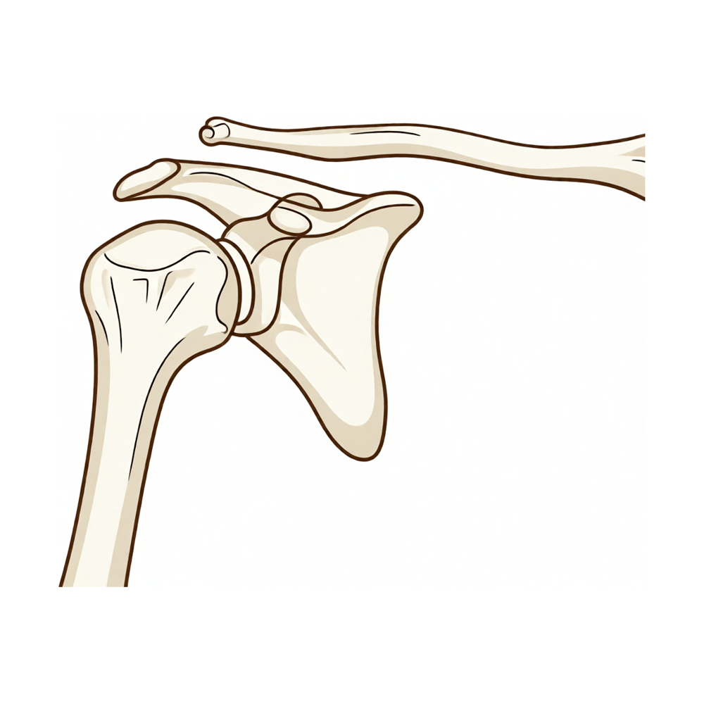
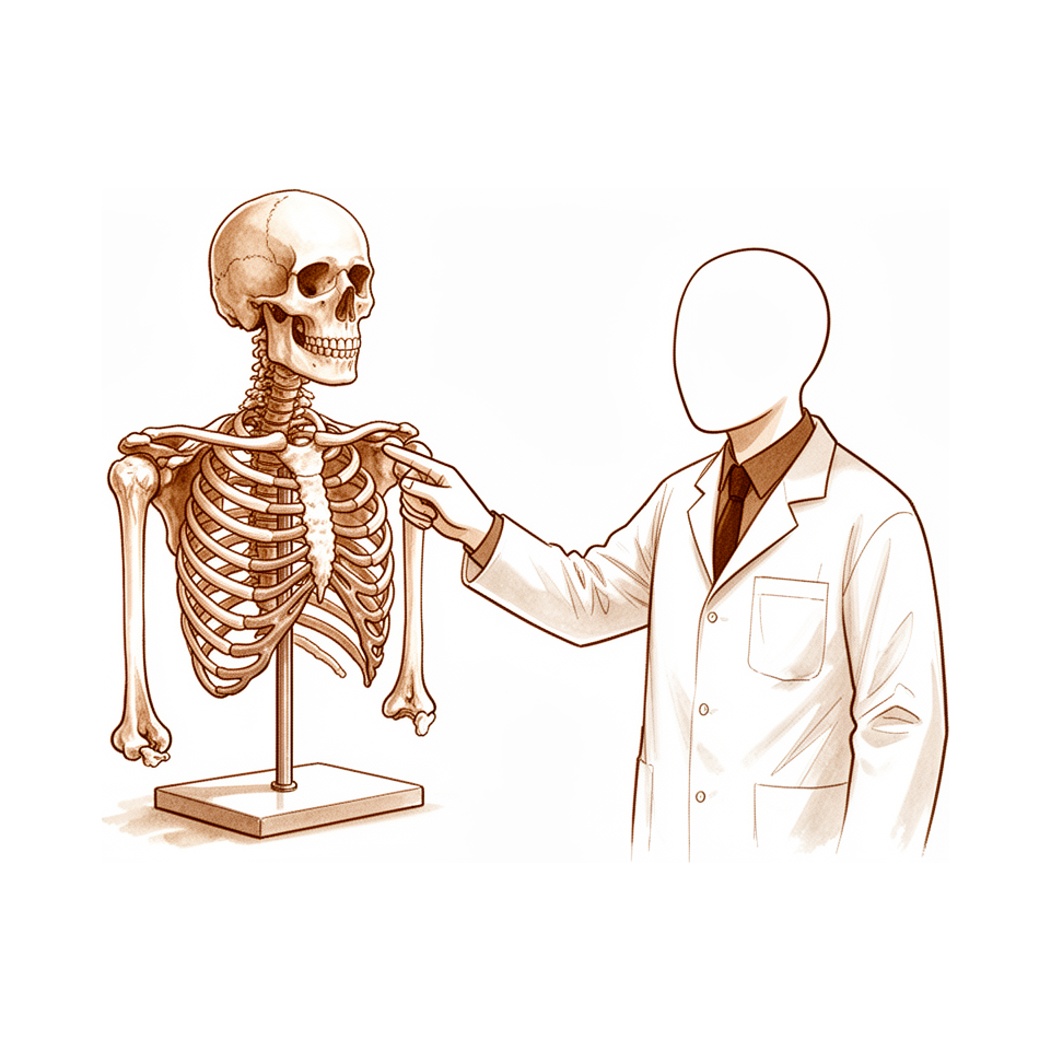
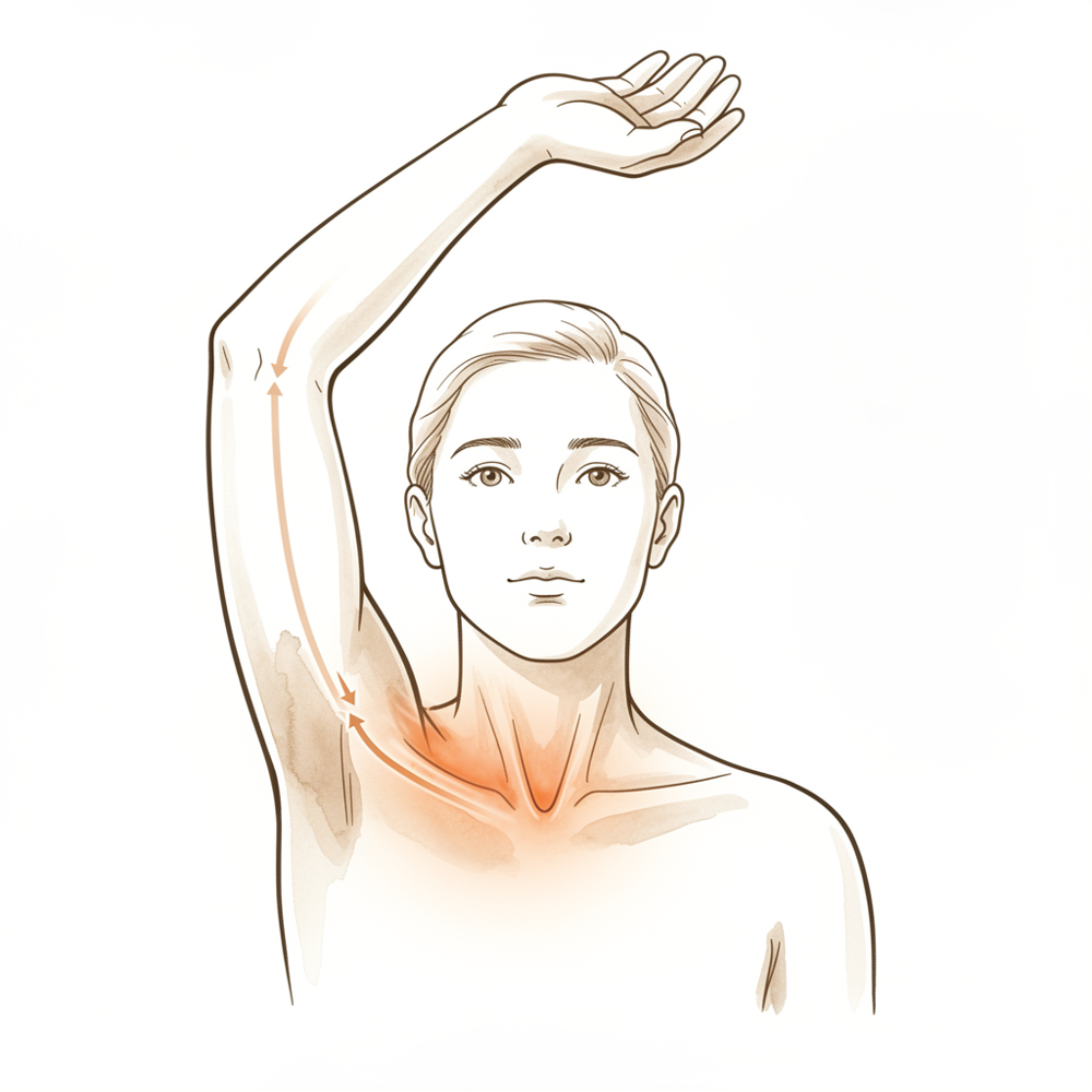
The shoulder possesses the most motion of any joint in the human body [37]. Shoulder function is highly dependent on unique anatomy and biomechanical properties [37]. Physical examination and radiologic workup of the shoulder require a thorough understanding of complex musculoskeletal interactions [37]. A clinical evaluation of altered shoulder kinematics remains complicated [25].
Kinematics and Biomechanics
A biomechanical shoulder model is consistent with clinical observations [24]. Observed glenohumeral translations and rotations characterize healthy shoulder motions but have technical shortcomings [34]. Asymptomatic rotator cuff pathology is associated with a plausible mechanical progression of kinematic and strength changes [56]. Biomechanical changes of passive glenohumeral joint motion occur with as little as 5% glenohumeral internal rotation deficit (GIRD) due to posterior capsular contracture [58]. Relating anatomic properties, kinematics, and muscle dynamics to subacromial volume is expected to identify predominant pathophysiological mechanisms in subacromial impingement syndrome (SIS) patients [59].
Scapular and Clavicular Mechanics
Clavicle shortening of >10% greatly affects scapular kinematics in vivo [65]. Scapular exercises resulted in superior migration of the humeral head and alterations in shoulder kinematics on radiographic analysis in healthy subjects under clinical fatigue conditions [67]. Altering scapula position may affect shoulder strength in asymptomatic individuals [82]. Trunk postures affect scapular kinematics and muscle activities during shoulder external rotation [80].
Throwing and Overuse Pathomechanics
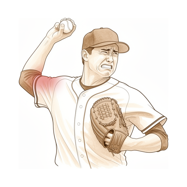
Updates on thrower's shoulder anatomy, mechanics, pathomechanics, and treatment are essential for clinicians and researchers [30]. Advances in understanding shoulder biomechanics, pathophysiology, and diagnostic techniques are necessary for preventing and treating common shoulder injuries in throwing athletes [52]. Improvements in surgical methods like arthroscopy are necessary for clinicians to properly prevent and treat common shoulder injuries in throwing athletes [52]. The combination of 80.6° of shoulder abduction and 10.7° of horizontal shoulder adduction minimized shear forces on the shoulder in adolescent baseball pitchers [79]. Future studies on shoulder overuse injuries in wheelchair athletes should be directed towards biomechanical modeling to develop knowledge of load and its effects [86].
Measurement and Assessment
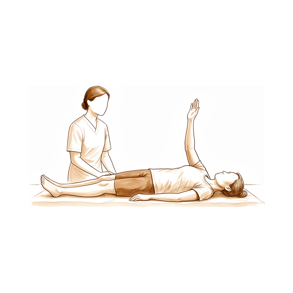
A valid and repeatable measurement of pectoralis minor extensibility is needed to improve studies evaluating its effect on shoulder movement and biomechanics [64]. Technology such as the Microsoft Kinect sensor plus Medical Interactive Recovery Assistant (MIRA) software may allow precise shoulder range of motion (ROM) measurement outside the clinic setting [83]. Passive shoulder external rotation range of motion changes with shoulder abduction angle, with greater external rotation ranges observed as abduction approaches 90° [84].
Classification
General Principles: A good classification system serves as a common language to define the severity of a condition, guide treatment, and facilitate clinical research [51]. The true value of a classification system can only be determined when it is used [40]. Unreliable classification may account for inconsistent treatment outcomes, suggesting a better classification system is needed for acromioclavicular joint dislocations [50].
Valgus Impacted Proximal Humerus Fractures: A prospective study with clearly defined inclusion criteria and a reliable classification system is needed to determine the best treatment for valgus impacted fractures of the proximal humerus [14].
Osteoporotic Thoracolumbar Vertebral Body Fractures: The addition of CT adds limited value compared to conventional radiographs and MRI only for the OF classification and the OF score of osteoporotic thoracolumbar vertebral body fractures [15].
Glenohumeral Instability: There are 16 categories within the FEDS classification that are clinically significant for classifying glenohumeral instability [36].
Acromioclavicular Joint Separations: The Rockwood classification is commonly used in Japan to assess severity of acromioclavicular joint separations, but there is some disagreement regarding the assessment for the diagnosis of type IV [57].
Septic Arthritis of the Shoulder: Patients with a classification of Grade III or higher in a novel MRI-based classification system for septic arthritis of the shoulder had higher reinfection rates than those with a classification of Grade I or II [72]. Patients with a classification of Grade III or higher in a novel MRI-based classification system for septic arthritis of the shoulder required more aggressive treatment to eradicate the infection [72].
Glenohumeral Synovitis: A new scoring system for the classification of glenohumeral synovitis as seen during arthroscopy has good reliability among a large range of surgeons [87].
Low Back Pain: The correlation between score on screening tools for risk classification of patients referred to secondary care for low back pain was good, while the classification agreement between the screening instruments was low [89].
Brachial Plexus Birth Injuries: The modified Mallet classification demonstrated strong to excellent agreement and interrater reliability across face-to-face, live tele-assessment, and video-based conditions for brachial plexus birth injuries [101]. The modified Mallet classification is appropriate for remote medical follow-up of brachial plexus birth injuries [101].
Intra-articular Hip Pathology: Arthroscopic classification of intra-articular hip pathology demonstrates at best moderate interrater reliability [104]. Further development and refinement of multifactorial grading systems for describing labral injury are indicated [104].
Elbow Rating Systems: Other elbow-specific clinical rating systems still have to be proven in the future to be as good as the Oxford Elbow Score for clinical or research purposes [106].
Clinical Presentation
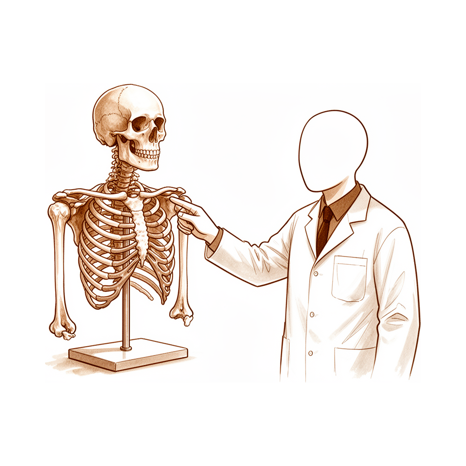
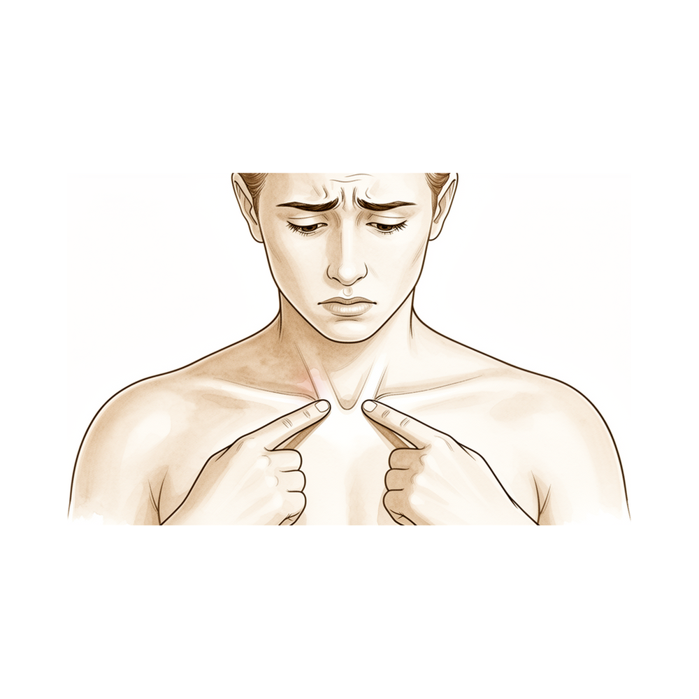
Clinical assessment of the pivot shift presents subjective variability and dependence on the tester's experience [1]. A thorough understanding of shoulder physical examination and specific diagnostic tests, emphasizing history-taking and examination, is important for ensuring diagnostic accuracy and optimizing patient outcomes [6]. Identification of whether a patient presents with pain or stiffness as their predominant symptom guides treatment selection for shoulder stiffness [27]. Careful clinical assessment can differentiate between causes of shoulder pain after a cerebrovascular accident or traumatic brain injury and guide best management [31].
A comprehensive assessment measuring both diagnostic capability and disease impact most accurately reflects the patient with femoroacetabular impingement syndrome (FAIS) [9]. The relative complexity of the final diagnostic tool for hip microinstability illustrates the difficulty clinicians face when making this diagnosis [32]. Clinical assessment accurately determined the existence of intra-articular abnormality in hip arthroscopy patients but was poor at defining its nature [39].
Clinical testing plays an important role in the initial identification of ulnar neuropathy lesions and determining subsequent changes from baseline [26]. Using the Resisted Supination External Rotation Test in the context of a thorough clinical history and physical examination allows for more reliable diagnosis of lesions of the superior labrum [42]. No single clinical test is sufficiently reliable to diagnose posterosuperior rotator cuff tears; clinicians should consider various combinations of patient characteristics and clinical tests, as well as imaging modalities, to confirm diagnosis and select the appropriate treatment option [41].
A systematic process of performing a comprehensive physical examination of the hand, including vascular, sensory, and motor assessments, is essential for appropriate treatment and providing the patient the opportunity for the best outcome [44]. Clinical evaluation is the first step in detecting feigned hand weakness, but it is not very specific; therefore, every suspected positive clinical result must be followed up with a more advanced test that has better specificity [33].
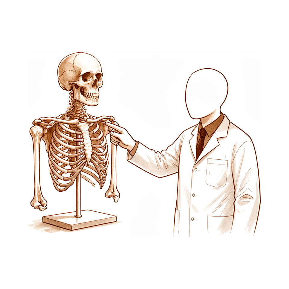
A thorough physical examination of the shoulder and cervical spine is critical in establishing a focused differential diagnosis of the pathology in and around the shoulder joint [45]. Knowledge of the characteristic clinical presentation and physical examination findings of neurologic, musculoskeletal, vascular, and other etiologies helps distinguish the source of upper extremity pain quickly to facilitate appropriate diagnostic measures and treatment [28]. Screening for medical problems in patients with upper extremity signs and symptoms involves assessing patient history, risk factors, and red flag signs to recognize potential underlying systemic pathology [4].
Clinical diagnoses of lumbopelvic pain agree with reference standards diagnoses more often than chance [43]. Appropriate clinical workup leads to earlier diagnosis and management of back pain in children and adolescents and avoids unnecessary cost [35]. A high index of suspicion is essential for early recognition and timely referral of aortic dissection disguised as a musculoskeletal condition, especially in patients with overlapping symptoms and vascular risk factors [29].
Muscle imaging aids in recognizing atypical clinical presentations of Pompe disease [2]. The complexity of presentation, diagnosis, and management of facioscapulohumeral muscular dystrophy (FSHD) emphasizes the necessity of a collaborative multidisciplinary team approach to maximize patient satisfaction and outcomes [47].
Investigations
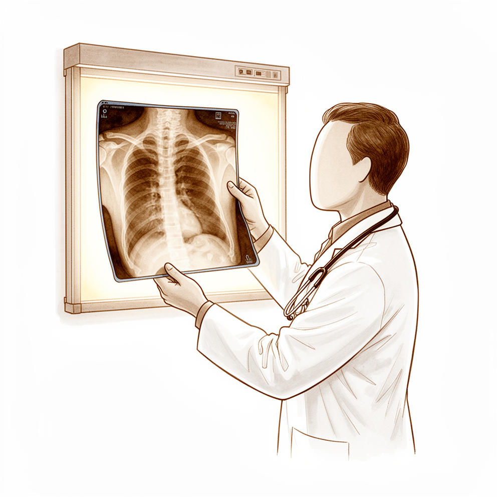
Plain radiography: Imaging studies should confirm the clinical impression derived from history and physical examination [18]. For critical shoulder angle (CSA) measurement, radiographs provide better reproducibility and accuracy compared to MRI [62]. In recurrent shoulder instability, appropriate preoperative imaging is essential for detection and quantification of osseous abnormalities to guide treatment decisions based on the extent of osseous deficiency and patient-specific factors [81]. Significant differences in bone loss measurement between imaging modalities, methods, and observers may lead to differences in treatment in up to 34% of cases [88]. For malunion of a head-splitting proximal humeral fracture, adequate radiologic investigation requires either three plain films obtained at 90° to each other or three-dimensional CT reconstruction [97]. In acromioclavicular joint osteoarthritis, patients with edema on MRI were more likely to present pain, and subchondral bone edema on histologic examination was more frequent in patients with pain [109]. Plain radiographs rarely alter diagnosis or management in atraumatic shoulder pain, particularly in patients younger than 50 years [107]. Roentgenographic evidence serves as an adjunct to define the degree and extent of involvement in osseous coccidioidomycosis [102].
MRI: Noninvasive imaging technologies, particularly MRI, provide objective measures to better understand disease natural history and mechanisms, optimize treatment, and assess joint tissue integrity [3]. Long-term follow-up studies are required to validate the predictive validity of these technologies for clinical outcomes [3]. MRI should be obtained at the discretion of the treating clinician based on specific clinical criteria rather than being mandated by insurers [10]. Advanced neurodiagnostic imaging should be refrained from until appropriate nonoperative management has failed [18]. Advanced imaging, such as MRI, can be used as necessary but should not replace the history and physical examination [38]. Clinicians should be aware of common anatomic findings on MRI when considering diagnostic and treatment planning for bilateral MRI findings in individuals with unilateral shoulder pain [54]. Orthopaedic surgeons are comfortable reviewing shoulder MRI scans without necessarily reading the MRI report prior to a surgical decision, but MRI scans should not be used without assessment of patient history or physical examination [110]. Anatomic abnormalities must be carefully correlated to symptoms when using imaging studies [18]. MRI findings are common in asymptomatic collegiate and professional hockey players; the significance of these findings must be cautiously interpreted in association with clinical presentation [96]. Preoperative MRI could be used to exclude subtle instability in patients with lateral epicondylitis, especially when patients have a history of multiple corticosteroid injections (≥3) or severe pain [99].
CT: The addition of CT adds limited value compared to conventional radiographs and MRI only in terms of the OF classification and the OF score for osteoporotic thoracolumbar vertebral body fractures [15]. Computed tomography is the imaging modality of choice for evaluation of sternoclavicular joint injuries [77].
Other Considerations: A comprehensive assessment measuring both diagnostic capability and disease impact most accurately reflects the patient with femoroacetabular impingement syndrome (FAIS) [9]. Tests for lumbar instability are simple to perform, reliable, and safe, and are recommended for use by physical therapists to assess suspected patients without immediate X-ray assessment [13].
Treatment
Non-Operative
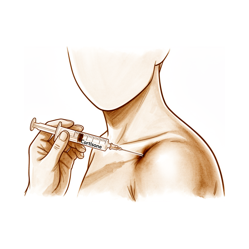
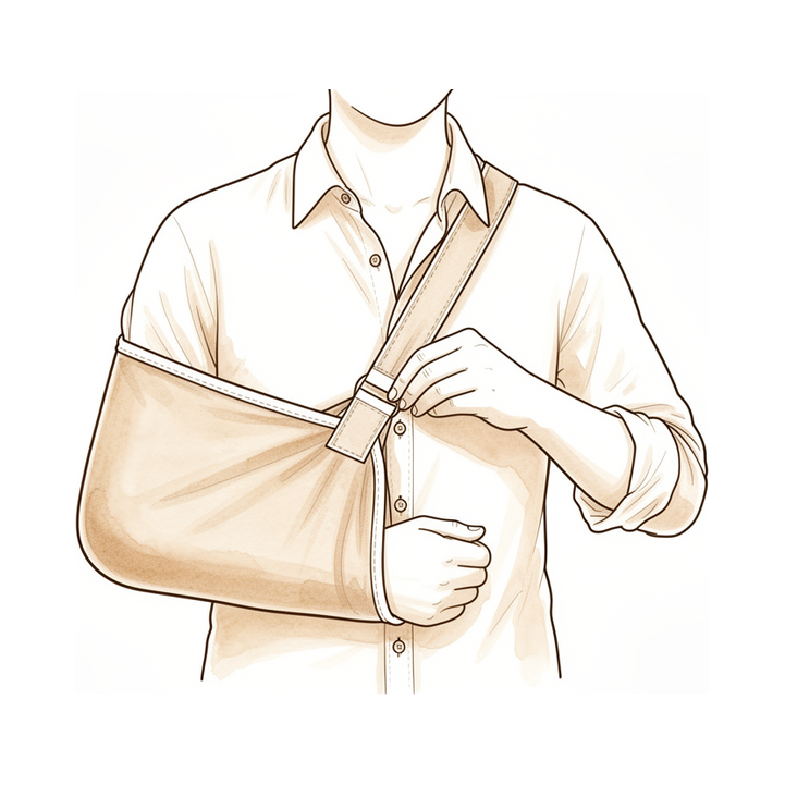
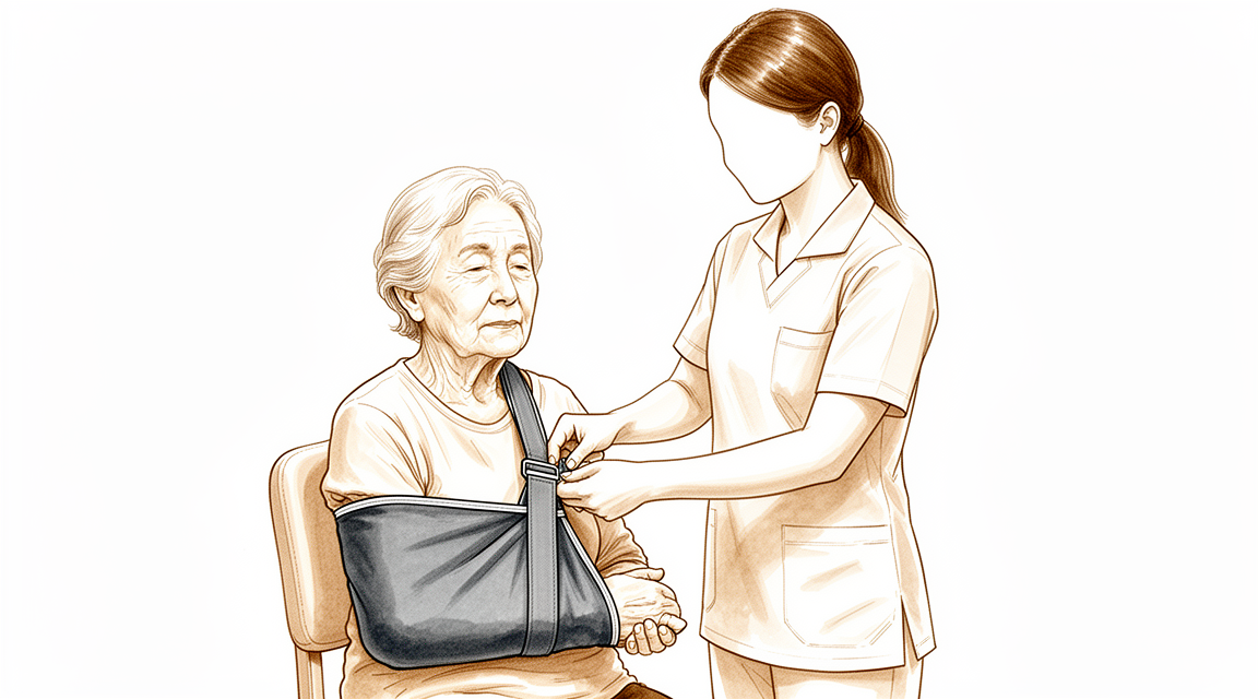
Nonoperative management serves as the first-line treatment for most young, active patients with SLAP tears lacking trauma history, mechanical symptoms, or overhead activity demands [85]. For quadrilateral space syndrome, nonoperative treatment for at least 6 months is recommended before pursuing operative intervention [91]. Adhesive capsulitis generally consists of conservative management, followed by surgical intervention if adequate function is not attained [92]. Nonoperative treatment is efficacious for chronic, massive, irreparable rotator cuff tears despite low-quality evidence [93] and is helpful for most painful acromioclavicular joint conditions, though osteolysis may require activity modification [94]. Corticosteroid injection offers potential long-term benefits in radial tunnel syndrome [95] and provides statistically and clinically relevant improvements in shoulder function and pain up to 4 months for glenohumeral arthritis [78]. In primary traumatic anterior glenohumeral dislocation, nonoperative treatment is preferred when recurrent instability risk is low or the patient has an aversion to surgery [98]. Bracing is not effective as an adjunct to standard nonoperative management for scholastic athletes with unstable shoulders [100]. For scapular winging due to rhomboid paralysis, conservative treatment in a multidisciplinary rehabilitation center is fundamental [74]. Nonoperative treatment is preferred in early-stage atraumatic osteonecrosis of the humeral head to prevent progression, though surgery is required in some cases [105]. Physical therapy for osteochondritis dissecans incorporates a full spectrum of conservative, nonoperative, and postoperative care [90]. Traditional Chinese medicine comprehensive therapy for exercise-related musculoskeletal injuries shows potential benefits, though randomized controlled trials are needed for definitive efficacy assessment [69].
Operative
Indications: Routine preoperative magnetic resonance imaging for hip arthroscopy should be obtained at the discretion of the treating clinician based on specific clinical criteria rather than being mandated by health care insurers [10]. Diagnostic tests for lumbar instability are simple, reliable, and safe, recommended for physical therapists to assess suspected patients without immediate X-ray assessment [13]. Each test for joint aspiration in diagnosing chronic periprosthetic joint infection has advantages and disadvantages and should be used with the overall clinical picture to guide further evaluation [11]. Evaluation protocols should focus on specific postsurgical therapy plans in addition to surgical and therapy decision making [7]. Understanding current evidence and appropriate indications of emerging technologies in orthopaedic trauma is critical for their utilization [16].
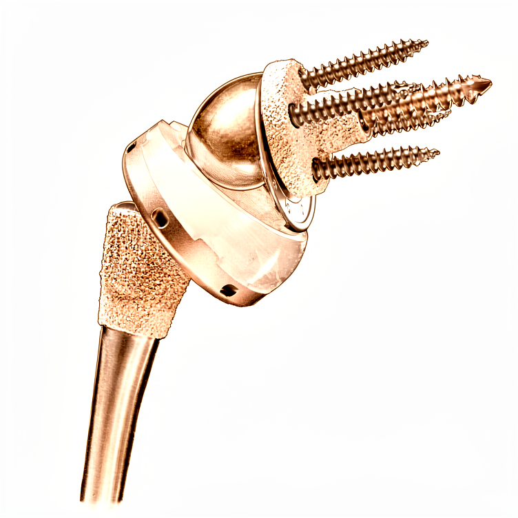
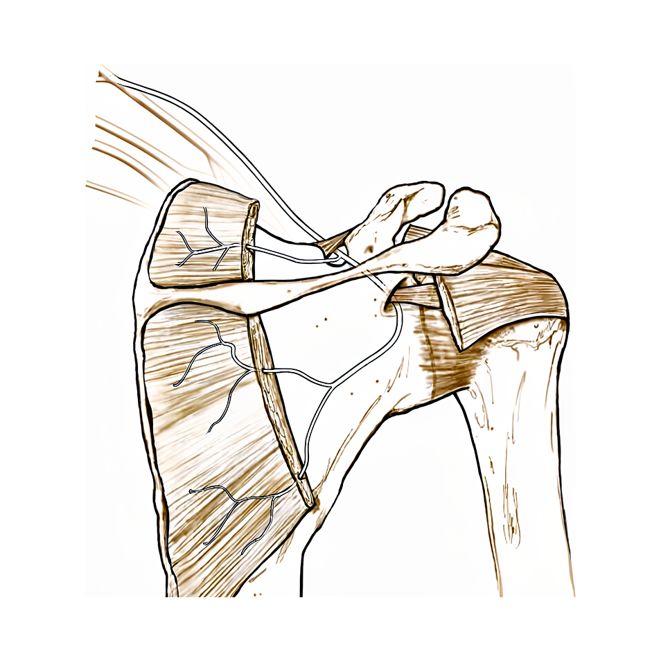
Surgical Approach / Technique: Clinical outcomes of Lima ProMade custom 3D-printed glenoid components in primary and revision reverse total shoulder arthroplasty with severe glenoid bone loss show modest but statistically significant improvements, though patients often do not achieve a normal shoulder [68]. Patients undergoing arthroscopic treatment for snapping scapula syndrome experienced clinically significant improvements in functional scores, pain, and quality of life [66]. The evidence supporting the suprascapular nerve block regarding efficacy and safety is compelling for clinical use of a landmark-based approach [71].
Pain Management: Pain self-efficacy did not moderate the relationship between treatment and outcome for shoulder pain treated with manual therapy, acupuncture, and electrotherapy [48]. Clinicians must be informed and educated about common drug interactions and contraindications in orthopaedic practice to ensure safe and effective clinical care [55].
Adjuncts: A rehabilitation program is currently being tested for efficacy as part of a randomized controlled trial for the treatment of multidirectional instability of the shoulder [5]. Clinical trials are needed to evaluate the effect of immobilization duration in external rotation on recurrence rates for acute anterior dislocations of the shoulder [60].
Other Considerations: The effects of comorbidity must be controlled in the evaluation of the functional status of patients and the effectiveness of treatment for glenohumeral degenerative joint disease [46]. Preoperative PROMIS PF scores predict postoperative outcome and were responsive to the functional improvements observed clinically in total shoulder arthroplasty patients [75]. The application and evaluation of the Dutch version of the Oxford Shoulder Score in clinical trials proved feasible and understandable [8]. Current studies on treatment modalities for proximal humeral fractures typically lack randomization, comparators, and independent evaluation, with a resultant inability to produce clinical conclusions [73].
Complications
Nerve palsy: Clinical outcomes of reverse total shoulder arthroplasty at a minimum follow-up of 1 year were similar in high- and lower-risk groups for iatrogenic suprascapular neuropathy by screw violation [118].
Other Considerations: Clinical assessment of the pivot shift presents subjective variability and dependence on the tester's experience [1]. Muscle imaging can help for recognition of atypical clinical presentations [2]. Noninvasive imaging technologies, particularly MRI, provide objective measures to better understand the natural history and mechanisms of diseases, optimize treatment, and assess the integrity of joint tissues [3]. Long-term follow-up studies are required to validate the predictive validity of noninvasive imaging technologies for clinical outcomes [3]. Screening involves assessing patient history, risk factors, and red flag signs to recognize potential underlying systemic pathology [4]. A thorough understanding of shoulder physical examination and specific diagnostic tests, emphasizing history-taking and examination, is important to ensure diagnostic accuracy and optimize patient outcomes for surgeons [6]. Short-term repeat assessment of pain was better than short-term change or baseline score at predicting long-term disability improvement across all cohorts [12]. Patient self-assessment of shoulder range of motion agrees with physician assessment, potentially allowing this method to replace routine clinic visits for short- and long-term follow-up [17]. Imaging studies should be used to confirm the clinical impression derived from history and physical examination, with careful correlation of anatomic abnormalities to symptoms [18]. Advanced neurodiagnostic imaging should be refrained from until appropriate nonoperative management has failed [18]. Three months follow-up is too early for outcome evaluation in shoulder conditions [19]. Longer-term follow-up is required to confirm promising short-term results of a hybrid cage glenoid compared to a cemented polyethylene glenoid in anatomic total shoulder arthroplasty [20]. The study on factors predicting long-term outcomes following physiotherapy in patients with subacromial pain syndrome provides statistical coefficients and model performance metrics but does not include an explicit conclusion statement [21]. Recent literature has enhanced understanding of pathoanatomy and natural history of brachial plexus birth palsy, leading to improved care [22]. The study on demographic and morphological features of rotator cuff disease provides insight into the natural history of rotator cuff disease by comparing morphological characteristics and prevalences in asymptomatic and symptomatic shoulders [23]. Short and long-term outcomes of traumatic hip dislocation are largely driven by the amount of time from injury to reduction and associated injuries [111]. Maintenance of reduction in arthroscopic treatment of posterior glenohumeral joint subluxation resulting from brachial plexus birth palsy requires continued follow-up over time [115]. Successful care of patients with neurologic and musculoskeletal problems begins with arriving at the proper diagnosis through a thorough history, complete physical examination, and appropriate imaging modalities to optimize safe, value-driven care [116]. SIRVA is a rare yet increasingly recognized complication of immunization that is primarily a clinical diagnosis [117]. Larger high-quality studies are needed to determine the natural history and optimal treatment of SIRVA [117]. All parameters related to history taking, physical exam and imaging studies for traumatic anterior shoulder instability should be considered according to the patient's age and the number of dislocation episodes [119]. Patients generally reported excellent 1-year outcomes following primary arthroscopic rotator cuff repair [120]. A sufficient basic diagnosis of overactive bladder including a detailed medical history and, if necessary, urodynamic evaluation is crucial [121].
Recovery
Light activity (weeks): Evidence does not provide specific week ranges for light activity or desk work. However, short-term repeat assessment of pain provides a more accurate prognosis than baseline information for low-back or shoulder pain [12]. For shoulder conditions, a 3-month follow-up is considered too early for outcome evaluation [19].
Full activity (months): Evidence does not provide specific month ranges for full activity or manual work. Patient self-assessment of shoulder range of motion could potentially replace routine clinic visits for short- and long-term follow-up [17].
Complete recovery / outcome plateau (months): Longer-term follow-up is required to confirm promising short-term results of a hybrid cage glenoid compared to a cemented polyethylene glenoid in anatomic total shoulder arthroplasty [20]. The goal of assessing the longer-term functional effect of femoral nerve block for anterior cruciate ligament reconstruction was accomplished [108]. Responsiveness of clinical tests for neck pain was generally low when using change in NDI score as the anchor from baseline to the 4-month follow up [113]. A cohort study demonstrated mild complaints at baseline and an overall stable course during one-year follow-up for arm, neck and/or shoulder complaints [122].
Rehabilitation protocol: Evidence does not provide specific details on PT phasing, immobilisation duration, or weight-bearing progression.
Functional milestones: The Oxford shoulder score may be considered an alternative for longer-term follow-up as it is easier to administer and correlates well with the Constant score [112]. Short-term repeat assessment of pain was better than short-term change or baseline score at predicting long-term disability improvement across all cohorts [12]. Physical outcome measures are being changed for the use of patient reported outcomes, and range of motion and strength are not as reliable measures as one would think [114].
Other Considerations: Clinical assessment of the pivot shift presents subjective variability and dependence on the tester's experience [1]. Muscle imaging can help for recognition of atypical clinical presentations, for understanding the natural history of the disease, and for determining patients suited for treatment [2]. Noninvasive imaging technologies, particularly MRI, provide objective measures to better understand the natural history and mechanisms of diseases, optimize treatment, and assess the integrity of joint tissues [3]. Long-term follow-up studies are required to validate the predictive validity of noninvasive imaging technologies for clinical outcomes [3]. Recent literature has enhanced understanding of pathoanatomy and natural history of brachial plexus birth palsy, leading to improved care [22]. The study on rotator cuff disease provides insight into the natural history of the condition by comparing morphological characteristics and prevalences in asymptomatic and symptomatic shoulders [23]. Early and aggressive treatment of rheumatoid arthritis is now standard, and complete resolution of signs and symptoms is achievable in perhaps 10% of patients [123]. Those with a worse prognosis may be monitored more frequently and the treatment plan modified if complaints persist in patients with shoulder pain [124]. Pain intensity, neck pain and longer duration of complaints predict poorer outcome in patients with shoulder pain [124]. At a population level, back beliefs were generally positive and relatively constant over time, but misconceptions about a poor prognosis were common [125]. Significant deficits in the adhesive capsulitis literature include a paucity of randomized controlled trials, failure to report response to treatment in a stage-based fashion, and an incomplete understanding of the disease's natural course [126].
Key Evidence
- [L2] Actual clinical assessment presents subjective variability and dependence on the tester's experience. [1] (10.1007/s00167-016-4130-1)
- [L5] Muscle imaging can help for recognition of atypical clinical presentations, for understanding the natural history of the disease, and for determining patients suited for treatment. [2] (10.1186/1471-2474-14-s2-o2)
- [L5] Noninvasive imaging technologies, particularly MRI, provide objective measures to better understand the natural history and mechanisms of diseases, optimize treatment, and assess the integrity of joint tissues, though long-term follow-up studies are required to validate their predictive validity for clinical outcomes. [3] (10.1177/0363546518817315)
- [L5] Screening involves assessing patient history, risk factors, and red flag signs to recognize potential underlying systemic pathology. [4] (10.1016/j.jht.2009.09.005)
- [L5] This clinical protocol is currently being tested for efficacy as part of a randomized controlled trial. [5] (10.1177/1758573216652086)
- [L5] This comprehensive review highlights the importance of a thorough understanding of shoulder physical examination and specific diagnostic tests, emphasizing history-taking and examination to ensure diagnostic accuracy and optimize patient outcomes for surgeons. [6] (10.5435/jaaos-d-25-00024)
- [L4] The evaluation protocol should focus on specific postsurgical therapy plans in addition to surgical/therapy decision making. [7] (10.1197/j.jht.2008.01.001)
- [L4] Application and evaluation in clinical trial proved feasible and understandable. [8] (10.1016/j.jse.2010.01.017)
- [L5] A comprehensive assessment measuring both diagnostic capability and disease impact most accurately reflects the patient with FAIS. [9] (10.1007/s00167-017-4484-z)
- [L5] The author concludes that MRI should be obtained at the discretion of the treating clinician based on specific clinical criteria rather than being mandated by insurers. [10] (10.1016/j.arthro.2022.04.009)
- [L5] Each available test has advantages and disadvantages and should be used in conjunction with the overall clinical picture to guide further evaluation and treatment. [11] (10.1186/s42836-023-00199-y)
- [L2] Short-term repeat assessment of pain was better than short-term change or baseline score at predicting long-term disability improvement across all cohorts. [12] (10.1186/s12891-017-1502-8)
- [L2] These tests are simple to perform, reliable, and safe, and are recommended for use by physical therapists to assess suspected patients without immediate X-ray assessment. [13] (10.1186/s12891-021-04854-w)
- [L4] A prospective study with clearly defined inclusion criteria and a reliable classification system is needed to determine the best treatment. [14] (10.1016/j.jhsa.2011.05.003)
- [L3] In terms of the OF classification and the OF score, the addition of CT adds limited value compared to conventional radiographs and MRI only. [15] (10.1186/s12891-022-06056-4)
- [L3] This method for short- and long-term follow-up could potentially replace routine clinic visits. [17] (10.1016/j.jse.2016.02.010)
- [L5] Imaging studies should be used to confirm the clinical impression derived from history and physical examination, with careful correlation of anatomic abnormalities to symptoms, and advanced neurodiagnostic imaging should be refrained from until appropriate nonoperative management has failed. [18] (10.5435/00124635-199609000-00002)
- [L2] Our results suggest that 3 months follow-up is too early for outcome evaluation. [19] (10.1186/s12891-021-04483-3)
- [L3] The authors note that longer-term follow-up is required to confirm these promising short-term results. [20] (10.1016/j.jse.2019.04.049)
- [L3] The provided text contains statistical coefficients and model performance metrics for predicting long-term outcomes but does not include the authors' explicit conclusion statement. [21] (10.1186/s12891-024-07686-6)
- [L5] Recent literature has enhanced understanding of pathoanatomy and natural history, leading to improved care. [22] (10.1016/j.jhsa.2009.11.026)
- [L3] The study provides insight into the natural history of rotator cuff disease by comparing morphological characteristics and prevalences in asymptomatic and symptomatic shoulders. [23] (10.2106/jbjs.e.00835)
- [L5] The biomechanical shoulder model is consistent with clinical observations. [24] (10.1016/j.jse.2016.05.031)
- [L2] A clinical evaluation of altered shoulder kinematics is still complicated. [25] (10.3390/ijerph17082974)
- [L5] Clinical testing plays an important role in the initial identification of a lesion and determining subsequent changes from baseline; however, empirical research studies are indicated to further quantify the relationship between testing outcomes and lesion severity. [26] (10.1016/j.jht.2008.10.010)
- [L5] Identification of whether a patient presents with pain or stiffness as their predominant symptom further guides treatment selection. [27] (10.1177/1758573215586152)
- [L5] Knowledge of the characteristic clinical presentation and physical examination findings of neurologic, musculoskeletal, vascular, and other etiologies can help distinguish the source of upper extremity pain quickly to facilitate appropriate diagnostic measures and treatment. [28] (10.5435/jaaos-d-11-00086)
- [Case_report] A high index of suspicion is essential for early recognition and timely referral, especially in patients with overlapping symptoms and vascular risk factors. [29] (10.1186/s12891-025-09135-4)
- [L5] Updates on the thrower's shoulder, including anatomy, mechanics, pathomechanics, and treatment, are essential for clinicians and researchers treating or investigating the shoulder. [30] (10.1016/j.arthro.2022.02.024)
- [L5] This review article describes how careful clinical assessment can differentiate between causes of shoulder pain and guide best management. [31] (10.1016/j.jse.2014.12.003)
- [L5] The relative complexity of the final diagnostic tool is illustrative of the difficulty clinicians face when making this diagnosis. [32] (10.1007/s00167-022-06933-4)
- [L5] Clinical evaluation is the first step in detecting feigned hand weakness, but it is not very specific; therefore, every suspected positive clinical result must be followed up with a more advanced test that has better specificity. [33] (10.1016/j.jhsa.2007.09.010)
- [L4] The observed glenohumeral translations and rotations characterize healthy shoulder motions but still have technical shortcomings. [34] (10.2519/jospt.2012.3584)
- [L5] Appropriate clinical workup leads to earlier diagnosis and management of back pain and avoids unnecessary cost. [35] (10.5435/jaaos-d-14-00130)
- [L4] There are 16 categories within the FEDS classification that are clinically significant. [36] (10.1016/j.jse.2018.08.014)
- [L5] Advanced imaging, such as MRI, can then be used as necessary but should not replace the history and physical examination. [38] (10.5435/jaaos-d-15-00464)
- [L4] Clinical assessment accurately determined the existence of intra-articular abnormality but was poor at defining its nature. [39] (10.1177/0363546504266480)
- [L5] The true value of the classification system can only be determined when it is used, and time will tell whether this classification system will be used. [40] (10.1016/j.jse.2010.07.008)
- [L2] As no single clinical test is sufficiently reliable to diagnose posterosuperior rotator cuff tears, clinicians should consider various combinations of patient characteristics and clinical tests, as well as imaging modalities, to confirm diagnosis and select the appropriate treatment option. [41] (10.1007/s00167-020-06136-9)
- [L2] By using this test in the context of a thorough clinical history and physical examination, lesions of the superior labrum can be more reliably diagnosed. [42] (10.1177/0363546504273050)
- [L3] Clinical diagnoses agree with reference standards diagnoses more often than chance. [43] (10.1186/1471-2474-6-28)
- [L5] This current concepts review presents a systematic process of performing a comprehensive physical examination of the hand including vascular, sensory, and motor assessments, which is essential for appropriate treatment and providing the patient the opportunity for the best outcome. [44] (10.1016/j.jhsa.2014.04.026)
- [L4] A thorough physical examination of the shoulder and cervical spine is critical in establishing a focused differential diagnosis of the pathology in and around the shoulder joint. [45] (10.5435/jaaos-d-17-00090)
- [L3] In the evaluation of the functional status of patients and the effectiveness of treatment, the effects of comorbidity must be controlled. [46] (10.2106/00004623-199808000-00007)
- [Case_report] The complexity of presentation, diagnosis, and management of FSHD emphasizes the necessity of a collaborative multidisciplinary team approach to maximize patient satisfaction and outcomes. [47] (10.1016/j.jseint.2020.04.022)
- [L2] Pain self-efficacy did not moderate the relationship between treatment and outcome. [48] (10.1177/17585732221105562)
- [L4] The authors suggest that the score should be redesigned with better standardization before being used to validate other assessments or compare outcomes between different testers. [49] (10.1016/j.jse.2007.06.024)
- [L4] Unreliable classification may account for inconsistent treatment outcomes, suggesting a better classification system is needed. [50] (10.1016/j.jse.2017.09.021)
- [L5] A good classification system serves as a common language to define the severity of a condition, guide treatment, and facilitate clinical research. [51] (10.5435/jaaos-d-15-00034)
- [L5] Advances in understanding shoulder biomechanics, pathophysiology, and diagnostic techniques, along with improvements in surgical methods like arthroscopy, are necessary for clinicians to properly prevent and treat common shoulder injuries in throwing athletes. [52] (10.1177/03635465000280022301)
- [L5] Clinicians and researchers must define their specific question to select the best assessment tool, ideally one with content validity and reliability, while considering normalization for specific populations and incorporating patient satisfaction. [53] (10.1016/j.jse.2009.03.010)
- [L3] Clinicians should be aware of the common anatomic findings on MRI when considering diagnostic and treatment planning. [54] (10.1016/j.jse.2019.04.001)
- [L5] This article discusses common drug interactions and contraindications in orthopaedic practice to highlight the importance of informing and educating clinicians for safe and effective clinical care. [55] (10.1302/0301-620x.97b4.35230)
- [L3] Furthermore, these findings suggest a plausible mechanical progression of kinematic and strength changes associated with the development of rotator cuff pathology. [56] (10.1016/j.jse.2016.11.048)
- [L4] The Rockwood classification is commonly used in Japan to assess severity, but there is some disagreement regarding the assessment for the diagnosis of type IV. [57] (10.1016/j.jseint.2019.11.006)
- [L5] Biomechanical changes of passive glenohumeral joint motion occur in the glenohumeral joint with as little as 5% GIRD. [58] (10.1177/0363546512462012)
- [L4] By relating anatomic properties, kinematics and muscle dynamics to subacromial volume, the study expects to identify one or more predominant pathophysiological mechanisms in every SIS patient to optimize future diagnostic and treatment strategies. [59] (10.1186/1471-2474-12-282)
- [L2] Clinical trials are needed to evaluate the effect of these results on recurrence rates. [60] (10.1177/0363546509331943)
- [L3] Radiographs provide better reproducibility and accuracy for CSA measurement compared to MRI. [62] (10.1007/s00167-015-3587-7)
- [L3] Improved clinical outcomes may be achieved for full-thickness RCTs when AAOS AUC recommendations are followed; however, because improved clinical outcomes may also be achieved when the recommendations are not followed, further investigation is needed in a population of patients in whom there is discordance between AAOS AUC recommendations and the treatment administered. [63] (10.1016/j.jse.2015.12.009)
- [Letter] A valid and repeatable measurement of extensibility would be a valuable addition to studies evaluating the effect of PM on shoulder movement and biomechanics, and additional work is needed to improve or develop new measurement instruments and methods. [64] (10.1016/j.jht.2017.06.007)
- [L4] Clavicle shortening of >10% greatly affects scapular kinematics in vivo. [65] (10.1016/j.jse.2017.03.013)
- [L3] Patients who underwent arthroscopic treatment for SSS experienced clinically significant improvements in functional scores, pain, and quality of life. [66] (10.1016/j.jse.2024.01.018)
- [L5] In the context of a clinical model of fatigue, these scapular exercises resulted in superior migration of the humeral head and alterations in shoulder kinematics on radiographic analysis in healthy subjects. [67] (10.1016/j.jse.2008.09.010)
- [L4] Clinical outcomes show modest but statistically significant improvements, though patients often do not achieve a normal shoulder. [68] (10.1016/j.jse.2023.04.020)
- [L4] These findings suggest potential benefits of this treatment approach, though randomized controlled trials are needed for definitive efficacy assessment. [69] (10.1186/s13018-025-05768-2)
- [L5] The AAOS Appropriate Use Criteria functions as a useful tool to aid in preoperative planning and intraoperative decision making, providing a framework to affirm that a selected treatment choice is appropriate based on clinical trials and expert consensus. [70] (10.5435/jaaos-21-12-772)
- [L5] The evidence in favor of the suprascapular nerve block both in efficacy and safety is compelling for clinical use of a landmark-based approach. [71] (10.1016/j.arthro.2019.05.031)
- [L3] Patients with a classification of Grade III or higher in the novel classification system had higher reinfection rates than those with a classification of Grade I or II and required more aggressive treatment to eradicate the infection. [72] (10.2106/jbjs.19.00951)
- [L2] Current studies typically lack randomization, comparators, and independent evaluation, with a resultant inability to produce clinical conclusions. [73] (10.1016/j.jse.2007.03.016)
- [L4] In the absence of a surgical solution, conservative treatment is fundamental and requires management in a rehabilitation center with intervention by a multidisciplinary team. [74] (10.1016/j.jse.2022.05.011)
- [L3] PROMIS PF scores were responsive to the functional improvements observed clinically. [75] (10.1016/j.jse.2018.08.040)
- [L3] This method of assessment is encouraged as a routine part of the preoperative evaluation of all patients under consideration for arthroscopic anterior stabilization. [76] (10.2106/jbjs.15.01099)
- [L4] Patients experienced statistically and clinically relevant improvements in shoulder function and pain up to 4 months after injection. [78] (10.1016/j.jse.2020.08.008)
- [L4] The combination of 80.6° of shoulder abduction and 10.7° of horizontal shoulder adduction minimized the shear forces on the shoulder at this point. [79] (10.1177/0363546518789626)
- [L5] Trunk postures affected scapular kinematics and muscle activities during external rotation. [80] (10.1016/j.jse.2019.04.059)
- [L5] Appropriate preoperative imaging is essential for detection and quantification of osseous abnormalities, and treatment decisions should be guided by the extent of osseous deficiency and patient-specific factors. [81] (10.2106/jbjs.j.00906)
- [L3] Altering scapula position may affect shoulder strength in asymptomatic individuals. [82] (10.1111/sae.12027)
- [L5] This technology, which can be easily set up, may also allow precise shoulder ROM measurement outside the clinic setting. [83] (10.1016/j.jse.2017.06.004)
- [L4] Passive shoulder external rotation range of motion changes with changes in shoulder abduction angle, with greater external rotation ranges observed as shoulder abduction approaches 90°. [84] (10.1177/17585732261420223)
- [L5] Nonoperative management is the first-line treatment for most young, active patients without history of trauma, mechanical symptoms, and/or demand for overhead activities. [85] (10.1016/j.arthro.2022.08.005)
- [L1] Future studies on shoulder overuse injuries of wheelchair athletes should be directed towards biomechanical modeling to develop knowledge of load and its effects. [86] (10.1371/journal.pone.0188410)
- [L4] This study defined a new scoring system for the classification of glenohumeral synovitis as seen during arthroscopy with good reliability among a large range of surgeons. [87] (10.1016/j.jse.2017.06.003)
- [L3] The significant differences in bone loss measurement between imaging modality, measurement method, and observers may lead to differences in treatment in up to 34% of cases. [88] (10.1016/j.arthro.2019.06.025)
- [L4] The correlation between score on the screening tools was good, while the classification agreement between the screening instruments was low. [89] (10.1186/s12891-018-2082-y)
- [Paper] Physical therapy management of osteochondritis dissecans can incorporate a full spectrum of conservative, nonoperative, and postoperative care. [90] (10.1016/j.csm.2014.01.001)
- [L5] Nonoperative treatment for at least 6 months is recommended before pursuing operative intervention. [91] (10.1016/j.jse.2017.10.024)
- [L5] Standard of care generally consists of conservative management which can be followed by surgical intervention if adequate function is not attained. [92] (10.1111/j.1758-5740.2010.00095.x)
- [L1] Despite low-quality evidence, nonoperative treatment has been shown to be efficacious for patients with chronic, massive, irreparable rotator cuff tears. [93] (10.1016/j.jse.2020.11.002)
- [L5] Nonoperative treatment is helpful for most patients, although those with osteolysis may have to modify their activities. [94] (10.5435/00124635-199905000-00004)
- [L4] Nonoperative management with corticosteroid injection can be used as a therapeutic measure with potential long-term benefits in the treatment of RTS. [95] (10.1177/1558944718787282)
- [L3] Given the high prevalence of MRI findings in asymptomatic hockey players, it is necessary to cautiously interpret the significance of these findings in association with clinical presentation. [96] (10.1177/0363546510388931)
- [L4] More importantly, this case highlights the need for adequate radiologic investigation, either by use of 3 plain films obtained at 90° to each other or by use of 3-dimensional CT reconstruction. [97] (10.1016/j.jse.2006.08.003)
- [L3] In clinical settings where the likelihood of recurrent instability is low after nonoperative care or when an informed patient has an aversion to surgery, nonoperative treatment may be the preferred treatment strategy. [98] (10.1016/j.jse.2011.01.031)
- [L4] Preoperative MRI could be used to exclude subtle instability, and surgeons should consider checking for subtle instability, especially when patients have a history of multiple corticosteroid injections (≥3) or severe pain. [99] (10.1186/s12891-018-2069-8)
- [L3] Bracing is not effective as an adjunct to standard nonoperative management in allowing athletes to return and complete a subsequent season. [100] (10.1016/j.jse.2018.02.027)
- [L3] The modified Mallet classification demonstrated strong to excellent agreement and interrater reliability across face-to-face, live tele-assessment, and video-based conditions, suggesting it is appropriate for remote medical follow-up. [101] (10.1177/17531934231196118)
- [L4] Roentgenographic evidence serves as an adjunct to define the degree and extent of involvement. [102] (10.2106/00004623-197153060-00012)
- [L4] Further development and refinement of multifactorial grading systems for describing labral injury are indicated. [104] (10.1007/s00167-020-06215-x)
- [L5] Nonoperative treatment is the preferred option in early-stage disease, and it may prevent disease progression, though surgical treatment is required in some cases. [105] (10.1016/j.xrrt.2022.02.005)
- [L2] Other rating systems still have to be proven in the future to be as good as the Oxford Elbow Score for clinical or research purposes. [106] (10.1016/j.jse.2013.04.013)
- [L2] Plain radiographs rarely alter the diagnosis or affect management in the setting of atraumatic shoulder pain, particularly in patients younger than 50 years. [107] (10.5435/jaaos-d-16-00884)
- [L3] They conclude that the goal of assessing the longer-term functional effect of FNB was accomplished and hope it serves as a catalyst for future prospective randomized studies. [108] (10.1177/0363546515601377)
- [L4] Patients with edema on MRI were more likely to present pain than patients without edema, and subchondral bone edema on histologic examination was more frequent in patients with pain. [109] (10.1016/j.jseint.2020.03.007)
- [L4] Orthopaedic surgeons are comfortable reviewing shoulder MRI scans without necessarily reading the MRI report prior to a surgical decision, but MRI scans should not be used without assessment of patient history and or physical examination. [110] (10.1186/s12891-022-05541-0)
- [L5] Short and long-term outcomes are largely driven by the amount of time from injury to reduction and associated injuries. [111] (10.5435/jaaos-d-23-01013)
- [L3] The OSS may be considered an alternative for longer-term follow-up as it is easier to administer and correlates well with the Constant score. [112] (10.1016/j.jse.2007.04.019)
- [L2] Responsiveness of the included clinical tests was generally low when using change in NDI score as the anchor from baseline to the 4-month follow up. [113] (10.1186/s12891-017-1918-1)
- [Paper] Physical outcome measures are being changed for the use of patient reported outcomes, and range of motion and strength are not as reliable measures as one would think. [114] (10.1016/j.injury.2019.11.017)
- [L4] The maintenance of reduction requires continued follow-up over time. [115] (10.1016/j.jse.2006.04.008)
- [L5] SIRVA is a rare yet increasingly recognized complication of immunization that is primarily a clinical diagnosis; while treatment modalities like physical therapy and corticosteroid injections provide modest improvement, larger high-quality studies are needed to determine the natural history and optimal treatment. [117] (10.5435/jaaos-d-21-00021)
- [L3] However, the clinical outcomes of RTSA at a minimum follow-up of 1 year were similar in the high- and lower-risk groups. [118] (10.1016/j.jse.2021.10.024)
- [L2] All parameters related to history taking, physical exam and imaging studies should be considered according to the patient's age and the number of dislocation episodes. [119] (10.1002/ksa.70336)
- [L2] Patients generally reported excellent 1-year outcomes following primary ARCR. [120] (10.1016/j.jse.2025.05.002)
- [L5] A sufficient basic diagnosis including a detailed medical history and, if necessary, urodynamic evaluation is crucial. [121] (10.1007/s00120-014-3718-7)
- [L2] The results demonstrate mild complaints at baseline and an overall stable course during one-year follow-up. [122] (10.1186/s12891-018-2116-5)
- [L5] Early and aggressive treatment of rheumatoid arthritis is now standard, and complete resolution of signs and symptoms is achievable in perhaps 10% of patients. [123] (10.1016/j.jhsa.2008.11.010)
- [L1] Those with a worse prognosis may be monitored more frequently and the treatment plan modified if complaints persist. [124] (10.1186/s12891-015-0738-4)
- [L3] At a population level, back beliefs were generally positive and relatively constant over time, but misconceptions about a poor prognosis were common. [125] (10.1186/s12891-019-2925-1)
- [L5] Significant deficits in the literature include a paucity of randomized controlled trials, failure to report response to treatment in a stage-based fashion, and an incomplete understanding of the disease's natural course. [126] (10.1177/0363546509348048)
See Also
- Shoulder Instability
- Fractures
- Rotator Cuff
- Cuff Pathology
- Total shoulder arthroplasty
- Suprascapular neuropathy
- Rotator Cuff Repair
References
[1] Analysis of the influence of anaesthesia on the clinical and quantitative assessment of the pivot shift: a multicenter international study. Knee Surgery, Sports Traumatology, Arthroscopy. 2016. DOI: 10.1007/s00167-016-4130-1
[2] Advances in imaging for the diagnosis and disease monitoring of Pompe disease. BMC Musculoskeletal Disorders. 2013. DOI: 10.1186/1471-2474-14-s2-o2
[3] Imaging and Biomechanics. The American Journal of Sports Medicine. 2018. DOI: 10.1177/0363546518817315
[4] Screening for Medical Problems in Patients with Upper Extremity Signs and Symptoms. Journal of Hand Therapy. 2010. DOI: 10.1016/j.jht.2009.09.005
[5] The treatment of multidirectional instability of the shoulder with a rehabilitation program: Part 1. Shoulder & Elbow. 2016. DOI: 10.1177/1758573216652086
[6] Upper Extremity Surgeon's Guide to the Evaluation of the Shoulder Girdle and Diagnosis of Associated Pathology. Journal of the American Academy of Orthopaedic Surgeons. 2025. DOI: 10.5435/jaaos-d-25-00024
[7] Pre- and Postsurgical Evaluation of Hand Function in Hemiplegic Cerebral Palsy: Exemplar Cases. Journal of Hand Therapy. 2008. DOI: 10.1197/j.jht.2008.01.001
[8] Validation of the Dutch version of the Oxford Shoulder Score. Journal of Shoulder and Elbow Surgery. 2010. DOI: 10.1016/j.jse.2010.01.017
[9] Important clinical descriptors to include in the examination and assessment of patients with femoroacetabular impingement syndrome: an international and multi-disciplinary Delphi survey. Knee Surgery, Sports Traumatology, Arthroscopy. 2017. DOI: 10.1007/s00167-017-4484-z
[10] Editorial Commentary: Routine Preoperative Magnetic Resonance Imaging for Hip Arthroscopy Is Wasting Health Care Dollars and Delaying Surgical Intervention: Decision Making Should Be at the Discretion of the Health Care Provider Not Mandated by Health Care Insurers. Arthroscopy. 2022. DOI: 10.1016/j.arthro.2022.04.009
[11] Joint aspiration for diagnosis of chronic periprosthetic joint infection: when, how, and what tests?. Arthroplasty. 2023. DOI: 10.1186/s42836-023-00199-y
[12] Brief pain re-assessment provided more accurate prognosis than baseline information for low-back or shoulder pain. BMC Musculoskeletal Disorders. 2017. DOI: 10.1186/s12891-017-1502-8
[13] A diagnostic tool for people with lumbar instability: a criterion-related validity study. BMC Musculoskeletal Disorders. 2021. DOI: 10.1186/s12891-021-04854-w
[14] Valgus Impacted Fracture of the Proximal Humerus. The Journal of Hand Surgery. 2011. DOI: 10.1016/j.jhsa.2011.05.003
[15] OF spine classification of osteoporotic thoracolumbar vertebral body fractures by MRI and conventional radiographs only leads to high inter-observer agreement rates-an additional CT adds limited information for the of classification and the OF score. BMC Musculoskeletal Disorders. 2022. DOI: 10.1186/s12891-022-06056-4
[16] Chapter 3 Emerging Technologies in Orthopaedic Trauma. 2021.
[17] Agreement between patient self-assessment and physician assessment of shoulder range of motion. Journal of Shoulder and Elbow Surgery. 2016. DOI: 10.1016/j.jse.2016.02.010
[18] Lumbar Spine Imaging: Role in Clinical Decision Making. Journal of the American Academy of Orthopaedic Surgeons. 1996. DOI: 10.5435/00124635-199609000-00002
[19] Responsiveness of five shoulder outcome measures at follow-ups from 3 to 24 months. BMC Musculoskeletal Disorders. 2021. DOI: 10.1186/s12891-021-04483-3
[20] Clinical and radiographic comparison of a hybrid cage glenoid to a cemented polyethylene glenoid in anatomic total shoulder arthroplasty. Journal of Shoulder and Elbow Surgery. 2019. DOI: 10.1016/j.jse.2019.04.049
[21] Factors predicting long-term outcomes following physiotherapy in patients with subacromial pain syndrome: a secondary analysis. BMC Musculoskeletal Disorders. 2024. DOI: 10.1186/s12891-024-07686-6
[22] Current Concepts in the Management of Brachial Plexus Birth Palsy. The Journal of Hand Surgery. 2010. DOI: 10.1016/j.jhsa.2009.11.026
[23] The Demographic and Morphological Features of Rotator Cuff Disease. The Journal of Bone & Joint Surgery. 2006. DOI: 10.2106/jbjs.e.00835
[24] Effects of glenoid inclination and acromion index on humeral head translation and glenoid articular cartilage strain. Journal of Shoulder and Elbow Surgery. 2017. DOI: 10.1016/j.jse.2016.05.031
[25] Scapular Dyskinesis: From Basic Science to Ultimate Treatment. International Journal of Environmental Research and Public Health. 2020. DOI: 10.3390/ijerph17082974
[26] A Review of Clinical Tests and Signs for the Assessment of Ulnar Neuropathy. Journal of Hand Therapy. 2009. DOI: 10.1016/j.jht.2008.10.010
[27] Physiotherapy assessment of shoulder stiffness and how it influences management. Shoulder & Elbow. 2015. DOI: 10.1177/1758573215586152
[28] Clinical Differentiation of Upper Extremity Pain Etiologies. Journal of the American Academy of Orthopaedic Surgeons. 2015. DOI: 10.5435/jaaos-d-11-00086
[29] Aortic dissection disguised as musculoskeletal condition: a case report and review of literature. BMC Musculoskeletal Disorders. 2025. DOI: 10.1186/s12891-025-09135-4
[30] Understanding the Disabled Throwing Shoulder Requires Updated Review of Anatomy, Mechanics, Pathomechanics, and Treatment. Arthroscopy: The Journal of Arthroscopic & Related Surgery. 2022. DOI: 10.1016/j.arthro.2022.02.024
[31] Management of shoulder pain after a cerebrovascular accident or traumatic brain injury. Journal of Shoulder and Elbow Surgery. 2015. DOI: 10.1016/j.jse.2014.12.003
[32] Diagnosing Hip Microinstability: an international consensus study using the Delphi methodology. Knee Surgery, Sports Traumatology, Arthroscopy. 2022. DOI: 10.1007/s00167-022-06933-4
[33] A Decision-Analysis Model to Diagnose Feigned Hand Weakness. The Journal of Hand Surgery. 2007. DOI: 10.1016/j.jhsa.2007.09.010
[34] Dynamic In Vivo Glenohumeral Kinematics During Scapular Plane Abduction in Healthy Shoulders. Journal of Orthopaedic & Sports Physical Therapy. 2012. DOI: 10.2519/jospt.2012.3584
[35] Evaluation and Diagnosis of Back Pain in Children and Adolescents. Journal of the American Academy of Orthopaedic Surgeons. 2016. DOI: 10.5435/jaaos-d-14-00130
[36] Epidemiology of the Frequency, Etiology, Direction, and Severity (FEDS) system for classifying glenohumeral instability. Journal of Shoulder and Elbow Surgery. 2019. DOI: 10.1016/j.jse.2018.08.014
[37] Chapter 23 Shoulder Anatomy and Biomechanics, Clinical Evaluation, Imaging. 2020.
[38] Physical Examination of the Knee: Meniscus, Cartilage, and Patellofemoral Conditions. Journal of the American Academy of Orthopaedic Surgeons. 2017. DOI: 10.5435/jaaos-d-15-00464
[39] Diagnostic Accuracy of Clinical Assessment, Magnetic Resonance Imaging, Magnetic Resonance Arthrography, and Intra-Articular Injection in Hip Arthroscopy Patients. The American Journal of Sports Medicine. 2004. DOI: 10.1177/0363546504266480
[40] Frozen shoulder: a consensus definition. Journal of Shoulder and Elbow Surgery. 2011. DOI: 10.1016/j.jse.2010.07.008
[41] Reliable diagnosis of posterosuperior rotator cuff tears requires a combination of clinical tests. Knee Surgery, Sports Traumatology, Arthroscopy. 2020. DOI: 10.1007/s00167-020-06136-9
[42] The Resisted Supination External Rotation Test. The American Journal of Sports Medicine. 2005. DOI: 10.1177/0363546504273050
[43] Agreement between diagnoses reached by clinical examination and available reference standards: a prospective study of 216 patients with lumbopelvic pain. BMC Musculoskeletal Disorders. 2005. DOI: 10.1186/1471-2474-6-28
[44] Physical Examination of the Hand. The Journal of Hand Surgery. 2014. DOI: 10.1016/j.jhsa.2014.04.026
[45] A Comprehensive Review of Physical Examination Tests of the Cervical Spine, Scapula, and Rotator Cuff. Journal of the American Academy of Orthopaedic Surgeons. 2019. DOI: 10.5435/jaaos-d-17-00090
[46] The Correlation of Comorbidity with Function of the Shoulder and Health Status of Patients Who Have Glenohumeral Degenerative Joint Disease. The Journal of Bone & Joint Surgery*. 1998. DOI: 10.2106/00004623-199808000-00007
[47] A team approach in the diagnosis and management of facioscapulohumeral muscular dystrophy: a case report. JSES International. 2020. DOI: 10.1016/j.jseint.2020.04.022
[48] Shoulder pain: Is the outcome of manual therapy, acupuncture and electrotherapy different for people with high compared to low pain self-efficacy? An analysis of effect moderation. Shoulder & Elbow. 2022. DOI: 10.1177/17585732221105562
[49] Evaluation of intratester and intertester reliability of the Constant-Murley shoulder assessment. Journal of Shoulder and Elbow Surgery. 2008. DOI: 10.1016/j.jse.2007.06.024
[50] Interobserver and intraobserver reliability of radiographic classification of acromioclavicular joint dislocations. Journal of Shoulder and Elbow Surgery. 2018. DOI: 10.1016/j.jse.2017.09.021
[51] Lumbar Spinal Stenosis: How Is It Classified?. Journal of the American Academy of Orthopaedic Surgeons. 2016. DOI: 10.5435/jaaos-d-15-00034
[52] Injuries to the Shoulder in the Throwing Athlete. The American Journal of Sports Medicine. 2000. DOI: 10.1177/03635465000280022301
[53] Assessing our patients: Commentary on “A brief fatigue inventory of shoulder health developed by quality function deployment technique” and “Normal shoulder outcome score values in the young, active adult”. Journal of Shoulder and Elbow Surgery. 2009. DOI: 10.1016/j.jse.2009.03.010
[54] Bilateral magnetic resonance imaging findings in individuals with unilateral shoulder pain. Journal of Shoulder and Elbow Surgery. 2019. DOI: 10.1016/j.jse.2019.04.001
[55] Common contra-indications and interactions of drugs in orthopaedic practice. The Bone & Joint Journal. 2015. DOI: 10.1302/0301-620x.97b4.35230
[56] Effects of asymptomatic rotator cuff pathology on in vivo shoulder motion and clinical outcomes. Journal of Shoulder and Elbow Surgery. 2017. DOI: 10.1016/j.jse.2016.11.048
[57] Methods used to assess the severity of acromioclavicular joint separations in Japan: a survey. JSES International. 2020. DOI: 10.1016/j.jseint.2019.11.006
[58] The Effect of Glenohumeral Internal Rotation Deficit Due to Posterior Capsular Contracture on Passive Glenohumeral Joint Motion. The American Journal of Sports Medicine. 2012. DOI: 10.1177/0363546512462012
[59] Study protocol subacromial impingement syndrome: the identification of pathophysiologic mechanisms (SISTIM). BMC Musculoskeletal Disorders. 2011. DOI: 10.1186/1471-2474-12-282
[60] How Long Should Acute Anterior Dislocations of the Shoulder be Immobilized in External Rotation?. The American Journal of Sports Medicine. 2009. DOI: 10.1177/0363546509331943
[61] 6. Complications of Proximal Humerus Fractures: Evaluation and Management. n.d..
[62] The critical shoulder angle is associated with rotator cuff tears and shoulder osteoarthritis and is better assessed with radiographs over MRI. Knee Surgery, Sports Traumatology, Arthroscopy. 2015. DOI: 10.1007/s00167-015-3587-7
[63] Evaluation of American Academy of Orthopaedic Surgeons Appropriate Use Criteria for the management of full-thickness rotator cuff tears. Journal of Shoulder and Elbow Surgery. 2016. DOI: 10.1016/j.jse.2015.12.009
[64] In response to the letter to the editor regarding: Resting pectoralis minor muscle length: An accurate way to determine if the muscle is shortened?. Journal of Hand Therapy. 2017. DOI: 10.1016/j.jht.2017.06.007
[65] Effects of short malunion of the clavicle on in vivo scapular kinematics. Journal of Shoulder and Elbow Surgery. 2017. DOI: 10.1016/j.jse.2017.03.013
[66] Preoperative patient factors that predict achieving the minimal clinically important difference following arthroscopic treatment of snapping scapula syndrome. Journal of Shoulder and Elbow Surgery. 2024. DOI: 10.1016/j.jse.2024.01.018
[67] Radiographic analysis of commonly prescribed scapular exercises. Journal of Shoulder and Elbow Surgery. 2009. DOI: 10.1016/j.jse.2008.09.010
[68] Clinical and radiologic outcomes of Lima ProMade custom 3D-printed glenoid components in primary and revision reverse total shoulder arthroplasty with severe glenoid bone loss: a minimum 2-year follow-up. Journal of Shoulder and Elbow Surgery. 2023. DOI: 10.1016/j.jse.2023.04.020
[69] Clinical study of traditional Chinese medicine comprehensive therapy for Exercise-Related musculoskeletal injuries using musculoskeletal ultrasound observation. Journal of Orthopaedic Surgery and Research. 2025. DOI: 10.1186/s13018-025-05768-2
[70] AAOS Appropriate Use Criteria: Optimizing the Management of Full-Thickness Rotator Cuff Tears. Journal of the American Academy of Orthopaedic Surgeons. 2013. DOI: 10.5435/jaaos-21-12-772
[71] Editorial Commentary: Mounting Evidence for a Landmark‐Based Approach to Suprascapular Nerve Block. Arthroscopy. 2019. DOI: 10.1016/j.arthro.2019.05.031
[72] The Prognostic Value of a Novel Magnetic Resonance Imaging-Based Classification for Septic Arthritis of the Shoulder. Journal of Bone and Joint Surgery. 2020. DOI: 10.2106/jbjs.19.00951
[73] Proximal humeral fractures: A systematic review of treatment modalities. Journal of Shoulder and Elbow Surgery. 2008. DOI: 10.1016/j.jse.2007.03.016
[74] Scapular winging due to rhomboid muscle paralysis: clinical assessment of 4 cases and anatomic study of the dorsal scapular nerve. Journal of Shoulder and Elbow Surgery. 2022. DOI: 10.1016/j.jse.2022.05.011
[75] Preoperative Patient-Reported Outcomes Measurement Information System (PROMIS) scores predict postoperative outcome in total shoulder arthroplasty patients. Journal of Shoulder and Elbow Surgery. 2019. DOI: 10.1016/j.jse.2018.08.040
[76] Clinical Validation of the Glenoid Track Concept in Anterior Glenohumeral Instability. Journal of Bone and Joint Surgery. 2016. DOI: 10.2106/jbjs.15.01099
[77] 19. Epidemiology, Clinical Evaluation, Imaging, and Classification of Sternoclavicular Joint Injuries. n.d..
[78] Efficacy of a single, image-guided corticosteroid injection for glenohumeral arthritis. Journal of Shoulder and Elbow Surgery. 2021. DOI: 10.1016/j.jse.2020.08.008
[79] Estimation of Shoulder Behavior From the Viewpoint of Minimized Shoulder Joint Load Among Adolescent Baseball Pitchers. The American Journal of Sports Medicine. 2018. DOI: 10.1177/0363546518789626
[80] Effect of different trunk postures on scapular muscle activities and kinematics during shoulder external rotation. Journal of Shoulder and Elbow Surgery. 2019. DOI: 10.1016/j.jse.2019.04.059
[81] Recurrent Shoulder Instability: Current Concepts for Evaluation and Management of Glenoid Bone Loss. Journal of Bone and Joint Surgery. 2010. DOI: 10.2106/jbjs.j.00906
[82] Altering Scapular Position Reduces Isometric Shoulder Strength. Shoulder & Elbow. 2013. DOI: 10.1111/sae.12027
[83] Can shoulder range of movement be measured accurately using the Microsoft Kinect sensor plus Medical Interactive Recovery Assistant (MIRA) software?. Journal of Shoulder and Elbow Surgery. 2017. DOI: 10.1016/j.jse.2017.06.004
[84] Is passive shoulder external range influenced by body posture or shoulder abduction angle?. Shoulder & Elbow. 2026. DOI: 10.1177/17585732261420223
[85] SLAP Tears: Treatment Algorithm. Arthroscopy. 2022. DOI: 10.1016/j.arthro.2022.08.005
[86] Shoulder complaints in wheelchair athletes: A systematic review. PLOS ONE. 2017. DOI: 10.1371/journal.pone.0188410
[87] Classifying glenohumeral synovitis: a novel intraoperative scoring system. Journal of Shoulder and Elbow Surgery. 2017. DOI: 10.1016/j.jse.2017.06.003
[88] Does Bone Loss Imaging Modality, Measurement Methodology, and Interobserver Reliability Alter Treatment in Glenohumeral Instability?. Arthroscopy: The Journal of Arthroscopic & Related Surgery. 2020. DOI: 10.1016/j.arthro.2019.06.025
[89] Risk classification of patients referred to secondary care for low back pain. BMC Musculoskeletal Disorders. 2018. DOI: 10.1186/s12891-018-2082-y
[90] Physical Therapy Management of Patients with Osteochondritis Dissecans. Clinics in Sports Medicine. 2014. DOI: 10.1016/j.csm.2014.01.001
[91] Quadrilateral space syndrome: a review. Journal of Shoulder and Elbow Surgery. 2018. DOI: 10.1016/j.jse.2017.10.024
[92] Clinical Commentary and Literature Review: Diagnosis, Conservative and Surgical Management of Adhesive Capsulitis. Shoulder & Elbow. 2010. DOI: 10.1111/j.1758-5740.2010.00095.x
[93] Nonoperative treatment of chronic, massive irreparable rotator cuff tears: a systematic review with synthesis of a standardized rehabilitation protocol. Journal of Shoulder and Elbow Surgery. 2021. DOI: 10.1016/j.jse.2020.11.002
[94] Painful Conditions of the Acromioclavicular Joint. Journal of the American Academy of Orthopaedic Surgeons. 1999. DOI: 10.5435/00124635-199905000-00004
[95] Prospective Evaluation of a Single Corticosteroid Injection in Radial Tunnel Syndrome. HAND. 2018. DOI: 10.1177/1558944718787282
[96] High Prevalence of Pelvic and Hip Magnetic Resonance Imaging Findings in Asymptomatic Collegiate and Professional Hockey Players. The American Journal of Sports Medicine. 2011. DOI: 10.1177/0363546510388931
[97] Arthroscopic treatment for malunion of a head-splitting proximal humeral fracture: The need for adequate initial radiologic investigation. Journal of Shoulder and Elbow Surgery. 2007. DOI: 10.1016/j.jse.2006.08.003
[98] Operative versus nonoperative treatment after primary traumatic anterior glenohumeral dislocation: expected-value decision analysis. Journal of Shoulder and Elbow Surgery. 2011. DOI: 10.1016/j.jse.2011.01.031
[99] Subtle elbow instability associated with lateral epicondylitis. BMC Musculoskeletal Disorders. 2018. DOI: 10.1186/s12891-018-2069-8
[100] Does Functional Bracing of the Unstable Shoulder Improve Return to Play in Scholastic Athletes?. Journal of Shoulder and Elbow Surgery. 2018. DOI: 10.1016/j.jse.2018.02.027
[101] Interrater reliability of face-to-face, tele- and video-based assessments with the modified Mallet classification in brachial plexus birth injuries. Journal of Hand Surgery (European Volume). 2023. DOI: 10.1177/17531934231196118
[102] Roentgenographic Features of Osseous Coccidioidomycosis and Differential Diagnosis. The Journal of Bone & Joint Surgery. 1971. DOI: 10.2106/00004623-197153060-00012
[103] 14. Complications of Scapular Fractures: Diagnosis and Management. n.d..
[104] Arthroscopic classification of intra‐articular hip pathology demonstrates at best moderate interrater reliability. Knee Surgery, Sports Traumatology, Arthroscopy. 2020. DOI: 10.1007/s00167-020-06215-x
[105] Atraumatic osteonecrosis of the humeral head: pathophysiology and current concepts of evaluation and treatment. JSES Reviews, Reports, and Techniques. 2022. DOI: 10.1016/j.xrrt.2022.02.005
[106] Elbow-specific clinical rating systems: extent of established validity, reliability, and responsiveness. Journal of Shoulder and Elbow Surgery. 2013. DOI: 10.1016/j.jse.2013.04.013
[107] Routine Plain Radiographs in the Setting of Atraumatic Shoulder Pain: Are They Useful?. Journal of the American Academy of Orthopaedic Surgeons. 2018. DOI: 10.5435/jaaos-d-16-00884
[108] Femoral Nerve Block for Anterior Cruciate Ligament Reconstruction: Letter to the Editor. The American Journal of Sports Medicine. 2015. DOI: 10.1177/0363546515601377
[109] Histologic and magnetic resonance image evaluation in acromioclavicular joint osteoarthritis. JSES International. 2020. DOI: 10.1016/j.jseint.2020.03.007
[110] Utilization of MRI in surgical decision making in the shoulder. BMC Musculoskeletal Disorders. 2022. DOI: 10.1186/s12891-022-05541-0
[111] Traumatic Hip Dislocation: Pediatric and Adult Evaluation and Management. Journal of the American Academy of Orthopaedic Surgeons. 2024. DOI: 10.5435/jaaos-d-23-01013
[112] A comparison of the Constant and Oxford shoulder scores in patients with conservatively treated proximal humeral fractures. Journal of Shoulder and Elbow Surgery. 2008. DOI: 10.1016/j.jse.2007.04.019
[113] Responsiveness of clinical tests for people with neck pain. BMC Musculoskeletal Disorders. 2017. DOI: 10.1186/s12891-017-1918-1
[114] Physical outcome measures: The role of strength and range of motion in orthopaedic research. Injury. 2020. DOI: 10.1016/j.injury.2019.11.017
[115] Arthroscopic treatment of posterior glenohumeral joint subluxation resulting from brachial plexus birth palsy. Journal of Shoulder and Elbow Surgery. 2007. DOI: 10.1016/j.jse.2006.04.008
[116] Chapter 47 Spine Evaluation, Clinical Examination, and Imaging. 2020.
[117] Shoulder Injury Related to Vaccine Administration. Journal of the American Academy of Orthopaedic Surgeons. 2021. DOI: 10.5435/jaaos-d-21-00021
[118] Three-dimensional analysis of baseplate screw penetration in reverse total shoulder arthroplasty: risk of iatrogenic suprascapular neuropathy by screw violation. Journal of Shoulder and Elbow Surgery. 2022. DOI: 10.1016/j.jse.2021.10.024
[119] Age‐ and time‐specific management of traumatic anterior shoulder instability: The 2024 ESSKA‐ESA formal consensus. Part 1: History taking, physical exam and imaging studies. Knee Surgery, Sports Traumatology, Arthroscopy. 2026. DOI: 10.1002/ksa.70336
[120] Predictors of patient-reported outcomes after primary arthroscopic rotator cuff repair. Journal of Shoulder and Elbow Surgery. 2026. DOI: 10.1016/j.jse.2025.05.002
[121] Diagnostik der überaktiven Blase (OAB). Der Urologe. 2015. DOI: 10.1007/s00120-014-3718-7
[122] The course of complaints of arm, neck and/or shoulder: a cohort study in a university population participating in work or study. BMC Musculoskeletal Disorders. 2018. DOI: 10.1186/s12891-018-2116-5
[123] TNF-α Antagonists and Other Recombinant Proteins for the Treatment of Rheumatoid Arthritis. The Journal of Hand Surgery. 2009. DOI: 10.1016/j.jhsa.2008.11.010
[124] Pain intensity, neck pain and longer duration of complaints predict poorer outcome in patients with shoulder pain – a systematic review. BMC Musculoskeletal Disorders. 2015. DOI: 10.1186/s12891-015-0738-4
[125] Back beliefs in patients with low back pain: a primary care cohort study. BMC Musculoskeletal Disorders. 2019. DOI: 10.1186/s12891-019-2925-1
[126] Adhesive Capsulitis. The American Journal of Sports Medicine. 2010. DOI: 10.1177/0363546509348048