Clinicians › Foot
Foot Anatomy & Biomechanics
Foot biomechanics & anatomy: key structures, common pathologies (flatfoot, hallux valgus), and impact on surgical decision-making.
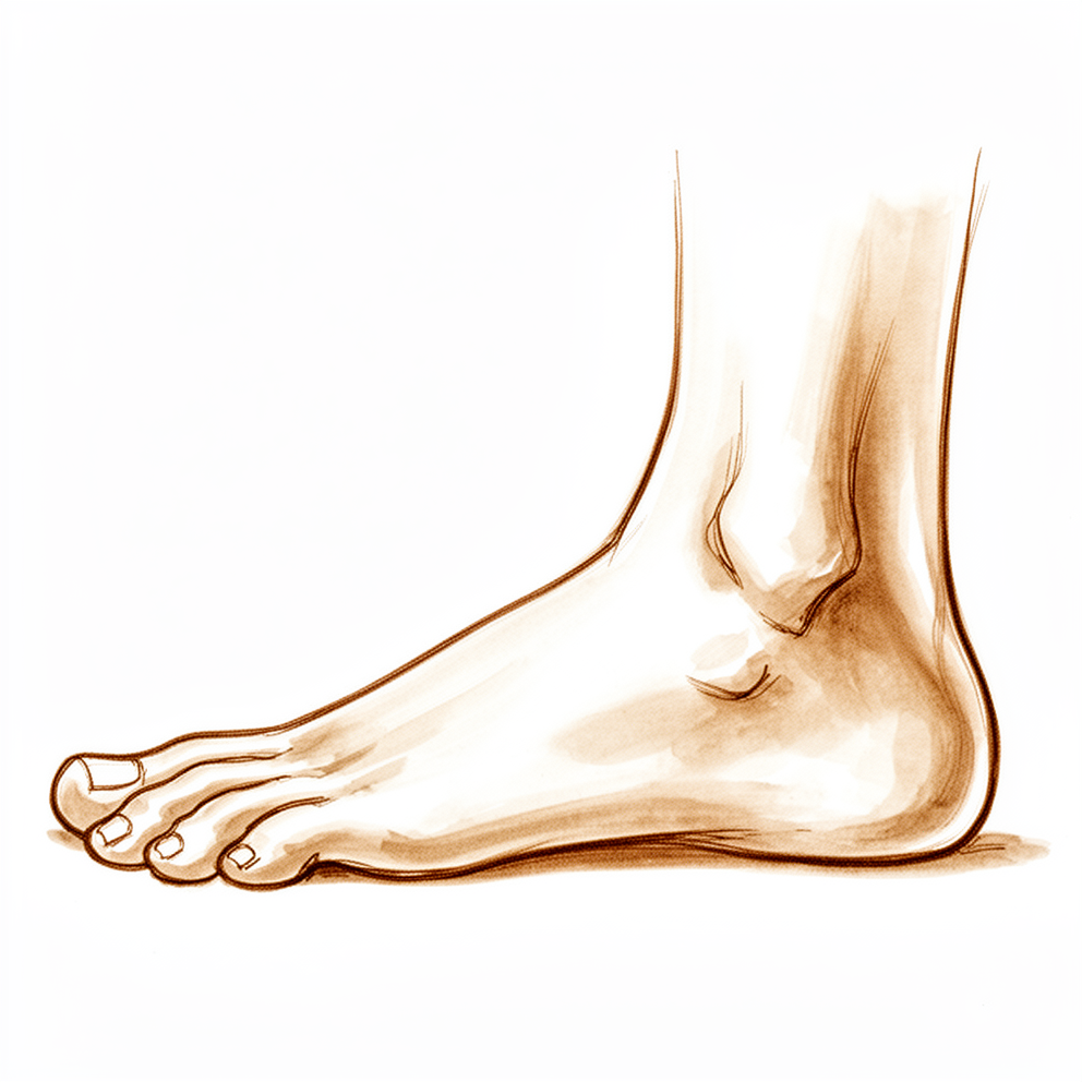
Overview
The foot and ankle serve the primary role of locomotion, defined as the orderly progression of the body through space while expending minimal energy [15]. During gait, muscle forces actively counteract gravity, with the foot transitioning from a flexible state at heel strike for impact absorption to a rigid lever at toe-off to propel the body forward [15]. Stance phase constitutes approximately 62% of the gait cycle, while swing phase accounts for 38% [15]. The magnitude of forces on the foot increases significantly with gait speed; initial ground contact force is approximately 80% of body weight when walking and 160% when jogging, with peak forces reaching 110% and 240% of body weight, respectively [15]. The intrinsic muscles act as a functional unit, stabilizing the foot during propulsion by paralleling progressive supination [14].
Biomechanical assessment distinguishes foot positions from motions, with varus and valgus defined for the hindfoot, abduction and adduction for the midfoot, and equinus and calcaneus for the ankle [8, 9]. Critical assessment determines the relationship of the forefoot to the hindfoot; a neutral hindfoot aligns the calcaneus with the tibial long axis and covers the talar head with the navicular [15]. Forefoot varus or valgus is an anatomic deformity observed when the hindfoot is neutral, where varus involves greater lateral plantar flexion and valgus involves greater medial plantar flexion [15]. Supination combines ankle plantar flexion, subtalar inversion, and transverse tarsal adduction, while pronation combines dorsiflexion, eversion, and abduction [15]. The majority of dorsiflexion and plantar flexion occurs at the ankle, inversion and eversion at the subtalar joint, and adduction and abduction at the transverse tarsal joint [15].
The foot comprises distinct bony columns and soft-tissue structures essential for stability. The forefoot includes structures distal to the tarsometatarsal joints, with the first metatarsal bearing 50% of weight during gait [8, 9]. The midfoot spans the Chopart and Lisfranc joint lines and consists of medial, middle, and lateral columns [59, 63]. The Lisfranc ligament connects the medial cuneiform to the second metatarsal base, while the spring ligament complex supports the talonavicular joint [13]. The plantar plate is the primary stabilizer of the lesser metatarsophalangeal joints, and the tibialis posterior tendon functions as a dynamic midfoot stabilizer during midstance and toe-off [8, 9, 59]. Pathologies such as cavus foot involve abnormally high arches frequently accompanied by hindfoot varus, often resulting from intrinsic weakness or neurologic disorders like Charcot-Marie-Tooth disease [22, 29].
Osseous Anatomy
Ankle
The ankle joint functions as a ginglymus (hinge) joint [30]. The distal tibia flares to form an inferior quadrilateral surface for articulation with the talus and a pyramid-shaped medial malleolus [24]. Laterally, the distal tibia forms a fibular notch that articulates with the fibula [24]. The lateral malleolus is a distal fibula expansion that extends beyond the distal tip of the medial malleolus and serves as a lateral buttress of the ankle joint [24]. The talar dome is biconcave with a central talar sulcus [30]. The radius of curvature of the talar dome is greater laterally [30].
Biomechanically, the ankle exhibits specific kinematic changes during motion. As the ankle moves from plantarflexion to dorsiflexion, the wider part of the talus enters the mortise, causing the distal fibula to externally rotate and translate proximally [24]. During plantarflexion, the talus becomes more narrow [24].
Foot
The foot is divided into the hindfoot (talus and calcaneus), midfoot (navicular, cuboid, and three cuneiforms), and forefoot (metatarsals and phalanges) [24]. The tarsals include the talus, calcaneus, cuboid, navicular, and three cuneiforms [24]. Throughout the growing period, the foot is relatively closer to its adult size than stature or long bones [46].
The talus has no muscular attachments [24]. Two-thirds of the talus is covered with cartilage [24]. A groove exists posteriorly on the talus for the tendon of the flexor hallucis longus (FHL) [24]. The os trigonum, if present, is located lateral to the FHL tendon [24]. The posterior process of the talus serves as an attachment site for the posterior talofibular ligament (PTFL) [24].
The talar body is wider anteriorly, conferring greater stability with the ankle in dorsiflexion [24]. The talar neck connects with the head, which articulates with the navicular distally and the calcaneus inferiorly [24]. The talar body and talar neck are defined by their relationship to the lateral process of the talus [24]. The region posterior to the lateral process of the talus is considered the talar body [24]. The region anterior to the lateral process of the talus is considered the talar neck [24].
Vascular supply to the talus is derived from multiple sources. The primary blood supply to the talar body is from the artery of the tarsal canal, which is a branch of the posterior tibial artery [24]. Additional blood supply to the talus comes from the superior neck vessels (anterior tibial artery) and the artery of the tarsal sinus (dorsalis pedis) [24].
Ligaments and Joint Capsule
Ankle and Subtalar Ligaments
The posterior talofibular ligament (PTFL) attaches to the posterior process of the talus [24]. The talar body is wider anteriorly, which confers greater stability when the ankle is in dorsiflexion [24]. Consequently, the foot is most stable in eversion and dorsiflexion and least stable in equinus position and inversion [20]. The subtalar joint plays a small but essential role in the motion occurring between the foot and the leg during the stance phase of normal walking [33]. Anterior subtalar arthroscopy offers a minimally invasive approach to address pathologies of this joint without requiring extensive resection of the ligamentous structures of the sinus tarsi [38].
Lisfranc Ligament and Tarsometatarsal Complex
The Lisfranc joint complex consists of a Roman archlike arrangement of osseous structures, including the three cuneiforms, the cuboid, and the five metatarsals, along with their respective joint capsules, a network of tarsometatarsal, intertarsal, and intermetatarsal ligaments, and secondary soft-tissue stabilizers [78]. Articular capsules separate this complex into three compartments: the medial (C1-M1), the central (C2, C3, M2, M3), and the lateral (cuboid, M4, M5) [78]. The ligamentous stabilizers comprise the tarsometatarsal, intermetatarsal, and anterior intertarsal ligaments, which consist of dorsal, interosseous, and plantar components [78].
The C1-M2 Lisfranc ligament is the most important and the most prone to injury [78]. Its components exhibit distinct anatomical features: * Dorsal component: The thinnest component, best seen on coronal and sagittal planes as a single hypointense stripe [78]. * Interosseous component: The strongest component, originating from the lateral surface of C1 and extending in an inferolaterodistal direction to insert at the medial-lower aspect of M2 [78]. * Plantar component: Arises from the inferolateral surface of C1 and bifurcates into a deep and superficial band to insert at the bases of M2 and M3, respectively [78].
The second tarsometatarsal (TMT) joint has the least motion, while the fourth and fifth have the most [13]. Injuries to the tarsometatarsal joints are usually a combination of fracture-dislocation, with dislocation without a fracture being rare [78]. Weight-bearing radiographs for Lisfranc injuries usually show lateral displacement of the lesser metatarsals, often with dorsal displacement [78]. The first metatarsal may dislocate in the same lateral direction (convergent or ipsilateral dislocation) or in the opposite medial direction (divergent) [78]. Nearly 25% of Lisfranc injuries are missed or occult on initial radiographs [78]. MRI is the most sensitive imaging modality for diagnosing Lisfranc injuries, delineating the presence of both osseous and soft-tissue involvement [78]. Following injury, the Lisfranc ligament may appear edematous and wavy in contour, with possible frank disruption of ligament fibers associated with avulsion fractures and osseous malalignment [78].
First Metatarsophalangeal Joint (MTPJ) Capsuloligamentous Complex
The first metatarsophalangeal joint is made up of the articular surface of the metatarsal head and the base of the proximal phalanx, along with the two sesamoid bones plantarly [23]. The static stability of this joint is provided by the collateral ligaments and the strong plantar plate, which consists of the plantar aponeurosis and the joint capsule [23]. Medially and laterally, the collateral ligaments stabilize the metatarsophalangeal joint and blend with the adductor and abductor hallucis tendons along the lateral and medial sides of the joint, respectively [23].
The sesamoids are connected by an intersesamoidal ligament, separated by a bony crista, and lie within the dual tendons of the flexor hallucis brevis [23]. They are stabilized by the firm attachment of the encapsulating plantar aponeurosis, which inserts into the base of the proximal phalanx [23]. Dorsally, the extensor hallucis longus tendon is stabilized by a medial and lateral hood mechanism similar to that present in the hand [23]. High-resolution 3T MRI allows accurate demonstration of the different anatomical details of the capsuloligamentous complex of the first MTPJ [93]. A correctly performed chevron osteotomy, either with or without a lateral capsular release, should not disrupt the vascular supply to the first metatarsal head [91].
Lesser Metatarsophalangeal Joint (MTPJ) Passive Restraints
The passive restraints of the lesser toes include the plantar plate, extensor hood, joint capsule, and collateral ligaments [8]. The plantar plate is the most important stabilizer of the lesser MTP joint and is disrupted in a hammer toe and crossover toe [8]. The extensor hood is the primary distal insertion point of the long extensors, which function to extend the MTP joint but not the PIP joint [8]. Intrinsic tendons pass plantar to the MTP joint axis proximally (providing a flexion force) and pass dorsal to the axis distally (providing an extension force) [8]. Plantar migration of the metatarsal head after a Weil (oblique shortening) osteotomy leads to a relatively dorsal position of the intrinsic tendons, causing them to lie dorsal to the axis of rotation and leading to a “floating” toe [8].
Muscles and Tendons
Intrinsic Muscles and Biomechanics
Intrinsic tendons pass plantar to the metatarsophalangeal (MTP) joint axis proximally, providing a flexion force, and pass dorsal to the axis distally, providing an extension force [8]. Plantar migration of the metatarsal head after a Weil osteotomy leads to a relatively dorsal position of the intrinsic tendons, causing them to lie dorsal to the axis of rotation and leading to a “floating” toe [8]. Weak intrinsic muscles become overpowered by stronger extrinsic muscles, leading to the development of hammer toes, claw toes, and distal migration of the fat pad [12].
The plantar aspect of the foot has historically been divided into four major compartments: medial/tibial, lateral/peroneal, central, and interosseous [35]. The medial foot compartment is confined by the plantar aponeurosis and the medial intermuscular septum and is located plantar and medial to the first metatarsal [35]. The lateral foot compartment is formed by the insertion of the lateral segment of the plantar aponeurosis and the lateral intermuscular septum and is located on the inferolateral aspect of the fifth metatarsal [35]. The interosseous plantar compartment is defined plantarly by the interosseous fascia, which is characterized as the upper limb of a horizontally oriented Y-shaped septum extending from the first to the fifth metatarsal [35]. The superficial central compartment has been recognized as independent and does not communicate with the surrounding compartments [35]. The intermediate central compartment is located dorsal to the superficial subunit and communicates proximally with the inferior calcaneal tunnel and indirectly via the tibiotalocalcaneal tunnel with the posterior compartment of the distal leg [35]. The medial compartment has no direct communication with the calcaneal tunnel or the central compartment [35]. The lateral compartment does not communicate with the calcaneal tunnel or the central compartment [35].
The medial compartment contains the M. flexor hallucis brevis, M. flexor hallucis longus tendons, and M. abductor digiti minimi [35]. The lateral compartment contains the M. flexor digitorum minimi brevis and M. opponens digiti minimi [35]. The superficial central compartment contains the M. flexor digitorum brevis, Mm. lumbricales, and M. flexor digitorum longus tendons [35]. The deep central compartment contains the M. adductor hallucis (oblique head), M. quadratus plantae, and the proximal segment of M. flexor digitorum longus/lumbricales [35]. The dorsal compartment contains the M. extensor digitorum brevis, M. extensor hallucis brevis, M. extensor digitorum longus tendons, and M. abductor hallucis [35].
The medial compartment is an isolated closed space with a landmark at the medial malleolus, a distance of approximately 6 cm, and a depth of approximately 11 mm [35]. The lateral compartment is an isolated closed space with a landmark at the lateral malleolus, a distance of approximately 11 cm, and a depth of approximately 11 mm [35]. The superficial central compartment is an isolated closed space with a landmark at the plantar fat pad insertion, a distance of approximately 11.5 cm, and a depth of approximately 10 mm [35]. The deep central compartment is a relatively isolated closed space with a landmark at the plantar fat pad insertion, a distance of approximately 11.5 cm, and a depth of approximately 21 mm [35]. The intermediate central compartment communicates with the posterior compartment of the distal leg via the tibio-talo-calcaneal tunnel, with a landmark at the medial malleolus, a distance of approximately 6 cm, and a depth of approximately 24 mm [35]. The dorsal compartment is not osseofascial and communicates with the anterior compartment of the distal leg [35]. The dorsal compartment has a depth of 0.8 mm dorsal and 10 mm plantar [35].
The medial compartment structures at risk include the superficial medial plantar artery and nerve [35]. The lateral compartment structures at risk include the superficial lateral plantar artery and nerve [35]. The superficial central compartment structures at risk include the common digital plantar arteries and nerves and the medial plantar nerve [35]. The deep central compartment structures at risk include the lateral plantar artery, medial and lateral plantar nerves, and the abductor digiti minimi nerve [35]. The dorsal compartment structures at risk include the cutaneous dorsalis lateralis, intermedius, and medius nerves and the dorsalis pedis, dorsales metatarsals, and plantaris profundus arteries [35]. Normative compartment pressures of the medial foot compartment are comparable with previously measured pressures of the leg [28]. The clinical relevance of the additional compartments beyond the 9 experimentally distinguished could not be shown [35].
Extrinsic Muscles and Tendons
The extensor digitorum longus (EDL) and extensor hallucis longus (EHL) tendons are both innervated by the deep peroneal nerve (L5) [74]. The EDL and EHL tendons travel underneath the superior and inferior extensor retinaculum before inserting onto the base of the distal phalanx of the respective toes [74]. The superficial location of the EDL and EHL tendons predisposes them to lacerations and closed rupture [74]. Closed rupture of EDL and EHL tendons is extremely rare [74]. Rupture of EDL and EHL tendons is either attritional or a result of high-energy eccentric contraction [74]. Attritional ruptures of EDL and EHL tendons have been reported in middle-aged patients, patients with repetitive microtrauma, and patients who have received previous steroid injections [74]. Physical examination for EDL and EHL tendon injury reveals an inability to extend the interphalangeal (IP) joints of the toes actively [74].
Acute EHL lacerations that are proximal to the extensor hood should undergo an end-to-end repair [74]. Partial lacerations or lacerations at or distal to the extensor hood may be treated closed with immobilization of the hallux in extension [74]. Chronic EHL injuries or acute attritional ruptures can be treated with débridement and repair, free tendon grafting, or tenodesis to a healthy extensor tendon [74]. Treatment of EDL injuries is controversial [74]. Repair of EDL injuries is more commonly done in younger, active individuals [74]. Authors who favor repair of EDL injuries cite preventing future formation of a claw toe deformity as the rationale for surgery [74].
The tibialis posterior tendon acts primarily as an invertor of the hindfoot and supinator of the forefoot during the stance phase of gait [74]. Activation of the tibialis posterior tendon during the toe-off phase of gait locks the transverse tarsal joints, creating a rigid lever arm for push off [74]. Tibialis posterior tendon dysfunction is the most common cause of an adult acquired flatfoot deformity [74]. Collapse of the medial longitudinal arch, hindfoot valgus, and forefoot abduction is the classic triad of foot deformity associated with tibialis posterior tendon insufficiency [74]. The “too many toes” sign and the inability to perform a single-limb heel rise are additional classic findings of tibialis posterior tendon insufficiency [74]. Nonsurgical treatment for stage II tibialis posterior tendon dysfunction consists of a UCBL-type orthosis [74]. Nonsurgical treatment for stage III or IV tibialis posterior tendon dysfunction consists of an AFO or Marzano or Arizona brace [74]. Surgical treatment of stage II tibialis posterior tendon dysfunction consists most commonly of FDL transfer in conjunction with a bony procedure, most commonly a medial calcaneal displacement osteotomy or a lateral column lengthening [74]. Surgical treatment of stage III tibialis posterior tendon dysfunction is a hindfoot arthrodesis, most commonly a triple arthrodesis [74].
Complete ruptures of the peroneal tendons are rare [74]. Peroneal tendon tears are often longitudinal in the tendon and typically seen in chronic situations [74]. Most peroneal tendon tears are in the peroneus brevis (PB) tendon at the level of the fibular groove [74]. Peroneal tendon tears are often caused by inversion injuries and chronic compression from the peroneus longus (PL) [74]. Patients with chronic peroneal tendon injuries are treated with tenodesis to the healthy tendon or transfer of the FHL when both tendons are involved [74]. Dislocation or subluxation of the peroneal tendons occurs during an inversion injury to a dorsiflexed ankle with rapid reflexive contraction of the PL and PB tendons [74]. Dislocation or subluxation of the peroneal tendons results from a disruption of the superior peroneal retinaculum (SPR) or fibrocartilage ridge [74].
Closed tibialis anterior tendon ruptures are either the result of strong eccentric contraction in younger individuals or attritional ruptures in older patients with musculoskeletal compromise [74]. The flexor hallucis longus (FHL) runs through its fibro-osseous tunnel, lateral to the posteromedial tubercle of the talus, under the sustentaculum tali and through the knot of Henry, before inserting onto the base of the proximal phalanx of the great toe [74]. Stenosing tenosynovitis of the FHL commonly occurs in the fibro-osseous tunnel posterior to the talus [74]. Stenosing tenosynovitis of the FHL is most common in dancers and gymnasts [74]. Stenosing tenosynovitis of the FHL may coexist with posterior ankle impingement and the presence of an os trigonum [74].
The muscles that produce plantar flexion are the gastrocnemius-soleus, flexor hallucis longus, flexor digitorum longus, peroneus longus, peroneus brevis, and posterior tibial [20]. The dorsiflexor muscles are the anterior tibial, extensor hallucis longus, extensor digitorum communis, and peroneus tertius [20]. The muscles that produce inversion are the posterior tibial, flexor hallucis longus, and anterior tibial [20]. The evertors of the foot are the peroneus brevis, peroneus tertius, extensor digitorum communis, and extensor hallucis longus [20]. The muscles that plantar flex the ankle and foot provide the force for forward propulsion of the body during locomotion [20]. The dorsiflexor muscle group clears the foot during the swing phase of gait [20]. Approximately two-thirds of the total musculature of the leg is constituted by the triceps surae [20]. The triceps surae acts on the foot as a first-class lever with the ankle joint as a fulcrum [20]. The working capacity of the triceps surae is 6.5 kg/m, whereas that of the dorsiflexors of the ankle joint is only 1.4 kg/m, or a relative ratio of 4:1 [20]. The strength of the calf muscles is a necessary antigravitational force against the elevated center of gravity of the body in the upright posture [20]. The center of gravity of the human body falls anterior to the ankle joint, requiring the triceps surae to counteract a strong rotatory component in ankle dorsiflexion [20]. The muscles that provide lateral stability to the foot in plantar flexion are the posterior tibial and peroneals [20]. In dorsiflexion, lateral stability is provided by the action of the anterior tibial and extensor digitorum communis [20].
The peroneus brevis attaches on the dorsal aspect of the tubercle of the fifth metatarsal [77]. The peroneus tertius attaches on the dorsal aspect at the proximal metaphyseal–diaphyseal junction of the fifth metatarsal [77]. The peroneus tertius acts as a balancing force during forefoot dorsiflexion counteracting the natural inversion tendency of the tibialis anterior [77]. The peroneus brevis serves as more of an antagonist to posterior tibialis function to maintain the position of the foot under the talus [77]. The abductor digiti quinti is a muscle inserting on the base of the fifth metatarsal [77]. There is a strong attachment of the plantar fascia to the plantar aspect of the tubercle of the fifth metatarsal [77]. The os peroneum is located within the tendon of the peroneus longus and can be found on the lateral border of the cuboid [77]. The os vesalianum is found just proximal to the base of the fifth metatarsal medial to the insertion of the peroneus brevis [77]. The sural nerve, the insertion of the peroneus brevis, and the insertion of the peroneus tertius are all within the surgical margins for access to the proximal tuberosity of the fifth metatarsal [77]. The lateral anchor for the extensor retinaculum is within the surgical margins for access to the proximal tuberosity of the fifth metatarsal [77]. The distance from the peroneal tendons sheath to the sural nerve at the posterior tip of the fibula decreases from proximal to distal [39].
A vascular watershed region in the Achilles tendon is found 2 to 6 cm above the calcaneal insertion [74]. This vascular watershed region is the typical location of a majority of Achilles tendon pathology, particularly acute ruptures [74]. Acute Achilles tendinitis is typically associated with younger, more active patients [74]. Patients with chronic Achilles tendinosis are typically older and more sedentary [74]. Skin problems, such as infection, necrosis, and adhesions, are the most common complications associated with the surgical treatment of Achilles tendon disorders [74]. Functional rehabilitation is a reasonable choice for the management of acute noninsertional Achilles ruptures, not just among the sedentary or elderly, but even among young, healthy, active individuals [74].
Ultrasound-guided partial plantar fasciotomy with a needle is safe because structures are under direct visualization of the surgeon and the risk of damage is minimal [11]. The plantar fascia and the auxiliary plantar flexors are important for maintaining normal strains in the metatarsals during gait [41]. A wedge under the lateral aspect of the forefoot decreases strain in the plantar aponeurosis [103]. A wedge under the medial aspect of the forefoot increases strain in the plantar aponeurosis [103]. Because of the close proximity of the medial plantar nerve to the tendon sheath, there is a significant risk of iatrogenic nerve injury when surgical procedures are performed in zone 2B of the flexor hallucis longus tendon sheath [17]. The ATFL is under increased strain in plantar flexion, inversion, and internal rotation [13]. The CFL is under increased strain in dorsiflexion and inversion [13]. The second TMT joint has the least motion, while the fourth and fifth have the most [13].
Neurovascular Anatomy
Nerve Anatomy and Surgical Safety
Surgical planning in the foot requires precise knowledge of neurovascular relationships to minimize iatrogenic injury. In zone 2B, the medial plantar nerve lies in close proximity to the flexor hallucis longus tendon sheath, creating a significant risk of nerve damage during procedures in that region [17]. To mitigate this risk, a 'high and inside' approach that remains superior to the superior border of the peroneus brevis tendon is anatomically safe and may decrease the chance of intraoperative nerve injury and postoperative irritation [26]. For arthroscopic debridement of the first metatarsophalangeal joint, an additional medial portal has been found to be safe from surrounding neurovascular structures [98]. During ankle surgery, the anatomic characteristics of the posteromedial neurovascular bundle must be considered to reduce the risk of injury to the medial neurovascular bundle [6]. Ultrasound-guided partial plantar fasciotomy with a needle is safe because structures are under direct visualization, resulting in minimal risk of damage [11].
Vascular Anatomy and Surgical Dissection
The dorsalis pedis artery continues distally as the first dorsal metatarsal artery [49]. The deep peroneal nerve is located lateral to the dorsalis pedis artery [49]. Tributaries to the greater saphenous system can be located on the medial side of the first metatarsal [49]. Factors impairing the restoration of microvascular blood supply to the talus may lead to osteonecrosis despite preserved macrovascular blood flow and a positive Hawkins sign [99].
Diabetic Neuropathy and Neurovascular Pathology
At the time of diagnosis, 10% of patients with diabetes mellitus have some form of sensory, motor, or autonomic dysfunction [31]. Neuropathy develops in 50% of diabetic patients within 25 years of diagnosis [31]. Sensory neuropathy is the most prevalent nerve dysfunction in diabetes, affecting as many as 70% of patients [31]. The critical triad of sensory neuropathy, trauma, and foot deformity was present in 63% of patients with lower extremity ulcers [31]. Pain is associated with 25% to 33% of diabetic neuropathies [31]. Sensory disturbances in diabetic neuropathy show a length-related pattern with stocking and glove distribution due to a “dying-back” distal axonopathy [31]. Protective sensation in diabetic neuropathy is indicated by the ability to perceive a 5.07 (10 g) Semmes-Weinstein monofilament applied perpendicular to the skin [31].
Motor neuropathy in the foot is evidenced by the development of claw toes from intrinsic muscle weakness and equinus contracture of the Achilles tendon [31]. Claw toes occur due to dysfunction of intrinsic muscles causing hyperextension of the metatarsophalangeal joints and flexion of the proximal and distal interphalangeal joints [31]. Autonomic neuropathy occurs when the autonomic system cannot control blood vessel tone and sweat glands in the foot [31]. Sweat gland dysfunction in autonomic neuropathy allows the skin to dry out and crack, allowing the ingress of microbes [31]. Standing foot pressure can be as high as 400 kPa, necessitating fine regulation of blood vessels to ensure adequate tissue oxygenation [31].
Histologic examination of plantar skin in diabetic patients showed significantly thicker elastic septae and dermal layers compared to nondiabetic patients [12]. Ultrasound examinations of Achilles tendons showed disorganized tendon fibers and calcification in 75% of diabetic patients without foot problems [12]. Dynamic pressures in the first metatarsocuneiform, medial and middle naviculocuneiform, and first intercuneiform joints were 46% higher in diabetic cadaver feet compared to nondiabetic feet during simulated walking [12]. As many as 65% of people with type 1 or 2 diabetes have evidence of peripheral neuropathy [12]. The presence of peripheral neuropathy is the most predictive factor for the development of diabetic foot ulcers [12].
Diabetic patients have glycation of arterial vessel walls, thickening of basement membranes, and reduced endothelial nitric oxide activity [12]. Examination of nerves in diabetic patients has shown multifocal ischemic proximal nerve lesions and epineural vessel atherosclerosis [12]. The absence of skin oils released by autonomic signals leads to increased susceptibility to fissuring of the skin from mild trauma [12]. Muscle contractures of the gastrosoleus complex increase the force load transmitted to the forefoot [12]. Diabetic individuals are four times more likely to suffer a stroke than nondiabetic individuals [12]. Diabetic individuals are twice as likely to develop peripheral arterial disease than nondiabetic individuals [12]. Diabetic patients with peripheral arterial disease are nine times more likely to develop a foot ulcer [12]. Diabetes impairs the immune system through alterations in the chemotaxis abilities of polymorphonuclear cells and cell wall abnormalities, making patients susceptible to secondary infections [12]. Subjects with concurrent neuropathy and claw toe deformity were associated with the smallest intrinsic foot muscle volumes and the thickest plantar aponeuroses [104].
Biomechanics and Function
Gait Cycle Mechanics
A full gait cycle, defined as one stride from heel strike to heel strike, comprises a stance phase (62% of the cycle) and a swing phase (38% of the cycle) [66]. During the swing phase, the anterior tibialis contracts concentrically [66]. Loss of anterior tibialis function results in footdrop and a steppage gait [66]. At heel strike, the anterior tibialis contracts eccentrically to control the rate of foot contact with the ground [66]. In patients with footdrop, rapid foot strike produces a loud “slap” during heel strike [66]. The hindfoot is locked and inverted at initial strike, then passively everts during the transition from heel strike to foot flat to facilitate energy absorption [66]. Failure of hindfoot eversion in cavovarus deformity increases forces to the lateral foot, resulting in fifth metatarsal stress fractures, callus formation, and ankle instability [66].
During the foot flat phase, the gastrocnemius-soleus complex contracts eccentrically to control forward body progression over the foot [66]. Loss of gastrocnemius-soleus function results in a calcaneus gait with heel pain [66]. The hindfoot is unlocked and everted during foot flat for ground accommodation [66]. As the foot progresses from heel strike to toe-off, it converts from a flexible shock absorber to a rigid propellant [66]. The plantar fascia tightens as metatarsophalangeal (MTP) joints extend, accentuating the longitudinal arch via the windlass mechanism [66]. At terminal stance, the flexor digitorum longus tendon is most active [66]. During toe-off, the gastrocnemius-soleus complex contracts concentrically [66]. The hindfoot supinates with firing of the posterior tibial tendon (PTT), and the transverse tarsal joint locks to provide a rigid lever arm [66]. Creation of this rigid lever arm is limited if the PTT is dysfunctional [66].
An equinus deformity from spasticity or contracture causes the toe to make initial contact with the ground rather than the heel [15]. Spasticity or contracture of the Achilles tendon delays the foot being flat on the ground at 7% of the gait cycle [15]. Heel rise of the standing foot begins at 34% of the gait cycle as the swinging leg passes the standing limb [15]. With spasticity, heel rise may occur earlier than normal [15]. With gastrocsoleus weakness, heel rise will be later than normal [15]. Heel strike of the opposite foot occurs at 50% of the cycle, ending single-limb support [15]. Toe-off of the opposite foot occurs at 62% of the cycle, at the beginning of the swing phase [15].
Joint Kinematics and Motion
The combined mobility of the foot and ankle equals that of a universal joint [20]. In inversion of the hindfoot, the os calcis is displaced forward, producing adduction and inversion of the forefoot [20]. When the hindfoot is everted, the os calcis moves backward, and the forefoot is abducted and everted [20]. When the ankle joint is plantar flexed, the hindfoot inverts [20]. In dorsiflexion of the ankle, the hindfoot everts [20]. The foot is most stable in eversion and dorsiflexion [20]. The foot is least stable in equinus position and inversion [20]. In plantarflexion, the talus becomes more narrow [24]. Three-dimensional kinematics under static full weightbearing were opposite between the ankle and subtalar joints on their respective axes [71]. Dynamic fluoroscopic assessment is a valuable tool for characterising the kinematics of the joints of the medial foot column during gait [4]. The first metatarsophalangeal joint has quantifiable parameters that define patterns of motion in normal feet and those with hallux valgus or hallux rigidus [5]. During weight-loading, the first metatarsal-cuneiform joint turns dorsiflexed, supinated, and internally rotated [57]. Hallux valgus is a dynamic condition, and the deformity could be more correlated with motions during weightbearing than with plain static measurements [81].
Forefoot and Midfoot Biomechanics
The first metatarsal is the widest and shortest and bears 50% of the weight during gait [8]. The second metatarsal is usually the longest and experiences more stress than the other lesser metatarsals [8]. The first metatarsophalangeal joint functions mainly as a weight-bearing structure and stabilizer of the medial aspect of the longitudinal arch [23]. Static stability of the first metatarsophalangeal joint is provided by the collateral ligaments and the strong plantar plate [23]. Dynamic stability is provided by the abductor hallucis and adductor hallucis muscles [23]. No muscle inserts into the first metatarsal head per se, suspending it in a sling of muscles and tendons [23]. If the windlass mechanism for the hallux is lost, pressure remains beneath the metatarsal heads rather than being transferred to the toes [23]. Metatarsalgia results from the transfer of load to the lesser metatarsal heads when the weight-bearing ability of the first metatarsal is disrupted [23]. A Keller arthroplasty disrupts the insertion of the plantar aponeurosis into the hallux, potentially causing a transfer lesion [23]. Prosthetic replacement of the first metatarsal joint results in loss of the windlass mechanism [23].
The midfoot functions in adduction and abduction [61]. The calcaneocuboid and talonavicular joints are collectively referred to as the midtarsal, transverse tarsal, or Chopart joint [61]. During foot flat (hindfoot valgus, forefoot abduction, and dorsiflexion of ankle), the transverse tarsal joints are parallel and supple, adapting to uneven ground [61]. During toe-off (hindfoot varus, forefoot adduction, and plantar flexion of ankle), the transverse tarsal joints become divergent and lock, providing stiffness for forward propulsion [61]. Failure of the posterior tibial tendon to invert the hindfoot and lock the transverse tarsal joints is the biomechanical etiology for the lack of a heel rise in patients with PTT dysfunction [61]. The primary stabilizer of the longitudinal arch is the interosseous ligaments, not the plantar fascia [61]. The plantar fascia is a secondary stabilizer of the longitudinal arch [61]. The second metatarsal extends more proximally than surrounding metatarsals, creating a “keystone” effect that imparts inherent bony stability to the Lisfranc joint complex [61]. The Lisfranc ligament travels from the medial cuneiform to the base of the second metatarsal [61]. The interosseous part of the Lisfranc ligament is the strongest of its three parts [61].
The midfoot columns are defined as follows: Medial column: Includes the first metatarsal, the medial cuneiform, and the navicular [61]. Middle column: Includes the second and third metatarsals, the middle cuneiform, and the lateral cuneiform [61]. Rigidity of the middle column allows for a rigid lever arm during push-off [61]. Lateral column: Includes the fourth and fifth metatarsals and the cuboid [61]. Sagittal mobility of the lateral column imparts flexibility necessary for walking on uneven ground [61].
Sagittal mobility varies by column: First TMT joint: 3.6 degrees of mobility [61]. Second TMT joint: 0.6 degrees of mobility [61]. Third TMT joint: 1.6 degrees of mobility [61]. Lateral column: Most sagittal mobility (~10 degrees) [61]. Middle column: Least sagittal mobility [61].
Plantar migration of the metatarsal head after a Weil (oblique shortening) osteotomy leads to a relatively dorsal position of the intrinsic tendons [8]. The plantar plate is the most important stabilizer of the lesser MTP joint [8]. The plantar plate is disrupted in a hammer toe and crossover toe [8]. Whenever a higher level of first metatarsal shortening is necessary, pushing down the distal metatarsal segment could be a compensatory procedure to maintain normal plantar force distributions [2]. Axial loading of the foot, external rotation, and pronation/supination are the most common conditions during injurious loading in tarsometatarsal injuries [36]. Sport-related movements load the plantar surface of the foot more than running straight [50]. High medial longitudinal arch may contribute to increased load on the lateral side of the foot [83]. Individuals with patellofemoral pain display a more medially oriented loading pattern of the forefoot compared to individuals without knee pain [85].
Muscle Function and Stability
The working capacity of the triceps surae is 6.5 kg/m [20]. The working capacity of the dorsiflexors of the ankle joint is 1.4 kg/m [20]. The relative ratio of working capacity between plantar flexors and dorsiflexors is 4:1 [20]. The tibialis posterior tendon acts as an invertor of the hindfoot and adducts and supinates the forefoot during the stance phase of gait [54]. The tibialis posterior tendon acts as a secondary plantar flexor of the ankle [54]. Activation of the tibialis posterior tendon allows locking of the transverse tarsal joints, creating a rigid lever arm for the toe-off phase of gait [54]. The tibialis posterior tendon contracts eccentrically during the stance phase to diminish forces on the supporting ligaments of the medial arch (spring ligament) [54]. The major antagonist to the tibialis posterior tendon is the peroneus brevis [54]. The normal excursion of the tibialis posterior tendon is relatively small (2 cm) [54]. The patient loses the ability to perform a single-limb heel rise due to inability to lock the transverse tarsal joints to create a rigid lever arm [54]. The patient loses the ability to perform a single-limb heel rise due to valgus displacement of the calcaneus that results in a weakened Achilles tendon moment arm [54].
Deformity Biomechanics
Forefoot varus or valgus is an anatomic deformity observed when the hindfoot is placed in neutral position [15]. With a flexible forefoot varus deformity, the foot lies flat on the floor during stance [15]. With a fixed forefoot varus deformity, excessive weight is borne on the lateral side of the foot [15]. In forefoot varus, as weight passes onto the forefoot, the calcaneus goes into valgus position [15]. Severe forefoot varus may result in lateral impingement of the calcaneus against the fibula [15]. In forefoot valgus deformity, the medial side of the foot has greater plantar flexion than the lateral side [15]. Forefoot valgus results in excessive weight bearing by the first metatarsal head [15]. To accommodate for forefoot valgus deformity, the calcaneus assumes a varus position [15]. Forefoot valgus may result in a feeling of instability at the ankle joint [15]. If the first ray is elevated, the forefoot is in varus position [8].
Common Sites of Injury
First Metatarsal and Forefoot
The first metatarsal is structurally stronger than the other metatarsals, accounting for approximately 1.5% of all metatarsal fractures [32]. Independent motion of the first metatarsal is permitted by the lack of interconnecting ligaments between the first and second metatarsals [32]. Biomechanically, the tibialis anterior inserts on the plantar medial aspect of the first metatarsal base to elevate it, while the peroneus longus attaches to the plantar lateral base to plantarflex the head [32]. During most activities, average peak pressures in the first metatarsal are the highest or among the highest levels recorded [32].
Fractures of the first metatarsal result from direct forces, which are more common in industrial settings, or indirect forces, which often occur in sports when the forefoot is fixed and the leg or foot is twisted [32]. Any fracture of the base of the first three metatarsals should raise suspicion of a midtarsal injury [32]. Specifically, small avulsion fractures involving the medial base of the first or second metatarsal suggest disruption of the tarsometatarsal (TMT) ligaments [32]. Vascular structures, including the arterial arch and dorsal and plantar metatarsal arteries, are particularly susceptible to injury in association with metatarsal fractures [32]. Compartment syndrome is relatively common with soft tissue trauma in the metatarsal area; consequently, compartment pressures should be monitored routinely, especially following direct trauma [32].
Stress fractures of the proximal fourth metatarsal can occur in young athletes with normal bone and without metatarsus adductus [90]. Furthermore, multiple, various-stage bone stress injuries of the ankle and foot may occur simultaneously in physically active young adults [60].
Midfoot and Tarsometatarsal Joints
Axial loading of the foot, external rotation, and pronation/supination are the most common conditions during injurious loading of the tarsometatarsal joints [36]. Severe deformities characterized by large amounts of midfoot pronation and hindfoot valgus may be better treated with nonanatomic reconstruction methods for the spring ligament [21].
Ankle and Hindfoot
The shape of the talus and calcaneus, along with the architecture within the calcaneus—especially the arrangement of the trabeculae—are essential factors for calcaneal fractures [64]. Osteochondral lesions of the talus are usually late sequelae of ankle trauma [89]. Given the functional significance of the talus and its limited capacity for repair, correct early diagnosis of osteochondral lesions is important [89]. Anatomic characteristics of the posteromedial neurovascular bundle should be kept in mind when ankle surgery is performed to reduce the risk of injury to the medial neurovascular bundle [6].
Diabetic Foot Pathology
Histologic examination of the plantar skin of diabetic patients showed significantly thicker elastic septae and dermal layers compared to nondiabetic tissue [12]. Ultrasound examinations of Achilles tendons have shown disorganized tendon fibers and calcification in 75% of diabetic patients without foot problems [12]. Peak midfoot joint pressures in diabetic cadaver feet during simulated walking showed 46% higher dynamic pressures in the first metatarsocuneiform, medial and middle naviculocuneiform, and the first intercuneiform joints compared to nondiabetic specimens [12].
Neuropathy is an independent predictor of ulcers [12]. The length of time that a patient has had diabetes and the level of metabolic control are the main predictors for the development, progression, and extent of neuropathy [12]. Joint contractures develop around the toes due to muscle imbalance from motor neuropathy [12]. Weak intrinsic muscles become overpowered by stronger extrinsic muscles, leading to the development of hammer toes, claw toes, and distal migration of the fat pad [12]. Diabetic individuals are four times more likely to suffer a stroke than nondiabetic ones [12]. Diabetic individuals are twice as likely to develop peripheral arterial disease than nondiabetic ones [12].
Surgical Anatomy
Neurovascular Structures
The posteromedial neurovascular bundle of the ankle possesses specific anatomic characteristics that must be considered during ankle surgery to reduce the risk of injury [6]. In the dorsomedial region, the area above the line connecting the upper 1/4 point at the base of the first metatarsal and the 1/2 point at the middle of the first metatarsal constitutes a dangerous zone for the dorsomedial cutaneous nerve [56]. A 'high and inside' approach that remains superior to the superior border of the peroneus brevis tendon is anatomically safe and may decrease the chance of intraoperative nerve injury and postoperative irritation [26]. During a dorsolateral incision placed over the fourth metatarsal extending proximally to the cuboid bone, the lateral cutaneous branch of the peroneal nerve should be identified and protected [79]. Conversely, the dorsalis pedis artery and the deep peroneal nerve must be avoided during a dorsolateral approach through the first–second intermetatarsal space [73]. In zone 2B, the medial plantar nerve is in close proximity to the flexor hallucis longus tendon sheath, creating a significant risk of iatrogenic nerve injury during surgical procedures in this area [17].
Ligaments and Joint Complexes
The Lisfranc joint complex has specific anatomical features that are reviewed to enhance understanding of injury patterns and support accurate diagnosis and management [7]. Anterior subtalar arthroscopy serves as a minimally invasive approach to pathologies of the subtalar joint that does not require extensive resection of the ligamentous structures of the sinus tarsi [38]. Severe deformities with large amounts of midfoot pronation and hindfoot valgus may be better treated with nonanatomic spring ligament reconstruction methods [21].
Muscles and Tendons
The surgical approach for fifth metatarsal fractures is along the lateral border of the fifth metatarsal just above the adductor minimi muscle [72]. For intramedullary screw fixation of proximal fifth metatarsal fractures, the incision is made proximal to the fifth metatarsal base between the peroneus brevis and longus tendons [72]. The insertion point for intramedullary fixation of the fifth metatarsal is 1 cm dorsal to the palpable inferior margin of the proximal tuberosity and just medial to the peroneus brevis insertion [72]. During a dorsomedial longitudinal incision in the web space between the first and second metatarsals, the tendons of the extensor hallucis longus and extensor digitorum brevis are identified and retracted laterally or medially [79].
Compartments
The foot contains 9 or 10 compartments, including the deep calcaneal compartment [79]. A three-incision approach is the most reliable way to decompress all 9 or 10 compartments of the foot [79]. A medial midfoot incision provides the quickest and most effective pressure reduction for all foot compartments, including the tarsal tunnel [79]. Neither a single nor a double dorsal fasciotomy achieved the short-term pressure reduction effects seen with a medial approach [79]. The dorsolateral incision for fasciotomy allows access to the dorsal compartment containing the extensor hallucis and digitorum brevis muscles, the lateral two interosseous compartments (intermetatarsal spaces 3/4 and 4/5), the lateral TMT joint complex, and the cuboid bone [79].
Bony Anatomy and Landmarks
The base of the second metatarsal is a landmark for the extramedullary tibial cutting guide in total knee arthroplasty, though it should be avoided in rheumatoid arthritis patients with foot involvement [87]. The lateral corner of the tarsal navicular turns downward to articulate with the cuboid and is difficult to visualize [76]. The articular surfaces of the talar head and the cuneiforms can be used as templates for anatomic articular reduction of the proximal and distal navicular articular surfaces [76]. In comminuted cuboid fractures, adjacent bone can be used as a template to restore joint congruity, with no tolerance for an articular step-off greater than 1 mm [37]. The thin lateral cortex of the cuboid is prone to collapse, making screw fixation alone a rare treatment option unless good bone stock is present [37]. The proximal tip of the tuberosity of the fifth metatarsal is identified during the approach for proximal fifth metatarsal fractures [72].
Key Evidence
- [L5] Whenever a higher level of shortening is necessary, pushing down the distal metatarsal segment could be a compensatory procedure to maintain normal plantar force distributions. [2] (10.1186/s12891-019-2973-6)
- [L4] Dynamic fluoroscopic assessment has been shown to be a valuable tool for characterisation of the kinematics of the joints of the medial foot column during gait. [4] (10.1186/1471-2474-13-14)
- [L5] This study describes methods for analysis of the kinematics of the first metatarsophalangeal joint and delineates quantifiable parameters that can be used to define patterns of motion for this articulation in normal feet as well as for those afflicted with hallux valgus or hallux rigidus. [5] (10.2106/00004623-198668030-00012)
- [L5] Anatomic characteristics should be kept in mind when ankle surgery is performed, thereby reducing the risk of injury to the medial neurovascular bundle. [6] (10.1016/j.arthro.2007.08.030)
- [L5] The manuscript provides a comprehensive anatomical review of the Lisfranc joint complex to enhance understanding of injury patterns and support clinicians in accurate diagnosis and effective management to achieve optimal clinical outcomes. [7] (10.1002/ksa.70260)
- [L4] Ultrasound-guided partial plantar fasciotomy with a needle is safe, since structures are under direct visualization of the surgeon and the risk of damage is minimal. [11] (10.1186/s13018-021-02302-y)
- [L4] The intrinsic muscles of the foot act as a functional unit that plays the principal active role in stabilizing the foot during propulsion by paralleling progressive supination. [14] (10.2106/00004623-196446030-00001)
- [L5] Because of the close proximity of the medial plantar nerve to the tendon sheath, there is a significant risk of iatrogenic nerve injury when surgical procedures are performed in zone 2B. [17] (10.1016/j.arthro.2009.11.007)
- [L5] Severe deformities with large amounts of midfoot pronation and hindfoot valgus may be better treated with nonanatomic reconstruction methods. [21] (10.1186/s13018-019-1154-5)
- [L5] A 'high and inside' approach that remains superior to the superior border of the peroneus brevis tendon is anatomically safe and may decrease the chance of intraoperative nerve injury and irritation postoperatively. [26] (10.1177/0363546512448320)
- [L4] Normative compartment pressures of the medial foot compartment are comparable with previously measured pressures of the leg. [28] (10.1177/03635465020300022001)
- [L5] The subtalar joint plays a small but essential role in the motion occurring between the foot and the leg during the stance phase of normal walking. [33] (10.2106/00004623-196446020-00010)
- [L4] Axial loading of the foot, external rotation, and pronation/supination are the most common conditions during injurious loading. [36] (10.1177/2325967114525347)
- [L5] Anterior subtalar arthroscopy is a minimally invasive approach to deal with pathologies of this joint without the need for extensive resection of the ligamentous structures of the sinus tarsi. [38] (10.1007/s00167-009-0917-7)
- [L5] This cadaveric study shows that the distance from the peroneal tendons sheath to the sural nerve decreases from proximal to distal. [39] (10.1007/s00167-019-05438-x)
- [L5] The plantar fascia and the auxiliary plantar flexors are important for maintaining normal strains in the metatarsals during gait. [41] (10.2106/00004623-199909000-00005)
- [L4] The foot is relatively closer to its adult size than stature or long bones throughout the growing period. [46] (10.2106/00004623-195638050-00004)
- [L4] Sport-related movements load the plantar surface of the foot more than running straight. [50] (10.1177/0363546507309315)
- [L5] The area above the line connecting the upper 1/4 point at the base of the first metatarsal and the 1/2 point at the middle of the first metatarsal is a dangerous zone for the DMCN. [56] (10.1186/s13018-023-04419-8)
- [L4] During weight-loading process, the first metatarsal-cuneiform joint turns dorsiflexed, supinated, and internally rotated. [57] (10.1186/s13018-015-0289-2)
- [L4] Multiple, various-stage bone stress injuries of the ankle and foot may occur simultaneously in physically active young adults. [60] (10.1177/0363546506295701)
- [L4] The shape of the talus and calcaneus and the architecture within the calcaneus, especially the arrangement of the trabeculae, are essential factors for calcaneal fractures. [64] (10.1186/s13018-022-02930-y)
- [L4] Three-dimensional kinematics under static full weightbearing were opposite between the ankle and subtalar joints on their respective axes. [71] (10.1186/s13018-019-1443-z)
- [L5] Hallux valgus is a dynamic condition, and the deformity could be more correlated with motions during weightbearing than with plain static measurements. [81] (10.1097/corr.0000000000002265)
- [L3] In addition, high medial longitudinal arch may contribute to increased load on the lateral side of the foot. [83] (10.1177/0363546519893365)
- [L3] Individuals with patellofemoral pain display a more medially oriented loading pattern of the forefoot compared to individuals without knee pain, which may be associated with the distribution of forces acting on the patellofemoral joint. [85] (10.1007/s00167-014-2943-3)
- [L2] Surgeons should aim toward the base of the second metatarsal but avoid using it in rheumatoid arthritis patients with foot involvement. [87] (10.1007/s00167-014-3254-4)
- [L4] Osteochondral lesions of the talus are usually late sequelae of ankle trauma; correct early diagnosis is important due to the functional significance of the talus and its limited capacity for repair. [89] (10.5435/00124635-201010000-00005)
- [L4] Stress fractures of the proximal fourth metatarsal can occur in young athletes with normal bone and without metatarsus adductus. [90] (10.1177/0363546506294467)
- [L5] A correctly performed chevron osteotomy, either with or without a lateral capsular release, should not disrupt the vascular supply to the first metatarsal head. [91] (10.2106/00004623-199502000-00005)
- [L4] High-resolution 3T MRI allows accurate demonstration of the different anatomical details of the capsuloligamentous complex of the first MTPJ from previous anatomical reports. [93] (10.1186/s13018-021-02795-7)
- [L5] The additional medial portal was found to be safe from the surrounding neurovascular structures. [98] (10.1016/j.arthro.2010.02.015)
- [L3] Factors impairing the restoration of microvascular blood supply to the talus may lead to osteonecrosis despite the presence of preserved macrovascular blood flow and an observed Hawkins sign. [99] (10.2106/jbjs.23.00906)
- [L5] A wedge under the lateral aspect of the forefoot decreases strain in the plantar aponeurosis, whereas a wedge under the medial aspect increases strain. [103] (10.2106/00004623-199910000-00005)
- [L4] Subjects with concurrent neuropathy and claw toe deformity were associated with the smallest intrinsic foot muscle volumes and the thickest plantar aponeuroses. [104] (10.1186/s12891-020-03503-y)
See Also
References
[2] Impact of first metatarsal shortening on forefoot loading pattern: a finite element model study. BMC Musculoskeletal Disorders. 2019. DOI: 10.1186/s12891-019-2973-6
[4] Investigation of first ray mobility during gait by kinematic fluoroscopic imaging-a novel method. BMC Musculoskeletal Disorders. 2012. DOI: 10.1186/1471-2474-13-14
[5] Kinematics of the first metatarsophalangeal joint.. The Journal of Bone & Joint Surgery. 1986. DOI: 10.2106/00004623-198668030-00012
[6] The Posteromedial Neurovascular Bundle of the Ankle: An Anatomic Study Using Plastinated Cross Sections. Arthroscopy. 2007. DOI: 10.1016/j.arthro.2007.08.030
[7] Anatomy of the Lisfranc joint complex: An illustrated review with surgical approach. Knee Surgery, Sports Traumatology, Arthroscopy. 2026. DOI: 10.1002/ksa.70260
[8] Miller S Review Of Orthopaedics. 2. Hindfoot and midfoot > 3. Forefoot.
[9] Miller S Review Of Orthopaedics. SECTION 16 PATELLAR TRACKING IN TOTAL KNEE ARTHROPLASTY > 2. Hindfoot and midfoot > 3. Forefoot.
[11] A novel closed technique for ultrasound-guided plantar fascia release with a needle: review of 107 cases with a minimum follow-up of 24 months. Journal of Orthopaedic Surgery and Research. 2021. DOI: 10.1186/s13018-021-02302-y
[12] Campbell S Operative Orthopaedics 4 Volume Set. COMBINED HAMMER TOE AND MALLET TOE DEFORMITY WITH ASSOCIATED DOUBLE CORNS > DIABETIC FOOT > BASIC SCIENCE.
[13] Aaos Comprehensive Orthopaedic Review 3. Anatomy and Biomechanics of the Foot and Ankle > III. Gait.
[14] Phasic Activity of Intrinsic Muscles of the Foot. The Journal of Bone & Joint Surgery. 1964. DOI: 10.2106/00004623-196446030-00001
[15] A Lange Medical Book Current Diagnosis Treatment In Orthopedics Fifth Edition. 8Foot and Ankle Surgery > BIOMECHANIC PRINCIPLES OF THE FOOT AND ANKLE.
[17] Cadaveric Study of Zone 2 Flexor Hallucis Longus Tendon Sheath. Arthroscopy. 2010. DOI: 10.1016/j.arthro.2009.11.007
[20] Tachdjian S Pediatric Orthopaedics From The Texas Scottish Rite Hospital For Children E Book. Iliopsoas Muscle Transfer for Lateral Stability of the Hip > Management of Specific Deformities of the Foot and Ankle > Normal Physiology.
[21] Nonanatomic versus anatomic techniques in spring ligament reconstruction: biomechanical assessment via a finite element model. Journal of Orthopaedic Surgery and Research. 2019. DOI: 10.1186/s13018-019-1154-5
[22] A Lange Medical Book Current Diagnosis Treatment In Orthopedics Fifth Edition. 10Pediatric Orthopedic Surgery > 4. Cavus Foot.
[23] A Lange Medical Book Current Diagnosis Treatment In Orthopedics Fifth Edition. 8Foot and Ankle Surgery > DEFORMITIES OF THE FIRST TOE.
[24] Miller S Review Of Orthopaedics. Genetics of musculoskeletal conditions and abnormalities are summarized in Table 1.27 > ANKLE AND FOOT > 1. Osteology.
[26] The Lateral Dorsal Cutaneous Branch of the Sural Nerve. The American Journal of Sports Medicine. 2012. DOI: 10.1177/0363546512448320
[28] Analysis of Pre- and Postexercise Compartment Pressures in the Medial Compartment of the Foot. The American Journal of Sports Medicine. 2002. DOI: 10.1177/03635465020300022001
[29] Aaos Comprehensive Orthopaedic Review 3. Pediatric Foot Conditions > Pes Cavus.
[30] Aaos Comprehensive Orthopaedic Review 3. Anatomy and Biomechanics of the Foot and Ankle > I. Anatomy.
[31] Aaos Comprehensive Orthopaedic Review 3. The Diabetic Foot and Ankle > I. Diabetic Peripheral Neuropathy.
[32] Rockwood And Green S Fractures In Adults. 67: Fractures and Dislocations of the Midfoot and Forefoot > First Metatarsal Fractures.
[33] Action of the Subtalar and Ankle-Joint Complex During the Stance Phase of Walking. The Journal of Bone & Joint Surgery. 1964. DOI: 10.2106/00004623-196446020-00010
[35] Rockwood And Green S Fractures In Adults. 67: Fractures and Dislocations of the Midfoot and Forefoot > Pathoanatomy and Applied Anatomy Related to Compartment Syndrome of the Foot.
[36] Etiology and Biomechanics of Tarsometatarsal Injuries in Professional Football Players. Orthopaedic Journal of Sports Medicine. 2014. DOI: 10.1177/2325967114525347
[37] Rockwood And Green S Fractures In Adults. 67: Fractures and Dislocations of the Midfoot and Forefoot > Open Reduction and Fixation of Tarsal Cuboid Fractures: Key Surgical Steps > Comminuted Fracture Pattern.
[38] Portal safety and efficacy of anterior subtalar arthroscopy: a cadaveric study. Knee Surgery, Sports Traumatology, Arthroscopy. 2009. DOI: 10.1007/s00167-009-0917-7
[39] The distance from the peroneal tendons sheath to the sural nerve at the posterior tip of the fibula decreases from proximal to distal. Knee Surgery, Sports Traumatology, Arthroscopy. 2019. DOI: 10.1007/s00167-019-05438-x
[41] Strains in the Metatarsals During the Stance Phase of Gait. The Journal of Bone & Joint Surgery. 1999. DOI: 10.2106/00004623-199909000-00005
[46] Lengths of the Growing Foot. The Journal of Bone & Joint Surgery. 1956. DOI: 10.2106/00004623-195638050-00004
[49] Campbell S Operative Orthopaedics 4 Volume Set. RESULTS OF SUTURE OF THE SCIATIC NERVE > PREOPERATIVE PLANNING > FOOT DISSECTION.
[50] Regional Foot Pressure during Running, Cutting, Jumping, and Landing. The American Journal of Sports Medicine. 2007. DOI: 10.1177/0363546507309315
[54] Aaos Comprehensive Orthopaedic Review 3. Tendon Disorders of the Foot and Ankle > II. Tibialis Posterior Tendon Disorders.
[55] Rockwood And Green S Fractures In Adults. 67: Fractures and Dislocations of the Midfoot and Forefoot > Introduction.
[56] The potentially dangerous zone of the dorsomedial cutaneous nerve in minimally invasive surgery for hallux valgus: a cadaveric study. Journal of Orthopaedic Surgery and Research. 2023. DOI: 10.1186/s13018-023-04419-8
[57] Mobility of the first metatarsal-cuneiform joint in patients with and without hallux valgus: in vivo three-dimensional analysis using computerized tomography scan. Journal of Orthopaedic Surgery and Research. 2015. DOI: 10.1186/s13018-015-0289-2
[58] Rockwood And Green S Fractures In Adults. Effect of Blast on the Musculoskeletal System > Foot.
[59] Rockwood And Green S Fractures In Adults. 67: Fractures and Dislocations of the Midfoot and Forefoot > Injuries of the Midfoot.
[60] Bone Stress Injuries of the Ankle and Foot. The American Journal of Sports Medicine. 2007. DOI: 10.1177/0363546506295701
[61] Miller S Review Of Orthopaedics. 2. Hindfoot and midfoot.
[63] Campbell S Operative Orthopaedics 4 Volume Set. COMBINED HAMMER TOE AND MALLET TOE DEFORMITY WITH ASSOCIATED DOUBLE CORNS > MIDFOOT.
[64] Calcaneal fracture maps and their determinants. Journal of Orthopaedic Surgery and Research. 2022. DOI: 10.1186/s13018-022-02930-y
[66] Miller S Review Of Orthopaedics. SECTION 16 PATELLAR TRACKING IN TOTAL KNEE ARTHROPLASTY > SECTION 1 BIOMECHANICS OF THE FOOT AND ANKLE.
[71] Three-dimensional kinematic change of hindfoot during full weightbearing in standing: an analysis using upright computed tomography and 3D-3D surface registration. Journal of Orthopaedic Surgery and Research. 2019. DOI: 10.1186/s13018-019-1443-z
[72] Rockwood And Green S Fractures In Adults. 67: Fractures and Dislocations of the Midfoot and Forefoot > Operative Treatment of Fifth Metatarsal Fractures > Open Reduction and Internal Fixation of Fifth Metatarsal Fractures.
[73] Rockwood And Green S Fractures In Adults. 67: Fractures and Dislocations of the Midfoot and Forefoot > Open Reduction and Internal Fixation > ORIF of First Metatarsal Fractures: Key Surgical Steps.
[74] Aaos Comprehensive Orthopaedic Review 3. Tendon Disorders of the Foot and Ankle > VI. Extensor Digitorum Longus and Extensor Hallucis Longus Tendon Disorders.
[76] Rockwood And Green S Fractures In Adults. 67: Fractures and Dislocations of the Midfoot and Forefoot > Reduction and Fixation of Tarsal Navicular Fractures: Preoperative Planning Checklist > Comminuted fracture pattern.
[77] Rockwood And Green S Fractures In Adults. 67: Fractures and Dislocations of the Midfoot and Forefoot > Fifth Metatarsal Fractures.
[78] Orthopaedic Knowledge Update Sports Medicine 6. Imaging of the Foot and Ankle > Lisfranc Ligament.
[79] Rockwood And Green S Fractures In Adults. 67: Fractures and Dislocations of the Midfoot and Forefoot > Treatment Options for Compartment Syndrome of the Foot > Operative Treatment of Compartment Syndrome of the Foot.
[81] Impact of First Metatarsal Hyperpronation on First Ray Alignment: A Study in Cadavers. Clinical Orthopaedics & Related Research. 2022. DOI: 10.1097/corr.0000000000002265
[83] Pathoanatomy of the Jones Fracture in Male University Soccer Players. The American Journal of Sports Medicine. 2019. DOI: 10.1177/0363546519893365
[85] Increased medial foot loading during drop jump in subjects with patellofemoral pain. Knee Surgery, Sports Traumatology, Arthroscopy. 2014. DOI: 10.1007/s00167-014-2943-3
[87] Accuracy of the second metatarsal as a landmark for the extramedullary tibial cutting guide in total knee arthroplasty. Knee Surgery, Sports Traumatology, Arthroscopy. 2014. DOI: 10.1007/s00167-014-3254-4
[89] Osteochondral Lesions of the Talus. American Academy of Orthopaedic Surgeon. 2010. DOI: 10.5435/00124635-201010000-00005
[90] Stress Fractures of the Base of the Fourth Metatarsal. The American Journal of Sports Medicine. 2007. DOI: 10.1177/0363546506294467
[91] The effect of chevron osteotomy with lateral capsular release on the blood supply to the first metatarsal head.. The Journal of Bone & Joint Surgery. 1995. DOI: 10.2106/00004623-199502000-00005
[93] High-resolution 3T magnetic resonance imaging and histological analysis of capsuloligamentous complex of the first metatarsophalangeal joint. Journal of Orthopaedic Surgery and Research. 2021. DOI: 10.1186/s13018-021-02795-7
[98] Arthroscopic Debridement for First Metatarsophalangeal Joint Arthrodesis With a 2‐ Versus 3‐Portal Technique: A Cadaveric Study. Arthroscopy. 2010. DOI: 10.1016/j.arthro.2010.02.015
[99] The Hawkins Sign of the Talus. Journal of Bone and Joint Surgery. 2024. DOI: 10.2106/jbjs.23.00906
[103] The Influence of Medial and Lateral Placement of Orthotic Wedges on Loading of the Plantar Aponeurosis. An in Vitro Study. The Journal of Bone & Joint Surgery*. 1999. DOI: 10.2106/00004623-199910000-00005
[104] Neuropathy, claw toes, intrinsic muscle volume, and plantar aponeurosis thickness in diabetic feet. BMC Musculoskeletal Disorders. 2020. DOI: 10.1186/s12891-020-03503-y